13-HODE, 9-HODE and ALOX15 as potential players in Rett syndrome OxInflammation
Alessandra Pecorelli, Carlo Cervellati, Valeria Cordone, Fernanda Amicarelli, Joussef Hayek, Giuseppe Valacchil
PII: S0891-5849(18)32541-3
DOI: https://doi.org/10.1016/j.freeradbiomed.2019.02.007 Reference: FRB 14158
To appear in: Free Radical Biology and Medicine
Received Date: 10 December 2018 Revised Date: 23 January 2019 Accepted Date: 6 February 2019
Please cite this article as: A. Pecorelli, C. Cervellati, V. Cordone, F. Amicarelli, J. Hayek, G. Valacchil, 13-HODE, 9-HODE and ALOX15 as potential players in Rett syndrome OxInflammation, Free Radical
Biology and Medicine (2019), doi: https://doi.org/10.1016/j.freeradbiomed.2019.02.007.
This is a PDF file of an unedited manuscript that has been accepted for publication. As a service to our customers we are providing this early version of the manuscript. The manuscript will undergo copyediting, typesetting, and review of the resulting proof before it is published in its final form. Please note that during the production process errors may be discovered which could affect the content, and all legal disclaimers that apply to the journal pertain.
M
AN
US
CR
IP
T
AC
CE
PT
ED
113-HODE, 9-HODE and ALOX15 as potential players in Rett Syndrome OxInflammation Alessandra Pecorelli1, Carlo Cervellati2, Valeria Cordone1-3, Fernanda Amicarelli3, Joussef Hayek4, Giuseppe Valacchi11-5*
1
Plants for Human Health Institute, Animal Science Dept., NC Research Campus, NC State University, 600 Laureate Way, Kannapolis, NC 28081, USA;
2
Department of Biomedical and Specialist Surgical Sciences, University of Ferrara, Via Luigi Borsari 46, 44121 Ferrara, Italy;
3
Department of Life, Health and Environmental Sciences, University of L’Aquila, L’Aquila, Italy;
4
Child Neuropsychiatry Unit, University General Hospital, Azienda Ospedaliera Universitaria Senese, Viale M. Bracci 16, 53100 Siena, Italy;
5
Department of Life Sciences and Biotechnology, University of Ferrara, Via Luigi Borsari 46, 44121 Ferrara, Italy.
*Address correspondence to:
Giuseppe Valacchi PhD
North Carolina State University Kannapolis Research Campus PHHI Building
M
AN
US
CR
IP
T
AC
CE
PT
ED
2 AbstractMutations in the MECP2 gene are the main cause of Rett syndrome (RTT), a pervasive neurodevelopmental disorder, that shows also multisystem disturbances associated with a metabolic component. The aim of this study was to investigate whether an increased production of oxidized linoleic acid metabolites, specifically 9- and 13-hydroxyoctadecadienoic acids (HODEs), can contribute to the altered the redox and immune homeostasis, suggested to be involved in RTT.
Serum levels of 9- and 13-HODEs were elevated in RTT and associated with the expression of arachidonate 15-Lipoxygenase (ALOX15) in peripheral blood mononuclear cells (PBMCs). Omega-3 polyunsaturated fatty acids supplementation has shown to lower HODEs levels in RTT. Statistically significant correlation was demonstrated between the increased plasma HODEs levels and the lipoprotein-associated phospholipase A2 (Lp-PLA2) activity.
Collectively, these findings reinforce the concept of the key role played by lipid peroxidation in RTT, and the possible ability of omega-3 polyunsaturated fatty acids supplementation in improving the oxinflammation status in RTT.
Keywords: Rett Syndrome; oxidative stress; hydroxyoctadecadienoic acids; inflammation;
M
AN
US
CR
IP
T
AC
CE
PT
ED
3 IntroductionRett syndrome (RTT; OMIM identifier #312750), although classified as a rare disease, is the second most prevalent cause of severe mental retardation in female gender (frequency: 1:10,000 live births). This neurodevelopmental disorder is characterized by 6-18 months of apparently normal neurodevelopment, followed by early neurological regression, with progressive cognitive impairment, and replacement of purposeful use of the hands with incessant stereotypies (hand wash like) (Armstrong, 1997). The classic form of RTT (affecting 95% of total cases) is caused by a specific mutations in the X-linked gene encoding the Methyl-CpG-binding protein 2 (MECP2) (Amir et al., 1999; Hagberg et al., 2002).
Cumulating evidence points to a complex, and still not fully known, pathogenic mechanism linking MECP2 dysfunction to disease manifestations. MECP2 is a ubiquitous protein and this, at least in part, accounts for the proposed multi-systemic nature of RTT, characterized by typical pathophysiological manifestations disseminated in the brain but also to other organs/tissues (Pecorelli et al., 2016). Indeed, reduced brain size and decreased number of cerebral synapsis are often accompanied by abnormalities in microvascular/endothelial system, bone, skin fibroblast, red blood cells, etc. (Valacchi et al., 2018).
We have recently proposed that a detrimental vicious cycle between inflammation and redox imbalance could contribute to the pathogenesis and clinical expression of RTT (Pecorelli et al. 2016). Indeed, clear signs of “OxInflammation” have been observed in the brain and in periphery of both animal models and RTT patients (Cortelazzo et al., 2014; Valacchi et al., 2017, 2018). Redox homeostasis derangement in RTT seems to stem from impaired enzymatic defensive activity, mitochondria dysfunction, endogenous production of H2O2 (NADPH oxidase activation), which parallel with an increased oxidative damage (Cervellati et al., 2015). Accumulation of by-products of lipid peroxidation, in particular 4-hydroxynonenal (4-HNE) and isoprostanes, and uncontrolled activation of NADPH oxidase (NOX), can affect the immune response and exacerbate the oxidative stress condition (Uchida, 2003; Valacchi et al., 2017). 4-HNE is an emblematic example of oxinflammation player, because it translates the original oxidative challenge in immunogenic biomolecules able to trigger both innate and adaptive immune responses (Kurien et al., 2006). Besides these reactive aldehydes, other bioactive lipid derivatives are potential mediators of inflammation and oxidative stress. Among them, 13- and
M
AN
US
CR
IP
T
AC
CE
PT
ED
4 9- hydroxyoctadecadienoic acid (13-HODE and 9-HODE, respectively) have been attracted great attention in this last two decades, most for the widely observed implication in the development of inflammatory related diseases (i.e. atherosclerosis) (Vangaveti et al., 2010). These stable oxidized lipids are produced through the interaction of omega-6 linoleic acid with reactive oxygen species (ROS) either free in solution or coordinated by enzymes including pro-oxidant 15-lipoxygenase (15-LOX) and, at lesser extent, heme-monoxygenases, such a (e.g., cytochrome P450s) (Wang et al., 2009).In vitro and animal studies have shown that HODEs are able to induce vasodilatation, suppress of cell proliferation and cause apoptosis. They also can upregulate NF-kB, induce ER stress, oxidative stress and perturb lipid homeostasis (mostly inverse cholesterol transport) (Vangaveti et al., 2010; Ogawa et al., 2011). The most important targets of HODEs action are macrophages and monocytes, especially those involved in the atherosclerotic processes (Vangaveti et al., 2010).
HODEs are bioactive lipids generated by the activation of arachidonate 15-lipoxygenase (ALOX15) from linoleic acid. ALOX15 is an enzyme, able to oxidize polyunsaturated fatty acids particularly omega-6 and -3 fatty acids, and to generate a number of bioactive lipid metabolites. Several scientific contributions have revealed the importance of ALOX15 role in oxidative and inflammatory responses. In vitro studies have demonstrated the ability of ALOX15 metabolites to induce the expression of various genes and production of cytokine related to inflammation and its resolution. In addition, knockout and transgenic animals for ALOX15 have shown its involvement in the pathogenesis of a variety of human diseases, including neurological and metabolic disorders. For instances it has been shown that ALOX15 levels and its metabolites have been increased in brain of Alzheimer and Parkinson’s patients due also to its high expression levels in the central nervous system (Singh and Rao, 2018). In addition, it has been shown that oxidative imbalance can increase ALOX15 levels and lead to a pro-inflammatory status, condition present also in RTT (Cortelazzo et al., 2014; Valacchi et al., 2017, 2018). In the present work, we were able to show that in RTT, both ALOX15 and HODEs levels are significantly increased respect to comparable healthy subjects, confirming the role of lipid mediators in this pathology and their contribute to the OxIflammation condition present in RTT.
M
AN
US
CR
IP
T
AC
CE
PT
ED
5Materials and Methods Subjects population
The subjects enrolled in the study included female patients with clinical diagnosis of typical RTT and MECP2 mutation (n=42; median age: 15) and healthy controls (n = 16; median age: 16). Twenty two RTT patients were supplemented with ω-3 PUFAs, administered in the form of fish oil (Norwegian Fish Oil AS, Trondheim, Norway, Product Number HO320-6; Italian importer: Transforma AS Italia, Forlì Italy; Italian Ministry Registration Code: 10 43863-Y) at a dose of 5 mL twice daily, corresponding to docosahexaenoic acid (DHA, 22 : 6 ω-3) 74.3 ± 6.8 mg/kg b.w./day and eicosapentaenoic acid (EPA, 20 : 5 ω-3) 119.7 ± 10.6 mg/kg b.w./day, with a total
ω-3 PUFAs of 248.2 ± 25.1 mg/kg b.w./day. Use of EPA plus DHA in RTT was approved by the AOUS Ethical Committee (main characteristics of sample groups are displayed in Table 1) All the patients were consecutively admitted to the Child Neuropsychiatry Unit of the “Azienda Ospedaliera Universitaria Senese” (AOUS, Siena, Italy). This research protocol was carried out in strict compliance with the Helsinki Declaration and conducted with the local Institutional Review Board approval. Written informed consent was obtained from either the parents or legal guardians of all enrolled patients. Blood samplings from RTT patients were obtained during periodic clinical checkups, while blood samples in the control group were carried out during routine health checks or blood donations. All subjects were on a typical Mediterranean diet.
Blood sampling
Fasting venous blood was collected at 8–10 AM following an overnight fast and all manipulations were carried out within 2 h. Blood was collected in tubes without anticoagulants and allowed to clot at RT. Following centrifugation at 1500×g for 10 min, the sera were transferred into clean tubes and stored at −80°C until analysis.
Biochemical determinations in serum
For detection of 9-HODE and 13-HODE, an UPLC system was coupled with Quattro Premier XE MS (Waters, Milford, MA) and the system was operated in electrospray ionization (ESI) negative mode. Serum sample preparation and analysis were performed as previously described (Nieman et al., 2016).
M
AN
US
CR
IP
T
AC
CE
PT
ED
6 Lipoprotein-associated phospholipase A2 (Lp-PLA2) activity was spectrophotometrically measured as previously described (Hayek et al., 2017).Peripheral blood mononuclear cells isolation
Human PBMC fractions were isolated as previously described (Pecorelli et al., 2016). Briefly, aliquots of venous blood from RTT patients (n = 5) and healthy controls (n = 5) were collected in heparinized tubes. Manipulations were carried out within 2 h after blood collection and PBMCs were separated by density gradient centrifugation using Ficoll-Paque PLUS (GE Healthcare Europe GmbH, Milan, Italy), according to the manufacturer’s instructions.
RNA extraction and RT-qPCR (Reverse Transcription Quantitative Real-Time PCR)
Total RNA was extracted from isolated PBMCs using the RNeasy mini kit (Qiagen, Hilden, Germany). Total RNA was quantified by a Bio-Rad SmartSpec Plus spectrophotometer (Bio-Rad, Laboratories, Inc., USA). RT-qPCR analysis was performed as previously described (Pecorelli et al., 2016). The primers used were: for ALOX15, forward primer, 5’-TGTGAAAGACGACCCAGAGC-3’; reverse primer, 5’-GGTCCCGAGCCTGTAAAGA-3’; for GAPDH, forward primer GACAGTCAGCCGCATCTTC-3’; reverse primer, 5’-GCGCCCAATACGACCAAAT-3’. Gene expression was calculated by using the ∆∆Ct method. The folds of increase or decrease were determined relative to a control, after normalizing to GAPDH (internal standard).
Statistical analysis
Mann-Whitney or Kruskall Wallis were used to evaluate the difference between two or more than two groups, respectively. Pearson’s correlation analysis was performed to evaluate the possible association between the variable of interest. A p<0.05 was considered statistically significant.
M
AN
US
CR
IP
T
AC
CE
PT
ED
7 ResultsSerum levels of 9- and 13-HODE in RTT patients
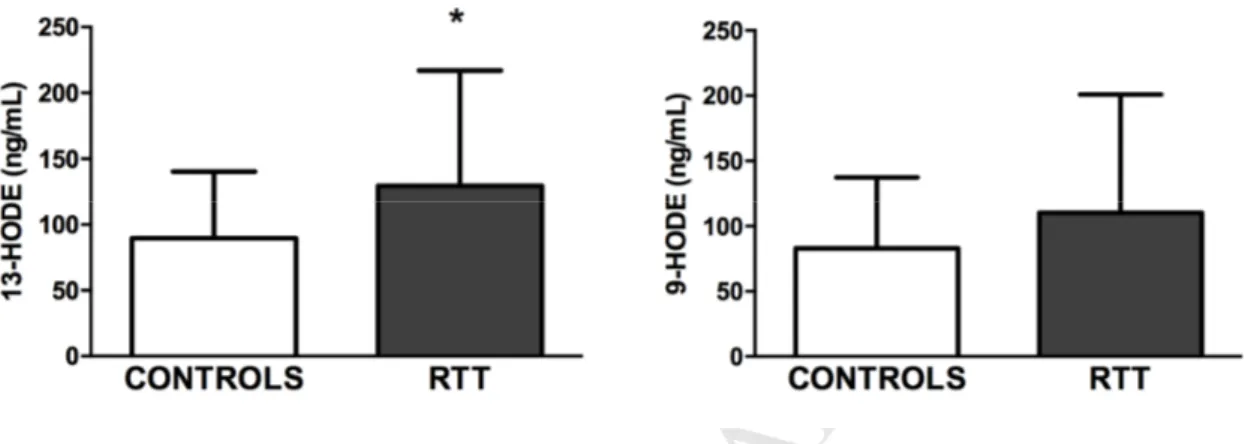
As shown in Fig. 1, 13-HODE levels were significantly increased in serum samples from RTT patients respect to the control group (Mann–Whitney, p < 0.05), and similar trend was observed for 9-HODE serum levels, although this result did not reach a significant difference.
Fig. 1. Serum levels of 9- and 13-HODE in controls and RTT patients. 13-HODE levels
significantly increased in RTT serum samples (n = 42) as compared to control subjects (n = 16). Conversely, no significant changes were observed for serum 9-HODE values. Data is provided as median ± SD. Mann–Whitney, *p < 0.05.
Increased ALOX15 gene expression in RTT PBMCs
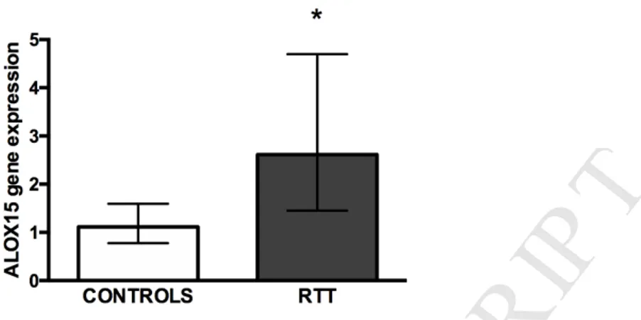
ALOX15 gene codifies for one of the most important enzymes involved in HODEs formation, 15-lipoxygenase-1 (Singh and Rao, 2018). As shown in Fig. 2, by the use of real time RT-PCR we were able to appreciate an over 2-fold upregulation of ALOX15 gene expression in PBMCs isolated from RTT patients.
M
AN
US
CR
IP
T
AC
CE
PT
ED
8Fig. 2. ALOX15 gene expression in RTT and control PBMCs. Real-time PCR shows the
upregulation of ALOX15 mRNA expression in RTT PBMCs (n = 5) compared to control subjects (n = 5). Unpaired t test with Welch's correction, *p < 0.05.
ω-3 PUFAs supplementation modulates 9- and 13-HODE serum levels in RTT patients Interestingly, ω-3 PUFA supplementation was able to affect 9- and 13-HODE serum levels in RTT patients respect to the untreated RTT group (Fig. 3). Notably, serum 13-HODE levels were significantly decreased in the supplemented RTT patients respect to the not supplemented group. This effect was more evident for the 13-HODE than for the 9-HODE although the trends were similar for both markers.
Fig. 3. Effects of dietary ω-3 PUFAs supplementation on serum HODEs levels in RTT patients. Following ω-3 PUFAs supplementation (n = 22), both serum HODEs levels decreased
M
AN
US
CR
IP
T
AC
CE
PT
ED
9 respect to un-supplemented RTT patients (n = 20). Data is provided as median ± SD. Kruskal-Wallis test, *p < 0.05 versus controls.9- and 13-HODE serum levels correlate with lipoprotein-associated phospholipase A2 (Lp-PLA2) activity
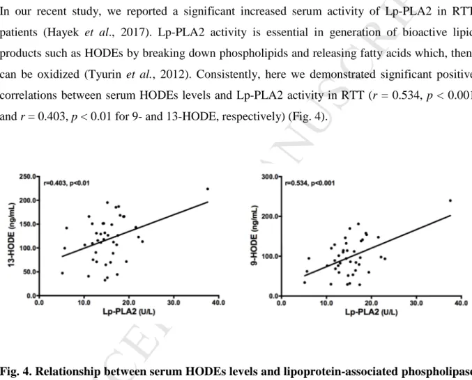
In our recent study, we reported a significant increased serum activity of Lp-PLA2 in RTT patients (Hayek et al., 2017). Lp-PLA2 activity is essential in generation of bioactive lipid products such as HODEs by breaking down phospholipids and releasing fatty acids which, then, can be oxidized (Tyurin et al., 2012). Consistently, here we demonstrated significant positive correlations between serum HODEs levels and Lp-PLA2 activity in RTT (r = 0.534, p < 0.001 and r = 0.403, p < 0.01 for 9- and 13-HODE, respectively) (Fig. 4).
Fig. 4. Relationship between serum HODEs levels and lipoprotein-associated phospholipase A2 (Lp-PLA2) activity. Serum levels of 9- and 13-HODE were significantly and positively
M
AN
US
CR
IP
T
AC
CE
PT
ED
10 DiscussionAlthough RTT is a well-recognized neurological disorder associated with mutations in MECP2 gene, there is growing evidence that a systemic metabolic component can contribute to its peculiar pathological phenotype (Kyle et al., 2018). In particular, a perturbed lipid homeostasis with increased serum cholesterol, triglycerides and LDLs has been observed in both RTT patients and animal models and linked to both neurological and systemic symptoms (Justice et al., 2013; Segatto et al., 2014; Buchovecky et al. 2013; Cobolli Gigli et al., 2016; Sticozzi et al. 2013; Ciernia et al., 2018). In the last few years, these new findings prompted a part of research efforts on RTT to focusing mainly on understanding the molecular mechanisms that could be responsible for the altered lipid metabolism. As a result of these studies, an alteration in the expression of genes involved in cholesterol biosynthesis has been observed in brain of Mecp2 mutant mice (Buchovecky et al. 2013; Urdinguio et al., 2008; Lopez et al., 2017). Similarly, an alteration of cholesterol regulatory network proteins has been detected also in fibroblasts isolated from RTT patients (Sticozzi et al., 2013; Segatto et al., 2014). Even more recently, RTT has been associated with fatty liver disease and dyslipidemia for an aberrant transcription of lipogenesis enzymes due to failed interaction of Mecp2 with the repressor complex containing NCoR1 and HDAC3 (Kyle et al., 2016).
However, once the lipid dyshomeostasis in RTT has been well established, besides understanding what causes these abnormalities, it is important to comprehend the impact of the altered lipid profile on RTT patient’s health. Of note, several lines of evidence highlighted a key role for an aberrant redox balance and a subclinical inflammation status, i.e. oxinflammation phenomena in RTT pathophysiology (Filosa et al., 2015; Pecorelli et al., 2016; Valacchi et al., 2017). Since lipids are among the main targets of free radicals, under a condition of redox imbalance a large variety of secondary byproducts of lipid oxidation can be generated (Frijhoff et al., 2015). Indeed, abnormal increased levels of isoprostanes (i.e. F2-isoprostanes, F4
-neuroprostanes, and F2-dihomo-isoprostanes) have been detected in plasma samples from RTT
patients and in whole brain from different RTT mouse models (Valacchi et al., 2017). In addition, our previous studies demonstrated high levels of 4-hydroxynonenal (4-HNE) protein adducts in both RTT patients and mouse models (Pecorelli et al., 2011; Valacchi et al., 2017). Interestingly, both F4-neuroprostanes and 4-HNE protein adducts were correlated to RTT
M
AN
US
CR
IP
T
AC
CE
PT
ED
11 biological mediators in RTT pathophysiology (Signorini et al., 2011, Pecorelli et al., 2011; Valacchi et al., 2017).In this study, we further implicated the putative role of oxidized lipids in RTT. Indeed, our findings demonstrated that HODEs concentrations, another important class of biologically active lipids, were significantly higher in serum of RTT patients. It is possible that the characteristic oxidative milieu of RTT and lack of a proper antioxidant defense coupled with the increased levels of LDL (Sticozzi et al., 2013; Segatto et al., 2014; Cervellati et al., 2015; Pecorelli et al., 2016; Valacchi et al., 2017) could drive the higher serum HODEs concentrations observed in our study. In addition, besides the free radical-mediated process, linoleic acid oxidation can proceed also via enzymatic mechanisms involving, among others, 15-lipoxygenase-1 (Singh and Rao, 2018). Of note, our findings indicate a significant upregulation of ALOX15 expression in RTT PBMC, thus suggesting a possible contribution of its enzymatic activity in the increasing HODEs formation. In addition, consistent with these results, our previous lipidomic analysis revealed a significant decreased content of linoleic acid in RTT erythrocyte membranes that could be ascribed to its increased enzymatic and non-enzymatic oxidation (Signorini et al., 2014). In addition, it has been demonstrated that 15-lipoxygenase-1 activity is associated with GSH depletion in brain cells (Li et al., 1997); therefore, the increased ALOX15 levels could be ascribed to the lower level of GSH detected in RTT cells (Signorini et al., 2014). Finally, ALOX15 expression can be also induced by several pro-inflammatory cytokines that have been showed to be sub-clinically detected in RTT (Singh and Rao, 2018; Pecorelli et al., 2016). As stable oxidized derivatives of linoleic acid, high HODEs levels are commonly found in oxidized LDL in pathological conditions characterized by both redox imbalance and inflammation (Vangaveti et al., 2010). Therefore, the activation of ALOX15/HODEs circuit could constitute another contributor factor to the typical oxinflammation status observed in RTT (Pecorelli et al., 2016; Valacchi et al., 2017, 2018). In fact, HODEs are not only indicators of lipid peroxidation but they are also able to modulate the inflammatory pathways with either beneficial or detrimental effects depending on their levels (Vangaveti et al., 2010). For example, ALOX15 and HODEs have received considerable attention as possible players in inflammatory responses associated with disorders such as atherosclerosis, hypertension, obesity and diabetes (Vangaveti, Baune and Kennedy, 2010), but also neurological conditions including Alzheimer disease and multiple sclerosis (Yoshida et al., 2009). HODEs can affect the expression of
M
AN
US
CR
IP
T
AC
CE
PT
ED
12 proinflammatory cytokines and cell adhesion molecules; modulate immune cells chemotaxis and monocyte adhesion to vascular endothelial cells; induce activation of transcription factors such as peroxisome proliferator activated receptor gamma (PPAR-γ) and nuclear factor-kappa B(NF-κB) (Ogawa et al., 2011). Of note, a subclinical inflammation status with a deregulated plasma cytokine profile and an aberrant NF-κB signaling have been reported in RTT (Cortelazzo et al., 2014; Pecorelli et al., 2016; Kishi et al., 2016; Jorge-Torres et al., 2018).
Notably, our results also indicated that serum HODEs levels in RTT were linearly and positively correlated with Lp-PLA2 activity, data that also agree with a recent study, where we showed an increase in serum PLA2 activity in RTT patients (Hayek et al., 2017). Elevated levels of Lp-PLA2 have been associated with an increased risk for cardiovascular disorders (Younus et al., 2017). Indeed, the main action of this enzyme involve the hydrolysis of fatty acids from oxidatively modified phospholipids present in oxidized LDL to produce free fatty acids. Among others, Lp-PLA2 is also able to release 13-HODE from ox-LDL in atherosclerotic lesions (Tyurin et al., 2012). Accordingly, the positive correlation observed in our study suggests that Lp-PLA2-mediated hydrolysis of oxidized linoleic acid can play a harmful role also in RTT, promoting an increased HODEs production.
Evidence indicated that adoption of healthy dietary intervention leads to the decline in plasma HODEs levels (Ramsden et al., 2012). In previous studies, we have shown that supplementation with ω-3 PUFAs improved clinical severity of RTT patients with a significant decrease of isoprostanes and 4-HNE plasma levels (Ciccoli et al., 2012; De Felice et al., 2012). Consistent with these observations, in this study, we confirmed the potential beneficial effect of ω-3 PUFAs on RTT, as indicated by the decreasing trend of serum HODEs levels in supplemented patients. To date, the exact mechanisms by which omega-3 PUFAs exerts their beneficial effects in several pathological conditions are not fully understood. Nevertheless, it is well known that eicosapentaenoic acid (EPA) and docosahexaenoic acid (DHA) compete with omega-6 PUFAs for the same molecular pathways involving enzymes like cyclooxygenases and lipoxygenases (Gabbs et al., 2015). Therefore, it is likely that the omega-3 fatty acids incorporated into the membrane phospholipids after the fish oil-enriched diet in RTT patients can reduce the metabolism of acid linoleic and, thus, the HODEs generation through a process of competitive inhibition. This possible shift in PUFAs-derived metabolites is confirmed by the work of Shearer et al. (2010) that demonstrated how a supplementation with omega-3 PUFAs for 4 weeks in
M
AN
US
CR
IP
T
AC
CE
PT
ED
13 healthy adults is able to lower the concentration of both HODE isomers by 15%. In addition, unlike omega-6 PUFAs metabolites, the omega-3 derived lipid mediators such as lipoxins, resolvins, and protectins show anti-inflammatory protective properties (Gabbs et al., 2015) that could account for the improved inflammation status in supplemented RTT patients (Signorini et al., 2014).Conclusions
The present study has been reported for the first time that endogenous formation of 9- and 13-HODE is increased in RTT patients. These findings concur with prior results obtained from our and other laboratories that indicate a clear key role of lipid peroxidation in RTT pathophysiology (Pecorelli et al., 2016; Valacchi et al., 2017, 2018). Indeed, given the ability of these compounds to modulate redox and immune homeostasis, together with 4-HNE protein adducts and isoprostanes, HODEs could contribute to metabolic abnormalities found in this disorder. In addition, our results confirm that dietary intervention with ω-3 PUFAs could revert, at least partially, the increased lipid peroxidation found in RTT, and potentially improve RTT oxinflammation condition in these patients. It should be mentioned that at this time, the RTT patients population of this study does not allow us to evaluate whether HODEs can have a role in the disease progression, since we could not collect plasma from patients belonging to 4 different stages of the disease, as previously reported for 4HNE levels (Pecorelli et al., 2011).
M
AN
US
CR
IP
T
AC
CE
PT
ED
14 References1. Amir RE, Van den Veyver IB, Wan M, Tran CQ, Francke U, Zoghbi HY. Rett syndrome is caused by mutations in X-linked MECP2, encoding methyl-CpG-binding protein 2. Nat Genet. 1999;23(2):185-8.
2. Armstrong, D. D. (1997) ‘Review of Rett syndrome.’ J Neuropathol Exp Neurol, 56(8), pp. 843–9.
3. Buchovecky CM, Turley SD, Brown HM, Kyle SM, McDonald JG, Liu B, Pieper AA, Huang W, Katz DM, Russell DW, Shendure J, Justice MJ. A suppressor screen in Mecp2 mutant mice implicates cholesterol metabolism in Rett syndrome. Nat Genet. 2013;45(9):1013-20. doi: 10.1038/ng.2714.
4. Cervellati, C. et al. (2015) ‘Impaired enzymatic defensive activity, mitochondrial dysfunction and proteasome activation are involved in RTT cell oxidative damage.’ Biochim Biophys Acta. 1852(10 Pt A), pp. 2066–74. doi: 10.1016/j.bbadis.2015.07.014. 5. Ciccoli L, De Felice C, Paccagnini E, Leoncini S, Pecorelli A, Signorini C, Belmonte G,
Valacchi G, Rossi M, Hayek J. Morphological changes and oxidative damage in Rett Syndrome erythrocytes. Biochim Biophys Acta. 2012;1820(4):511-20. doi: 10.1016/j.bbagen.2011.12.002.
6. Ciernia AV, Yasui DH, Pride MC, Durbin-Johnson B, Noronha A, Chang A, Knotts T, Rutkowsky J, Ramsey JJ, Crawley JN, LaSalle JM. MeCP2 isoform e1 mutant mice recapitulate motor and metabolic phenotypes of Rett syndrome. Hum Mol Genet. 2018. doi: 10.1093/hmg/ddy301.
7. Cobolli Gigli C, Scaramuzza L, Gandaglia A, Bellini E, Gabaglio M, Parolaro D, Kilstrup-Nielsen C, Landsberger N, Bedogni F. MeCP2 Related Studies Benefit from the Use of CD1 as Genetic Background. PLoS One. 2016;11(4):e0153473. doi: 10.1371/journal.pone.0153473.
8. Cortelazzo, A. et al. (2014) ‘Subclinical inflammatory status in Rett syndrome.’ Mediators Inflamm. 2014, p. 480980. doi: 10.1155/2014/480980.
9. De Felice C, Signorini C, Durand T, Ciccoli L, Leoncini S, D'Esposito M, Filosa S, Oger C, Guy A, Bultel-Poncé V, Galano JM, Pecorelli A, De Felice L, Valacchi G, Hayek J. Partial rescue of Rett syndrome by ω-3 polyunsaturated fatty acids (PUFAs) oil. Genes Nutr. 2012;7(3):447-58. doi:10.1007/s12263-012-0285-7.
M
AN
US
CR
IP
T
AC
CE
PT
ED
15 10.Filosa S, Pecorelli A, D'Esposito M, Valacchi G, Hajek J. Exploring the po,sible link between MeCP2 and oxidative stress in Rett syndrome. Free Radic Biol Med. 2015;88(Pt A):81-90. doi: 10.1016/j.freeradbiomed.2015.04.019.11.Frijhoff J, Winyard PG, Zarkovic N, Davies SS, Stocker R, Cheng D, Knight AR, Taylor EL, Oettrich J, Ruskovska T, Gasparovic AC, Cuadrado A, Weber D, Poulsen HE, Grune T, Schmidt HH, Ghezzi P. Clinical Relevance of Biomarkers of Oxidative Stress. Antioxid Redox Signal. 2015;23(14):1144-70. doi:10.1089/ars.2015.6317.
12.Gabbs M, Leng S, Devassy JG, Monirujjaman M, Aukema HM. Advances in Our Understanding of Oxylipins Derived from Dietary PUFAs. Adv Nutr. 2015;6(5):513-40. doi: 10.3945/an.114.007732.
13.Hagberg B. Clinical manifestations and stages of Rett syndrome. Ment Retard Dev Disabil Res Rev. 2002;8(2):61-5.
14.Hayek, J. et al. (2017) ‘Lactonase Activity and Lipoprotein-Phospholipase A2as Possible Novel Serum Biomarkers for the Differential Diagnosis of Autism Spectrum Disorders and Rett Syndrome: Results from a Pilot Study.’ Oxid Med Cell Longev. 2017, p. 5694058. doi: 10.1155/2017/5694058.
15.Jorge-Torres OC, Szczesna K, Roa L, Casal C, Gonzalez-Somermeyer L, Soler M, Velasco CD, Martínez-San Segundo P, Petazzi P, Sáez MA, Delgado-Morales R, Fourcade S, Pujol A, Huertas D, Llobet A, Guil S, Esteller M. Inhibition of Gsk3b Reduces Nfkb1 Signaling and Rescues Synaptic Activity to Improve the Rett Syndrome Phenotype in Mecp2-Knockout Mice. Cell Rep. 2018;23(6):1665-1677. doi: 10.1016/j.celrep.2018.04.010.
16.Justice MJ, Buchovecky CM, Kyle SM, Djukic A. A role for metabolism in Rett syndrome pathogenesis: New clinical findings and potential treatment targets. Rare Dis. 2013;1:e27265. doi: 10.4161/rdis.27265.
17.Kishi N, MacDonald JL, Ye J, Molyneaux BJ, Azim E, Macklis JD. Reduction of aberrant NF-κB signalling ameliorates Rett syndrome phenotypes in Mecp2-null mice. Nat Commun. 2016;7:10520. doi: 10.1038/ncomms10520.
18.Kurien, B. T. et al. (2006) Oxidatively modified autoantigens in autoimmune diseases. Free Rad Biol Med, 41(4), pp. 549–56. doi: 10.1016/j.freeradbiomed.2006.05.020.
M
AN
US
CR
IP
T
AC
CE
PT
ED
16 19.Kyle SM, Saha PK, Brown HM, Chan LC, Justice MJ. MeCP2 co-ordinates liver lipid metabolism with the NCoR1/HDAC3 corepressor complex. Hum Mol Genet, 2016;25(14):3029-3041.20.Kyle SM, Vashi N, Justice MJ. Rett syndrome: a neurological disorder with metabolic components. Open Biol, 2018;8(2). pii: 170216. doi: 10.1098/rsob.170216.
21.Li Y, Maher P, Schubert D. A role for 12-lipoxygenase in nerve cell death caused by glutathione depletion. Neuron, 1997;19(2):453-63.
22.Lopez AM, Chuang JC, Posey KS, Turley SD. Suppression of brain cholesterol synthesis in male Mecp2-deficient mice is age dependent and not accompanied by a concurrent change in the rate of fatty acid synthesis. Brain Res, 2017;1654(Pt A):77-84. doi: 10.1016/j.brainres.2016.10.021.
23.Nieman DC, Meaney MP, John CS, Knagge KJ, Chen H. 9- and 13-Hydroxy-octadecadienoic acids (9+13 HODE) are inversely related to granulocyte colony stimulating factor and IL-6 in runners after 2h running. Brain Behav Immun. 2016; 56:246-52. doi: 10.1016/j.bbi.2016.03.020.
24.Ogawa, E. et al. (2011) ‘Epidermal FABP (FABP5) Regulates Keratinocyte Differentiation by 13(S)-HODE-Mediated Activation of the NF-κB Signaling Pathway’, J Invest Dermatol, 131(3), pp. 604–612. doi: 10.1038/jid.2010.342.
25.Pecorelli A, Ciccoli L, Signorini C, Leoncini S, Giardini A, D'Esposito M, Filosa S, Hayek J, De Felice C, Valacchi G. Increased levels of 4HNE-protein plasma adducts in
Rett syndrome. Clin Biochem, 2011;44(5-6):368-71.
doi:10.1016/j.clinbiochem.2011.01.007.
26.Pecorelli, A. et al. (2016) ‘OxInflammation in Rett syndrome’, Int J Biochem Cell Biol, 81, pp. 246–253. doi: 10.1016/j.biocel.2016.07.015.
27.Pecorelli A, Cervellati F, Belmonte G, Montagner G, Waldon P, Hayek J, Gambari R, Valacchi G. Cytokines profile and peripheral blood mononuclear cells morphology in Rett and autistic patients. Cytokine, 2016; 77:180-8. doi:10.1016/j.cyto.2015.10.002. 28.Ramsden CE, Ringel A, Feldstein AE, Taha AY, MacIntosh BA, Hibbeln JR,
Majchrzak-Hong SF, Faurot KR, Rapoport SI, Cheon Y, Chung YM, Berk M, Mann JD. Lowering dietary linoleic acid reduces bioactive oxidized linoleic acid metabolites in humans.
M
AN
US
CR
IP
T
AC
CE
PT
ED
17 Prostaglandins Leukot Essent Fatty Acids, 2012;87(4-5):135-41. doi: 10.1016/j.plefa.2012.08.004.29.Segatto M, Trapani L, Di Tunno I, Sticozzi C, Valacchi G, Hayek J, Pallottini V. Cholesterol metabolism is altered in Rett syndrome: a study on plasma and primary cultured fibroblasts derived from patients. PLoS One, 2014;9(8):e104834. doi: 10.1371/journal.pone.0104834. e Collection 2014. Erratum in: PLoS One. 2014;9(11):e114654.
30.Shearer GC, Harris WS, Pedersen TL, Newman JW. Detection of omega-3 oxylipins in human plasma and response to treatment with omega-3 acid ethyl esters. J Lipid Res, 2010;51(8):2074-81. doi: 10.1194/M900193-JLR200.
31.Signorini C, De Felice C, Leoncini S, Durand T, Galano JM, Cortelazzo A, Zollo G, Guerranti R, Gonnelli S, Caffarelli C, Rossi M, Pecorelli A, Valacchi G, Ciccoli L, Hayek J. Altered erythrocyte membrane fatty acid profile in typical Rett syndrome: effects of omega-3 polyunsaturated fatty acid supplementation. Prostaglandins Leukot Essent Fatty Acids, 2014;91(5):183-93. doi:10.1016/j.plefa.2014.08.002.
32.Signorini C, De Felice C, Leoncini S, Giardini A, D'Esposito M, Filosa S, Della Ragione F, Rossi M, Pecorelli A, Valacchi G, Ciccoli L, Hayek J. F₄-neuroprostanes mediate neurological severity in Rett syndrome. Clin Chim Acta, 2011;412(15-16):1399-406. doi: 10.1016/j.cca.2011.04.016.
33.Signorini C, Leoncini S, De Felice C, Pecorelli A, Meloni I, Ariani F, Mari F, Amabile S, Paccagnini E, Gentile M, Belmonte G, Zollo G, Valacchi G, Durand T, Galano JM, Ciccoli L, Renieri A, Hayek J. Redox imbalance and morphological changes in skin fibroblasts in typical Rett syndrome. Oxid Med Cell Longev, 2014;2014:195935. doi: 10.1155/2014/195935.
34.Singh NK, Rao GN. Emerging role of 12/15-Lipoxygenase (ALOX15) in human pathologies. Prog Lipid Res, 2018. pii: S0163-7827(18)30038-9. doi:10.1016/j.plipres.2018.11.001.
35.Sticozzi C, Belmonte G, Pecorelli A, Cervellati F, Leoncini S, Signorini C, Ciccoli L, De Felice C, Hayek J, Valacchi G. Scavenger receptor B1 post-translational modifications in Rett syndrome. FEBS Lett, 2013;587(14):2199-204. doi: 10.1016/j.febslet.2013.05.042.
M
AN
US
CR
IP
T
AC
CE
PT
ED
18 36.Tyurin, V. A. et al. (2012) ‘Specificity of Lipoprotein-Associated Phospholipase A 2 toward Oxidized Phosphatidylserines: Liquid Chromatography–Electrospray Ionization Mass Spectrometry Characterization of Products and Computer Modeling of Interactions’, Biochemistry, 51(48), pp. 9736–9750. doi: 10.1021/bi301024e.37.Uchida, K. (2003) ‘4-Hydroxy-2-nonenal: A product and mediator of oxidative stress’, Prog Lipid Res, pp. 318–343. doi: 10.1016/S0163-7827(03)00014-6.
38.Urdinguio RG, Lopez-Serra L, Lopez-Nieva P, Alaminos M, Diaz-Uriarte R, Fernandez AF, Esteller M. Mecp2-null mice provide new neuronal targets for Rett syndrome. PLoS One. 2008;3(11):e3669. doi: 10.1371/journal.pone.0003669.
39.Valacchi, G. et al. (2017) ‘4-hydroxynonenal protein adducts: Key mediator in Rett syndrome oxinflammation.’, Free Rad Biol Med, 111, pp. 270–280. doi: 10.1016/j.freeradbiomed.2016.12.045.
40.Valacchi, G. et al. (2018) ‘OxInflammation: From Subclinical Condition to Pathological Biomarker.’, Front Physiol, 9, p. 858. doi: 10.3389/fphys.2018.00858.
41.Vangaveti, V., Baune, B. T. and Kennedy, R. L. (2010) ‘Review: Hydroxyoctadecadienoic acids: Novel regulators of macrophage differentiation and atherogenesis’, Ther Adv Endocrinol Metab, doi: 10.1177/2042018810375656.
42.Wang, L. et al. (2009) ‘Triglyceride-rich lipoprotein lipolysis releases neutral and oxidized FFAs that induce endothelial cell inflammation’, J Lipid Res, doi: 10.1194/jlr.M700505-JLR200.
43.Yoshida Y, Yoshikawa A, Kinumi T, Ogawa Y, Saito Y, Ohara K, Yamamoto H, Imai Y, Niki E. Hydroxyoctadecadienoic acid and oxidatively modified peroxiredoxins in the blood of Alzheimer's disease patients and their potential as biomarkers. Neurobiol Aging. 2009; 30(2):174-85.
44.Younus A, Humayun C, Ahmad R, Ogunmoroti O, Kandimalla Y, Aziz M, Malik R, Saand AR, Valdes C, Badlani R, Younus MA, Ali SS, Chen Y, Nasir K. Lipoprotein-associated phospholipase A2 and its relationship with markers of subclinical cardiovascular disease: A systematic review. J Clin Lipidol, 2017;11(2):328-337. doi: 10.1016/j.jacl.2017.02.005.
M
AN
US
CR
IP
T
AC
CE
PT
ED
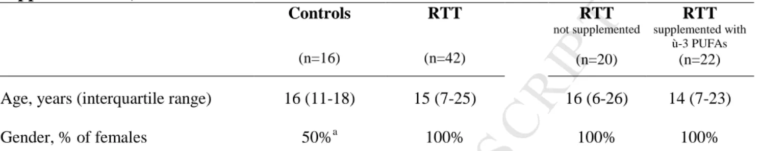
19Table 1. Age and gender of Controls and RTT patients (all, with and without ù-3 PUFAs supplementation) Controls (n=16) RTT (n=42) RTT not supplemented (n=20) RTT supplemented with ù-3 PUFAs (n=22)
Age, years (interquartile range) 16 (11-18) 15 (7-25) 16 (6-26) 14 (7-23) Gender, % of females 50%a 100% 100% 100%
Data presented are expressed as: % within the group for categorical variables; median (interquartile range) for continuous variables
M
AN
US
CR
IP
T
AC
CE
PT
ED
Rett syndrome (RTT) is a neurodevelopmental rare diseases OxInflammation status characterizes this pathology
Role of lipid mediators in RTT pathogenesis has been suggested
Increase levels of HODEs, ALOX1 and PLA2 activity confirm the oxidative lipid role in RTT