https://doi.org/10.1177/1758835918793569 https://doi.org/10.1177/1758835918793569 Therapeutic Advances in Medical Oncology
journals.sagepub.com/home/tam 1
Ther Adv Med Oncol
2018, Vol. 10: 1 –9 DOI: 10.1177/ 1758835918793569 © The Author(s), 2018. Article reuse guidelines: sagepub.com/journals-permissions
Introduction
Cardiac tumors are a very rare disease. Thus, optimal diagnosis and treatment management are not well established, to date. Here, we report an overview on current available data in litera-ture on these tumors, focusing on the role of imaging, with particular reference to 18-fluorine-fluorodeoxyglucose positron emission tomography/ computed tomography (PET/CT).
Epidemiology of cardiac tumors
Cardiac tumors account only for a small fraction of all cardiac masses, mostly represented by pseu-dotumors like thrombi, vegetations, abscesses, aneurysms.
The estimated frequency of cardiac tumors ranges from 0.0017% to 0.33%, mostly represented by benign tumors (75%), with cardiac myxomas being the most common, accounting for nearly half of them.1
Primary malignant cases account for 25% of all cardiac tumors, while secondary cardiac tumors, either by metastatic spread or by direct invasion, are far more common (40–50 times more frequent).
Autopsy series reviews found a 2.9–8.4% fre-quency of metastatic tumors involving the heart.2,3 Cardiac metastases originate mainly from lung, breast, kidney and cutaneous melanoma.
Among the primary malignant tumors of the heart, the most frequent are sarcomatous in nature. All types of sarcomas may be observed in the heart with a predominance of rhabdomyosar-coma in childhood and angiosarrhabdomyosar-comas or undif-ferentiated sarcomas in adulthood. They affect mostly patients in the fourth decade of life and survival remains extremely poor: in older retro-spective case series, regardless of the type of treat-ment, most patients died within 12–16.5 months after initial diagnosis.4 In more recent series,
18
F-FDG-PET/CT imaging in cardiac tumors:
illustrative clinical cases and review of the
literature
Maristella Saponara , Valentina Ambrosini, Margherita Nannini , Lidia Gatto, Annalisa Astolfi, Milena Urbini, Valentina Indio, Stefano Fanti and Maria Abbondanza Pantaleo
Abstract: Cardiac tumors are a very rare condition. Mostly, they are benign tumors (75%),
with myxomas being the most frequent. The remaining 25% are malignant; either primary malignant sarcoma or secondary metastases. Given the small number of cases reported and the lack of prospective and randomized clinical trials, the level of evidence for the optimal multimodal treatment of primary cardiac sarcomas is very low and the optimal imaging diagnostic workup is not well established. In particular, 18F-FDG-PET/CT is not yet included
in routine diagnosis of cardiac masses. Here, we report four illustrative clinical cases and a review of the literature on the current available data on the role of 18F-fluorodeoxyglucose
PET/CT imaging in cardiac tumors.
Keywords: 18F-FDG PET/CT, cardiac tumor, dietary recommendations, imaging workup, PET
preparation, rare tumor, sarcoma
Received: 14 February 2018; revised manuscript accepted: 23 May 2018. Correspondence to:
Maristella Saponara Division of Medical Oncology, Department of Specialized, Experimental and Diagnostic Medicine, Sant’Orsola-Malpighi Hospital, University of Bologna, Via Massarenti 9, Bologna 40138, Italy maristella.saponara@ unibo.it Valentina Ambrosini Stefano Fanti Division of Diagnostic Imaging and Radiotherapy, University of Bologna, Bologna, Italy Margherita Nannini Lidia Gatto Division of Medical Oncology, University of Bologna, Bologna, Italy
Annalisa Astolfi Milena Urbini Valentina Indio
‘Giorgio Prodi’ Cancer Research Center, University of Bologna, Bologna, Italy Maria Abbondanza Pantaleo Division of Medical Oncology, University of Bologna, Bologna, Italy ‘Giorgio Prodi’ Cancer Research Center, University of Bologna, Bologna, Italy
survival reaches 38.8 months but only for patients who underwent complete resection in referral centers.5
Treatment of cardiac sarcomas
Given the small number of cases reported and the lack of randomized clinical trials, the level of evi-dence for the optimal multimodal management of primary cardiac sarcomas is low. There is no standardized evidence-based approach which is then often extrapolated from that of extracardiac soft tissue sarcomas with the inevitable limits due to the cardiac location itself.
Complete surgical resection is rarely achievable in practice, given the aggressive and infiltrative nature of these tumors (only 13.3% of cases were resectable in the largest series of primary cardiac sarcomas reported by the French Sarcoma Group in a multi-institutional retrospective study).5 Moreover, it is easy to understand how dose-lim-iting problems are related to radiotherapy. The value of preoperative or postoperative radiother-apy (an effective adjuvant tool for sarcomas aris-ing at other sites) is very limited in the treatment of cardiac sarcomas considering that the heart is usually more sensitive than the tumor to radiation injury, and that the ventricular wall cannot be treated with a radical radiation dose without sub-stantial risk of cardiomyopathy and chronic pericarditis.
Generally, surgical resection represents the cor-nerstone of therapy in primary localized cardiac sarcoma. Only surgical resection with pathologi-cally negative margins (R0 resection) is associated with a prospect of prolonged survival. However, despite complete surgical resection, tumor recur-rence and metastases occur early, mostly within 6–12 months, and frequently. A high rate of both relapse and distant metastases within the follow-up period is reported in a series of malignant car-diac tumors seen over 15 years (45.5% and 72.2%, respectively).6
Neoadjuvant or adjuvant chemotherapy may be administered in order to downsize the tumor and facilitate R0 resection or to reduce risk of relapse and distant metastases. However, the rarity of these tumors hinders generation of reliable evi-dence in favor of or against neoadjuvant and adjuvant chemotherapy in cardiac sarcoma. In the literature, the optimal treatment approach
regarding postoperative adjuvant therapy remains unclear: some studies found that the adjuvant approach failed to modify the natural course of the disease,7,8 while others have demonstrated a better survival for patients whose surgery was fol-lowed by adjuvant chemotherapy.6,9
Overall, anthracyclines (in particular, doxoru-bicin) and nitrogen mustard alkylating agents (in particular, ifosfamide), which represent the pre-ferred chemotherapeutic choice to treat sarcomas originating from other sites, are the most com-mon chemotherapy agents used also for cardiac sarcomas. However, the typical cardiotoxicity of anthracyclines should be kept in mind when adju-vant treatment is proposed to a patient.
In brief, given the limited and conflicting results of adjuvant treatment, this approach may be consid-ered for a patient–physician shared decision mak-ing in a subset of selected fit and high-risk patients and, in any case, a strict follow up must be planned, even after apparently completed treatment.
In case of unresectable disease or presence of metastases, palliative chemotherapy should be offered, although in some cases, palliative surgical debulking may also be proposed, with the aim of relieving rapidly progressing symptoms.
As reported so far, cardiac sarcomas show a high degree of complexity: due to their location and dismal prognosis, a multidisciplinary approach is crucial from the very beginning of patient man-agement. Because of their rarity, diagnosis, imag-ing techniques, surgical, radiotherapy or chemotherapy treatments and follow-up strategy still pose a serious challenge for physicians. Imaging techniques
With regard to noninvasive diagnostic modalities, echocardiography and magnetic resonance imag-ing (MRI) have always represented the two most used and sensitive techniques to detect and attempt to characterize suspected cardiac masses. Echocardiography, especially the transesophageal route, is usually the initial imaging modality. It may show the tumor, its extent and its hemody-namic consequences. Cardiac MRI provides fur-ther information about morphology, location and extent of the mass. In case of suspected cardiac malignancies, CT scan is also useful to assess ext-racardiac extent and distant metastasis, if any.
In particular, the multiplanar assessment of anat-omy, tissue composition, and functional impact afforded by MRI, allows accurate confirmation of the presence of a space-occupying lesion, locali-zation and assessment of the extent of involve-ment, evaluation of the functional impact of the lesion, as well as tissue characterization. Such information is important not only for diagnosis but also for prognosis determination and therapy planning. As such, cardiac MRI has rapidly evolved as the reference standard technique for early assessment and differentiation between a non-neoplastic mass and a tumor mass, be it benign or malignant.
A quick diagnosis facilitates the initiation of a proper treatment, which may in turn improve outcome of patients affected by these rare and aggressive neoplasms. Therefore, a high level of suspicion is required, and, over the years, efforts have been made to define criteria for malignancy of cardiac masses. Findings suggestive of a malignant cardiac tumor at MRI include: right atrial location; involvement of more than one cardiac chamber, size > 5 cm, hemorrhagic per-icardial effusion, broad base of attachment, extension into the mediastinum or great vessels and a ‘moderate,’ ‘strong,’ or heterogeneous delayed enhancement pattern.10
18F-fluorodeoxyglucose positron emission
tomography/computed tomography
Scattered case reports in literature describe how integrated PET/CT with FDG imaging can inci-dentally show cardiac neoplasia, particularly metastatic involvement,11–17 or may be used in case of suspected aggressive cardiac malignancy to strengthen the diagnosis and detect possible occult distant disease.18–26
However, unlike many other solid tumors, FDG PET does not have an established role in the rou-tine evaluation of cardiac tumors, which is prob-ably due both to their low frequency and to the physiologically high uptake of FDG in the myo-cardium, although with interindividual and intraindividual variability.27
In the normal myocardium, metabolism is primar-ily oxidative and uses various substrates, including glucose, free fatty acids and lactate. The choice of substrate depends on its availability and the physi-ological and pathologic condition of the myocar-dium. The European Association of Nuclear
Medicine (EANM) guidelines suggest that, in case of evaluation of a lesion in the heart or very close to the myocardium, dietary recommenda-tions can be helpful.28 Several reports indicate that a low-carbohydrate diet, associated with pro-longed fasting, is adequate for shifting myocar-dium metabolism toward fatty acid consumption. The EANM 2015 guidelines recommend a low-carbohydrate diet for 24 h prior to the PET/CT study or, at least, a low-carbohydrate meal before starting the 6 h fasting period preceding the study. In order to standardize the high-fat/low- carbohydrate meal, a very recent report by the Society of Nuclear Medicine and Molecular Imaging-American Society of Nuclear Cardiology (SNMMI-ASNC) Expert Consensus indicates in detail the foods permitted and prohibited before PET scanning.29
Two larger studies have been conducted to assess the diagnostic value of 18F-FDG PET (integrated with CT or MRI imaging) in the diagnostic algo-rithm and in the noninvasive preoperative determi-nation of malignancy and metastatic spread of cardiac tumors, with the ultimate aim of stratifying patients and selecting and monitoring therapies.30,31 In the study by Rahbar and Colleagues, 18F-FDG PET/CT scans (whole body imaging with low-dose CT) of 24 consecutive patients with newly diagnosed cardiac tumors were retrospectively analyzed.30 The patients underwent standard oncological preparation for PET examination (with a fasting period of at least 6 h). As a result, background myocardial and blood pool uptake corresponded to a mean standardized uptake value (SUV) of 2.1 ± 0.6 and 1.6 ± 0.4, respec-tively. The maximum standardized uptake values (SUVmax) of the tumors were measured and com-pared between patients affected by benign and malignant cardiac tumors (both cardiac primaries or metastases). The study showed that mean SUVmax was 2.8 ± 0.6 in benign cardiac tumors and significantly higher both in malignant primary and in secondary cardiac tumors (8.0 ± 2.1 and 10.8 ± 4.9, respectively), (p < 0.001), assessing an SUVmax cutoff with high sensitivity at 3.5. In the study by Nensa and Colleagues, 20 patients were prospectively assessed using integrated car-diac 18F-FDG PET/MRI to evaluate whether this integrated imaging provided significant benefit over 18F-FDG PET or MRI alone.31 In order to optimally suppress physiologic glucose uptake in
the myocardium, patients were adequately pre-pared with a high-fat, low-carbohydrate diet for a period of 24 h before the examination, and 50 IU/ kg of unfractionated heparin was intravenously administered 15 min before 18F-FDG injection. Blood glucose levels at the time of tracer injection were less than 120 mg/dl. As a result, the mean SUV in the blood pool was 1.3 ± 0.3. The study showed that the mean SUVmax in all nonmalignant cases (benign tumors, thrombi, and scar tissue) was 2.3 ± 1.2, whereas the mean SUVmax in pri-mary and secondary malignant tumors was signifi-cantly higher at 13.2 ± 6.2 (p < 0.0004). Despite the obvious bias of statistical power related to the poor sample size, in this study, the combination of 18F-FDG PET and MRI yielded 100% sensitivity and specificity. In fact, there were only two cases (both with an SUVmax of 5.2) that could not be
differentiated using 18F-FDG PET alone, then unequivocally identified as malignancy and patch tissue by MRI, respectively. In another case, MR imaging misclassified scar tissue as local relapse of cardiac angiosarcoma, but a low SUVmax of 2.2 made malignant relapse unlikely.
Illustrative cases on the role of
18F-fluorodeoxyglucose positron emission
tomography/computed tomography in cardiac tumors
Herein we report four illustrative clinical cases of patients affected by cardiac tumors, referred to our institution. 18F-FDG PET/CT imaging was per-formed during the patients’ oncologic examina-tion, proving to be a very useful tool in detecting neoplasms and guiding physicians’ choices.
Figure 1. 18F-fluorodeoxyglucose positron emission tomography/computed tomography images.
(a) Transaxial low-dose CT; (b) transaxial fused PET/CT; (c) MIP of a 23-year-old patient, showing heterogenous FDG uptake (SUVmax = 12.3) in the right atrium, suggestive of malignant lesion. Transbronchial biopsy revealed an angiosarcoma of
the heart. After diagnosis, the patient underwent three cycles of chemotherapy with epirubicin and ifosfamide, with good response. Of note, the absence of physiological myocardium activity allows a better visualization of pathologic findings. CT, computed tomography; FDG, fluorodeoxyglucose; MIP, maximum intensity projection; PET, positron emission tomography; SUV, standardized uptake value.
The study protocol was approved by the ethics committee of Sant’Orsola Hospital-Bologna, Italy (approval no. 164/2017/O/Oss) as part of a large retrospective analysis of patients with rare tumors. All patients provided written informed consent for inclusion in the study (Figures 1–4).
Conclusion
Cardiac tumors are very uncommon. Given their rarity and complexity, optimal diagnosis and treatment management are still unknown.
Within the noninvasive diagnostic modalities to characterize suspected cardiac masses, echocardi-ography and MRI have represented the two most used and sensitive techniques.
18F-FDG PET has not an established role in the imaging workup of cardiac tumors, while it may have a substantial meaning in the diagnostic algo-rithm and in the noninvasive preoperative deter-mination of malignancy and metastatic spread of cardiac tumors.
Although based on small case series, the few avail-able studies, as well as our illustrative cases reported, suggest that PET imaging can add useful information to CT or MRI alone, improving the diagnostic workup in patients with cardiac masses both in the preoperative and post-treatment set-ting, enabling definition of surgical indication and early identification of local or distant recurrences that are key elements in the overall treatment strategy.
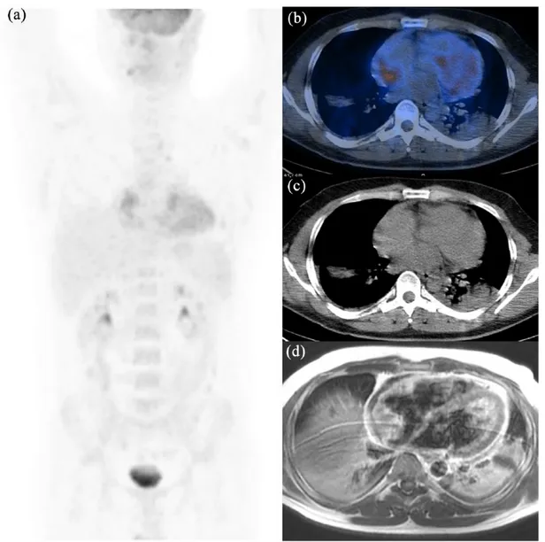
Figure 2. 18F-fluorodeoxyglucose positron emission tomography/computed tomography images of a
74-year-old man with abnormal uptake in the right ventricule.
(a) Transaxial fused PET/CT; (b) transaxial PET; (c) transaxial low-dose CT. Fused PET/CT (a) and PET (b) images show intense uptake (SUVmax = 12) at the periphery of the lesion, also clearly evident on corresponding transaxial MRI images
(d), while the hypointense core (b) reflects the presence of central necrosis. Bioptic sampling then revealed metastasis of a previous urothelial carcinoma. Because of the patient’s age and comorbidities, both surgery and chemotherapy were excluded and a palliative radiation treatment was performed instead.
In brief, by reviewing available literature and based on our reported experience, PET/CT, unlike other instrumental investigations, through the functional study of the heart, allows: (a) obtaining information about the malignant poten-tial of doubtful masses, based on SUVmax evalua-tion; (b) estimation of disease stage, also revealing potential myocardial involvement and pericardial spread; (c) evaluation of potential postoperative residual disease (in order to avoid false positives, the examination should be performed after an appropriate time period of 3/4 weeks from
surgery); (d) performing early evaluation of response to chemotherapy, especially in light of the recent introduction of target therapies for some types of these tumors.
Of course, for patients that are going to be scanned with PET/CT with FDG to assess the myocardium, a specific and accurate preparation (high-fat/low-carbohydrate diet plus prolonged fasting) is mandatory. Future studies are eagerly awaited to better define and validate this role on larger series.
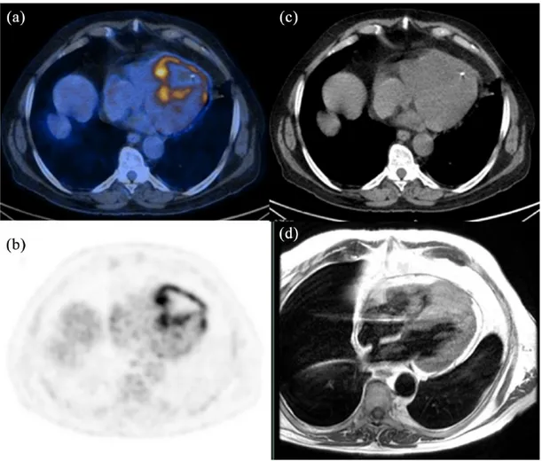
Figure 3. 18F-fluorodeoxyglucose positron emission tomography/computed tomography images of a
38-year-old man with abnormal uptake (SUVmax = 8.7) in the right atrium.
(a) MIP; (b) transaxial PET fused; (c) transaxial low-dose CT. FDG uptake (b) corresponds to the voluminous neoplastic mass on the transaxial MRI (d). Given the characteristics of malignancy shown by the imaging tests, the patient underwent surgery, a partial resection of the lesion. Histological examination revealed an angiosarcoma of the right atrium. Radio- and chemotherapy were then performed, obtaining disease stabilization and relief of symptoms.
CT, computed tomography; FDG, fluorodeoxyglucose; MIP, ; MRI, magnetic resonance imaging; PET, positron emission tomography; SUV, .
Funding
This research received no specific grant from any funding agency in the public, commercial, or not-for-profit sectors.
Conflict of interest statement
The authors declare that there is no conflict of interest.
ORCID iDs
Saponara M https://orcid.org/0000-0003-07 15-171X
Margherita Nannini https://orcid.org/0000-00 02-2103-1960
References
1. Travis WD. Pathology and genetics of tumours of the
lung, pleura, thymus and heart. Lyon: IARC Press,
2004.
2. Abraham KP, Reddy V and Gattuso P. Neoplasms metastatic to the heart: review of 3314 consecutive autopsies. Am J Cardiovasc
Pathol 1990; 3: 195–198.
Figure 4. 18F-fluorodeoxyglucose positron emission tomography/computed tomography images.
(a) MIP; (b–d) transaxial PET fused images of a 73-year-old man showing intense FDG uptake (SUVmax = 34.7) in the right
atrium (b) with infiltration of pericardial fat and pericardium (c) and several prevascular nodes (d). With diagnostic intent, the patient underwent explorative sternotomy and the surgical biopsy of the lesion revealed a poorly differentiated angiosarcoma of the heart with extensive areas of necrosis. Because of the disease spread, surgery was excluded and the patient started an antracyclin-based first-line chemotherapy. Of note, the absence of physiological myocardium activity allows a better visualization of pathologic findings.
3. Silvestri F, Bussani R, Pavletic N, et al. Metastases of the heart and pericardium. G Ital
Cardiol 1997; 27: 1252–1255.
4. Donsbeck AV, Ranchere D, Coindre JM, et al. Primary cardiac sarcomas: an immunohistochemical and grading study with long-term follow-up of 24 cases. Histopathology 1999; 34: 295–304.
5. Isambert N, Ray-Coquard I, Italiano A, et al. Primary cardiac sarcomas: a retrospective study of the French Sarcoma Group. Eur J Cancer 2014; 50: 128–136.
6. Habertheuer A, Laufer G, Wiedemann D,
et al. Primary cardiac tumors on the verge of
oblivion: a European experience over 15 years. J
Cardiothorac Surg 2015; 10: 56.
7. Llombart-Cussac A, Pivot X, Contesso G, et al. Adjuvant chemotherapy for primary cardiac sarcomas: the IGR experience. Br J Cancer 1998; 78: 1624–1628.
8. Simpson L, Kumar SK, Okuno SH, et al. Malignant primary cardiac tumors: review of a single institution experience. Cancer 2008; 112: 2440–2446.
9. Behi K, Ayadi M, Mezni E, et al. Two years survival of primary cardiac leiomyosarcoma managed by surgical and adjuvant therapy. Clin
Sarcoma Res 2017; 7: 5.
10. Randhawa K, Ganeshan A and Hoey ET. Magnetic resonance imaging of cardiac tumors: part 2, malignant tumors and tumor-like conditions. Curr Probl Diagn Radiol 2011; 40: 169–179.
11. Hori Y, Funabashi N, Miyauchi H, et al. Angiosarcoma in the right atria demonstrated by fusion images of multislice computed tomography and positron emission tomography using F-18 fluoro-deoxyglucose. Int J Cardiol 2007; 123: e15–e17.
12. Moulin-Romsee G, De Wever W, Verbeken E,
et al. Atrial metastasis of esophageal carcinoma
detected by follow-up FDG PET/CT. Clin Nucl
Med 2007; 32: 393–395.
13. Orcurto MV, Delaloye AB, Letovanec I, et al. Detection of an asymptomatic right-ventricle cardiac metastasis from a small-cell lung cancer by F-18-FDG PET/CT. J Thorac Oncol 2009; 4: 127–130.
14. Coccia P, Ruggiero A, Rufini V, et al. Cardiac metastases of Ewing sarcoma detected by 18F-FDG PET/CT. J Pediatr Hematol Oncol 2012; 34: 236–238.
15. Derlin T, Clauditz TS and Habermann CR. Adrenal epithelioid angiosarcoma metastatic to the epicardium: diagnosis by 18F-FDG PET/CT.
Clin Nucl Med 2012; 37: 914–915.
16. Vatankulu B, Dirlik Serim B, Sonmezoglu K,
et al. Right ventricle metastasis of pleomorfic
undifferentiated sarcoma detected by FDG PET/ CT and three-dimensional echocardiography.
Echocardiography 2016; 33: 1103–1104.
17. Crombé A, Lintingre PF, Le Loarer F, et al. Multiple skeletal muscle metastases revealing a cardiac intimal sarcoma. Skeletal Radiol. Epub ahead of print 8 September 2017. DOI: 10.1007/ s00256-017-2768-5.
18. Zhang M, Li B and Jiang X. PET/CT imaging in a case of cardiac liposarcoma. J Nucl Cardiol 2008; 15: 473–475.
19. Higashiyama S, Kawabe J, Hayashi T, et al. Effectiveness of preoperative PET examination of huge angiosarcoma of the heart. Clin Nucl Med 2009; 34: 99–102.
20. Ak I, Ciftçi OD, Ustünel Z, et al. Atrial angiosarcoma imaged by F-18 FDG PET/CT.
Anadolu Kardiyol Derg 2011; 11: E17.
21. Goto T, Ohte N, Tani T, et al. Malignant nature of cardiac liposarcoma revealed by fluorine-18 fluorodeoxyglucose positron emission tomographic imaging. Intern Med 2012; 51: 1367–1370.
22. Bilski M, Kamiński G and Dziuk M. Metabolic activity assessment of cardiac angiosarcoma by 18FDG PET-CT. Nucl Med Rev Cent East Eur 2012; 15: 83–84.
23. Tan H, Jiang L, Gao Y, et al. 18F-FDG PET/ CT imaging in primary cardiac angiosarcoma: diagnosis and follow-up. Clin Nucl Med 2013; 38: 1002–1005.
24. Tokmak H, Demir N and Demirkol MO. Cardiac angiosarcoma: utility of [(18) F]fluorodeoxyglucose positron emission tomography-computed tomography in
evaluation of residue, metastases, and treatment response. Vasc Health Risk Manag 2014; 10: 399–401.
25. Santiago-Chinchilla A, Ruiz-Carazo E, Moral-Ruiz A, et al. Findings of the (18)F-FDG PET-CT in a cardiac angiosarcoma complicated by a cardiac rupture. Rev Esp Med Nucl Imagen
Mol 2014; 33: 227–230.
26. Jain A, Simon S and Elangovan I. (18)F-fluoro-deoxyglucose positron emission tomography-computed tomography in initial assessment and diagnosis of right atrial angiosarcoma with
widespread visceral metastases: a rare case report and review of the literature. Indian J Nucl Med 2015; 30: 51–54.
27. De Groot M, Meeuwis AP, Kok PJ, et al. Influence of blood glucose level, age and fasting period on non-pathological FDG uptake in heart and gut. Eur J Nucl Med Mol Imaging 2005; 32: 98–101.
28. Boellaard R, Delgado-Bolton R,
Oyen WJ, et al.; European Association of Nuclear Medicine (EANM). FDG PET/CT: EANM procedure guidelines for tumour imaging: version 2.0. Eur J Nucl Med Mol Imaging 2015; 42: 328–354.
29. Chareonthaitawee P, Beanlands RS, Chen W,
et al.; Name of Collab Group. Joint
SNMMI-ASNC expert consensus document on the role of (18)F-FDG PET/CT in cardiac sarcoid detection and therapy monitoring. J Nucl Med 2017; 58: 1341–1353.
30. Rahbar K, Seifarth H, Schäfers M, et al. Differentiation of malignant and benign cardiac tumors using 18F-FDG PET/CT. J Nucl Med 2012; 53: 856–863.
31. Nensa F, Tezgah E, Poeppel TD, et al. Integrated 18F-FDG PET/MR imaging in the assessment of cardiac masses: a pilot study. J Nucl Med 2015; 56: 255–260.
Visit SAGE journals online journals.sagepub.com/ home/tam