Contents lists available atScienceDirect
Journal of Pediatric Surgery Case Reports
journal homepage:www.elsevier.com/locate/epscTraumatic buccal fat pad herniation in an infant
Giulia De Giorgi
a, Rossella Angotti
b, Giulia Fusi
b, Lorenzo Salerni
a, Mario Messina
b,
Francesco Molinaro
b,∗aDivision of Otorhinolaryngology, Department of Medical, Surgical and Neurological Sciences, University of Siena, Siena, Italy bDepartment of Medical Sciences, Surgery and Neuroscience, Section of Paediatric Surgery, University of Siena, Siena, Italy
A R T I C L E I N F O
Keywords: Buccal fat pad Herniation Children Trauma
A B S T R A C T
Traumatic herniation of buccal fat pad (BFP) is very rare, usually seen in young children, from 5 months to 12 years of age. A minor injury or perforation of the buccinator muscle and buccal mucosa can cause the extrusion of the buccal fat pad into the oral cavity. A differential diagnosis is very important but a history of trauma, an absence of masses before the accident, anatomical site and fatty appearance should suggest the correct diagnosis. The treatment options are usually excision or repositioning of the herniated fat. For the present case report, a 7 month-old boy, diagnosed with traumatic buccal fat pad herniation, was successfully treated with surgical ex-cision.
1. Introduction
The buccal fat pad (BFP), also known as‘suctorial pad’ or ‘buccal fat pad of Bichat’, refers to an encapsulated biconvex fat mass located within the buccinator and masseter muscles [1].
The fatty nature of the BFP wasfirst explained by Bichat in 1802. Its body is located below the zygomatico-maxillary buttress, which lies anteriorly, and several facial muscles, including buccinator, zygoma-ticus major and quadrate muscle of the upper lip [2].
The BFP functions tofill the deep tissue spaces and to act as gliding pads when masticatory and facial muscles contract. It also needs as protection and cushioning for the facial neurovascular bundles and plays an important part in facial aesthetics [3].
The volume of the buccal fat pad changes throughout a person life; it could decrease in relation to facial growth from childhood, when it is large and prominent, to maturity.
In infants, this anatomic feature, the sucking activity and their tendency to put foreign objects into the oral cavity may are predis-posing factors to a traumatic herniation of the BFP; moreover only a thin layer of mucosa and buccinator muscle is present between the BFP and the oral cavity in case of penetrating injuries. The size of herniated mass is usually very large compared to the size of the perforation [4]. Traumatic herniation of BFP is however very rare, usually seen in infants and young children ranging from 5 months to 12 years of age. Important features for diagnosis are: patient's age, history of trauma, absence of prolapse before the injury, specific location of the
mucosal perforation and appearance of adipose tissue.
The herniated BFP is treated mainly by surgical excision. However such treatment could compromise aesthetics and functions, like natal sucking and mastication in infants. Alternative to excision is the re-positioning of the herniated fat in its anatomical location, if noticed early [5].
2. Case report
A 7 month-old boy was brought to the Emergency Department of ‘Santa Maria alle Scotte’ Hospital in Siena following a fall from the stroller while he was in the garden with his mother.
She had removed a bamboo stick from her child's mouth and she had noticed a new mass inside it.
At the first inspection he showed bleeding from the mouth, drooling, difficulty in the sucking activity and an inconsolable crying. Intraoral examination revealed a soft and well-pedunculated mass, of size 2 × 2 cm, on right buccal mucosa.
The history of trauma, clinical appearance, absence of prolapse before the injury, anatomic location and young age of the patient led to an intraoral laceration on the buccal mucosa with a suspected herniated BFP.
We planned, in agreement and in collaboration with pediatric sur-geons, a surgical approach under general anesthesia. We tried to re-place the herniated fat in its anatomical location, but the mass was full of slivers of bamboo and at high risk of superinfection. So we excised
https://doi.org/10.1016/j.epsc.2019.02.007
Received 1 February 2019; Accepted 19 February 2019
∗Corresponding author. Department of Medical Sciences, Surgery and Neuroscience, Section of Pediatric Surgery, University of Siena, Policlinico Le Scotte, Viale Bracci 14, 53100 Siena, Italy.
E-mail address:[email protected](F. Molinaro).
Journal of Pediatric Surgery Case Reports 43 (2019) 74–76
Available online 22 February 2019
2213-5766/ © 2019 Published by Elsevier Inc. This is an open access article under the CC BY-NC-ND license (http://creativecommons.org/licenses/BY-NC-ND/4.0/).
the mass intraorally and we closed the wound using 5-0 Vicryl suture material (Figs. 1–3).
The definitive histological examination revealed adipose tissue with lobules, septal inflammation and fat necrosis; all these features were compatible with traumatic herniation of the buccal fat pad (traumatic pseudolipoma) [1,6].
No complications, asymmetry or facial dysmorphism was detected at follow-up were observed during follow-up.
3. Discussion
Traumatic herniation of the buccal fat pad, as for the literature, is very rare [5,9]. It occurs most frequently in infants and young children following a fall with a sharp object in the mouth or a laceration of the buccal tissues from occlusal trauma. The herniated BFP initially pre-sents as a reddish or yellowish, and when thrombosis and necrosis occur, it gradually darkens to purple or dark blue.
Differential diagnosis includes: pyogenic granuloma, inflammatory pseudotumor, foreign body granuloma, traumatic neuroma, lipoma, hemangioma and salivary neoplasm [1].
The size of herniated mass is very large when compared to the size of perforation because its major portion lies deep to the thin buccal mucous membrane and it is closely related to buccinator muscle.
BFP herniation has been reported throughout the literature for decades with a lack of uniformity in nomenclature. Khadilkar et al. [7] proposed a new classification system where post-traumatic craniofacial fatty masses can be divided into two groups: simple herniation of normal fatty tissues from their normal location and the growth of fatty tumors after trauma. Simple herniation can be divided, moreover, into ‘herniation’ with perforation of the buccal mucosa and buccinators muscle and‘pseudoherniation’, in which fat tissue is contained within the mucosa with no perforations involved.
In our case we had a real herniation of BFP into oral cavity and we decided to apply surgical excision because of the high risk of infections; indeed, indications for this kind of treatment, according to the litera-ture, are: inflammatory reaction, the presence of necrotic tissue, in-fection from bacterial aggregation, salivary contamination, delay in treatment [8].
Another treatment is surgical relocation of the BFP and antibiotic supportive care with the advantage of maintaining facial contour, sucking, masticatory and oral functions [5].
In both cases, care should be taken during surgical procedure to prevent injury to vital structures, like parotid duct and facial nerve.
Our patient was very young and he didn't show any kind of com-plication.
Moreover, the removal of the herniated tissue did not cause facial asymmetries to require a second plastic surgery.
Patient consent
Consent to publish the case report was not obtained. This report does not contain any personal information that could lead to the identification of the patient.
Authorship
All authors attest that they meet the current ICMJE criteria for Authorship.
Conflict of interest
The following authors have nofinancial disclosures: GDG, RA, GF, LS, MM, FM.
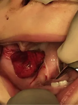
Fig. 1. Intraoperatively herniated buccal pad of fat.
Fig. 2. Excision of the BFP.
Fig. 3. Oral cavity after excision.
G. De Giorgi, et al. Journal of Pediatric Surgery Case Reports 43 (2019) 74–76
Funding
No funding or grant support. Appendix A. Supplementary data
Supplementary data to this article can be found online athttps:// doi.org/10.1016/j.epsc.2019.02.007.
References
[1] Gadhia K, Rehman K, Williams RW, Sharp I. Traumatic pseudolipoma: herniation of buccal fat pad, a report of two cases. Int J Oral Maxillofac Surg 2009;38(6):694–6. [2] Kim SY, Alfafara A, Kim JW, Kim SJ. Traumatic buccal fat pad herniation in young children: a systematic review and case report Elsevier Inc J Oral Maxillofac Surg 2017;75(9):1926–31. Available from:https://doi.org/10.1016/j.joms.2017.05.019. [3] Zhang HM, Yan YP, Qi KM, Wang JQ, Liu ZF. Anatomical structure of the buccal fat
pad and its clinical adaptations. Plast Reconstr Surg 2002;109(7):2509–18. [4] Desai RS, Vanaki SS, Puranik RS, Thanuja R. Traumatic herniation of buccal fat pad
(traumatic pseudolipoma) in a 4-year-old boy: a case report. J Oral Maxillofac Surg 2005;63(7):1033–4.
[5] Gadipelly S, Sudheer MVS, Neshangi S, Harsha G, Reddy V. Traumatic herniation of buccal fat pad in 1 Year old child: case report and review of literature. J Maxillofac Oral Surg 2015;14(S1):435–7. Available from:http://link.springer.com/10.1007/ s12663-014-0664-2.
[6] Ribas MO, Martins WD, de Sousa MH, Braga AM. Traumatic pseudolipoma of the oral cavity: report of a case. J Contemp Dent Pract 2006;7(4):89–97.
[7] Khadilkar AS, Goyal A, Gauba K. The enigma of“traumatic pseudolipoma” and “traumatic herniation of buccal fat pad”: a systematic review and new classification system of post-traumatic craniofacial fatty masses. J Oral Maxillofac Surg 2018;76(6):1267–78.
[8] Singhal M, Sagar S. Post traumatic buccal fat pad injury in a child: a missed entity in ER. Oman Med J 2010;25(3) Available from:http://www.omjournal.org/ CaseReports/FullText/201007/FT_5PostTraumaticBuccalFat.html.
[9] Patil R, Singh S, Subba Reddy VV. Herniation of the buccal fat pad into the oral cavity: a case report. J Indian Soc Pedod Prev Dent 2003;21(4):152–4. Available from:http://www.ncbi.nlm.nih.gov/pubmed/14765616.
G. De Giorgi, et al. Journal of Pediatric Surgery Case Reports 43 (2019) 74–76