1
Università degli Studi di Ferrara
DOCTORATE COURSE IN
BIOMEDICAL SCIENCE
CICLE XXVI
COORDINATOR: Professor Silvano Capitani
Ventilatory pattern efficiency in
different physiologic and pathologic conditions
Disciplinary Scientific Sector MED 10 /
RESPIRATORY DISEASES
PhD candidate Supervisor
MSc Mandolesi Gaia Professor Cogo Annalisa
2
TABLE OF CONTENTS
INTRODUCTION 1. RESPIRATORY MECHANICS - Lung volumes 2. VENTILATION - Alveolar ventilation - Control of breathing- Ventilatory Response to Carbon Dioxide (CO2) - Ventilatory Response to Oxygen (O2)
- Respiratory muscles - Work of breathing
3. VENTILATORY RESPONSE TO EXERCISE
- Adaptation in the pulmonary system during exercise
- Respiratory system causes of exercise performance limitation
4. PHYSIOLOGICAL SETTING: THE IPOXIC ENVIRONMENT AND EXERCISE
- Ventilatory response in hypoxic conditions
- The hemoglobin affinity for oxygen and oxygen dissociation curve - Hematocrit and plasma hemoglobin concentration
- Other Physiological Changes at High Altitude
5. PHATOLOGICAL SETTINGS: ASTHMA AND OBESITY 5.1 ASHTMA - Definition of Asthma - Pathophysiology of Asthma - Pathogenesis of asthma 4 7 8 8 9 10 10 11 11 13 14 14 16 17 18 20 21 22 23 23 23 23 25
3
- Measures of asthma assessment and monitoring - Diagnosis of asthma
5.2 OBESITY
- Definition of obesity
- Effects of obesity on lung functions
5.3 INTERACTION BETWEEN ASTHMA AND OBESITY
- Exercise in asthmatics and obese
6. STUDY 1: VENTILATORY EFFICIENCY IN SKYRUNNERS DURING A RACE SIMULATION AT ALTITUDE
- Background - Introduction
- Materials and Methods - Data and statistical analysis - Results
- Discussion
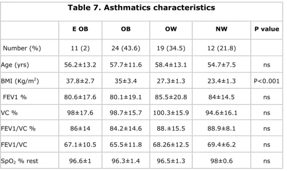
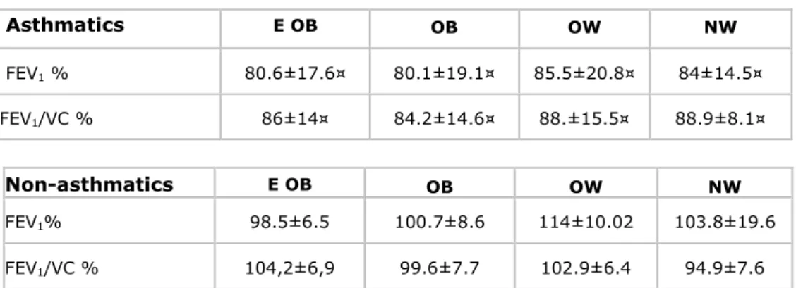
7. STUDY 2: VENTILATORY PATTERN AND EXERCISE CAPACITY IN ASTHMATICS AND NON-ASTHMATICS IN RELATIONSHIP TO BMI
- Background
- Materials and Methods - Data and statistical analysis - Results - Discussion REFERENCES 27 27 28 28 30 35 37 39 39 40 41 43 44 48 51 51 52 56 57 63 66
4
INTRODUCTION
The respiratory system consists primarily of the lungs, whose main function is to ensure gas exchanges with the environment, and the thoracic wall, which moves as a result of continual muscle action sustained by ventilatory drive. Between the components that make possible the breathing, a fundamental role is insured by the thoraco-abdominal area composed by the rib cage and abdomen wall that are separated by the diaphragm. Thus, normal thoraco-abdominal motion consists of expansion and retraction of these compartments during inspiration and expiration, respectively. Although the rib cage and abdomen move in unison, each of the compartments has independence of movement. When the displacement between the compartments is harmonious, the thoraco-abdominal motion is synchronous.
The ventilatory efficiency is defined as the amount of ventilation, composed by the tidal volume (VT) and the breathing frequency (RR), required to achieve a given level of oxygen saturation under spontaneous breathing [1].The efficiency of breathing is given by the thoraco and abdominal contributions to the minute ventilation (VE): a higher coordination between two sections corresponds to a higher efficiency [2]. Another aspect that contributes to the breathing efficiency is the ventilatory pattern represented by the ratio VE/VT. As a fact it has been demonstrated in COPD patients that a deeper and lower ventilation is more efficient in terms of gas exchange, because there is a smaller fraction of anatomic dead space that is rebreathed in each breath[3]. A more efficient respiratory pattern, as the slow yoga breathing, characterized by a lower respiratory rate and a higher tidal volume, looks like to maintain a satisfactory oxygen transport. This has been seen both in healthy subjects during exposure to high altitude, in which a good oxygen saturation has been maintained without the need of marked increases in ventilation and ventilatory drive [1,4] and chronic obstructive pulmonary disease (COPD) patients [5]. Also in subjects suffering of chronic heart failure it has been demonstrated that a slow respiratory rate reduces the dyspnoea improving the resting pulmonary gas exchange and exercise performances [6].
Breathing pattern and thoraco-abdominal motion may be influenced by several factors, such as the individual’s positioning, age, sex, respiratory overload, neuromuscular diseases, lung diseases associated with increased airway resistance and chronic obstructive pulmonary disease [7].
5
Data on breathing pattern and thoraco-abdominal synchrony are important sources of information on ventilatory efficiency and represent an important tool in functional evaluations of athlete performances (i.e endurance-trained runners) and of subjects suffering of respiratory diseases (i.e asthma and COPD), especially in evaluation of exercise training programs.
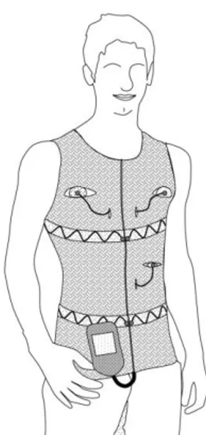
The ventilatory pattern can be analysed by a spirometer with the use of a mouth piece or a mask. But this modality of measure can influence the breathing pattern. To overcome this problem we can use an unobtrusive monitoring technique that evaluates both the breathing pattern and the thoraco-abdominal motion, the inductive pletysmography. The portable respiratory inductive pletysmography measures the displacement of thoraco-abdominal compartments, the changes in time and in pulmonary volumes and accurately estimates the ventilation during exercise. It allows therefore to identify the differences in breathing patterns among healthy subjects and patients with pulmonary or cardiac diseases [8].
To the best of our knowledge, despite the importance of these aspects there are few available studies on breathing pattern and thoraco-abdominal motion among healthy adults [9, 10, 11].
Our group performed a pilot study on elite climbers at high altitude that evidenced a reduction of thoraco-abdominal coordination associated to an oxygen desaturation with increasing of ground slope during exercise. To discriminate if this reduction in breathing coordination was due simply to exercise or also to hypoxic condition we conducted a study at sea level on trained volunteers performing a similar exercise. The results show a decrease of thoraco-abdominal coordination associated to an increase in treadmill’s slope during intense incremental exercise at sea level (max slope reached: 25%), then we hypothesized that an higher slope may have prompted subjects to change their posture, leading to a thoraco-abdominal asynchrony. This was the start point of the design of Study 1 [12].
Especially for what concerns the ventilatory pattern we found that obesity is a para-physiological condition that could modify the breathing efficiency at rest and during exercise. In fact obesity has a profound effect on the physiology of breathing: tidal volumes are often reduced in severe obesity and breathing follow a rapid, shallow pattern both at rest as well as during an exercise. This implies a limited ventilatory efficiency. Having the opportunity to study asthmatics subjects, and being obesity a risk factor of asthma, we decided in
6
Study 2 to investigate if the presence of obesity together with the bronchial obstruction could have had a further influence on the ventilatory pattern and breathing efficiency.
In conclusion the present research aims to:
analyse the breathing coordination and ventilatory parameters in elite athletes (skyrunners) during an heavy-intensity exercise at altitude to verify if a better thoraco-abdominal coordination is associated to a more efficient breathing pattern and endurance performance. (Study 1)
describe the breathing pattern at rest and during exercise in subjects with a different body mass index (BMI), both healthy and asthmatics in order to identify the influence of bronchial obstruction and elevated BMI on ventilation and exercise capacity. (Study 2)
The first study is composed by two parts: a descriptive research on retrospective pulmonary function data of asthmatic patients with the purpose to identify a possible role of obesity on the airflow severity, and a randomized non-blinded parallel case-control designed as an observational research that compares matched asthmatic and healthy control subjects to highlight the differences in breathing efficiency, exercise capacity and functional health-related lifestyle and quality of life parameters.
The second study is an observational study in which evaluations during heavy intensity exercises were performed to analyse the breathing pattern efficiency and thoraco-abdominal synchrony related to exercise performance at altitude of elite athletes.
Ethical approval by the Committee of the Ethics of the University Hospital of Ferrara, and informed consent by patients and subjects evaluated were obtained.
7
1. RESPIRATORY MECHANICS
The chest wall is formed by structures that protect the lungs: the rib cage, sternum, vertebrae, muscles and connective tissues. Chest wall muscles responsible for breathing are the diaphragm that separates the thoracic from the abdominal cavity and the internal and external intercostal, inserted on the coasts. Inspiration and expiration are driven by a pressure gradient that causes the movement of the air from high pressure areas to low pressure areas. Inspiration occurs when the atmospheric pressure is greater than the alveolar pressure, resulting in a pressure gradient that moves the air inside of the alveoli; expiration occurs when the atmospheric pressure is less than the alveolar one [13]. These air flows determined by breathing muscles change the volume of the lungs.
The relationship governing pressure and volume of a gas follows Boyle's law which states that given a certain amount of gas within a container, the pressure is inversely proportional to the volume of the container. So if the lung volume increases, the air pressure decreases [14]. When the lungs are at rest and all respiratory muscles are relaxed, the alveoli contain a volume of air called residual functional capacity (FRC) and the pressure is equal to the atmospheric pressure. At the beginning of inspiration the lungs expand as a result of the inspiratory muscles contraction. This contraction causes an increase in the volume of the alveoli and then a decrease of the pressure inside them: for this reason, thanks to the pressure gradient the air is conveyed in the lungs. During expiration the opposite occurs: the thorax and lungs tend to return to the rest position, for the elastic recoil, whereby the air leaves the lungs when the pressure is greater in the alveoli. Volume changes in the alveoli take place thanks to the change in volume in the chest cavity, by means of respiratory muscles.
The lung’s elastic behavior is the tendency of the lungs to return to its resting volume after distension. One factor responsible of the elastic recoil is the elastic tissue of lungs constituted of fibers of elastin and collagen present in the alveolar walls and around vessels and bronchi. The elastic recoil decreases with age and in respiratory diseases (i.e emphysema), this change is caused by changes in this elastic tissue.
8
Lung volumes
The conducting airways (bronchi up to terminal bronchiole) represent the
anatomic dead space. This leads into the gas-exchanging region of the lung,
which is bounded by the blood-gas interface and the pulmonary capillary blood. With each inspiration, about 500 ml of air enters the lung (tidal volume, VT). The anatomic dead space represents a small proportion of the total lung volume as the volume of capillary blood is very small compared with that of alveolar gas.
The static volumes of the lung can be measured with a spirometer. During quiet respiration the normal breathing can be seen (VT). Next, the subject is asked to take a maximal inspiration followed by a maximal expiration. The total volume is called the vital capacity (VC). However, some gas remains in the lung after a maximal expiration; this is the residual volume (RV). The volume of gas in the lung after a normal expiration is the FRC. When a forced inspiration and expiration is performed (FVC) the forced expiratory volume in the first second (FEV1) can be measured, which represents a dynamic measurement of lung function. The ratio FEV1/VC indicates the pulmonary airflow capacity and the presence of airway obstruction. [15]
As with many anatomic and physiologic measures, lung volumes vary with age, gender, height and body size. Common practice evaluates lung volumes in relation to established standards that consider these factors.
2. VENTILATION
The primary function of the lung is the gas exchange. In order to transfer gases from the lung in an efficient manner, blood and air must be brought into close proximity in the lung. Ventilation is the movement of fresh air from the outside to the alveoli for gas exchange, along with the subsequent movement of alveolar air back to the outside. The total ventilation or minute ventilation (VE = volume exhaled per minute, L/min) can be determined multiplying the volume of gas exhaled during one normal respiratory cycle (tidal volume (VT)) by the breathing frequency:
9
The dynamic VE depends from the maximum air volume (VC) and the speed of the moving a volume of air (RR). In turn, the airflow velocity depends on the airflow pulmonary resistance and by the lung compliance, the stiffness imposed by the mechanical properties of the chest and lung tissue to a change in shape during breathing.
Alveolar ventilation
A gas exchange occurs in the alveolar acinus when fresh air comes in close proximity to capillary blood. However, not all the air inspired reaches the alveoli to participate in gas exchange. The conducting airways (nose, mouth, pharynx, larynx, trachea, bronchi and bronchioles) conduct air from atmosphere to alveoli but they do not participate in gas exchange since they contain no alveoli. At the end of inspiration, the volume of air remaining in the conducting airways is called the anatomic dead space. In addition to the conducting airways any alveoli that are ventilated with air but not perfused with blood do not participate in gas exchange. The volume of ventilation going to these alveoli also acts as dead-space ventilation and is called alveolar dead
space. The sum of anatomic and alveolar dead space makes up the physiologic dead space. The volume of air that does participate in gas exchange because it
is on contact with perfused alveoli is the alveolar ventilation (VA). [16]
The ventilatory efficiency is very important for the gas exchange since the body’s carbon dioxide (CO2) production is eliminated only by ventilation and the level of alveolar O2 reflects a balance of two processes: the oxygen delivery to the alveoli by ventilation and the oxygen removal from the alveoli by capillary blood. A representation of lung volumes and flows is in the Figure 1. [15]
10
Control of breathing
The normal automatic process of breathing originates in impulses that come from the brainstem. The central pattern generator that comprises groups of neurons located in the pons and medulla is responsible for generating the rhythmic pattern of inspiration and expiration and it receives the input from chemoreceptors, lung and other receptors, and the cortex. The cortex can override these centers if voluntary control is desired. These complex nervous mechanism regulates the respiratory rhythm in order to maintain normal levels of the partial pressure of O2 and CO2 in the arterial blood, in spite of widely differing demands for O2 uptake and CO2 output made by the body in different conditions. The nervous stimulation of respiratory muscles occurs through the release of acetylcholine from motoneurons in the neuromuscular junction. [16]
Unlike all other processes essential for life, breathing is a semi-automatic and semi-voluntary activity: normally occurs without conscious participation and autonomously, but we can step in with a conscious action to change its rhythm and depth. It, through the forced ventilation, can be enhanced further by recruiting other muscles voluntarily. At this stage of hyperventilation is highlighted even more the work of the internal and external intercostal muscles, aided by other muscle groups. During this work the shoulder blades are stabilized by trapezius and elevator of scapula and rhomboid muscles; the small pectoral and serratus anterior raise the coasts; upper limbs can be stabilized through the use of the pectoralis major.
Ventilatory Response to Carbon Dioxide (CO
2)
The most important factor in the control of ventilation under normal conditions is the partial pressure of CO2 of the arterial blood (PaCO2). Arterial PCO2 is the most important stimulus to ventilation under most conditions and is normally tightly controlled: most of the stimulus comes from the central chemoreceptors, but the peripheral chemoreceptors also contribute and the response is magnified if the arterial PO2 is lowered. The ventilatory response to CO2 is reduced by sleep, increasing age, and genetic. Trained athletes tend to have a low CO2 sensitivity. The ventilatory response to CO2 is also reduced if the work of breathing is increased. [15]
11
The reduced ventilatory response to CO2 and the consequent CO2 retention in some patients with lung disease can also be partly explained by an increasing in work of breathing. The main stimulus to increase ventilation when the arterial PCO2 rises comes from the central chemoreceptors, which respond to the increased H+ concentration of the brain extracellular fluid near the receptors. An additional stimulus comes from the peripheral chemoreceptors, because of both the rise in arterial PCO2 and the fall in pH.
Ventilatory Response to Oxygen (O
2)
Raising the PaCO2 increases the ventilation at any PaO2. When the PaCO2 is increased, a reduction in PaO2 below 100 mmHg causes some stimulation of ventilation, unlike the situation in which the PaCO2 is normal. Thus, the combined effects of both stimuli exceed the sum of each stimulus given separately; this is referred to as interaction between the high CO2 and low O2 stimuli. Large differences in response occur between individual subjects. Because in healthy subjects at sea level the PaO2 can normally be reduced so far without evoking a ventilatory response, the role of this hypoxic stimulus in the day-to-day control of ventilation is small. [15]
However, in the presence of low PaO2 (i.e. on ascent to high altitude or as a consequence of respiratory and cardiac diseases), a large increase in ventilation occurs in response to the hypoxia. Hypoxemia reflex stimulates ventilation by its action on the carotid and aortic body chemoreceptors. It has no action on the central chemoreceptors; however, prolonged hypoxemia can cause mild cerebral acidosis, which, in turn, can stimulate ventilation.
Respiratory muscles
The respiratory muscles are morphologically and functionally skeletal muscle, and their primary task is to displace the chest wall rhythmically to pump gas in and out of the lung. The actions of diaphragm, the muscles of rib cage and abdominal muscles normally are simultaneous and coordinated. Rib cage and abdomen, the two compartments that compose the chest wall are mechanically arranged in parallel and are separated from each other by a thin musculotendinous structure, the diaphragm. [16]
The diaphragm is the most important muscle of inspiration. It is supplied by the phrenic nerves from cervical segments. During inspiration the diaphragm
12
contracts and its dome descends. This motion expands the thoracic cavity, causing a fall in the pleural pressure and an increase in the lung volumes, and displaces the abdominal visceral mass downward and forward increasing the abdominal pressure. The action of diaphragm increases the vertical dimension of thoracic cavity. In addition, the rib margins are lifted and moved out, causing an increase in the transverse diameter of the thorax. The contraction of external intercostal muscles increases in both the lateral and the anteroposterior diameters of the thorax by a lifting and rotating effects on the ribs. The intercostal muscles are supplied by intercostal nerves that come off the spinal cord at the same level. The accessory muscles of inspiration include the scalene muscles, which elevate the first two ribs, and the sternomastoids, which raise the sternum. There is little, if any, activity in these muscles during quiet breathing, but during exercise, they may contract vigorously. When the lungs expands the alveolar pressure decrease below the atmospheric pressure level and the air flows in the alveoli until the two pressures reaches the same level.
The expiration is a passive process during quiet breathing. The lung and chest wall are elastic and tend to return to their equilibrium positions after being actively expanded during inspiration. During exercise and voluntary hyperventilation, expiration becomes active. The most important muscles of expiration are those of the abdominal wall, including the rectus abdominis,
internal and external oblique muscles, and transversus abdominis. When these
muscles contract, intra-abdominal pressure raises, and the diaphragm is pushed upward. The internal intercostal muscles assist active expiration by pulling the ribs downward and inward (opposite to the action of the external intercostal muscles), thus decreasing the thoracic volume. This causes an increase of alveolar pressure that becomes higher than atmospheric pressure and assure the air movement out of the lungs. Thus, moving the chest wall during breathing is a complex, integrated process that involves many muscles. The control mechanism that promote coordinated use of these different muscles are critically important to maintaining both the work of breathing and the VA within acceptable limits. The regulation of gas exchange is made possible by the carefully control of the level of ventilation.
13
Work of breathing
Work is required to move the lung and chest wall. During inspiration, the work done on the lung is given by the work required to overcome the elastic forces and the work overcoming viscous (airway and tissue) resistance.
The higher the airway resistance or the inspiratory flow rate, the more negative would be the intrapleural pressure excursion. On expiration, the work is required to overcome airway and tissue resistance and this work can be accomplished by the energy stored in the expanded elastic structures and released during a passive expiration. There is also a little work dissipated as heat.
The total work done moving the lung and chest wall (Total work of breathing) can be calculated by measuring the O2 cost of breathing and assuming a figure for the efficiency as given by:
Efficiency % = Useful work_____________ × 100 Total energy expended (or O2 cost)
The efficiency is believed to be about 5% to 10%.
The O2 cost of quiet breathing is extremely small, being less than 5% of the total resting O2 consumption. With voluntary hyperventilation, it is possible to increase this to 30%. In patients with obstructive lung disease, the O2 cost of breathing may limit their exercise ability. [15]
Concerning the breathing pattern, the higher the respiratory rate, the faster the flow rates and the larger is the viscous work. On the other hand, the larger the tidal volume, the larger is the elastic work. When pulmonary compliance is reduced (stiff lungs), breaths tend to be small rapid, whereas in presence of severe airway obstruction sometimes breath is slow and these patterns tend to reduce the work done on the lungs.
14
3. VENTILATORY RESPONSE TO EXERCISE
Adaptations in the pulmonary system during exercise
The lung has a primary role in the ability to perform exercise.
The respiratory system represents, together with musculoskeletal and cardiovascular systems, the integrated structure that enables the execution of the exercise. The role of the respiratory system is basically to provide supplies and diffusion of O2 and CO2 elimination. [9]
During exercise, the increased demands of O2 by the muscle and the need to eliminate the CO2 produced increase the ventilation. In addition, during heavy exercise, higher levels of lactic acid are produced by anaerobic glycolysis, and additional CO2 is therefore eliminated from bicarbonate. The increased CO2 elimination because the increased H+ concentration stimulates the peripheral chemoreceptors, thus further increasing ventilation.
The gas exchange demands of the lung are then enormously increased by exercise. Typically, the resting oxygen consumptions of 300 ml/min can rise to about 3000 ml/min in a moderately fit subject (and as high as 6000 ml/min in a top level athlete). Similarly, the resting CO2 output of 240 ml/min increases to about 3000 ml/min.
Essential to meeting these tasks is an increase in breathing, specifically an increase in VA. The increase in pulmonary ventilation required to meet these needs must be achieved efficiently with minimal oxygen consumption (VO2) by the respiratory muscles. As the work rate (or power) is increased, ventilation increases linearly initially, but at high VO2 values, it increases more rapidly because lactic acid is liberated, and this increases the ventilatory stimulus. Unfit subjects produce lactate at relatively low work levels, and they have an earlier ventilation increase, whereas well trained subjects can reach fairly high work levels before substantial anaerobic glycolysis occurs.
During a maximal exercise, the increase of metabolic requests, in particular the oxygen request, from the muscles involved in the effort can be considerable: in untrained healthy subjects oxygen consumption can increase about 10 times compared to the measured value in rest condition, and in trained individuals and athletes can increase by more than 20 times. The increased oxygen demand by the skeletal muscles leads to a progressive
15
increase in ventilation. This increase in healthy subjects is initially obtained by an increase in the depth of breathing (VT), through the use of both a portion of the expiratory reserve volume and inspiratory reserve volume. Afterwards, it will occur even an increase in RR through the respiratory muscle capacity (inspiratory and expiratory) to increase its activity. [17] Ventilation cannot increase indefinitely. The maximum ventilation that a subject can produce is described as maximum voluntary ventilation (MVV). Its measurement is carried out asking to the subject to ventilate at maximum speed and depth for 12 seconds. An healthy subject during quiet breathing (at VT level at rest), has a wide ventilatory reserve, defined as the difference between the maximum predicted ventilation and the maximum ventilation reached during a maximal exercise. During an exercise the VE never reaches the ventilatory capacity in healthy subjects; in fact generally the exercise is stopped for muscular exhaustion or for the achievement of maximum heart rate. At the end of the exercise we can then highlight the ventilatory reserve. The possible ventilatory limit to effort is generally determined by the ventilatory reserve: it states that when the ventilatory reserve is at least 15 l/min, the ventilation system is not the limiting element to exercise. The maximum theoretical ventilation can be obtained directly through the manoeuvre of MVV or indirectly by multiplying the FEV1 by 0.35 to 0.40. During an exercise the ventilatory pattern analysis, the analysis of the prevalence of RR and/or VT and of the movements of the chest and abdominal compartment and their coordination are very important. Greater respiratory coordination is in fact associated to a greater ventilatory efficiency. [9, 18, 19, 10]
Moreover, the oxygen dissociation curve of hemoglobin moves to the right in exercising muscles because of the increase in PCO2, H+ concentration, and temperature. This assists the unloading of oxygen to the muscles. When the blood returns to the lung, the temperature of the blood falls a little and the curve shifts leftward somewhat. The arterial PO2, PCO2, and pH are poorly affected by moderate exercise whereas at very high work levels, PCO2 often falls, while PO2 rises, and pH falls because of lactic acidosis.
Many other functions of the respiratory system change in response to exercise. The diffusing capacity of the lung increases because of increases in both the diffusing capacity of the membrane and the volume of blood in the pulmonary capillaries. These changes are brought about by recruitment and distension of pulmonary capillaries, particularly in the upper parts of the lung. Nevertheless,
16
some elite athletes at extremely high work levels show a fall in arterial PO2 caused by diffusion limitation because of the reduced time available for the loading of oxygen in the pulmonary capillary. The cardiac output increases approximately linearly with the work level, this implies an increase in pulmonary arterial and venous pressures, which account for the recruitment and distension of pulmonary capillaries and a pulmonary vascular resistance falls. However there is a much larger increase in VE than blood flow and the change in cardiac output is only about a quarter of the increase in VE.
Respiratory system causes of exercise performance limitation
Three are the mechanism primarily under the control of respiratory system that contribute to exercise limitation: the respiratory muscle work and fatigue, the arterial oxygen desaturation and cyclical fluctuations in intrathoracic pressure. [20, 21]
Heavy intensity sustained exercise can cause a time dependent arterial oxygen desaturation in arterial blood which can be evident in some elite athletes (see above). This is also due to a rightward shift in the oxygen dissociation curve because of a progressive metabolic acidosis and a rise in blood temperature which occurs to varying extents in all subjects. This implies an increase in peripheral fatigue and in the rate of perception of limb discomfort with a decrease in exercise time to exhaustion.
At the same time, the hyperventilation intensity dependent, that occurs during sustained high intensity exercise, requires the progressive recruitment of inspiratory and expiratory muscles. If the exercise intensity exceeds 80% of the maximum (and is sustained for a long time), a significant diaphragm and expiratory muscle fatigue can occur.
This exercise induced respiratory muscle fatigue does not limit the hyperventilatory response throughout exercise. However, according to Dempsey [22] it might trigger a metaboreflex from the fatiguing diaphragm which in turn causes sympathoexcitation and vasoconstriction of the vasculature of the exercise limb, resulting in a reduced limb flow. In Figure 2 is shown as during exercise at intensities greater than 80% of maximal oxygen consumption (VO2max) the diaphragm shows a significant fatigue, in part due to the high levels of diaphragmatic work and in part because it must compete with the locomotor muscles for the available blood flow during heavy intensity
17
exercise. This promotes an inadequate oxygen transport and an higher fatigue of the diaphragm. The consequences of a fatiguing diaphragm and other respiratory accessory muscles (inspiratory and expiratory), is that the fatiguing contractions and accumulation of metabolites in the inspiratory and expiratory muscles activates type IV phrenic afferents, which in turn increases sympathetic vasoconstrictor activity via a supra-spinal reflex. The consequence is a gradual increase in limb muscle sympathetic nerve activity and a reduction in vascular conductance and blood flow in the resting limb.
Figure 2
A decreased respiratory muscle work affects the performance through a relief of dyspnea perceptions and a reduction of peripheral locomotor muscle fatigue via augmented limb blood flow and O2 transport. [23]
4.
PHYSIOLOGICAL SETTING : THE HYPOXIC ENVIRONMENT
Hypoxia, is a pathological condition in which the body as a whole (generalized hypoxia) or a region of the body (tissue hypoxia) is deprived of adequate oxygen supply.
At high altitude, differences in barometric pressure result in insufficient oxygen in the air, thereby causing hypoxia. People at high-altitude for short periods of time, who are not adapted to that environment, are at increased risk for acute altitude sickness involving pulmonary and cerebral edema. Newcomers to high altitude frequently complain of headache, fatigue, dizziness, palpitations,
18
insomnia, loss of appetite, and nausea. This is known as acute mountain sickness (AMS) and is attributable to the hypoxemia and alkalosis.
The more important responses of the organism to hypoxia are those implemented to prevent tissue hypoxia. These include changes in ventilation, heart rate, pH and hematocrit and hemoglobin concentration. All the beneficial changes implemented in response to hypoxia at high altitude take the name of acclimatization.
Variations in arterial oxygen concentrations can be also part of the normal physiology, for example, during strenuous physical exercise. A mismatch between oxygen supply and its demand at the cellular level may result in a hypoxic condition.
Mountain medicine distinguishes five height zones based on various limits on the acclimatization and exercise. [24]
sea level (0-500 m): no effect on fit
low altitude (500-2000 m): no effect of altitude on fit, but possible
reductions in athletic performance completely reversible with acclimatization.
moderate altitude (2000-3000 m): possible symptoms of altitude sickness,
after a period of acclimatization the exercise capacity turns back to previous levels of exposure to altitude.
high altitude (3000-5500 m): altitude sickness is common; rare
manifestations of pulmonary and cerebral edema. Despite a good acclimatization is achieved, athletic performances are reduced.
extreme altitude (over 5500 m): altitude-related disorders are numerous;
the total acclimatization above 5400 m is considered impossible.
Ventilatory response in hypoxic conditions
Altitude’s physiologic challenge comes from the decreased PO2 and air density progressively with the ascent above sea level. The PO2 falls as the gas moves from the atmosphere in which we live to the mitochondria where it is utilized. The PO2 of air is 20.93% of the total dry gas pressure. At sea level, the barometric pressure is 760 mmHg, and at the body temperature of 37°C, the water vapor pressure of most inspired gas (which is fully saturated with water vapor) is 47 mmHg. Thus, the PO2 of inspired air at sea level is (760 − 47) x 20.93, or 150 mmHg. The barometric pressure decreases with distance above
19
the earth’s surface in an approximately exponential manner. At the summit of Mount Everest (8848 m), the inspired PO2 is only 43 mm Hg. A remarkable degree of acclimatization occurs when humans ascend to these altitudes; indeed, climbers have lived for several days at altitudes that would cause unconsciousness within a few seconds in the absence of acclimatization.
At altitude we can reach very high ventilations (up to 200 L/min), especially during exercise, due to the lower density of the air and to the increase of the maximum breathing capacity.
The maximum O2 uptake (VO2max) declines starting at about 1200 m; at 1800 m it is about of 10% and it progressively loss the 10% each 1000 m of altitude. It can change depending on individual acclimatization level, training and ethnicity. The hypoxic ventilatory response (HVR) is defined as the increase of ventilation caused by hypoxic stimulus. As it is well known, the main stimulus to ventilation is an increase in PaCO2 detected at the level of the aortic and carotid bulbs. These stimulate breathing so that the PaCO2 is maintained within very narrow limits (variations of 2-3%); outside these values the system gain is very high and breathing is strongly inhibited or stimulated. The hypoxic stimulus causes an increase in ventilation only when the PaO2 drops below 60 mmHg, a variation of as much as 40% compared to normal value 100 mmHg. This apparently too wide margin is actually appropriate, since it operates around the values of PaO2 whereby hemoglobin dissociation curve becomes steeper.
The reduction in PaCO2 caused by increased ventilation, following the hypoxic stimulus, constitutes an inhibitory stimulus on the breath, partially masking the ventilatory response to hypoxia either directly or because of the increase in blood pH due to depletion of CO2. Ventilatory response to hypoxia is not quantitatively equal in all subjects. The individual variations are important, especially in the early days of exposure to high altitude. In particular categories, such as athletes trained at sea level and elderly, there is a minor HVR. The subjects exposed to intermittent hypoxia show a sort of basic acclimation to hypoxia with an increase of HVR. In high mountain populations the values of PaO2 to which breathing is stimulated can be extremely low, probably due to some degree of tissue tolerance to hypoxia; the fact that babies born at altitude show a HVR equal to that of the habitants to sea level suggests that the phenomenon is mainly due to an individual acclimatization, and only secondarily to genetic factors. Even in individuals born at low
20
altitudes not belonged to high mountain populations, in fact, it can be seen a decrease of HVR after decades of residence, also not catching up the local values, whose physique has some adaptations not yet clarified. The return at low altitude of subjects acclimated shows a rapid increase in PaCO2: ventilation decreases to values between the values at high altitudes and those in normoxia conditions. The fact that ventilation remains high is partly a consequence of the lowering of the set-point of peripheral chemoreceptors of CO2, which persists for a few days after the removal of the hypoxic stimulus.
Exercise in hypoxic environment intensifies the hyperventilatory response and the work of breathing and diaphragm fatigue accordingly. Since it is recognized that the respiratory muscle strength is reduced during the first days of exposition at altitudes above 4000 m, we cannot exclude a role of the respiratory muscles in exercise limitation. [25]
The hemoglobin affinity for oxygen and oxygen dissociation
curve
Under hypoxic conditions despite the impediment to the release of O2 to tissue level, an increase in the affinity of hemoglobin for oxygen has beneficial effects, especially during exercises. In conditions of limited diffusion a greater oxygen affinity of hemoglobin increases O2 extraction by alveolar air more than it prevents the release, increasing the tissue oxygenation.
O2 in fact forms an easily reversible combination with hemoglobin (Hb) to give oxyhemoglobin: O2 + Hb = Hb O2. The maximum amount of O2 that can be combined with Hb is called the O2 capacity. This is when all the available binding sites are occupied by O2. The O2 saturation of Hb is the percentage of the available binding sites that have O2 attached and is given by:
O2 combined with Hb × 100 O2 capacity
At sea level in normal conditions, the O2 saturation of arterial blood (SpO2) with PO2 of 100 mm Hg is about 97.5%, whereas that of mixed venous blood with a PO2 of 40 mm Hg is about 75%. The curved shape of the O2 dissociation curve has several physiological advantages (Figure 3). [15]
21
The flat upper portion means that even if the PO2 in alveolar gas falls somewhat, loading of O2 will be little affected. The steep lower part of the dissociation curve means that the peripheral tissues can withdraw large amounts of O2 for only a small drop in capillary PO2.
Figure 3
During exercise the O2 dissociation curve is shifted to the right, that is, the O2 affinity of Hb is reduced, due to an increase in H+ concentration, PCO2 (Bohr effect) and temperature. A rightward shift means more unloading of O2 at a given PO2 in a tissue capillary. An exercising muscle is acid, hypercarbic, and hot, and it benefits from increased unloading of O2 from its capillaries.
In hypoxic conditions, at moderate altitudes (2000-3000 m), there is a rightward shift of the O2 dissociation curve that results in a better unloading of O2 in venous blood at a given PO2. The cause of the shift is an increase in concentration of 2,3-diphosphoglycerate (DPG), an end product of red cell metabolism that occurs in chronic hypoxia, which develops primarily because of the respiratory alkalosis. At higher altitudes, there is a leftward shift in the dissociation curve caused by the respiratory alkalosis, and this assists in the loading of O2 in the pulmonary capillaries. The number of capillaries per unit volume in peripheral tissues increases, and changes occur in the oxidative enzymes inside the cells.
Hematocrit and plasma hemoglobin concentration
The amount of oxygen carried depends for the most part by the saturation of hemoglobin and its concentration; to the portion carried by red blood cells
22
must be added the proportion of oxygen dissolved in plasma. Normally, the latter contributes to arterial O2 content only for 1.5%. The hypoxic stimulus induces the synthesis of erythropoietin through the activation of the hypoxia inducible factor (HIF-1 gene), and possibly through other mechanisms. In subjects fully acclimatized to altitudes above 5000 m oxygen content is the same as at sea level (≈200 ml/L) due to the increase in the concentration of hemoglobin. In the first weeks of exposure to high altitude the hemoglobin concentration grows rapidly because of changes in plasma volume.
The plasma volume decreases significantly secondarily to an increase in diuresis, remaining below the normal values even during definitively acclimatization, especially in subjects that do not exhibit symptoms of acute mountain sickness. The effects of reduced plasma volume on total blood volume are partially offset by an increase of the mass of erythrocytes.
Other Physiological Changes at High Altitude
Pulmonary vasoconstriction occurs in response to alveolar hypoxia. This increases the pulmonary arterial pressure and the work done by the right heart. The pulmonary hypertension is further increased by the polycythemia, which raises the viscosity of the blood. There is no physiological advantage in this response, except that the topographical distribution of blood flow becomes more uniform with an improvement of VE/Qc ratio. The pulmonary hypertension is sometimes associated with pulmonary edema, although the pulmonary venous pressure is normal. The probable mechanism is that the arteriolar vasoconstriction is uneven, and leakage occurs in unprotected, damaged capillaries. The edema fluid has a high protein concentration, indicating that the permeability of the capillaries is increased.
23
5. PHATOLOGICAL SETTING: ASTHMA AND OBESITY
5.1 ASTHMA
Definition of Asthma
Asthma is a common complex chronic disorder of the airways characterized by variable and recurring symptoms, airflow obstruction, bronchial hyperresponsiveness, and an underlying inflammation. The interaction of these features of asthma determines the clinical manifestations and severity of asthma and the response to treatment. Asthma is defined by its clinical, physiological, and pathological characteristics. The predominant feature of the clinical history is episodic shortness of breath often accompanied by non- productive cough. Wheezing appreciated on auscultation of the chest is the most common physical finding.
The dominant pathological feature is airway inflammation, sometimes associated with airway structural changes. Asthma has significant genetic and environmental components, but since its pathogenesis is not clear, much of its definition is descriptive.
Based on the functional consequences of airway inflammation, an operational description of asthma is: “Asthma is a chronic inflammatory disorder of the
airways in which many cells and cellular elements play a role. The chronic inflammation is associated with airway hyperresponsiveness that leads to recurrent episodes of wheezing, breathlessness, chest tightness, and coughing, particularly at night or in the early morning. These episodes are usually associated with widespread, but variable, airflow obstruction within the lung that is often reversible either spontaneously or with treatment.”
There is now good evidence that the clinical manifestations of asthma symptoms, sleep disturbances, limitations of daily activity, impairment of lung function, and use of rescue medications, can be controlled with appropriate treatment. When asthma is controlled, there should be no more than occasional recurrence of symptoms and severe exacerbations should be rare. [26, 27]
Pathophysiology of Asthma
Airflow limitation in asthma is recurrent and caused by a variety of changes in the airway. These include:
24
Bronchoconstriction
In asthma, the dominant physiological event leading to clinical symptoms is airway narrowing and a subsequent interference with airflow. In acute exacerbations of asthma, bronchial smooth muscle contraction (bronchoconstriction) occurs quickly to narrow the airways in response to exposure to a variety of stimuli including allergens or irritants. In addition, other stimuli (including exercise, cold air, and irritants) can cause acute airflow obstruction. The mechanisms regulating the airway response to these factors are less well defined, but the intensity of the response appears related to the level of underlying airway inflammation.
Airway edema
As the disease becomes more persistent and inflammation more progressive, other factors further limit airflow. These include edema, inflammation, mucus hyper secretion with the formation of inspissated mucus plugs, as well as structural changes including hypertrophy and hyperplasia of the airway smooth muscle.
Airway hyperresponsiveness
Airway hyperresponsiveness is an exaggerated bronchoconstrictor response to a wide variety of stimuli, and it is a major, but not necessarily unique, feature of asthma. The degree to which airway hyperresponsiveness can be defined by contractile responses to challenges with methacholine correlates with the clinical severity of asthma. The mechanisms influencing airway hyperresponsiveness are multiple and include inflammation, dysfunctional neuroregulation, and structural changes. Inflammation appears to be a major factor in determining the degree of airway hyperresponsiveness and it has a central role in the pathophysiology of asthma. Airway inflammation involves an interaction of many cell types and multiple mediators with the airways that eventually results in the characteristic pathophysiological features of the disease: bronchial inflammation and airflow limitation that result in recurrent episodes of cough, wheeze, and shortness of breath. The processes by which these events occur and lead to clinical asthma are still under investigation. Treatment directed toward reducing inflammation can reduce airway hyperresponsiveness and improve asthma control.
Airway remodeling
In some persons whit asthma, airflow limitation may be only partially reversible. Permanent structural changes can occur in the airway and these
25
are associated with a progressive loss of lung function that is not prevented by or fully reversible by current therapy. Airway remodeling involves an activation of many of the structural cells, with consequent permanent changes in the airway that increase airflow obstruction and airway responsiveness and render the patient less responsive to therapy. These structural changes can include thickening of the sub-basement membrane, subepithelial fibrosis, airway smooth muscle hypertrophy and hyperplasia, blood vessel proliferation and dilation, and mucous gland hyperplasia and hypersecretion.
Pathogenesis of asthma
Factors that influence the risk of asthma can be divided into those causing the development of asthma and those triggering asthma symptoms; some have both effects. The former include host factors (which are primarily genetic) while the latter are usually environmental factors. However, the mechanisms whereby they influence the development and expression of asthma are complex and interactive. In addition, developmental aspects, such as the maturation of the immune response and the timing of infectious exposures during the first years of life, are emerging as important factors modifying the risk of asthma in the genetically susceptible person.
Host Factors
Genetic: It is well recognized that asthma has an inheritable component to its expression, but the genetics involved in the eventual development of asthma remain a complex and incomplete picture.
Obesity: Asthma is more frequently observed in obese subjects (Body Mass Index > 30 kg/m2) and is more difficult to control. Obese people with asthma have lower lung function and more co-morbidities compared with normal weight people with asthma131. The use of systemic glucocorticosteroids and a sedentary lifestyle may promote obesity in severe asthma patients, but in most instances, obesity precedes the development of asthma. How obesity promotes the development of asthma is still uncertain but it may result from the combined effects of various factors. It has been proposed that obesity could influence airway function due to its effect on lung mechanics, development of a pro-inflammatory state, in addition to genetic, developmental, hormonal or neurogenic influences. In this regard, obese
26
patients have a reduced expiratory reserve volume, a pattern of breathing that may possibly alter airway smooth muscle plasticity and airway function. In fact, to overcome the reduced total respiratory compliance and respiratory muscle inefficiency associated to obesity, obese subjects may breathe rapidly and shallowly. This pattern of breathing is similar to that seen among patients with neuromuscular and musculoskeletal disorders. This pattern of breathing is also associated with increased oxygen cost of breathing (Sood 2009)
Furthermore, the release by adipocytes of various pro-inflammatory cytokines and mediators (i.e interleukin-6, tumor necrosis factor (TNF)-α, leptin) combined with a lower level of anti-inflammatory adipokines in obese subjects can favor a systemic inflammatory state although it is unknown how this could influence airway function.
Sex: In early life, the prevalence of asthma is higher in boys. At puberty,
however, the sex ratio shifts, and asthma appears predominantly in women.
Environmental Factors
Allergens: Sensitization and exposure to house-dust mite and Alternaria are
important factors in the development of asthma in children. Early studies showed that animal dander, particularly dog and cat, were associated with the development of asthma. Exposure to cockroach allergen, for example, a major allergen in inner-city dwellings, is an important cause of allergen sensitization and is a risk factor for the development of asthma. In addition, allergen exposure can promote the persistence of airway inflammation and likelihood of an asthma exacerbation.
Infections. During infancy, a number of respiratory viruses have been associated with the inception or development of the asthma. In early life, respiratory virus and parainfluenza virus in particular, cause bronchiolitis that parallels many features of childhood asthma. Symptomatic rhinovirus infections in early life also are emerging as risk factors for recurrent wheezing.
Other environmental exposures. Tobacco smoke, air pollution, occupations,
and diet have also been associated with an increased risk for the onset of asthma, although the association has not been as clearly established as with allergens and respiratory infections.
27
Measures of asthma assessment and monitoring
Diagnosing a patient as having asthma is the first step in reducing the symptoms, functional limitations, impairment in quality of life, and risk of adverse events that are associated with the disease. The ultimate goal of treatment is to enable a patient to live with none of these manifestations of asthma, and an initial assessment of the severity of the disease allows an estimate of the type and intensity of treatment needed. An important point linking asthma severity, control, and responsiveness is that the goals are identical for all levels of baseline asthma severity: in well-controlled asthma, the manifestations of asthma are minimized by therapeutic intervention.
Diagnosis of asthma
To establish a diagnosis of asthma, the clinician should determine that:
- Episodic symptoms of airflow obstruction or airway hyperresponsiveness are present.
- Airflow obstruction is at least partially reversible. - Alternative diagnoses are excluded.
Recommended methods to establish the diagnosis are: - Detailed medical history.
- Physical exam focusing on the upper respiratory tract, chest, and skin.
- Spirometry to demonstrate obstruction and assess reversibility. Reversibility is determined by an increase in FEV1 of both ≥12 percent from baseline and ≥ 200mL after inhalation of a short-acting bronchodilator.
Pulmonary function testing (spirometry)
The gold standard for the diagnosis of asthma is spirometry ,particularly the demonstration of reversibility of bronchial obstruction.
Usually a forced expiration curve is performed with the measurement of forced expiratory volume in the first second (FEV1) and forced vital capacity (FVC), FEV1, FVC, FEV1/FVC measurements are recommended before and after the patient inhales a short-acting bronchodilator. These measurements help to determine whether there is airflow obstruction, its severity, and whether it is reversible over the short term. Patients’ perception of airflow obstruction is
28
highly variable, and spirometry sometimes reveals obstruction much more severe than would have been estimated from the history and physical examination. Spirometry typically measures the maximal volume of air exhaled from the point of maximal inhalation (FVC) and the volume of air exhaled during the first second of this maneuver (FEV1).
Airflow obstruction is indicated by a reduction in the values for both the FEV1 and the FEV1/FVC relative to reference or predicted values. Predicted values of FEV1, FVC, and PEF based on age, sex, and height have been obtained from population studies. They are useful for judging whether a given value is abnormal or not.
Significant reversibility is indicated by ATS standards as an increase in FEV1 of >200 mL and ≥12 percent from the baseline measure after inhalation of a short-acting bronchodilator (e.g., albuterol, 2–4 puffs of 90 mcg/puff).
A reduced ratio of FEV1/FVC (<88% in M and 89% in F or below the 5th percentile) indicates the presence of obstruction to the flow of air from the lungs, whereas a proportionately reduced FVC with a normal or increased FEV1/FVC ratio suggests a restrictive pattern. The severity of abnormality of spirometric measurements is evaluated by comparison of the patient’s results with reference values based on age, height, sex, and race.
Spirometry should be performed using equipment and techniques that meet standards developed by the American Thoracic Society (ATS) [28]. Correct technique, calibration methods, and maintenance of equipment are necessary to achieve consistently accurate test results. Maximal effort by the patient in performing the test is required to avoid important errors in diagnosis and management.
5.2 OBESITY
Definition of obesity
Obesity is a complex multifactorial chronic disease that develops from an interaction of genotype and the environment. The development of obesity involves the integration of social, behavioral, cultural, physiological, metabolic and genetic factors. Obesity is generally acknowledged as a global phenomenon that increases morbidity and reduces life expectancy.
29
This disease is a major risk factor for many acute and chronic disorders, including cardiovascular and cerebrovascular disease, and diabetes. Higher body weights are also associated with increases in all-cause mortality. Most observational studies have shown a U- or J-shaped relationship between BMI and mortality, with individuals at very low and very high weights at increased risk, after adjusting for confounding factors such as smoking or preexisting illness. The number of overweight and obese men and women has risen since 1960; in the last decade the percentage of people in these categories has increased to 54.9 % of adults age 20 years or older.
Obesity classification
The primary classification of obesity is based on the measurement of BMI. This classification is designed to relate BMI to risk of disease. The BMI, which describes relative weight for height, is significantly correlated with total body fat content. The relation between BMI and disease risk varies among individuals and among different populations. In addition, susceptibility to risk factors at a given weight varies among individuals. Some individuals may have multiple risk factors with mild obesity, whereas others may have fewer risk factors with more severe obesity. BMI is a practical indicator of the severity of obesity, and it can be calculated from existing tables (Figure 4). [29]
BMI is a direct calculation based on height and weight, regardless of gender. The relationship between BMI and body fat content varies somewhat with age, sex, and possibly ethnicity because of differences in factors such as composition of lean tissue, sitting height, and hydration state. The limitations of BMI as a measure of total body fat, nonetheless, must be recognized.
The BMI is calculated as follows:
30 Figure 4
The patient’s risk status is assessed by determining the degree of overweight or obesity based on BMI, the presence of abdominal obesity based on waist circumference, and the presence of concomitant cardiovascular diseases risk factors or comorbidities. Some obesity-associated diseases and risk factors place patients in a very high risk category for subsequent mortality. Obesity also has an aggravating influence on several cardiovascular risk factors.
Effects of obesity on lung functions
Obesity has a profound effect on the physiology of breathing. [30, 31, 32] Obese people are at increased risk of respiratory symptoms, such as breathlessness, particularly during exercise, even if they have no obvious respiratory illness. Obesity has a clear potential to have a direct effect on respiratory well-being, since it increases oxygen consumption (VO2) and carbon dioxide production (VCO2), while at the same time it stiffens the respiratory system and increases the mechanical work of breathing.
The effects of obesity and being overweight on the control of breathing, pulmonary mechanics and their consequences on lung function tests and symptoms are discussed hereafter.
LUNG VOLUMES
The most consistently reported effect of obesity on lung function is a reduction in the functional residual capacity (FRC). This effect reflects a shift in the balance of inflationary and deflationary pressures on the lung due to the mass load of adipose tissue around the rib cage and abdomen and in the visceral cavity. There is an exponential relationship between BMI and FRC with a reduction in FRC detectable even in overweight individuals. However, the effects of obesity on total lung capacity (TLC) and RV, are modest: TLC is usually maintained above the lower limit of normal, and the RV is usually well preserved. The reduction in FRC is manifested by an increase in inspiratory capacity (IC) and a very marked decrease in the expiratory reserve volume (ERV) [33] (Figure 5). The reasons for the reduction in TLC are not known, but it is probably due to a mechanical effect of the adipose tissue.
31 Figure 5
FAT DISTRIBUTION AND BODY COMPOSITION
Abdominal and thoracic fat are likely to have direct effects on the downward movement of the diaphragm and on chest wall properties, while fat on the hips and thighs would be unlikely to have any direct mechanical effect on the lungs. Both abdominal fat and thoracic or upper body fat are associated with reductions in lung volumes.
RESPIRATORY SYSTEM MECHANICS
An important respiratory abnormality in obesity is a decrease in total respiratory system compliance. Obesity is indeed characterized by a stiffening of the total respiratory system, which is presumed to be due to a combination of effects on lung and chest wall compliance.
The reduction in lung compliance in obese individuals, exponentially related to BMI, is likely to be the result of decreased lung volumes while the decrease in chest-wall compliance is associated with the obese individual’s accumulation of fat in and around the ribs, the diaphragm and the abdomen. Both respiratory resistance and airway resistance rose significantly with the level of obesity, which appeared to be inversely related to changes in FRC. Although considerable energy may be spent in overcoming the reduction in chest wall compliance, it accounts for only one-third of the increased work of breathing and the remaining increase is likely due to an increase in non-elastic work,
32
performed to overcome the air flow limitation and the airway resistance that are increased, or an inefficiency of the respiratory muscles.
AIRWAY FUNCTION
Spirometric variables, such as FEV1 and FVC, tend to decrease with increasing BMI. However, the effect is small, and the values usually within the normal range in healthy, obese adults and children. The FEV1/FVC ratio is usually well preserved and this implies that the major effect of obesity is on lung volumes, with no direct effect on airway obstruction (Figure 6) [34]. Respiratory resistance is increased in the obese, indicating that airway caliber is reduced and this apparent reduction is attributable to the reduction in lung volumes rather than to airway obstruction.
Figure 6
RESPIRATORY MUSCLES
Obese individual’s respiratory muscles must work constantly against a less compliant chest wall and higher airway resistance, and their MVV is reduced (especially in severe obesity). This may result from diaphragm dysfunction due to increased abdominal and visceral adipose tissue deposition. It has been suggested that the additional load causes a length-tension disadvantage for the diaphragm due to fiber overstretching. Obese subjects may demonstrate inefficiency of respiratory muscles, particularly the diaphragm. Reduced respiratory muscle strength and endurance, as suggested by static maximal inspiratory pressure (MIP) values of 60-70% of normal subjects. Recent
33
studies have confirmed that obese subjects are at greater risk for inspiratory muscle fatigue both at rest and with exercise [35]. A possible cause of impaired respiratory muscle function in obesity includes increased elastic load which the respiratory muscles are required to overcome during inspiration. An overstretched diaphragm would place this respiratory muscle at a mechanical disadvantage, leading to decreased inspiratory muscle strength and efficiency.
BREATHING PATTERN At rest:
Tidal volumes are often reduced in severe obesity, and breathing is characterized by a rapid, shallow pattern. This pattern is likely to be a response to the increased stiffness of the respiratory system associated to increased elastic loads. Despite the reduction in VT, the increase in RR is such that VE is significantly increased and was shown to be 11 L/min or greater in most studies [31]. However, in mild-moderate obesity, tidal volumes at rest are often in the normal range, and the frequency and magnitude of regular sighs and deep inspirations appear similar to those in normal weight subjects [32].
Breathlessness at rest may also be reported by obese individuals but it is unclear if this is due entirely to obesity.
During exercise:
The breathing pattern observed during exercise is similar to that at rest, with a rapid, shallow breathing pattern. Obese subjects preferentially increase their breathing frequency more, and VT less, than non-obese subjects [36, 32]. This is predominantly due to the mechanical constraints (increased elastic load) placed on the chest wall from excessive adipose tissue deposition. Since obese individuals have a higher basal metabolic rate than lean subjects, it is not surprising that they have a higher oxygen consumption during exercise for any given work rate. Obese subjects increase their oxygen intake during exercise by increasing their VT and RR similar to normal-weight subjects, however, as they burn more O2, they need to augment their VE to an even greater extent than normal-weight subjects. This is achieved mainly through a higher RR, as their VT are not generally greater. This could probably be related to body fat distribution leading to impaired diaphragmatic excursion, and therefore an inability to augment VT during exercise any further.
34
In obese subjects the oxygen cost of breathing is increased, since the oxygen cost of breathing increases parabolically with breathing frequency and the relative dead space (VD/VT) increases: rapid breathing ultimately is uneconomical. Morbidly obese patients dedicate a disproportionately high percentage of total VO2 for respiratory work, even during quiet breathing. This relative inefficiency would suggest a decreased ventilatory reserve, since their breathing reaches earlier the maximum level of ventilation and a predisposition to respiratory failure in the setting of even mild pulmonary or systemic insults [30]. Anyway, despite the abnormalities imposed on the respiratory system, it is unusual for obese individuals to demonstrate ventilatory limitation based on traditional measures used to determine ventilatory reserve. Ventilatory reserve is the difference between maximum voluntary ventilation (MVV) and the VE achieved at peak exercise. It can be directly measured or predicted from the forced expiratory volume in 1 sec (FEV1) multiplied for 35 or 40. The ventilatory reserve normally ranges from 20 to 60 L/min, with an extreme lower limit of normal of 11 L/min, although caution should be applied to this measure given the unreliability of calculated MVV.
DYSPNOEA
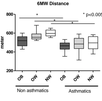
Breathlessness during exercise is a common complaint among morbidly obese individuals (BMI > 35), almost 80% of subjects, and obesity has long been thought to be a cause of dyspnoea, but data supporting this have been scarce and the mechanisms that drive these symptoms are not well defined. In severely obese subjects, Dempsey et al. [36] found that even during maximal exercise on a cycle ergometer these subjects were able to increase their ventilation sufficiently to avoid hypercapnia. [37] showed that during cycle exercise VO2 and VE are greater in obese subjects than normal weight at all work rates and that the relationships between breathlessness scores and these two values is not different in two groups. The implication of this finding is that the determinants of breathlessness were similar in obese and normal weight subjects, and that respiratory mechanical factors related to obesity did not contribute to breathlessness in the obese subjects. Concerning the walk exercise Hulens [38] showed that morbidly obese were more exerted and complained more frequently of dyspnea than obese and lean subjects during the six minute walking test (6MWT). In COPD patients during 6MWT obese