Maria Palmieri, MS,aAurora Currò, MD,a,bAndrea Tommasi, MD,a,bLaura Di Sarno, MS,b
Gabriella Doddato, MS,aMargheria Baldassarri, MD,bElisa Frullanti, MS,aAnn Rita Giliberti, MS,a
Chiara Fallerini, MS,aAngelo Spinazzola, MD,cAnna Maria Pinto, MD, PhD,bAlessandra Renieri, MD, PhD,a,b andMassimo Vaghi, MD,c,dSiena and Crema, Italy
ABSTRACT
Objective: Somatic mosaicism ofKRASgene is currently recognized as the only established molecular basis of arteriove-nous malformations (AVM). However, given the limitations of the current technologies,KRASsomatic mutations are detected only in a limited proportion of AVMs and tissue biopsy remains an invasive high risky, sometimes life-threatening, diagnostic procedure. Next-generation sequencing liquid biopsy using cell-free DNA (cfDNA) has emerged as an innovative noninvasive approach for early detection and monitoring of cancer. This approach overcomes the space-time profile constraint of tissue biopsies opens a new scenario for vascular malformations owing to somatic mosaicism. Here, we pro-pose a new approach as a fast noninvasive reliable tool in order to investigate the cfDNA coming from the AVMs.
Methods: A group offive patients suffering from AVM were selected. Blood samples from peripheral vein and efferent vein from vascular malformation were collected and cfDNA was extracted. The cfDNA libraries were performed using Oncomine Pan-Cancer Cell-Free Assay. We used Ion Proton for sequencing and Ion Reporter Software for analysis (Life Technologies, Carlsbad, Calif).
Results: In all cases, either G12D or G12V mutations inKRASwere identified. The mutational load was higher in the efferent vein than in peripheral blood, confirming the causative role of the identified mutation at a somatic level. Conclusions: We demonstrate that cfDNA next-generation sequencing liquid biopsy is able to identify theKRAS mu-tation detected in affected tissues. Moreover, we have shown that blood sample withdrawal at the lesion site increases variant allele frequency with an order of magnitude above the limit of detection (usually 0.05%), decreasing the risk of a false negative. Finally, the noninvasiveness of the method avoids any risk of bleeding, being easily performed also in children. We propose this technique as the method of choice to better investigate AVMs and consequently to identify the therapy tailored to the genetic defect. (JVSeVascular Science 2020;1:176-80.)
Clinical Relevance: This article highlights the importance of using liquid biopsy as a new method to investigate the molecular profile of AVMs. In view of the frequent inaccessibility of vascular tissues owing to the invasiveness of solid biopsy and the relative high incidence of biopsies with low diagnostic power, here we evaluated the efficacy of detecting cfDNA fragments released into the bloodstream from the affected tissue cells. Through a simple blood draw from the efferent vein at the vascular malformation site, the liquid biopsy allowed us to identifyKRAS
pathogenic mutations piloting a personalized therapeutic approach and opening a new scenario for new therapeutic strategies.
Keywords: Arteriovenous malformation; Liquid biopsy; cf-DNA; KRAS mutation; Noninvasive technique
Arteriovenous malformations (AVM) are fast-flow vascular malformations composed of tangles of abnor-mally developed vasculature. The absence of capillaries between arteries and veins often leads to high blood
pressure and rupture. They can occur in some part of the body, including the brain.1,2In the majority of cases an activating KRAS mutation has been identified.1-5 TheseKRAS variants have been previously described as
From the Medical Genetics, University of Siena,aand the Genetica Medica,
Azienda Ospedaliera Universitaria Senese,bSiena; and the Radiologia
inter-ventistica, Ospedale Maggiore di Crema,cand the Chirurgia Vascolare,
Ospe-dale Maggiore di Crema, Largo Ugo Dossena,dCrema.
Funded by ONG ILA.
Author conflict of interest: none.
Correspondence: Massimo Vaghi, MD, Chirurgia Vascolare, Ospedale Maggiore di Crema, Largo Ugo Dossena, Cremaa, Italy (e-mail:[email protected]); and Alessandra Renieri, MD, PhD, Medical Genetics Unit, University of Siena, Poli-clinico“Santa Maria alle Scotte”, Viale Bracci, 2, 53100 Siena, Italy (e-mail:
The editors and reviewers of this article have no relevantfinancial relationships to disclose per the JVS-Vascular Science policy that requires reviewers to decline review of any manuscript for which they may have a conflict of interest. 2666-3503
CopyrightÓ 2020 by the Society for Vascular Surgery. Published by Elsevier Inc. This is an open access article under the CC BY-NC-ND license (http:// creativecommons.org/licenses/by-nc-nd/4.0/).
https://doi.org/10.1016/j.jvssci.2020.08.002
gain-of-function mutations in cancer and Nikolaev et al3 have recently shown that hot spot mutations inKRAS, namely p.G12V are associated with arteriovenous brain malformations through an increase in angiogenesis, migration and cell proliferation.6 During the last years, next-generation sequencing (NGS) liquid biopsy has emerged as an innovative noninvasive technique for the identification of key mutations that are responsible for tumor growth allowing to optimize diagnosis, moni-toring, and therapeutic choice.7-9 Therefore, cell-free DNA (cfDNA) analysis has the possibility to overcome the space-time profile constraint of physical biopsies and it opens a new scenario for vascular malformations where tissue biopsy represents an invasive, high-risk, sometime life-threatening, diagnostic procedure. The use of liquid biopsy would also improve the opportunity to monitor illness evolution at a molecular level.
In the present study, we performed a comprehensive analysis offive patients with AVMs to determine if nonin-vasive NGS liquid biopsy from the efferent vein at the lesion site could detect the key variant bypassing the need for a high-risk, life-threatening tissue biopsy. The blood from the efferent vein at the vascular malforma-tion site was sampled during embolizamalforma-tion procedures before the injection of embolizing materials or liquids without causing further discomfort to the patient. For this reason, we define this technique as noninvasive. The NGS-liquid biopsy at the venous malformation site detected pathogenic mutations in KRAS gene in each patient. This study was consistent with Institutional guidelines and approved by the ethical committees of Azienda Ospedaliera Senese, Siena.
METHODS
Patient enrollment and sample collection. Five pa-tients affected by AVM were enrolled at the Medical Ge-netics Unit of the Azienda Ospedaliera Universitaria Senese, Siena, Italy, for a new diagnostic approach. Writ-ten informed consent for genetic analysis was obtained from all patients. Clinical information as well as genea-logic trees and cancer family history were collected on a genetic consultation setting. For all patients, liquid bi-opsy withdrawal from the lesion efferent vein was per-formed by Vascular Surgery of Ospedale Maggiore di Crema. All the blood specimens were taken during embolization procedures before the injection of embol-izing materials or liquids. All the patients underwent a complete hemodynamic and radiologic evaluation including computed tomography angiography or mag-netic resonance angiography. According to the Yakes Classification, these shunts belong to category II or III.
Extraction of cfDNA from plasma. Blood samples (10 mL) were collected from each patient and placed into cfDNA BCT blood collection tube (Streck, Neb). The cfDNA was extracted from 4 ml of plasma using
MagMAX cell-free Total Nucleic Acid Isolation Kit (Ther-moFisher Scientific, Waltham, Mass), according to the manufacturer’s instructions. The quality and quantity of cfDNA were verified respectively using the Agilent High Sensitivity DNA Kit (Agilent Technologies, Palo Alto, Calif) on Agilent2100 Bioanalyzer (Agilent Technologies) and Qubit dsDNA HS Assay Kits on Qubit 2.0 fluorometer (Invitrogen, Carlsbad, Calif).
NGS sequencing on cfDNA. The cfDNA sequencing was performed using Oncomine Pan-Cancer Cell-Free Assay (ThermoFisher Scientific) on Life Technologies Ion Proton sequencer (Life Technologies, Carlsbad, Calif). This tech-nology identifies various types of alterations, including single nucleotide variants, insertions/deletions, gene fu-sions and copy number variations in cancer-related genes (clinical actionable mutations) with a reportable range up to 0.05%. Sequencing analysis was performed using Ion Reporter Server System (Thermo Fisher Scientific). RESULTS
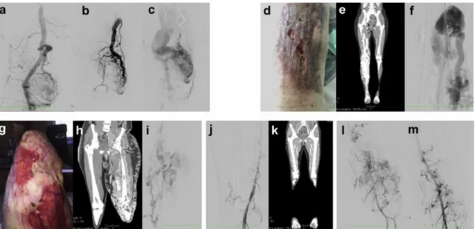
Patient 1 is a 56-year-old man with a congenital port wine stain angioma on the lower right leg. He also suffers from varicose veins since the age of 18. At the age of 54, after meniscal injury, magnetic resonance imaging of the right knee showed an AVM with the presence of flow voids and enlargement of the arterial and venous vessels in the medial and lateral compartment, and bone involvement at femoral and tibial level; the morphology of the shunt is compatible with type III B ac-cording to the classification of Wayne Yakes
Patient 2 is a 40-year-old woman with a congenital port wine stain angioma on the external part of the lower right leg, associated with hypertrophy. Angiography and computed tomography angiography showed a complex angiodysplasia of the right limb, which pre-sented with multiple arteriovenousfistulas, high-flow fis-tulas involving the tibial bone malformation, and abnormal communication between the common iliac
ARTICLE HIGHLIGHTS
d Type of Research: multicenter, prospective study d Key Findings: Cell-free DNA next-generation
sequencing liquid biopsy is able to identify somatic KRAS mutations in 100% of investigated cases without the need of tissue biopsy. The mutational load is higher in the plasma sample collected in the efferent vein from vascular malformation than in peripheral vessel, confirming the causative role of the identified mutation.
d Take Home Message: We propose this noninvasive
technique as the method of choice for arteriovenous malformation investigation and identification of tailored therapy.
artery and vein. Over the time, the patient underwent multiple embolizations and resection of arteriovenous fistulas. Because of limb length discrepancy, the patient underwent the elongation of the contralateral limb with the Ilizarov technique. Previous molecular analysis with a targeted panel did not identify any RASA1 patho-genic mutation, excluding Parkes-Weber syndrome.
Patient 3 is a 29-year-old man with congenital angio-dysplasia of the lower left limb, extending to the groabdominal region. Several endovascular and surgical in-terventions have been performed since childhood. He underwent a below-the-knee amputation at the age of 14 because of life-threatening bleeding and experienced a recurrence of AV shunts on the stump 10 years later. The patient reported episodes of nose bleeding, also pre-sent in other family members, but he did not satisfy Curacao criteria for a clinical diagnosis of hereditary hemorrhagic telangiectasia.
Patient 4 is a 45-year-old woman with congenital red vascular malformation in the upper right limb associated with limb hypermetry. The patient underwent several devascularization procedures, both surgical and endo-vascular, and resections over time.
Patient 5 is a 40-year-old woman who was referred for red-purple spots on the left lower limb, with hypertrophy and dysmetry. Thefirst clinical manifestation dates back to the age of 11 years, with deep venous thrombosis in the left lower limb. The patient underwent multiple endo-vascular procedures, and surgical removal of an aneu-rysm at the level of the femoral artery. The sampling for the cfDNA analysis was taken from the extremity AVM. Magnetic resonance imaging identified an additional he-patic arteriovenousfistula. Clinical features of all patients are shown in theFig.
In patients 1, 3, and 5, NGS-liquid biopsy analysis detected the same pathogenic mutation inKRAS gene c.35G>A; p.(Gly12Asp) (Table). In patients 2 and 4, NGS liquid biopsy analysis identified the mutation in KRAS gene c.35G>T; p.(Gly12Val). The variant allele frequency (VAF) ranged from 0,19% to 4,10%. In patient 1, who had the lowest VAF, a second blood draw from the efferent vein of AVMs of the knee was performed. The percentage of VAF in this experiment increased of one order of magnitude from 0.19% to 1.63%. Clinical features and molecularfindings are shown in theFigand theTable. DISCUSSION
AVMs are a nontumor subset of vascular anomalies owing to a dysmorphogenesis in the developmental process.
In view of the increasing role of endovascular treat-ments, the frequent inaccessibility of vascular tissues, the invasiveness of solid biopsy, and the relative high inci-dence of nondiagnostic biopsies, we evaluated the effi-cacy of detecting cfDNA fragments that are released into the bloodstream from the affected tissue cells.
Different cells, including normal healthy cells and he-matopoietic cells, contribute to the cfDNA in the blood. However, in efferent venous blood, major cfDNA is released by cells; thus, this technique is highly sensitive for circulating cfDNA, allowing the identification of path-ogenic mutations inKRAS genes, even with a very low VAF percentage in all patients.
In patient 1, liquid biopsy from the efferent vein detected an enrichment for the causativeKRAS mutation, allowing a search for a causative role in the angiodysplastic process. Taken together, our data strengthen the idea thatKRAS somatic mosaicism for gain-of-function mutations is the key genetic driver involved in the development of AVMs. Furthermore, our data suggest that our novel approach, based on the combination of NGS and liquid biopsy from the efferent vein at the vascular malformation site, al-lows detecting even low-grade somatic mosaicism responsible of the vascular phenotype, thus bypassing the need for a high-risk tissue biopsies.
Interestingly, the identified KRAS mutations (p.Gly12Asp and p.Gly12Val) are cancer hotspot mutations, which in-crease MAPK-ERK pathway activation, thus inducing endothelial cell proliferation3and enhancing their migra-tory behavior.10-15Noteworthy, although individuals who harborRAS mutations at a germline level present with an increased risk of tumors such a juvenile myelomono-cytic leukemia, acute leukemia, neuroblastoma, and rhab-domyosarcoma,16 no increased risk for cancer development has so far been clearly shown for patients with AVMs who harbor somaticKRAS mutations. Accord-ing to recent lines of evidence, drugs that specifically target a cancer driver gene represent innovative repurposing-based treatments readily available for use in different settings for hereditary conditions or somatic mosaic syndromes that carry the same driver genomic ab-erration.14,17Thus, in the new era of personalized medicine, agents that inhibit the MAP-ERK pathway commonly used in phase II clinical trials for the treatment of several solid tumors18could be likely considered as a therapy for sporadic brain AVMs caused by KRAS mutations. In conclusion, the diagnostic use of liquid biopsy for nonon-cologic diseases will forward the development of person-alized therapeutic approach for AVMs and will open the door to new therapeutic strategies potentially able to block or slow down disease progression.
We thank AVMs patients and ILA association (Italian as-sociation of childhood angiodysplasias and hemangi-omas). The“Cell lines and DNA bank of Rett Syndrome, X-linked mental retardation and other genetic diseases”, member of the Telethon Network of Genetic Biobanks (project no. GTB12001 and GFB18001), funded by Tele-thon Italy, and of the EuroBioBank network provided us with specimens. We thank SienaGenTest srl, a Spin-off of the University of Siena (www.sienagentest.dbm.unisi. it) for assessment of data analysis.
AUTHOR CONTRIBUTIONS Conception and design: AR, MV
Analysis and interpretation: MP, LD, EF, AS, AP, AR, MV Data collection: MP, AC, AT, LD, GD, MB, EF, AG, CF, AP,
AR, MV
Writing the article: MP, AC, AT, LD, GD, MB, EF, AG, CF, AS, AP, AR, MV
Critical revision of the article: EF, AP, AR, MV
Final approval of the article: MP, AC, AT, LD, GD, MB, EF, AG, CF, AS, AP, AR, MV
Statistical analysis: Not applicable Obtained funding: Not applicable Overall responsibility: AR
REFERENCES
1. Oka M, Kushamae M, Aoki T, Yamaguchi T, Kitazato K, Abekura Y, et al. KRAS G12D or G12V mutation in human
brain arteriovenous malformations. World Neurosurg
2019;126:e1365-73.
2. Nikolaev SI, Vetiska S, Bonilla X, Boudreau E, Jauhiainen S, Jahromi BR, et al. Somatic activating KRAS mutations in arteriovenous malformations of the brain. N Engl J Med 2018;378:250-61.
3. Nikolaev SI, Vetiska S, Bonilla X, Boudreau E, Jauhiainen S, Rezai Jahromi B, et al. Somatic activating KRAS mutations in arteriovenous malformations of the brain. N Engl J Med 2018;378:250-61.
4. Cheng F, Nussinov R. KRAS activating signaling triggers arteriovenous malformations. Trends Biochem Sci 2018;43: 481-3.
5. Priemer DS, Vortmeyer AO, Zhang S, Chang HY, Curless KL, Cheng L. Activating KRAS mutations in arteriovenous mal-formations of the brain: frequency and clinicopathologic correlation. Hum Pathol 2019;89:33-9.
6. Starke RM, McCarthy D, Komotar RJ, Connolly ES. Somatic KRAS mutation found in sporadic arteriovenous malforma-tions. Neurosurgery 2018;83:e14-5.
Table. Patient clinical features and molecularfindings
Patient
Code
number Gender Age, years Phenotype Peripheral NGS liquid biopsy Efferent vein NGS liquid biopsy 1 2023/19 Male 56 AVM dx leg KRAS(p.(G12D)) 0.19% KRAS(p.(G12D)) 1.63%
2 4417/19 Female 40 AVM dx leg na KRAS(p.(G12V)) 4.11% 3 4477/19 Male 29 AVM sx leg na KRAS(p.(G12D)) 1.18% 4 4820/19 Female 45 AVM dx leg na KRAS(p.(G12V)) 4.19% 5 3859/19 Female 40 AVM sx leg na KRAS(p.(G12D)) 1.18%
AVM, Arteriovenous malformation; dx, dextrum; na, not applicable; NGS, next-generation sequencing; sx, sinistrum.
Fig. Clinical features of the patients. a-c, Patient 1: intra-articular arteriovenous shunt and outflow vein where the blood specimen was taken. d-f, Patient 2: right lower limb arteriovenous malformation (AVM) with leg ulceration (d). Computed tomography (CT) scan (e) and angiography (f) of diffuse arteriovenous shunt at the tibial artery level. g-i, Patient 3: left lower stump AVM with leg ulceration (g). CT scan (h) and angiography (i) with huge diffuse arteriovenous shunting. j, k, Patient 4: angiography of the right lower limb (j) and CT reconstruction of arterio-venous shunt at the knee level (k). l, m, Patient 5: angiography of diffuse arterioarterio-venous shunt.
7. Palmieri M, Baldassarri M, Fava F, Fabbiani A, Gelli E, Tita R, et al. Two point-NGS analysis of cancer genes in cell free-DNA of metastatic cancer patients. Cancer Med 2020;9:2052-61. 8. Crowley E, Di Nicolantonio F, Loupakis F, Bardelli A. Liquid
biopsy: monitoring cancer-genetics in the blood. Nat Rev Clin Oncol 2013;10:472-84.
9. Corcoran RB, Chabner BA. Application of cell-free DNA analysis to cancer treatment. N Engl J Med 2018;379:1754-65. 10. McDonald J, Bayrak-Toydemir P, Pyeritz RE. Hereditary hemorrhagic telangiectasia: an overview of diagnosis, man-agement, and pathogenesis. Genet Med 2011;13:607-16. 11. Gallione CJ, Repetto GM, Legius E, Rustgi AK, Schelley SL,
Tejpar S, et al. A combined syndrome of juvenile polyposis and hereditary haemorrhagic telangiectasia associated with mutations in MADH4 (SMAD4). Lancet 2004;363:852-9. 12. Bayrak-Toydemir P, McDonald J, Markewitz B, Lewin S,
Miller F, Chou LS, et al. Genotypeephenotype correlation in hereditary hemorrhagic telangiectasia: mutations and manifestations. Am J Med Genet A 2006;140:463-70. 13. Revencu N, Boon LM, Mulliken JB, Enjolras O, Cordisco MR,
Burrows PE, et al. Parkes Weber syndrome, vein of Galen aneurysmal malformation, and other fast-flow vascular
anomalies are caused by RASA1 mutations. Hum Mutat 2008;29:959-65.
14. Luks VL, Kamitaki N, Vivero MP, Uller W, Rab R, Bovée JVMG, et al. Lymphatic and other vascular malformative/over-growth disorders are caused by somatic mutations in PIK3CA. J Pediatr 2015;166:1048-54.e1-5.
15. Do Prado LB, Han C, Paul Oh S, Su H. Recent advances in basic research for brain arteriovenous malformation. Int J Mol Sci 2019;20:5324.
16. Kratz CP, Franke L, Peters H, Kohlschmidt N, Kazmierczak B, Finckh U, et al. Cancer spectrum and frequency among children with Noonan, Costello, and cardio-facio-cutaneous syndromes. Br J Cancer 2015;112:1392-7.
17. Burotto M, Chiou VL, Lee JM, Kohn EC. The MAPK pathway across different malignancies: a new perspective. Cancer 2014;120:3446-56.
18. Braicu C, Buse M, Busuioc C, Drula R, Gulei D, Raduly R, et al. A comprehensive review on MAPK: a promising therapeutic target in cancer. Cancers (Basel) 2019;11:1618.