UNIVERSITY OF MESSINA
Ph.D. Course in “Surgical and Medical Biotechnologies” XXXII Cycle
(Coordinator: Prof. Giovanni Squadrito)
Evaluation of M1 and M2 macrophages in ovarian
endometriomas from women affected by endometriosis at
different stages of the disease
Ph.D. Candidate:
Dr. Antonio Simone LAGANA’
___________________________
Supervisor: Prof. Roberta GRANESE
______________________________
who support me in every action of my life with dedication and kindness To my family, who allowed me to study and become doctor, teaching me to help the others To Prof. Roberta Granese, Dr. Francesca Maria Salmeri and Prof. Vincenza Sofo who believed in me and in this research project To Dr. Helena Ban Frangež and Prof. Eda Vrtačnik-Bokal of the Department of Reproduction of the University Medical Center Ljubljana, who taught me the surgical skills in minimally invasive gynecological surgery To Prof. Fabio Ghezzi and the Surgical Team of the “Filippo Del Ponte” Hospital, who welcomed me in a wonderful work setting and enhanced my surgical skills
Pag. 3 a 50
INDEX
1. Subtypes and functions of macrophages
1.1. Regulation of the macrophage polarization 1.2. Actions of M1 macrophages1.3. Actions of M2 macrophages
1.4. The M1-M2 phenotypic switch in pathological conditions 1.5. M2a macrophages
1.6. M2b macrophages 1.7. M2c macrophages
1.8. Role of macrophages in the regulation of metabolic pathways
2. Endometriosis
2.1. Epidemiology of endometriosis 2.2. Classification of endometriosis 2.3. Etiopathogenesis of endometriosis
3. Aims of the study
4. Materials and methods
4.1. Study design and population4.2. Sample collection and macrophages characterization 4.3. Statistical analysis
4.4. Ethical and methodological standards
5. Results
6. Discussion
7. Conclusion
8. Declaration of interests
9. Acknowledgements
10. References
Pag. 4 a 50
1. Subtypes and functions of macrophages
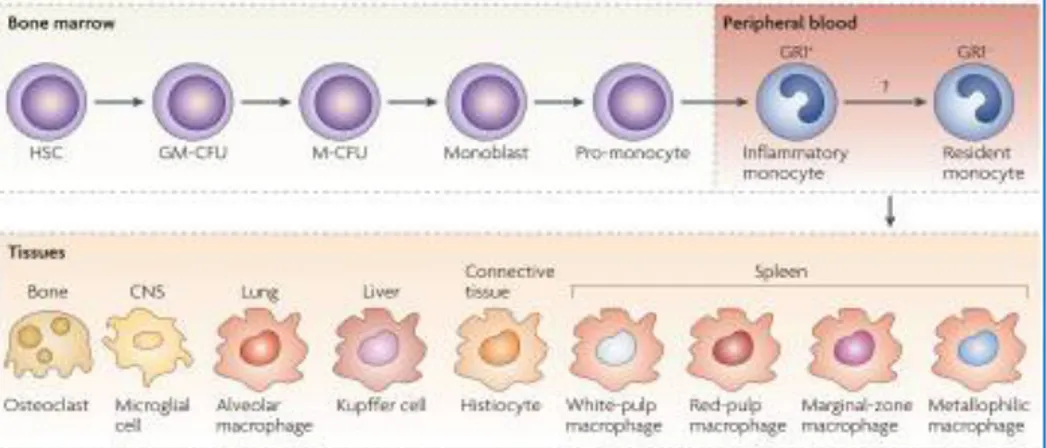
Monocytes (Mo) originate in the bone marrow from a common Hematopoietic Stem Cell (HSC). HSC undergoes differentiation stages during which it is directed towards the myeloid line and then towards the monocytic line. In response to Macrophage Colony-Stimulating Factor (M-CSF) it divides and differentiates into monoblast and then into pro-monocyte, before to become monocyte, which leaves the bone marrow and enters the bloodstream. Monocytes, unlike neutrophils, are less abundant (500-1000 cells/µL) and survive for long periods in extravascular tissues, where they can migrate and differentiate into macrophages. The resident macrophages are present in the connective tissues and in every organ where they perform functions similar to those of the newly recruited macrophages. They can take different forms and functions depending on the site (Figure 1): microglia in the central nervous system, Kupfer cells in the liver, alveolar macrophages in the lung, osteoclasts in the bone and histiocytes in the connective tissue. The circulating monocytes resident macrophages represent two differentiation stages of the same cellular precursor and play a pivotal role in homeostasis and in the defense of the organism [1].
Pag. 5 a 50
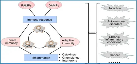
Macrophages are considered master regulators in homeostasis and host defense: the main role is phagocytosis in the innate immune response and the antigen (Ag) presentation to T lymphocytes within acquired immunity. In addition, macrophages participate and orchestrate also inflammatory and autoimmune processes and carcinogenesis (Figure 2).
Figure 2. Roles and functions of macrophages.
When not stimulated, macrophages are quiescent; nevertheless, external and/or endogenous stimuli (microbial products, damaged cells, activated cells and their soluble mediators) can re-program them and direct their plasticity towards a reversible polarization spectrum [2].
In response to toll-like receptor (TLR) and cytokine ligands, i.e. interferon-gamma (IFN-g) alone or with microbial co-stimuli or with other cytokines (such as tumor necrosis factor - TNF-a), interleukin (IL)-4 and IL-13, macrophages undergo classical activation and become M1, or alternative activation and become M2, a situation that mimics the T-helper (Th)1/Th2 polarization of lymphocytes. During the last decade, significant progresses have been made in defining the molecular mechanisms responsible for the polarization of
Pag. 6 a 50
macrophages [3]. Typically, M1 macrophages have an IL-12high, IL-23high,
IL-10low phenotype, play phagocytic functions through the production of effector
molecules, such as IL-1α, IL-6, IL-12 and TNF-α, and reactive species of oxygen (ROS) and nitrogen (RNS), participate in Th1 responses, acting as inducers and effectors and mediating resistance against intracellular parasites and tumors. The different forms of M2 cells, on the other hand, show IL-12low,
IL-23low, IL-10high phenotype, participate in Th2 responses and produce
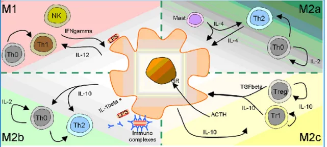
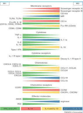
transforming growth factor-beta (TGF-β) and pro-angiogenic factors (factor XIII and Vascular Endothelial Growth Factor - VEGF), inhibit inflammatory processes, promote tissue remodeling, angiogenesis and tumor progression and regulate the immune response [4–7]. Generally, M2 macrophages have high levels of scavenger receptors (SRs), low levels of IL-1b and low Caspases-1, high levels of IL-1Ra and type II decoy receptors [8]. M1 and M2 macrophages also have different chemokine profiles: M1 express chemokines that attract Th1 cells, such as C-X-C motif ligand 9 (CXCL9) and CXCL10, while M2 produce chemokines (C-C motif) ligand 17 (CCL17), CCL22 and CCL24 [9,10]. M1 subpopulation shows potent microbicidal and tumoricidal properties and promotes strong Th1 responses mediated by IL-12, while M2 responses are Th2 type and have a role in the resolution of inflammation. The macrophages can also polarize towards a further "M2-like" state and can be further subdivided in different M2 subtypes: M2a (after stimulation by 4 or IL-13), M2b (stimulated by immune complexes in combination with IL-1b or lipopolysaccharides) and M2c (after stimulation by IL-10, TGF-b or glucocorticoids). These phenotypes share some but not all the characteristics and markers of M2 cells (Figure 3 and 4).
Pag. 7 a 50
Figure 3. Polarization of the macrophages according to the theory of the type 1 or type 2 response.
The macrophages show different functional phenotypes, some antagonists among them, under the influence of specific mediators. M1: Bacterial lipopolysaccharides in combination with IFN-g leads to classical activation by triggering the IL-12/IFN-g pathway. M2a: IL-4 and IL-13 are both capable of triIL-12/IFN-gIL-12/IFN-gerinIL-12/IFN-g the alternative activation of macrophages. M2b: the binding of Fc receptors (FcR) in the presence of Toll-like stimuli is able to induce an antagonistic polarization to M1, which promotes the Th2 response through the IL-10 pathway. M2c: deactivation is required to end the inflammatory process and is triggered by IL-10 or glucocorticoids.
Figure 4. The polarization of macrophages is associated with the expression of distinct genes.
M1 show receptors for cytokines while M2 are characterized by high levels of
non-opsonic receptor. M1 also have a high ratio of CXCR3 ligands while M2a express ligands for CCR8 and CCR3. M2b and M2c are less characterized populations that produce large quantities of CCL1 and CCL18, respectively.
Pag. 8 a 50 1.1. Regulation of the macrophage polarization
A coordinated action of the various modulators of inflammation, signal molecules and transcription factors is involved in the regulation of the polarization of the macrophages [11,12]. At the cellular level, specialized or polarized T cells (Th1, Th2, Th17 and Treg) play a role in the polarization of macrophages. The Interferon Regulatory Factors/Signal Transducer and Activator of Transcription (IRF/STAT) signaling pathway has a key role in modulating and controlling the polarization of the macrophages [13]. The activation of IRF/STAT signaling pathway by IFN and TLR directs the function of macrophages towards the phenotype M1 (via STAT1), while the activation of the IRF/STAT signal pathway (via STAT6) by IL-4 and IL-13 directs the function of the macrophages towards the M2 phenotype [14]. Signals initiated by IL-10, glucocorticoid hormones, molecules released by apoptotic cells and immune complexes can profoundly influence the functional status of macrophages. Their polarization is also modulated by local microenvironmental conditions, such as hypoxia. In addition, the polarization of M1/M2 macrophages is a highly dynamic process and the phenotype of polarized macrophages can still vary under physiological and pathological conditions. The macrophage’s phenotype switch remains, however, a still unclear mechanism. Furthermore, the imbalances in the polarization of M1/M2 macrophages are associated with various pathological conditions, with M1 macrophages involved in promoting and sustaining inflammation and M2 macrophages related to the resolution of chronic inflammation.
1.2. Actions of M1 macrophages
The TLR signaling pathway, in particular TLR4 stimulated by lipopolysaccharide and other microbial ligands, preferentially leads macrophages towards M1
Pag. 9 a 50
phenotype. Two adapters, myeloid differentiation primary response 88 (MYD88) and TIR-domain-containing adapter-inducing interferon-β (TRIF), mediate the downstream signaling of TLR4 [15]. The signal pathway through the MYD88 adapter results in the activation of a kinase cascade, including interleukin-1 receptor-associated kinase 4 (IRAK4), TNF receptor-receptor-associated factor 6 (TRAF6), and inhibitor of nuclear factor kappa-B kinase subunit beta (IKK-β), which eventually leads to the activation of the nuclear factor kB (NF-kB) [16]. NF-kB regulates the expression of a large number of inflammatory genes including TNF-a, IL-1b, cyclooxygenase 2 (COX-2), IL-6 and IL-12p40 [17]. The activity of NF-kB is modulated by the activation of the trimer kinase-kappa B complex inhibitor (IKK) (two kinases, IKKa, IKKb, and a regulatory protein, IKKg). When the upstream signals converge to the IKK complex, they first activate IKKβ via phosphorylation and further activated IKKβ phosphorylates the inhibitory molecule (I-kB). This results in the proteosomal degradation of I-kB and the release of the NF-kB p65/p50 heterodimer from the NF-kB/I-kB complex [18]. The NF-kB p65/p50 heterodimer is then translocated to the nucleus and binds to the promoters of inflammatory genes [19]. Signaling via the TRIF adapter pathway activates the transcription of interferon-responsive factor 3 (IRF3), which leads to the expression and secretion of type I interferons, such as IFN-a and IFN-b. Type I secreted IFNs bind to their own receptor with STAT1 activation. The IFN-stimulated genes include chemokines CXCL9 and CXCL10 [20], characteristics of the classic activation of M1 macrophages.
Pag. 10 a 50 1.3. Actions of M2 macrophages
Macrophages can be directed to an M2 phenotype from stimuli such as IL-4, IL-13 and IL-10. IL-4 and IL-13 polarize macrophages towards the M2 phenotype by activating STAT6 [21], through the IL-4 alpha receptor (IL-4Rα), while IL-10 promotes the M2 phenotype by activating STAT3 [22], through the IL-10 receptor. In the IL-4 and IL-13 pathways, IL-4 receptor binding activates Janus Kinase (JAK)1 and JAK3 [23], which lead to activation and translocation of STAT6. The M2 macrophage phenotype is promoted by several transcription factors, including the peroxisome proliferation-activated receptor (PPAR)-γ and the Krueppel-like factor 4 (KLF-4) [24]. PPARs play an important role in modulating the polarization of M2 macrophages induced by IL-4 or IL-13. Studies with PPAR-deficient macrophages have demonstrated the role of this nuclear receptor in promoting M2 activation to protect mice from insulin resistance [25]. A similar role was also found for PPAR-δ in determining macrophage polarization [26]. Taken together, these data suggest that STAT6, PPAR- d , KLF-4, and IRF4 can coordinate the polarization of M2 macrophages. This polarization is also closely related to the different expression of the various TLRs. The TLR4/TLR2 ratio is significantly higher in M1 than in M2 cells, while TLR4 deficiency promotes alternative M2 activation. Chronic signal activation through the TLR4 pathway induces various negative regulators such as IRAK-M, Suppression of Tumorigenicity 2 (ST2) gene, Suppressor Of Cytokine Signaling 1 (SOCS1) gene, short version of MYD88 (MyD88sh) and SHIPs [27]. These negative regulators inhibit TLR-mediated signaling and therefore address the macrophages towards immunosuppressive and endotoxin-tolerant M2 phenotype. The transition from a MYR88-dependent TLR4 pathway to a TRIF-dependent pathway in macrophages probably serves to shift the phenotype from an inflammatory to an
anti-Pag. 11 a 50
inflammatory state. In this way, the interaction of signal molecules and transcription factors can influence the polarization phenotype. In particular, STAT-mediated activation of macrophages is regulated by the SOCS genes. Members of the SOCS family are inducible inhibitors of cytokine signals and, therefore, play a fundamental role in limiting inflammatory responses. SOCS proteins could be induced by cytokine signaling pathways, due to several mechanisms. For example, IL-4 and IFN-γ, the latter together with the TLR stimulation, up-regulates SOCS1 and SOCS3, which in turn inhibits the action of STAT1 and STAT3, respectively [28]. SOCS proteins can also be directly induced by TLR signals. In macrophages, SOCS proteins not only regulate the sensitivity of cells to cytokines, but also modulate signals through TLRs. Since SOCS3 is a molecule downstream of the Notch signal, it is likely that the Notch signaling pathway determines the polarization of M1 macrophages with respect to M2, via SOCS3 (although the role of SOCS3 is controversial) [29]. Similarly, SOCS3 deficiency promotes macrophage M1 polarization and inflammation.
1.4. The M1-M2 phenotypic switch in pathological conditions
Macrophages differentiation is a highly dynamic process. In response to microenvironmental factors, macrophages can rapidly pass from one phenotype to another, even in different pathological conditions. Studies have shown that spatial-temporal activation of transcription factor NF-kB is a key regulator of macrophage plasticity observed during various disease progressions. For example, during the first stage of carcinogenesis, activation of NF-kB in M1 cells is a critical factor in the link between cancer and inflammation. However, in the late stage of tumorigenesis, the macrophages are reprogrammed as M2, with low NF-kB activation but greater
Pag. 12 a 50
immunosuppressive capacity [30]. An analogous situation of macrophages polarization is observed in the progression of sepsis, in which the activation of NF-kB in M1 macrophages leads to the initial phase of the inflammatory process, while during the late phase of endotoxin tolerance, the macrophages are polarized towards the M2 anti-inflammatory phenotype in promoting tumor growth [31].
1.5. M2a macrophages
The M2a-subtype macrophage activation is what is commonly considered as alternative activation. This subtype differs in terms of receptor expression, cytokine production and effector function compared to M1 macrophages. Alternative macrophage activation induced in culture by IL-4 or IL-13 exposure is associated with tissue repair. These macrophages in fact produce high levels of fibronectin and a matrix-associated protein, βIG-H3 [32]. The induction of arginase in these cells can lead to the biosynthesis of polyamine and proline promoting cell growth, collagen formation and tissue repair. Alternative activation does not produce nitric oxide (NO) and consequently the ability to kill intracellular microbes with respect to the classic phenotype is compromised [33]. Although these cells overexpress class II of major histocompatibility complex (MHC) molecules, they are less efficient in presenting Ag. In many cases they inhibit the proliferation of T cells, participate in the humoral immune response, regulating the Th2 type effector response and stimulate the B cell to produce immunoglobulins [34]. The cytokines by these alternately activated cells are IL-10 and the receptor antagonist for IL-1. Based on these data, the M2a phenotype is considered anti-inflammatory and immune-regulatory.
Pag. 13 a 50 1.6. M2b macrophages
M2b represents an intermediate phenotype between the classical and the alternative pathways [35]. The macrophage differentiation in this subtype occurs following exposure to immunocomplexes (IC), TLRs agonists and the IL-1 receptor. Thus, activated cells secrete high levels of anti-inflammatory cytokines such as IL-10 and low IL-12 levels and are involved both in the activation of the innate immune response, determined by TLR ligands, and in the increase in the production of proinflammatory cytokines and synthesis of inducible nitric oxide synthase (iNOS) and ROS [36]. In this regard, this type of activation has hybrid characteristics between classic and alternative activation.
1.7. M2c macrophages
The process of "extinguishing" inflammatory and immune activity can be initiated by the presence of anti-inflammatory cytokines, such as IL-10, or glucocorticoid hormones. Indeed, these molecules bind a cytosolic receptor for steroid hormones which causes a repression of the transcription of genes such as iNOS, COX-2 and TNF. In high concentrations, glucocorticosteroids have immunosuppressive effect by inhibiting the MHC I and II and therefore the Ag processing. Some effects of the abovementioned anti-inflammatory molecules are mediated by the activation in the transcription of anti-inflammatory genes such as annexin 1 (or lipocortin1) [37]. This protein binds phospholipase A2 substrates that are found on the cell membrane, down-regulating the formation of arachidonic acid, a fundamental substrate for the synthesis of prostaglandins (PG) and leukotrienes (LT). During the resolution of the inflammatory process, the production of PG and LT decreases in favor of anti-inflammatory products, such as 15-Deoxy-delta-12,14-prostaglandin J2
Pag. 14 a 50
(15dPGJ2) and lipoxins [38]. The first inhibits the pro-inflammatory pathways mediated by NFkB, AP-1 and STAT, while the second participates in neutrophil deactivation. 15dPGJ2 is able to increase macrophage apoptosis and reduce the production of oxygen free radicals, besides its known role PPARγ agonist. The PPARγ, besides being involved in the maintenance of lipid homeostasis, plays an important role in the regulation of the inflammatory process. For this reason, PPARγ agonists seem to be involved in the alternative macrophage activation [39]. It has also been shown that CD200 receptor inhibition is able to stop signal transduction via the Mitogen-Activated Protein Kinase (MAPK), extracellular-signal-regulated kinase (ERK) and JNK46 pathways [40]. For these reasons, macrophage deactivation is associated with both immunosuppression processes, activating the cells of the Th2 effector response, and matrix deposition and tissue remodeling. Some authors [40,41], however, do not entirely agree with this clear distinctive scheme, suggesting to consider M1 and M2 as the two extremes of a continuum of functional states (Figure 5).
Pag. 15 a 50
1.8. Role of macrophages in the regulation of metabolic pathways
M1 and M2 also have different actions with regard to the metabolism of iron, glucose and amino acids in response to different polarizing stimuli in the tissue microenvironment, in normal and pathological conditions [10]. This creates a two-way cross-talk between metabolism and macrophages: indeed, macrophages exert not only "extrinsic" effects on metabolism regulation (through the release of cytokines), but also "intrinsic" effects whereby the metabolic state of these cells influence their phenotype. Recent studies on both mice and humans have shown strong differences between M1 and M2 in iron metabolism [42,43]. M1 express high levels of proteins involved in iron deposition, such as ferritin, while they have low levels of ferroportin, an iron transporter. In contrast, M2 show low ferritin levels and high ferroportin levels. These divergences in iron metabolism may be related to the functional results of the respective actions. Iron sequestration by M1 cells could have a bacteriostatic effect (iron is essential for bacterial growth) and thus support host protection against infections. In contrast, the release of iron from M2 cells could promote the resolution of inflammation and tissue repair but, unfortunately, also tumor growth. Polarized macrophages exhibit a different regulation of glucose metabolism: in response to M1 stimuli, they show a metabolic shift towards the path of anaerobic glycolysis, while exposure to M2 type stimuli (such as IL-4) has minor effects [44]. The use of specific metabolic pathways could be functionally related to different tasks: M1, for example, are often associated with acute infections and need to quickly trigger their microbicidal activity, in the hypoxic tissue microenvironment [45]. In this context, an anaerobic process (such as glycolysis) is more suitable for their rapid energy demand. In contrast,
Pag. 16 a 50
functions related to M2 polarization, such as tissue remodeling and repair require an important energy supplement, which can be satisfied by the oxidative metabolism of glucose (fatty acid oxidation), the metabolic pathway of choice of M2 cells [46]. In particular, macrophages adapt to hypoxia by directing their metabolism towards anaerobic glycolysis. When they are recruited at the sites of inflammatory processes, they face a condition of hypoxia, which can directly influence their polarization. Hypoxia acts on macrophages through the two isoforms of factors inducible hypoxia, HIF-1 and HIF-2. The activation of HIF-1 and HIF-2 causes profound functional modifications, including the expression of chemokines and chemokine receptors (CXCR4 and CXCL12) and of VEGF [47]; in this way, macrophages contribute to the organization of the tissue response in hypoxic conditions [48]. The up-regulation of these genes due to hypoxia would lead to the recruitment of more macrophages in hypoxic zones in diseases such as atherosclerosis, obesity and cancer where they can both attenuate inflammation and promote tumor progression. Furthermore, pro-inflammatory cytokines such as TNF-a, IL-1β, MIF, CCL3 and COX2, as well as M2 markers, such as IL-10 and arginase 1, are also induced by hypoxia.
Amino acid metabolism is closely linked to the functional phenotype of myelomonocytic cells: M1 are characterized by the expression of nitric oxide-synthetase 2 (NOS2) and by the production of NO, while M2 do not produce NO but express high levels of arginase 1, which catalyzes the production of polyamine, necessary for collagen synthesis, cell proliferation, fibrosis and other cellular remodeling functions [49]. These actions, taken collectively, show that the metabolic adaptation represents an integral aspect of the polarization of the macrophages and their functional diversity.
Pag. 17 a 50
2. Endometriosis
Endometriosis is a pathological, estrogen-dependent condition, characterized by the presence and growth of functional endometrial-like tissue, glands and stroma, outside the uterine cavity [50]. The endometrial-like tissue could implant in the pelvic area, especially at the level of the ovaries, Fallopian tubes, Douglas pouch, bladder, bowel, rectum and colon; more rarely, endometriotic foci can be found in vagina, vulva, scars from pelvic and extra-pelvic surgery.
Under the influence of the estrogens and progesterone, secreted by the ovary during the menstrual cycle, the endometrium undergoes regular cyclic changes (proliferative phase, secretory phase, menstruation). At each menstrual cycle, under the effect hormonal stimuli, the endometriotic tissue follows the cyclical changes of the physiological endometrium and undergo shedding and consequent bleeding. In the eutopic site this process is followed by a phase of remodeling of the endometrium itself, but in the ectopic tissue there is an inflammatory process of the surrounding tissues, which gives rise to the formation of fibrotic tissue and adhesions. This condition may cause acute and chronic pelvic pain, dyspareunia, dysmenorrhea, metrorrhagia and infertility [51].
2.1. Epidemiology of endometriosis
Endometriosis affects mainly women in reproductive age, with a peak incidence between 30 and 40 years, with a prevalence of about 10% in the general female population [52]. Although endometriosis is a benign pathology, it causes important repercussions on the quality of life of the woman, above all for the clinical implications that it involves. Approximately 30-40% of women with endometriosis are infertile. The disease can occur from the first menstruation and exceptionally even
Pag. 18 a 50
before the menarche. After the age of 40, endometriotic tissue growth seems slower. The disease develops independently of parity, with a prevalence for women who did not have pregnancy.
2.2. Classification of endometriosis
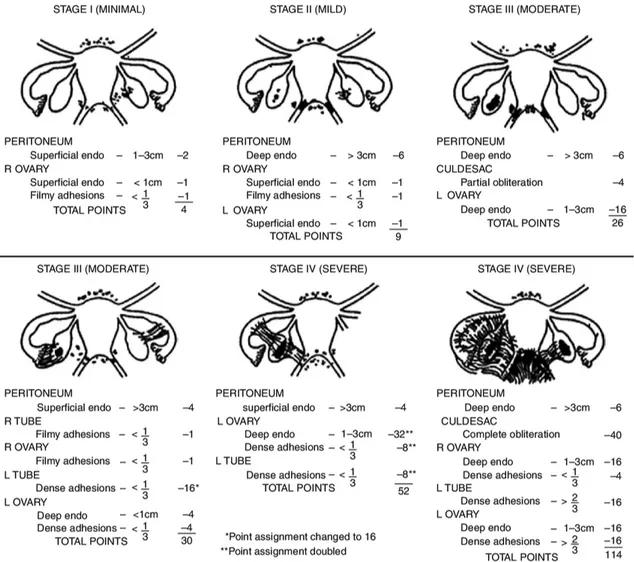
Several classifications of endometriosis have been proposed over time. To date, the most used is that of the American Society of Reproductive Medicine (Figure 6) [53].
Figure 6. Revised American Society of Reproductive Medicine classification of endometriosis.
Pag. 19 a 50
The staging of endometriosis is based on the following parameters:
1. the appearance, size and depth of peritoneal and ovarian lesions;
2. the presence, extent and type of lesion (red, white and chocolate/black); 3. the presence, extent and type of adhesions at the level of the appendages and the degree of obliteration of the Douglas pouch.
2.3. Etiopathogenesis of endometriosis
The etiopathogenesis of endometriosis is very complex and still not well defined. There are numerous hypotheses formulated to explain the ectopic cellular dislocations, typical of the disease [54].
• Retrograde menstruation [55]: groups of endometrial cells shed at the time of menstruation and refluxed through the tubes in the abdominal cavity would be able to implant themselves on the peritoneum to form endometriotic islands. This phenomenon, however, is very frequent (observed in about 90% of women of reproductive age with patent Fallopian tubes) and it contrasts with the relatively modest incidence of the disease.
• "Metastatic" dissemination in blood or lymphatic vessels [56]: the theory of hematogenous metastases has been proposed to explain the implants of endometrial-like cells in areas very far from those commonly observed in the pelvis and not reachable by retrograde transport. According to this theory, endometriotic cells are transported through the venous drainage of the uterus, resulting in possible migration to any part of the body. Nevertheless, venous blood drained from the uterus pass through the pulmonary capillary vessels,
Pag. 20 a 50
and therefore hematogenous diffusion should also lead to a high incidence rate of secondary pulmonary endometriosis, unless there are concomitant atrial defects or ventricular septum that may explain a possible bypass of the pulmonary vessels. Since pulmonary endometriosis is extremely rare, the theory of hematogenous diffusion remains purely speculative. The theory of lymphatic diffusion has been confirmed by the finding of endometriosis in the lymph nodes, even if data published so far are not conclusive. Lymph nodal endometriosis, however, could also be explained by deposition of endometrial precursor cells within nodal lymphatic tissue during early organogenesis.
• Iatrogenic implants [57,58]: endometriotic foci have been observed on surgical scars after cesarean section, episiotomy or other gynecological surgery. The possibility of surgical dissemination in case of rupture of an endometrioma also remain to be elucidated: nevertheless, current guidelines do not recommend to avoid spillage as mandatory during the stripping of ovarian endometriomas [59].
• Coelomatic metaplasia [60]: peritoneal mesothelial cells, of coelomic origin, would undergo metaplasia turning into endometrial cells. This hypothesis would explain the exceptional formation of endometriotic islands in the bladder and prostate of male subjects. The latter, however, could be the result of hyperplasia of prostatic utricle cells, the embryonic residue of Muller's ducts, in patients treated with high doses of estrogens for prostate cancer [60]. This hypothesis has therefore lost some of its credit.
Pag. 21 a 50
• Embryologic origin: according to this theory, endometriosis would be determined by the presence of primitive endometrial tissue outside the uterus, resulting from a dislocation of this tissue due to a defect during organogenesis. In this regard, it has been shown that the diminished expression of the Homeobox A10 (HOXA-10) gene [61] is a potential mechanism that causes the increase of adhesion and implantation of ectopic endometriotic cells.
• Environmental pollutants: endometriosis is strongly influenced by the hormonal stimuli and is therefore exposed to the action of the so called "endocrine disruptors" [62]. These are exogenous substances that alter the functions of both the endocrine and immune systems. These substances can be of natural origin (such as phytoestrogens) or of artificial origin (such as xenoestrogens, pesticides, fungicides, herbicides or accidental combustion products). The association of endometriosis with environmental factors has been demonstrated by exposure to dioxins, a class of heterocyclic organic compounds classified as carcinogenic substances and included in the International Agency for Research on Cancer (IARC) group 1, defined as "definitely carcinogenic to humans". Dioxins reach humans mainly through food. They are organic pollutants, persistent, capable of bioaccumulation and, being lipophilic in nature, they accumulate in the adipose tissue. Dioxins and dioxin-like compounds seem to induce local estrogen-like activity that promotes growth of endometriotic foci [63]. Furthermore, these substances induce the expression of genes for CYP1A1 and γ-sinuclein, which are up-regulated in the advanced stages of the disease
Pag. 22 a 50
[64]. In addition, robust suggest that some polymorphisms in receptor genes commonly linked by dioxin and for detoxifying protein genes seem to cause susceptibility to the disease [65].
• Hormonal factors: endometriosis is estrogen-dependent and, consequently, estrogens are crucial for the development and growth of endometriotic lesions [66]. Indeed, the interruption of the menstrual cycle through hormonal therapy or after menopause determines a significant regression of the symptoms and signs of the disease. The exposure of the endometriotic foci to estrogens is high, since the ectopic endometrium is able to convert the androstenedione to estrone and estradiol. This conversion is due to the 17β-HSD enzyme of type 1: consequently, the ectopic endometrium synthesizes estradiol. In the normal endometrium, on the other hand, large amounts of 17β-HSD type 2 are expressed. Estradiol within endometriotic foci is able to stimulate matrix metalloproteases (MMPs) and induce the activation of COX-2 enzyme, which will lead to an increase in the synthesis of prostaglandins (PGs), especially PGE2, with intense pro-inflammatory activity [67]. PGE2 also stimulates the production of the aromatase enzyme, which catalyzes the transformation of androstenedione to estrone and estradiol. PGE2 and aromatase, in this way, have a positive feedback in order to enhance the production and release of estrogens within the endometriotic foci. Conversely, progesterone may counteract many of the biological effects of estrogen, suppressing the positive feedback between PGE2 and aromatase by downregulating estrogen.
Pag. 23 a 50
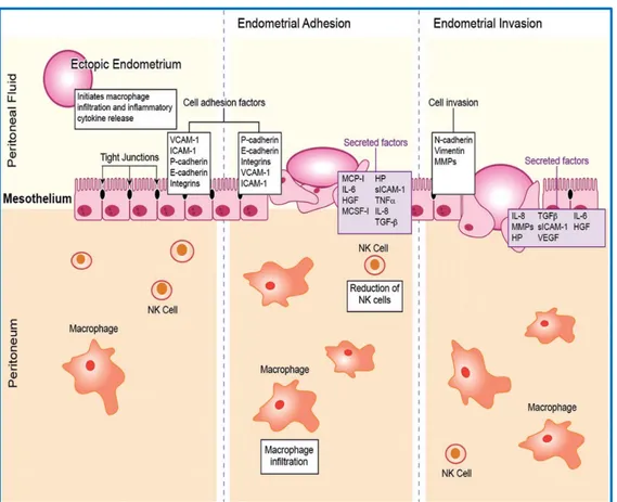
• Changes in the immune system. According to recent data, the immune system plays a predominant role in the pathogenesis of the disease. Numerous local (peritoneal cavity) and systemic (peripheral blood) alterations of the immune network have been demonstrated in women suffering from endometriosis [68,69]. Immune dysfunctions generate a cascade of phenomena that explain most of the histological and symptomatologic characteristics of endometriosis. In the peritoneal microenvironment, a breakdown of immunological homeostasis occurs during endometriosis, with a consequent alteration of cell-mediated immunity, humoral immunity and angiogenesis which allow the implantation and survival of endometriotic cells. In women with endometriosis, the peritoneal fluid is characterized by the presence of inflammatory activity, as evidenced by the increased level activated macrophages [70]. These activated mononuclear cells secrete several inflammatory cytokines and chemokines with pleiotropic activity and numerous growth factors. Therefore, the recruitment of macrophages within the lesions may represent not only an early event in the development of the disease but a necessary step for the subsequent maintenance of the lesions [71].
The endometriotic cells should be recognized, attacked and eliminated by the immune system through all the mechanisms of innate and adaptive immunity. On the contrary, these cells "escape" from the immune surveillance, implant and proliferate in the pelvic cavity. The study of the peritoneal microenvironment is therefore important to monitor the development and progression of endometriosis and associated infertility [72,73].
Pag. 24 a 50
Figure 7. Potential role of macrophages for the development of endometriotic implants within the peritoneum.
Pag. 25 a 50
3. Aims of the study
Although recent data suggest a pivotal role of the tumor associated M1/M2 macrophages polarization in oncology, data about endometriosis are still scarce and do not allow to draw a firm conclusion about their specific roles in the different stages of the disease. Based on these elements, the aim of our study was to evaluate M1 and M2 macrophages in ovarian endometriomas in women affected by endometriosis at different stages of the disease.
Pag. 26 a 50
4. Materials and methods
4.1. Study design and population
We prospectively enrolled women in reproductive age affected by ovarian endometriomas or by a single ovarian functional cyst persistent for more than 6 months (controls), who underwent laparoscopic surgical procedures from December 2016 to December 2018. We excluded women affected by significant comorbidities, including endocrine, cardiovascular, metabolic, auto-immune, oncological or other gynecological concomitant diseases. In addition, we excluded patients taking any kind of hormonal or non-hormonal drug (including anti-inflammatory or immunomodulatory medications) in the preceding three months from the enrolment (wash-out period). All the cases of ovarian endometriomas and ovarian functional cysts, suspected according to preoperative evaluation using the International Ovarian Tumor Analysis (IOTA) Group simple descriptors and simple rules for classifying adnexal masses [74], were confirmed by histological analysis. Endometriosis stage was scored according to the revised American Society for Reproductive Medicine (ASRM) classification of endometriosis [53], following the World Endometriosis Society consensus [75]. Both endometriomas and functional cysts were managed by laparoscopic approach, performed always during the proliferative phase of the menstrual cycle, through stripping technique [76], in order to preserve the residual ovarian parenchyma avoiding its electrocoagulation as much as possible. For each woman, we recorded age, body mass index (BMI) and clinical symptoms and signs, including infertility, chronic pelvic pain, dyspareunia, intermenstrual spotting, rectorrhagia and hematuria.
Pag. 27 a 50
4.2. Sample collection and macrophages characterization
For each patient, we collected a biological sample of the cyst (ovarian endometriomas for cases and ovarian functional cyst as control) during laparoscopy, following the recommendations of the World Endometriosis Research Foundation Endometriosis Phenome and Biobanking Harmonisation Project [77]. All samples were transported to the laboratory in sterile containers containing Roswell Park Memorial Institute (RPMI)-1640, supplemented by Penicillin/Streptomycin, for immediate analysis. The endometriotic tissue was weighed, reduced to small pieces, extensively washed in PBS to remove debris and aggregates of red blood cells. Visible vessels and connective tissue were carefully removed by mechanical dissociation; the tissue samples were further cut with scissors to obtain small fragments and washed in Phosphate-Buffered Saline (PBS) to minimize blood contamination.
The tissue fragments were digested with a mix of DNAse I (100 µgr/ml), Collagenase type IV (1 mg/ml), Hyaluronidase type V (1 mg/ml) in RPMI-1640 supplemented with Penicillin/Streptomycin for 90 minutes in thermostat at 37 °C. The digested tissue was filtered through 100 nm filters and subsequently washed and centrifuged in PBS to remove excess enzymes. Cell viability was tested by Trypan blue and was considered satisfactory above 95%.
The obtained cells were stratified on Histopaque-1077 and centrifuged at 400xg for 40 minutes at room temperature. The cell pellet, containing lymphocytes and macrophages, was recovered, washed and resuspended in 1 ml of PBS. The cells were subsequently counted in Bürker's chamber, and the number of cells per gram of tissue was calculated. The obtained cells were incubated with the following monoclonal antibodies in a 1: 100 dilution (according to the manufacturer's
Pag. 28 a 50
indications - Miltenyi Biotec GmbH, Germany) and characterized by flow cytometry (MACSQuant™ Analyzer) as follows: M1 macrophages were identified by CD14 conjugated with fluorescein isothiocyanate (FITC), CD68 conjugated with Biotin, CD197 (CCR7/REA546) conjugated with phycoerythrin-Violet 770 (PE-Vio770), CD80 conjugated with allopicocyanin (APC); M2 macrophages were identified by CD14 conjugated with FITC, CD68 conjugated with Biotin, CD163 conjugated with PE-Vio770, CD206 conjugated with APC.
4.3. Statistical analysis
Descriptive statistics were reported according to data distribution as mean and standard deviation (SD) for continuous variables; the categorical variables were reported as absolute number and percentage (%). The assumption of normal distribution for continuous variables was tested by Kolmogorov-Smirnov test for goodness of fit. Wilcoxon signed-rank test and Friedman test were used to compare non-parametric and ordinal variables, as appropriate. Normally distributed variables were compared using the t-test or ANOVA and Bonferroni post-hoc analysis. Proportions were analyzed with Fisher’s exact test. Statistical significance was set for p <0.05. Statistical analysis was performed using IBM SPSS version 24 (IBM Corp., Armonk, NY).
4.4. Ethical and methodological standards
The study design was registered on ClinicalTrials.gov (NCT03136978) before to start the enrolment. The design, analysis, interpretation of data, drafting and revisions followed the Helsinki Declaration and the strengthening the reporting of observational studies in epidemiology (STROBE) statement [78], available through the enhancing
Pag. 29 a 50
the quality and transparency of health research (EQUATOR) network (www.equator-network.org). The study was approved by the independent Institutional Review Board of the University of Messina (6/2016). Each patient enrolled in this study signed an informed consent for all the procedures and to allow data and biological sample collection and analysis for research purpose. The study was non-advertised, and no remuneration was offered to encourage patients to give consent for collection and analysis of their data. An independent data monitoring committee evaluated the interim and final data analysis of the study.
Pag. 30 a 50
5. Results
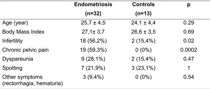
During the study period, we enrolled 32 women in reproductive age affected by ovarian endometriomas and 13 women with a single ovarian functional cyst persistent for more than 6 months (controls) who met the inclusion/exclusion criteria and signed informed consent for the study procedures. In the endometriosis group, 9 (28.125%) women were in stage I, 11 (34.375%) in stage II, 7 (21.875%) in stage III, 5 (15.625%) in stage IV. Endometriosis (n=32) Controls (n=13) p Age (year) 25,7 ± 4,5 24,1 ± 4,4 0.29
Body Mass Index 27,1± 3,7 26,6 ± 3,5 0.69
Infertility 18 (56,2%) 2 (15,4%) 0.02
Chronic pelvic pain 19 (59,3%) 0 (0%) 0.0002
Dyspareunia 9 (28,1%) 2 (15,4%) 0.47
Spotting 7 (21,9%) 3 (23,1%) 1
Other symptoms
(rectorrhagia, hematuria)
3 (9,4%) 0 (0%) 0.54
Table 1. Clinical characteristics in the endometriosis group and controls. Data are reported as means and standard deviations (continuous variables) or by percentages (dichotomous variables).
As shown in Table 1, we did not find significant differences between the two groups for age (p=0.29), BMI (p=0.69) and for the rate of dyspareunia (p=0.47), spotting (p=1) and other symptoms such as rectorrhagia and hematuria (p=0.54), whereas the rate of infertility (p=0.02) and chronic pelvic pain (p=0.0002) was significantly higher in the endometriosis group than controls.
The mean number of mononuclear cells per gram of tissue was 0,79 ± 0,26 x10⁶ in
the control group; 2,42 ± 0,36 x10⁶ in samples of ovarian endometrioma from women
Pag. 31 a 50
in stage IV. As expected, we found that endometriosis group showed a significantly higher number of mononuclear cells than controls, regardless of the stage (p<0.0001 for each stage versus controls).
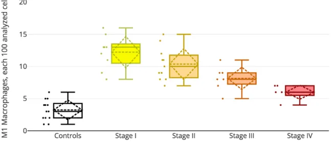
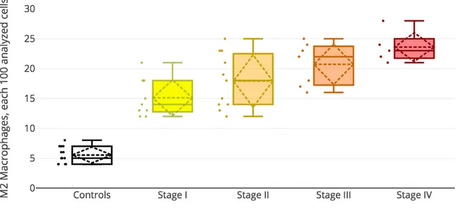
Controls (n=13) Stage I (n=9) Stage II (n=11) Stage III (n=7) Stage IV (n=5) p* M1 macrophages 3,23 ± 1,59 12,22 ± 2,59 10,36 ± 2,54 8,14 ± 1,86 6 ± 1,22 0.0002 M2 macrophages 5,54 ± 1,39 15,11 ± 3,18 17,91 ± 4,59 20,71 ± 3,64 23,6 ± 2,70
Table 2. M1 and M2 macrophages, expressed as quantity each 100 analyzed mononuclear cells, in tissue samples from ovarian endometriomas at different stages of the disease and from ovarian functional cysts (controls).
* ANOVA test for endometriosis groups (results of the Bonferroni post-hoc analysis are reported in the main text).
Table 2 reports the number of M1 and M2 macrophages, expressed as quantity each 100 analyzed mononuclear cells, identified in tissue samples from ovarian endometriomas at different stages of the disease and from ovarian functional cysts (controls). In this regard, we found that the number of both M1 and M2 macrophages was significantly higher in endometriosis group than controls, regardless of stage (p<0.0001 for each stage versus controls). The ANOVA test reported significant differences among the four stages (p<0.0002), which were further investigated using Bonferroni post-hoc analysis.
We did not find significant differences for M1 macrophages quantity between stage I and stage II (p=0.1237) and between stage II and stage III (p=0.0641); conversely, the number of M1 macrophages was significantly higher in stage I respect to stage III (p=0.0035) and stage IV (p=0.0003), in stage II compared to stage IV (p=0.0029), and finally in stage III compared to stage IV (p=0.0493).
Pag. 32 a 50
Similarly, we did not find differences for M2 macrophages quantity between stage I and stage II (p=0.1391), between stage II and stage III (p=0.1927) and between stage III and stage IV (p=0.1652); conversely, the number of M2 macrophages was significantly lower in stage I compared to stage III (p=0.0054), in stage I compared to stage IV (p=0.0003) and in stage II compared to stage IV (p=0.0232).
Moreover, our data analysis shows a trend in progressive decrease of M1 macrophages from stage I to stage IV (Figure 8).
Figure 8. Decreasing trend of M1 macrophages quantity in tissue samples from ovarian endometriomas from stage I to stage IV.
On the contrary, M2 macrophages show a specular trend compared to M1 macrophages, with a progressive increase from stage I to stage IV (Figure 9).
Pag. 33 a 50
Figure 9. Increasing trend of M2 macrophages quantity in tissue samples from ovarian endometriomas from stage I to stage IV.
This trend is consistent with a decreasing M1:M2 macrophages ratio from stage I to stage IV: 0.81 for stage I; 0.58 for stage II; 0.39 for stage III; 0.25 for stage IV.
Pag. 34 a 50
6. Discussion
Endometriosis causes chronic inflammatory condition of the peritoneal microenvironments, which leads to reduced apoptosis rate of endometriotic cells [79] and decreased killing by cytotoxic immune cells [80]. These immunological alterations, in the later stages of the disease cause the typical induction of pro-fibrotic shift in cytokine production and lead to the development of thick adhesions within the pelvis [81]. In particular, accumulating evidence suggests that Vascular Cell Adhesion Molecule 1, aV and b3 integrins, Fibroblast Growth Factor 2, Interleukin
(IL)-1a and Macrophage Inflammatory Protein 1b expression is increased during endometriosis [82]; in addition, a recent characterization of peritoneal cytokine profiles in peritoneal fluid with multiplex immunoassays found that Stem Cell Growth Factor-b, IL-8, Hepatocyte Growth factor, and Monocyte Chemoattractant Protein-1 were significantly higher, while IL-13 was significantly lower in women affected by endometriosis compared to women without endometriosis [83], suggesting a clear role of dysregulated Th1/Th2 response and mobilization and proliferation of hematopoietic stem cells in the pathogenesis of the disease.
In this scenario, recently Chen et al. [84] found CXCL1 and CXCL2, two cytokines known to be released by macrophages and to have significant neutrophil chemoattractant activity, as strongly elevated in the peritoneal fluid even in early stage (I-II) of the disease. In addition, others highlighted that both the percentage of Fas(CD95)-expressing macrophages and the concentration of soluble Fas in the peritoneal fluid were significantly higher in all stages of endometriosis compared with women without the disease [85], confirming also for this immune population what we previously found analyzing Fas/FasL system in mononuclear cells [86].
Pag. 35 a 50
Despite this growing body of evidence, data published so far about the activity and polarization of macrophages in endometriosis are not robust enough to draw a firm conclusion. It has been reported that endogenous macrophages are involved in tissue remodeling during the development of endometriosis, and M1 to M2 phenotypic transition is required for the growth of ectopic lesion in a mouse model [70]. More recently, Nie et al. [87] found a decrease in the percentage of CD86+
macrophages and an increase in the ratio of CD163+/CD86+ macrophages in
peritoneal washings of endometriosis patients, especially those with advanced disease (stages III-IV); in addition, their data analysis suggested that ectopic endometrial homogenate could promote M1 to M2 macrophage polarization, as showed by an increased percentage of CD163+ macrophages and elevated IL-10
expression as well as a decreased percentage of CD86+ cells and lower expression
of IL-12. These results are consistent with the previous report by Mei et al. [88], who found that macrophages expressed higher levels of CD163 and CD209 but lower levels of HLA-DR and CD11c, and showed an increased secretion of IL-10 and decreased secretion of IL-12, when co-cultured with endometrial stromal cells (ESCs). Interestingly, the upregulation of Smad2/Smad3 was found in macrophages after treatment with eutopic and ectopic endometrial homogenates or serum of women with endometriosis in response to lipopolysaccharide stimulation, whereas inhibition of Smad2/Smad3 seems to reverse the macrophage polarization from M1 to M2 [87], suggesting the involvement of the transforming growth factor-β-SMAD2/3 signal transduction pathway in the M1 to M2 polarization.
Nevertheless, most of the investigations performed so far rely on animal models of endometriosis or immune cells isolated from the peritoneal fluid of women affected by endometriosis, and data about M1 and M2 macrophages isolated from endometriotic
Pag. 36 a 50
tissue are still scarce. In this scenario, our study may shed new lights on the topic. Indeed, our data analysis found that the number of both M1 and M2 macrophages was significantly higher in endometriosis group, regardless of stage, than controls. In addition, for the first time to the best of our knowledge, we showed a trend in progressive decrease of M1 macrophages from stage I to stage IV, whereas M2 macrophages progressive increased from stage I to stage IV. Interestingly, our findings are consistent with the transition from classical M1 macrophage activity to an alternate M2 profile recently found in the mouse model [89,90], which correlates to histological hallmarks of initially acute inflammation followed by tissue remodeling during lesion development which occur in humans [81,91].
Despite our results may be considered a step forward in understanding the pathogenesis of endometriosis, some limitations of our study should be taken into account for a proper data interpretation: first of all, we enrolled a limited number of patients, so our findings need to be confirmed in a large dataset; in addition, we used samples from ovarian functional cysts persistent for more than 6 months as controls, since the collection of biopsies of healthy ovarian parenchyma was not possible to ethical reasons; last, and most important, we should consider that polarization of macrophages may vary over the time between a spectrum of epigenetic changes from M1 to M2 activity [92,93]. This is an element of paramount importance, considering also the novel makers to characterize macrophage subpopulations [94], which may further amplify the methodological differences found among the studies published so far.
Pag. 37 a 50
7. Conclusion
Our data analysis suggests that M1 macrophages are more abundant in ovarian endometriomas stages I-II, whereas M2 macrophages are found in large amount in stages III-IV of endometriosis. This may contribute, at least in part, to the pro-inflammatory microenvironment characteristic of the early stages of the disease, whereas the switch to M2-polarization may account for the pro-fibrotic activity of the advanced stages. Despite these results, we take the opportunity to solicit future studies on a larger cohort in order to clarify the behavior of macrophage subpopulations among the different stages of endometriosis.
Pag. 38 a 50
8. Declaration of interests
The authors have no proprietary, financial, professional or other personal interest of any nature in any product, service or company. The authors alone are responsible for the content and writing of the paper.
9. Acknowledgements
The research project was developed and supported by the Ph.D. program of the University of Messina (CIP n. 2014.IT.05.SFOP.014/3/10.5/9.2.02/0006 - CUP n. G47E16000030009).
Pag. 39 a 50
10. References
[1] Sica A, Larghi P, Mancino A, Rubino L, Porta C, Totaro MG, et al. Macrophage polarization in tumour progression. Semin Cancer Biol 2008;18:349–55. doi:10.1016/j.semcancer.2008.03.004.
[2] Gordon S, Martinez FO. Alternative activation of macrophages: Mechanism and functions. Immunity 2010;32:593–604. doi:10.1016/j.immuni.2010.05.007. [3] Natoli G, Lawrence T. Transcriptional regulation of macrophage polarization:
enabling diversity with identity. Nat Rev Immunol 2011;11:750–61. doi:10.1038/nri3088.
[4] Song E, Ouyang N, Hörbelt M, Antus B, Wang M, Exton MS. Influence of Alternatively and Classically Activated Macrophages on Fibrogenic Activities of Human Fibroblasts. Cell Immunol 2000;204:19–28. doi:10.1006/cimm.2000.1687.
[5] Hume DA. The Many Alternative Faces of Macrophage Activation. Front Immunol 2015;6:370. doi:10.3389/fimmu.2015.00370.
[6] Mosser DM, Edwards JP. Exploring the full spectrum of macrophage activation. Nat Rev Immunol 2008;8:958–69. doi:10.1038/nri2448.
[7] Brancato SK, Albina JE. Wound macrophages as key regulators of repair: origin, phenotype, and function. Am J Pathol 2011;178:19–25. doi:10.1016/j.ajpath.2010.08.003.
[8] Dinarello CA. Blocking IL-1 in systemic inflammation. J Exp Med 2005;201:1355–9. doi:10.1084/jem.20050640.
[9] Mantovani A. From phagocyte diversity and activation to probiotics: back to Metchnikoff. Eur J Immunol 2008;38:3269–73. doi:10.1002/eji.200838918. [10] Gleissner CA, Shaked I, Little KM, Ley K. CXC chemokine ligand 4 induces a
unique transcriptome in monocyte-derived macrophages. J Immunol 2010;184:4810–8. doi:10.4049/jimmunol.0901368.
Pag. 40 a 50
[11] Mantovani A, Biswas SK, Galdiero MR, Sica A, Locati M. Macrophage plasticity and polarization in tissue repair and remodelling. J Pathol 2013;229:176–85. doi:10.1002/path.4133.
[12] Wang N, Liang H, Zen K. Molecular mechanisms that influence the macrophage m1-m2 polarization balance. Front Immunol 2014;5:614. doi:10.3389/fimmu.2014.00614.
[13] Zhou D, Huang C, Lin Z, Zhan S, Kong L, Fang C, et al. Macrophage polarization and function with emphasis on the evolving roles of coordinated regulation of cellular signaling pathways. Cell Signal 2014;26:192–7. doi:10.1016/j.cellsig.2013.11.004.
[14] Rojo R, Pridans C, Langlais D, Hume DA. Transcriptional mechanisms that control expression of the macrophage colony-stimulating factor receptor locus. Clin Sci 2017;131:2161–82. doi:10.1042/CS20170238.
[15] Ni W, Zhang Q, Liu G, Wang F, Yuan H, Guo Y, et al. Escherichia coli maltose-binding protein activates mouse peritoneal macrophages and induces M1 polarization via TLR2/4 in vivo and in vitro. Int Immunopharmacol 2014;21:171–80. doi:10.1016/j.intimp.2014.04.025.
[16] Wang F, Lu Z, Hawkes M, Yang H, Kain KC, Liles WC. Fas (CD95) induces rapid, TLR4/IRAK4-dependent release of pro-inflammatory HMGB1 from macrophages. J Inflamm 2010;7:30. doi:10.1186/1476-9255-7-30.
[17] Gregorelli A, Sgarbossa A, Khan S, Soriente A, De Rosa M, Saturnino C, et al. Three Arachidonoylamide Derivatives Inhibit Pro-Inflammatory Genes Expression by Modulating NF-kB and AP1 Activities. Med Chem 2016;12:662– 73.
[18] Lin Y-T, Chen L-K, Jian D-Y, Hsu T-C, Huang W-C, Kuan T-T, et al. Visfatin Promotes Monocyte Adhesion by Upregulating ICAM-1 and VCAM-1 Expression in Endothelial Cells via Activation of p38-PI3K-Akt Signaling and Subsequent ROS Production and IKK/NF-κB Activation. Cell Physiol Biochem 2019;52:1398–411. doi:10.33594/000000098.
Pag. 41 a 50
[19] Simon PS, Sharman SK, Lu C, Yang D, Paschall A V., Tulachan SS, et al. The NF-κB p65 and p50 homodimer cooperate with IRF8 to activate iNOS transcription. BMC Cancer 2015;15:770. doi:10.1186/s12885-015-1808-6. [20] Wang F, Cai R, He D, Zhao Y, Ye Y, Zhang X. Serum IFN-γ-inducible
chemokines CXCL9 and CXCL10 are elevated in nonimmediate drug hypersensitivity reactions. Asian Pacific J Allergy Immunol 2016;34:236–41. doi:10.12932/AP0679.
[21] Hu X, Wang H, Han C, Cao X. Src promotes anti-inflammatory (M2) macrophage generation via the IL-4/STAT6 pathway. Cytokine 2018;111:209– 15. doi:10.1016/j.cyto.2018.08.030.
[22] Yin Z, Ma T, Lin Y, Lu X, Zhang C, Chen S, et al. IL-6/STAT3 pathway intermediates M1/M2 macrophage polarization during the development of hepatocellular carcinoma. J Cell Biochem 2018;119:9419–32. doi:10.1002/jcb.27259.
[23] Gremese E, Alivernini S, Tolusso B, Zeidler MP, Ferraccioli G. JAK inhibition by methotrexate (and csDMARDs) may explain clinical efficacy as monotherapy and combination therapy. J Leukoc Biol 2019:JLB.5RU0519-145R. doi:10.1002/JLB.5RU0519-2019:JLB.5RU0519-145R.
[24] Luo W, Xu Q, Wang Q, Wu H, Hua J. Effect of modulation of PPAR-γ activity on Kupffer cells M1/M2 polarization in the development of non-alcoholic fatty liver disease. Sci Rep 2017;7:44612. doi:10.1038/srep44612.
[25] Odegaard JI, Ricardo-Gonzalez RR, Goforth MH, Morel CR, Subramanian V, Mukundan L, et al. Macrophage-specific PPARγ controls alternative activation and improves insulin resistance. Nature 2007;447:1116–20. doi:10.1038/nature05894.
[26] Namgaladze D, Brüne B. Macrophage fatty acid oxidation and its roles in macrophage polarization and fatty acid-induced inflammation. Biochim Biophys Acta 2016;1861:1796–807. doi:10.1016/j.bbalip.2016.09.002.
Pag. 42 a 50
Biomarkers and Pathways during LPS-Induced M1 Polarization. Mediators Inflamm 2016;2016:6986175. doi:10.1155/2016/6986175.
[28] Norkina O, Dolganiuc A, Catalano D, Kodys K, Mandrekar P, Syed A, et al. Acute Alcohol Intake Induces SOCS1 and SOCS3 and Inhibits Cytokine-Induced STAT1 and STAT3 Signaling in Human Monocytes. Alcohol Clin Exp Res 2008;32:1565–73. doi:10.1111/j.1530-0277.2008.00726.x.
[29] Wang Y-C, He F, Feng F, Liu X-W, Dong G-Y, Qin H-Y, et al. Notch Signaling Determines the M1 versus M2 Polarization of Macrophages in Antitumor Immune Responses. Cancer Res 2010;70:4840–9. doi:10.1158/0008-5472.CAN-10-0269.
[30] Sun K, Xu L, Jing Y, Han Z, Chen X, Cai C, et al. Autophagy-deficient Kupffer cells promote tumorigenesis by enhancing mtROS-NF-κB-IL1α/β-dependent inflammation and fibrosis during the preneoplastic stage of hepatocarcinogenesis. Cancer Lett 2017;388:198–207. doi:10.1016/j.canlet.2016.12.004.
[31] Kumar V. Targeting macrophage immunometabolism: Dawn in the darkness of
sepsis. Int Immunopharmacol 2018;58:173–85.
doi:10.1016/j.intimp.2018.03.005.
[32] Gratchev A, Kzhyshkowska J, Utikal J, Goerdt S. Interleukin-4 and Dexamethasone Counterregulate Extracellular Matrix Remodelling and Phagocytosis in Type-2 Macrophages. Scand J Immunol 2005;61:10–7. doi:10.1111/j.0300-9475.2005.01524.x.
[33] Wang Y, Wehling-Henricks M, Samengo G, Tidball JG. Increases of M2a macrophages and fibrosis in aging muscle are influenced by bone marrow aging and negatively regulated by muscle-derived nitric oxide. Aging Cell 2015;14:678–88. doi:10.1111/acel.12350.
[34] Kubota T, Inoue M, Kubota N, Takamoto I, Mineyama T, Iwayama K, et al. Downregulation of macrophage Irs2 by hyperinsulinemia impairs IL-4-indeuced M2a-subtype macrophage activation in obesity. Nat Commun 2018;9:4863. doi:10.1038/s41467-018-07358-9.
Pag. 43 a 50
[35] Wang L, Zhang S, Wu H, Rong X, Guo J. M2b macrophage polarization and its roles in diseases. J Leukoc Biol 2018. doi:10.1002/JLB.3RU1018-378RR. [36] Philipp D, Suhr L, Wahlers T, Choi Y-H, Paunel-Görgülü A. Preconditioning of
bone marrow-derived mesenchymal stem cells highly strengthens their potential to promote IL-6-dependent M2b polarization. Stem Cell Res Ther 2018;9:286. doi:10.1186/s13287-018-1039-2.
[37] Zagoura D, Trohatou O, Makridakis M, Kollia A, Kokla N, Mokou M, et al. Functional secretome analysis reveals Annexin-A1 as important paracrine factor derived from fetal mesenchymal stem cells in hepatic regeneration. EBioMedicine 2019;45:542–52. doi:10.1016/j.ebiom.2019.07.009.
[38] Ishii T. Close teamwork between Nrf2 and peroxiredoxins 1 and 6 for the regulation of prostaglandin D 2 and E 2 production in macrophages in acute inflammation. Free Radic Biol Med 2015;88:189–98. doi:10.1016/j.freeradbiomed.2015.04.034.
[39] Bouhlel MA, Derudas B, Rigamonti E, Dièvart R, Brozek J, Haulon S, et al. PPARgamma activation primes human monocytes into alternative M2 macrophages with anti-inflammatory properties. Cell Metab 2007;6:137–43. doi:10.1016/j.cmet.2007.06.010.
[40] Mantovani A, Sica A, Sozzani S, Allavena P, Vecchi A, Locati M. The chemokine system in diverse forms of macrophage activation and polarization. Trends Immunol 2004;25:677–86. doi:10.1016/j.it.2004.09.015.
[41] Mantovani A, Sozzani S, Locati M, Allavena P, Sica A. Macrophage polarization: tumor-associated macrophages as a paradigm for polarized M2 mononuclear phagocytes. Trends Immunol 2002;23:549–55.
[42] Corna G, Campana L, Pignatti E, Castiglioni A, Tagliafico E, Bosurgi L, et al. Polarization dictates iron handling by inflammatory and alternatively activated
macrophages. Haematologica 2010;95:1814–22.
doi:10.3324/haematol.2010.023879.
Pag. 44 a 50
al. Differential regulation of iron homeostasis during human macrophage polarized activation. Eur J Immunol 2010;40:824–35. doi:10.1002/eji.200939889.
[44] Rodríguez-Prados J-C, Través PG, Cuenca J, Rico D, Aragonés J, Martín-Sanz P, et al. Substrate fate in activated macrophages: a comparison between innate, classic, and alternative activation. J Immunol 2010;185:605–14. doi:10.4049/jimmunol.0901698.
[45] Nizet V, Johnson RS. Interdependence of hypoxic and innate immune responses. Nat Rev Immunol 2009;9:609–17. doi:10.1038/nri2607.
[46] Vats D, Mukundan L, Odegaard JI, Zhang L, Smith KL, Morel CR, et al. Oxidative metabolism and PGC-1beta attenuate macrophage-mediated inflammation. Cell Metab 2006;4:13–24. doi:10.1016/j.cmet.2006.05.011.
[47] Arnold L, Henry A, Poron F, Baba-Amer Y, van Rooijen N, Plonquet A, et al. Inflammatory monocytes recruited after skeletal muscle injury switch into antiinflammatory macrophages to support myogenesis. J Exp Med 2007;204:1057–69. doi:10.1084/jem.20070075.
[48] Perdiguero E, Sousa-Victor P, Ruiz-Bonilla V, Jardí M, Caelles C, Serrano AL, et al. p38/MKP-1-regulated AKT coordinates macrophage transitions and resolution of inflammation during tissue repair. J Cell Biol 2011;195:307–22. doi:10.1083/jcb.201104053.
[49] Lisi L, Ciotti GMP, Braun D, Kalinin S, Currò D, Dello Russo C, et al. Expression of iNOS, CD163 and ARG-1 taken as M1 and M2 markers of microglial polarization in human glioblastoma and the surrounding normal
parenchyma. Neurosci Lett 2017;645:106–12.
doi:10.1016/j.neulet.2017.02.076.
[50] Giudice LC, Kao LC. Endometriosis. Lancet 2004;364:1789–99. doi:10.1016/S0140-6736(04)17403-5.
[51] DiVasta AD, Vitonis AF, Laufer MR, Missmer SA. Spectrum of symptoms in women diagnosed with endometriosis during adolescence vs adulthood. Am J
Pag. 45 a 50
Obstet Gynecol 2018;218:324.e1-324.e11. doi:10.1016/j.ajog.2017.12.007. [52] Parazzini F, Esposito G, Tozzi L, Noli S, Bianchi S. Epidemiology of
endometriosis and its comorbidities. Eur J Obstet Gynecol Reprod Biol 2017;209:3–7. doi:10.1016/j.ejogrb.2016.04.021.
[53] Revised American Society for Reproductive Medicine classification of endometriosis: 1996. Fertil Steril 1997;67:817–21. doi:10.1016/S0015-0282(97)81391-X.
[54] Laganà AS, Vitale SG, Salmeri FM, Triolo O, Ban Frangež H, Vrtačnik-Bokal E, et al. Unus pro omnibus, omnes pro uno: A novel, evidence-based, unifying theory for the pathogenesis of endometriosis. Med Hypotheses 2017;103:10– 20. doi:10.1016/j.mehy.2017.03.032.
[55] D’Hooghe TM, Debrock S. Endometriosis, retrograde menstruation and peritoneal inflammation in women and in baboons. Hum Reprod Update 2002;8:84–8. doi:10.1093/humupd/8.1.84.
[56] Russell P, van der Griend R, Anderson L, Yu B, O’Toole S, Simcock B. Evidence for lymphatic pathogenesis of endosalpingiosis. Pathology 2016;48:72–6. doi:10.1016/j.pathol.2015.11.004.
[57] Paşalega M, Mirea C, Vîlcea ID, Vasile I, Pleşea IE, Calotă F, et al. Parietal abdominal endometriosis following Cesarean section. Rom J Morphol Embryol 2011;52:503–8.
[58] Sepilian V, Della Badia C. Iatrogenic endometriosis caused by uterine morcellation during a supracervical hysterectomy. Obstet Gynecol 2003;102:1125–7.
[59] Saridogan E, Becker CM, Feki A, Grimbizis GF, Hummelshoj L, Keckstein J, et al. Recommendations for the surgical treatment of endometriosis—part 1: ovarian endometrioma. Gynecol Surg 2017;14:27. doi:10.1186/s10397-017-1029-x.
Pag. 46 a 50
in MRKH cases as a proof for the coelomic metaplasia hypothesis? Reproduction 2019;158:R41–7. doi:10.1530/REP-19-0106.
[61] Zanatta A, Rocha AM, Carvalho FM, Pereira RM, Taylor HS, Motta EL, et al. The role of the Hoxa10/HOXA10 gene in the etiology of endometriosis and its related infertility: A review. J Assist Reprod Genet 2010;27:701–10. doi:10.1007/s10815-010-9471-y.
[62] Porpora MG, Resta S, Fuggetta E, Storelli P, Megiorni F, Manganaro L, et al. Role of environmental organochlorinated pollutants in the development of endometriosis. Clin Exp Obstet Gynecol 2013;40:565–7.
[63] Sofo V, Götte M, Laganà AS, Salmeri FM, Triolo O, Sturlese E, et al. Correlation between dioxin and endometriosis: an epigenetic route to unravel the pathogenesis of the disease. Arch Gynecol Obstet 2015;292:DOI 10.1007/s00404-015-3739-5. doi:10.1007/s00404-015-3739-5.
[64] Singh MN, Stringfellow HF, Taylor SE, Ashton KM, Ahmad M, Abdo KR, et al. Elevated expression of CYP1A1 and gamma-SYNUCLEIN in human ectopic (ovarian) endometriosis compared with eutopic endometrium. Mol Hum Reprod 2008;14:655–63. doi:10.1093/molehr/gan056.
[65] Wu C-H, Guo C-Y, Yang J-G, Tsai H-D, Chang Y-J, Tsai P-C, et al. Polymorphisms of Dioxin Receptor Complex Components and Detoxification-Related Genes Jointly Confer Susceptibility to Advanced-Stage Endometriosis in the Taiwanese Han Population. Am J Reprod Immunol 2012;67:160–8. doi:10.1111/j.1600-0897.2011.01077.x.
[66] Bulun SE, Lin Z, Imir G, Amin S, Demura M, Yilmaz B, et al. Regulation of aromatase expression in estrogen-responsive breast and uterine disease: from bench to treatment. Pharmacol Rev 2005;57:359–83. doi:10.1124/pr.57.3.6. [67] Yang M, Jiang C, Chen H, Nian Y, Bai Z, Ha C. The involvement of osteopontin
and matrix metalloproteinase- 9 in the migration of endometrial epithelial cells in patients with endometriosis. Reprod Biol Endocrinol 2015;13:95. doi:10.1186/s12958-015-0090-4.