Depression of Renshaw recurrent inhibition by
activation
of
corticospinal
fibres in human upper and lower limb
R.
Mazzocchio,
A. Rossi
and
J.
C.
Rothwell*
Laboratorio
di
Neurofisiologia,
Istituto di
Scienze Neurologiche, Universita degli Studi
di
Siena, Viale Bracci,
1-53100 Siena, Italy and *MRC Human Movement
and
Balance Unit, Institute
of Neurology, Queen Square, London WC1N 3BG, UK
1. This study tested whether the recurrent inhibition of soleus and wrist flexor motoneurones could be modified by transcranial magnetic stimulation in human subjects.
2. Magnetic stimulation was given through a circular coil centred at the vertex. The intensityof the magnetic stimulus was subthreshold for evoking a motor responseinthe active soleus and wrist flexor muscles. The recurrent inhibition brought about by a conditioningHi reflex dischargewasestimatedbyatest H'reflex. The modifications of the recurrent inhibition after cortical stimulation were distinguished from the motoneuronalchangesbycomparingH'to areferenceHreflex.
3. In the soleus motoneurones, the reference H reflex was inhibited at a minimum conditioning-test interval of -2ms(Hreflex stimulusbeforemagneticstimulation). In contrast, the H' reflexwasfacilitatedat minimumconditioning-test intervals of +1ms.
In the wrist flexor motoneurones, both H' and reference H reflexes were facilitated. However, at lower cortical stimulus intensities, only the H'reflex was facilitated at
minimumconditioning-test intervals of +1ms.
4. Inboth motoneurone pools, H'facilitationstarted3-4mslaterthan the earliestchanges in the reference H reflex. Also, the threshold of H' facilitation waslower than that of
reference Hreflex.
5. Itisconcluded that facilitationof the H' reflex is produced by corticospinal inhibition of Renshaw cells viaashort interneuronal chaininboth the upper and lower limb.
It is well known from animal studies that Renshaw cell activity can be influenced from various supraspinal
structures(for references see Baldissera, Hultborn & Illert, 1981). In humans, characteristic changes in recurrent inhibition have been observed during various voluntary and postural contractions: (1) weak tonic contraction of triceps surae, quadriceps andpretibialflexors increases the excitability of the respective Renshaw cells more than can be explained by the input from active motor axon collaterals (Hultborn & Pierrot-Deseilligny, 1979; Rossi &
Mazzocchio, 1991), whereas strong tonic contraction of triceps surae decreases the excitability of Renshaw cells whichcanonly be explainedbyasimultaneousinhibitory convergence on the Renshaw cells (Hultborn &
Pierrot-Deseilligny, 1979); (2) before and at the beginning of phasic ramp contractionsof triceps surae there is some indication of Renshaw cell facilitation which shifts progressively to an inhibition that peaks at the end of the ramp (Katz, Pierrot-Deseilligny & Hultborn, 1982); (3) tonic or phasic ramp contractions of the pretibial flexors increase the
recurrent inhibition of soleus motoneurones (Katz &
Pierrot-Deseilligny, 1984); (4) postural contractions while standingunsupported (Pierrot-Deseilligny, Morin,Katz& Bussel, 1977) and after backward tilt of the head-body (Rossi,
Mazzocchio
& Scarpini, 1987) increase the excitability of Renshaw cells projecting to soleusmotoneurones. From all these studies it appears that
varioussupraspinal inputscanmodifytransmissioninthe
recurrentpathway during natural movements. However,
thedescending pathways mediatingtheseeffectscanonly
be hypothesized. In addition, although the presence and distribution ofrecurrent inhibition in the human upper limb has beenrecently reported(Rossi&Mazzocchio, 1992; Katz, Mazzocchio, Penicaud & Rossi, 1993), it is not yet
knownwhether Renshawcellactivityintheupper limbis subjected to the same control as in the lower limb. We therefore tested whether the activity of Renshaw cells could be modified by cortical stimulation in humans at rest.
When the human motor cortex is stimulated
transcranially using an electromagnetic stimulator, the initial short-latency facilitatory effects on
R. Mazzocchio, A. Rossi and J C. Rothwell motoneurones-interneurones at spinal cord level have
been attributed to activity in the fast-conducting component of the corticospinal pathway(Day et al. 1989; Brouwer &Ashby, 1992; Palmer &Ashby, 1992). Weused this tool to explore the effects of the activation of a
'relatively specific' descending tract on the activity of
Renshaw cells oftwofunctionally different motor nuclei: the soleusin the lower limb and the flexorsof the wrist in the upper limb. It will be shown thatcortical stimulation
produces depression of Renshaw cell activity in both motoneurone pools. The type of motor activity in which the descending control of Renshaw cells is involved is discussed.
METHODS
Stimulationandrecording technique
Experiments were performed on twelve normal subjects (23-38years old) with their informed consent and Poeal Ethical Committeeapproval. The muscles investigated were
theflexors of the wrist inthe upper limb (18 experiments on 6subjects)and the soleusinthe lower limb(10 experimentson
6 subjects. The subjects were seated and relaxed with the elbow semiflexed (120deg), the forearm pronated and the handorthe footimmobilized.Surfaceelectrodeswereusedfor both stimulation and recording. The soleus Hreflex was
elicitedby stimulatingtheposteriortibialnerve.The cathode wasahalf-ball(2cmindiameter)inthepoplitealfossa, while
the anode was fixed to the anterior aspect of the knee. The
H reflex from wrist flexors was elicited by stimulating the
median nervethrough bipolarelectrodesplaced2 cmapartin the cubital fossa. Rectangular pulses of 05-1 ms duration
were delivered by constant-current stimulators. The reflex responses were recorded by non-polarizable disc electrodes (09cm indiameter)and measured in termsof the amplitude of musclepotential.
The brain was stimulated using a magnetic stimulator (Magstim 200; The MagstimCo. Ltd, Whitland, Dyfed, UK) witha13cmexternal diameter coil centredatthevertexwith the electric current flowing clockwise in the coil. Intensities
were expressedas apercentageof themaximumoutputof the stimulator.Subthreshold stimuliweredefinedasshocksbelow the intensity needed to produce a motor-evoked response during a small background voluntary contraction of the
soleus muscle or the wrist flexor muscles (approximately
5-10% ofmaximum).In mostsubjectsit was infactdifficultto
evoke a clear motor-evoked potential in the relaxed soleus muscle.
Stimuli weregiven randomly every 6-8 swith orwithout subthreshold shockstothescalp.
Electrophysiologicalmethodforestimatingrecurrent inhibition
The method for exploring the recurrent inhibition of
a-motoneurones in human subjects was that originally
developed in the soleus muscle by Bussel & Pierrot-Deseilligny (1977). The dischargeof the a-motoneuronesby a
group I afferent volley(S1) produces, through theirrecurrent
collaterals, orthodromic activation of the Renshaw cells and an EMG response called conditioning HIreflex. The
inhibitory effectof Renshaw activation is assessed by a second H reflex called test H'reflex elicited by a stimulation supramaximalfor a-motor fibres of the same nerve after the S1 volley. The effect of giving the Si conditioning and the supramaximal test stimuli together at adequate conditioning-test intervals is twofold: on the one hand, it eliminates by collision the descending HI reflex discharge; on the other hand, it opens the wayfor the H' reflex response producedby the orthodromic reactivationof the same motoneurones. Since the H'reflex can only pass along the motor axons in which previous collision has taken place, only motoneurones which have already fired in the first conditioning discharge have their excitability assessed by the following test H' volley. Thus the testH'reflex can be at the very most equal to the HI reflex. In the soleus motor nucleus (Bussel & Pierrot-Deseilligny, 1977), quadriceps and pretibial motor nuclei (Rossi & Mazzocchio, 1991) and flexor carpi radialis and extensorcarpi radialis motor nuclei (Rossi & Mazzocchio, 1992; Katz et al. 1993), when the HI reflex increases beyond a
certainvalue,theabsolute valueof the testH'reflex decreases indicating that an increasingly smaller fraction of the motoneuroneswhich have given rise to HI are involvedinthe
H' response. Provided that the conditioning-test interval is more than 9 ms, the effect of Ib fibre stimulation by the conditioning stimulus can be eliminated since firstly, it has been demonstrated that in humans the duration of lb inhibition from triceps to soleus motoneurones is less than 10ms (Pierrot-Deseilligny et al. 1977), and secondly, the
amountof inhibition of thetestH'reflexisindependent of the conditioning stimulus strength and is only related to the size of the conditioning HIreflex (Pierrot-Deseilligny & Bussel, 1975; Bussel& Pierrot-Deseilligny, 1977). This reduction of the fraction of the motoneurones involved in Hi which is
recruited by the supramaximal test stimulus may well be explained by recurrent inhibition, the amount of which depends upon the size of the conditioning discharge (Ryall, Piercey, Polosa& Goldfarb, 1972). However, the absolute size of thetestH'reflex also dependsontherelationshipbetween
the strength of the Iaconnections and the size of the after-hyperpolarization (AHP). Itcould be expected that the AHP would prevent the firing of the motoneurones with the weakestexcitatoryactionfrom the Ia testvolley (see Awiszus &Feistner, 1993).Accordingly,thelargertheamplitudeofHi,
the greater the number of motoneurones with AHP which prevent excitation by the Iatest volley and the smallerthe
fraction ofmotoneuronesinvolved in HI which isactivatedby the supramaximal stimulus. Ifone disregards the recurrent
inhibition itcould be postulated that the slope of the curve
relating thesizeofH' tothesizeofHi shoulddecrease with increasing HI. However, increasing HI results in recruiting fasterconductingmotoneuroneswhich havealesspronounced AHP than the smallest motoneurones (for references see
Bussel&Pierrot-Deseilligny, 1977). Therefore the AHP alone
cannotgiveadecrease ofH'becausetheindividualAHPs are not enhanced when HI is increased. It follows that if AHP
wasactingalone, there would beafixed limittothe number of
motoneurones available for the testH'reflex. Increasing the conditioning reflex should notdecrease this number, and the
variations in test reflex versus those in conditioning reflex
should exhibit a plateau.Since the value of the testH' reflex
is seen to decrease progressively as HI increases beyond a
specifie value (as illustrated in Fig. 6), which implies a
reduction in the number of motoneuronesinvolved in the test reflex when the conditioning reflex is increased, then this decrement should be due to the recurrent inhibition elicited by the conditioning reflex. Thisconclusion has been recently confirmed using a pharmacological approach to test the validitv of the electrophysiological method (Mazzocchio & Rossi, 1989). As a corollary, it is highly probable that the motoneurone population which is recruited by the supramaximal stimulus is restricted to the motoneurones
*which are the most susceptible to the Ia excitatory effects.
Therefore the motoneurones available for the test H' reflex should not be very different fromeach other regarding their AHP and may constitute a relatively homogeneous fraction within the various motornuclei (see Rossi &Mazzocchio, 1991, 1992).
Experimental procedure with transcranial magnetic stimulation
Cowan, Day,Marsden& Rothwell (1986) using H reflex testing
described changes in spinal cord excitability following subthresholdelectrical stimulation of thescalp. In the relaxed
wristflexor musclesan initial peakfacilitation of the H reflex (lasting 2-5 ms on average) was followed by a more variable and longer-lasting (from 5 to 20 ms) phase offacilitation. The samefacilitatory eventhas beenrecentlydescribed alsowhen
using transcranial magnetic stimulation (Mazzocchio,
Rothwell, Day & Thompson, 1994). In the soleus muscle, in
contrast to the forearm flexors, Cowan et al.(1986) found that the initial event was mainly an inhibition of the H reflex. It follows that changes in the size of the test H' reflex during
scalp stimulationcan beascribed to variation incorticospinal
control of Renshaw cell activity provided that: (a) the size of the HIconditioning reflex is maintained constant, giventhat its variability would per se affect the amplitude ofthe test H' reflex; and (b) the background excitability of theexplored motoneurones is simultaneously estimated, as these are also
subjected to the effectof the corticospinal volley. This can be monitored byusing a reference H reflex having the same size as the H' reflex. However, the sensitivity of these reflexes to
excitation or inhibition is not the same. It has been shown
that identical excitatory inputs to soleusmotoneurones cause a smaller increase of the test H' reflex than of a reference
H reflex of similar size (see Hultborn & Pierrot-Deseilligny,
1979). To verify whether this was also the case for the wrist
flexor motoneurones, the sensitivities of H' and reference H reflexes of wrist flexor muscles tocombined stimulation of
the same nerve were compared. Conditioning stimulation to
the median nerve wasapplied through the same electrode as
the test stimulus. The intensity of thisconditioning stimulus
was just below the threshold of the motor response.
Conditioning-test intervals between 2-5 and 5 ms were
explored.
Experimental protocol
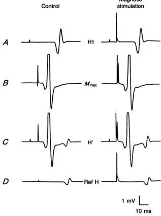
The basic experimental procedure is illustrated in Fig. 1. The sequence of responses on the left represents the control
situation, i.e. in the absence of scalp stimulation. It consists
of: A, a conditioning stimulus alone so as to elicit the
HI reflex. Its sizewasalways made largeenough tobe within the range in which the corresponding H' amplitude was
decreasing (see above). The intensity of the conditioning
stimulus was adjusted so that there was no, or very little, direct M wave; B, the supramaximal test stimulus alone
which produced a maximal direct response; C, the combined
conditioning and supramaximaltest stimuliinorder toelicit thetestH'reflex; D, a stimulus of suchintensityas toevoke the reference H reflex. This was always given at the same
timing as that for the test H'reflex. In the lower limb, conditioning-test intervals of 10, 15 and 20ms were used. It
hasbeen demonstrated that the amplitude of thesoleus test
H'reflex is relatively stable when the conditioning-supramaximal stimulus intervalislargerthan 8ms
(Pierrot-Deseilligny,Bussel, Held&Katz,1976).Inthe upperlimb,the reflex arc being muchshorter, interstimulus intervals of10,12
and 15ms were used(see Katz et al. 1993). Attheseintervals,
possible Ib effects from theconditioningS1stimulusshould be
over, if the durationof Ibinhibitionofsoleusmotoneuronesis
taken as a parameter (seePierrot-Deseilligny, Katz&Morin,
1979). All phases were then repeated during subthreshold magneticstimulation of the scalpdelivered atdifferenttime
intervals with respect tothe supramaximal stimulus for the
H'reflex. The time of the scalp shock was considered to be 0ms; so if the stimulus for theH'reflex wasgiven first, the interstimulus intervalwasnegative.Inthecaseillustratedin
Fig. 1, it was +1 ms. Theamplitude ofHI was keptconstant
by readjusting the stimulusstrength when necessary so asto
be the same as in control conditions. Adifferential neteffect between the H'reflex and the reference Hreflex was looked for as evidence ofcorticospinal control oftransmission in the
recurrent pathway. The amplitude of the two reflexes may vary inversely or inthesame direction. TheamplitudesofH'
and reference Hreflexes obtained during magnetic
stimulation were expressed as a percentage ofthoserecorded under control conditions. The same phases were also studied whilechanging the intensity of themagneticshock whichwas
delivered at a fixed interstimulus interval usually
corresponding to the peak of the effect. At least twenty-four
reflexes (with and without magnetic stimulation) were
measured in every series for later statistical analysis (mean,
standard errorof themeananddifferences between groups by
Student's paired t test). The above protocol was performed in
all subjects. In some subjects, the size of the HIreflex was
changed (while keeping the same cortical stimulus intensity
and thesame delay between the magneticshock and the test
reflex) soas toevokeH'reflexes ofdifferentamplitude. In this case, both reflexes were expressed as a percentage of the maximumdirect response(Mlmax)
The conditioning S1 stimulus to the median nerve always
produced a local sensation without paraesthesia radiating to
the fingers. The possible effects due to the activation of cutaneous fibres weretherefore considered. A pure cutaneous
stimulation mimicking the sensation evoked by the median
nerve stimulus was provided by electrical stimulation of the
skin laterally on the arm. Single shocks of 0 5 ms duration were delivered through a pair of discs (of 1cm diameter)
placedatthe same levelasthe testelectrodes. Thestimulation intensitycorresponded to1P35 timestheperception threshold. Sixteen to thirty-two trials each of control Hreflexes and
R. Mazzocchio, A. Rossi andJ C. Rothwell
collected at a conditioning-test interval of 10ms. Blocks of trials werealso collected using asconditioning stimulation a
sub-motor-threshold cortical shock whichwasdelivered either at the same time as thecontrol H reflex or2msbefore it. The intensity of the cortical shock was such as to produce facilitation of the Hreflex. The effects of the cutaneous
stimulation were then tested on the amount of cortically inducedfacilitation.
RESULTS
The effect of magnetic stimulation of the scalp
on soleus
motor nucleus
Cowan et al. (1986) using subthreshold electrical scalp stimuli showed that the initial effect of the cortical descending volley on the soleus motoneurones was an
inhibition of theHreflex. Inour case theeffectofcortical
stimulation (subthreshold for the motor-evoked response
in the active muscle) was simultaneously studied on two
Control
Hreflexes of comparable size, the reference H and the H'reflexes, the onlydifference between them being due to the effects elicited by the Hi discharge on the H' reflex.
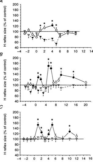
Figure 2 illustrates the time course of cortical action on the reference H reflex (triangles) and on the H' reflex (circles) of the soleus muscle fromthree subjects. The intensity of the magnetic stimulus was about10% below the threshold for the evoked response in the active soleus muscles. In the subjects illustrated in Fig. 2A and B, cortical stimulation depressed the reference Hreflexes at a minimum
conditioning-test interval of-1and -2ms, respectively,
asreported previously by Cowan et al. (1986) and byIles& Pisini (1992). In the subject illustrated in Fig. 2C, there was
no significantchange in thesize of the reference H reflex using this intensity of cortical stimulation. In contrastto its effect on the reference H reflex, cortical stimulation facilitated the H'reflex at minimum conditioning-test intervals of +1msor so. There was no sign of any inhibition
Magnetic stimulation A Hi B
Mmax
C u / H' D - 4---J~-RefH 1 mv L 10msFigure 1. Comparison between soleus EMG responses without and with magnetic brain stimulation
A circular coil centred at the vertex was used to deliver the magnetic shock with a stimulus intensity of70% of the maximum output of the stimulator (about 10% below the threshold for obtainingamotor-evoked responseintheactivesoleus muscleinthissubject). TheEMGresponses from the soleus musclewereobtainedby electrical stimulation of the posterior tibialnerve(PTN)at
thepopliteal fossa with different intensities. A, isolated conditioning IPTN stimulusproducingan
HIreflex of nearly maximal size. B, isolated test PTN stimulus (supramaximal to the ac-motor fibres)causing maximalmotor(Mmax)response. C, combined conditioning and test stimuli (A + B)
at10 msintervalproducinganH'reflex response. D, isolated PTN stimulusproducing areference
Hreflex (Ref H) of thesame sizeand latency as theH'reflex. Magnetic stimulation precedes the supramaximal teststimulusfor theH'reflex and the stimulus for the refHreflex by 1ms. (The time course of the cortical effect for this subject is illustrated in Fig. 2C.)Each trace, with the exception of those showingtheMmax(n=2),isthe averageof12responses.
J. Physiol.481.2
ofthe H' reflexatshorter intervals. In allsubjects the size of both the reference H and H' reflex had returnedtobaseline levelsatconditioning-test intervals of +8ms.
Results similar to those illustrated in Fig. 2B were
obtained in three other subjects, and the mean data
showing theonsetof corticaleffectsareshown from all five
subjects in Fig. 3A. Cortical stimulation produced its earliesteffecton the referenceH reflexatconditioning-test
intervals of -2ms, and thereafter at +1 and +2ms. There
was no effect on the H' response until conditioning-test
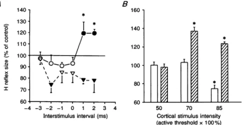
intervals of +1ms.The corticaleffectonthetworesponses was significantly different at conditioning-test intervals of +1 and +2ms(P<0-05). Figure 3B shows theeffectof different cortical stimulus intensities on the size of the
reference H (open columns) and H' (hatched columns) reflexes of the relaxed soleus muscle in all five subjects. The interstimulus interval used corresponded to the first peak of the facilitatory effect on the H' reflex. This varied
from +1to+4ms. At these intervals the referenceHreflex wasconsistently inhibited in all the subjects but the one shown in Fig.2C. Since motor cortical threshold varied among subjects, the cortical stimulus intensity was
expressedas apercentage of theaverageliminal intensity (indicated as 100%) necessary to obtain a motor-evoked
response in the active soleus muscle. It can be seen that
the threshold of the cortical effect on the H'reflex was
lower than that on the reference Hreflex. In fact, on
decreasing the cortical stimulus intensity below 85%, at which thetworeflexes showed opposite changes, therewas
still asignificant facilitatory effect on the H' reflex while
the referenceHreflexwas no longer inhibited. Indeed, at an intensity of 70%, cortical stimulation produced a greater facilitation of the H' response than seen at an
intensity of85%. A possible explanation for this is that theH' response is subject to opposite influences from the
cortex: an inhibition, (which is alsoseen by thereference
Hreflex), andanexcitation(limitedtothe H' reflex itself. Suchopposingeffectsmayalso explain whythesubjectin Fig. 2A who had the largest inhibitory effect on the
reference H reflex also had the smallest excitatory effect
on the H' response. The subjects in Fig. 2B and C had smalleror no inhibitory effectson the reference Hreflex,
and hadlarger excitatoryeffectsonthe H'reflex.
In a sixth subject, subthreshold cortical stimulation delivered between 0 and +4ms ofinterstimulus interval
produced facilitation rather than inhibition ofreference Hreflex whichoverlappedwiththeparallelfacilitation of H'reflex. Since the threshold of the cortical facilitation of
A
Figure2.Theeffectof magneticbrainstimulationonthe sizeof the soleus H'andreference H reflexesat restinthree different subjects
Magnetic stimulator outputwasat80 % inA, 60 % in B and 70%in C.Inall threecasesthiscorrespondedto10% belowthe threshold
for the motor-evokedresponsein the activesoleus muscle. The cortical shockwasgivenattimezero.Control H reflex size is
represented bythe horizontal continuousline. Eachpoint
representsthemeanof 12observations;S.E.M.areexpressed by the vertical bars.Open and filled symbols indicate non-significant and significant differences for the reference H and the H' reflexversus therespectivecontrolvalues;asterisksindicate thepointswhere therewas asignificative difference in the cortical effectonthe reference H and the H' reflex(P<005).
B 200 - 180 8 160 o 140 120 ,, 100 e 80 60 40 200 180 8 160 o 140 120 ,, 100 a 80 60 40 200 180 160 140 120 C 0 cJ 0 0 .I-0 N U) x a) * * _ T,0 \T p 0 \TV/ -6ys01, \ -'?_ s / 4 -2 0 2 4 6 8 10 12 14 *
T*
* Tnn*
i/\ i
- -s.v,1f7aIffvvTv-*~ /w_ *
c
-4 v0i4 8 1A6--4 0 4 8 12 16 20 * * \*57-,V_ o \vZ n OT/ 1uu ¢-V-v-¢0-i - s - v v v 80- 60-40 1--4 -2 0 2 4 6 8 10 12 14 16Interval betweenHreflex stimulusand
corticalshockgivenattime zero(ms)
R.
Mazzocchio,
A. Rossi andJ C. Rothwell A B 140 160 -130 T * * 0 1 12 -1 14 -0 0 0 100 T( 10 u~90-00-6,
x V-80- ~ ' vv8 I 70 j 60.60 -4-3 -2-1 0 1 23450 70 85Interstimulusinterval(ins) Cortical stimulusintensity (activethresholdx 100%)
Figure3. The effect ofmagneticbrainstimulationonthe sizeof the soleus H' and reference Hreflexesinallfivesubjects
A, theonsetof the cortical effectonthe reference H(triangles). andtestH'(circles)reflexes. The magnetic shockwasgiven attimezero. Theintensity of the magneticstimulatorwas onaverage 15% below the threshold for the active evoked response in the soleus muscle. Suchavaluealways
producedasignificantinhibition of thereference H reflexat aconditioning-testinterval of-2 ms inall subjectsbut theoneshown inFig.2C. Control H reflexsize isrepresented bythe horizontal
continuousline. Eachpointisthegrandmeanof the values obtainedatconditioning-testintervals of-3to+2ms;S.E.M.areexpressed bythe bars.Openand filledsymbolsand asterisksasinFig. 2.
B,comparisonof themeanthresholdintensityof the facilitation and inhibition oftestH'(hatched
columns)andreference H(open columns)reflexesatrest.Tonormalize cortical stimulationstrength betweendifferentsubjects,intensityisgivenas apercentageof the averageintensity(indicatedas
100%)needed toproducealiminal motor-evoked responseintheactivesoleus muscle. The size of the conditioned soleus H reflex as a percentage of the control reflex size was measured at
conditioning-testintervalscorrespondingtothefirstpeakof H' facilitation. Theconditioning-test interval thusvariedforthefivesubjectsbetween +1and+4ms. Each vertical bar representsone
standard error of the grand mean. Asterisks referto significant differences betweencontrol and conditionedHreflexes(P<005). A B Magnetic 160 * * Control stimulation 140 * _10 * I .\ 1 Ti 8 120 T Mmax - | > 4 X,,H1I V 0 60 _ refH' 40A -4 -2 0 2 4 6 8 10
Interval between H reflex stimulus and 1
mVL
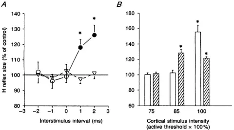
corticalshockgivenattimezero(ms) 10ms Figure 4. The effect ofmagneticbrain stimulationonthesizeofwristflexorH'(circles)and reference H(triangles)reflexesat restThe cortical shockwasgiven attimezero. The intensityof themagnetic stimuluswasset at30%
(10%
below the threshold for the activeevokedresponse).ControlHreflexsize isrepresented bythe horizontal continuous line. Each point represents the mean of 12 observations; S.E.M. arerepresented bythe vertical bars. OpenandfilledsymbolsandasterisksasinFig.2. B,tracesshow
the responsesobtained from thewristflexor muscles under control conditionsand with magnetic stimulation. The latterwasapplied3 msbefore the stimuli for H' and reference H reflexes. Traces
arearrangedasinFig.1.
H'reflex is lower than that of reference H reflex (see Fig.3B), we decreased the cortical stimulus intensity to look for a quantitativedifferential effect between the two reflexes. However, it was not possible to show such an effect.
The
effect of magnetic stimulation on wrist
flexor motor nucleus
In wrist flexor muscles, a subthreshold magnetic scalp
shock delivered through a circular coil centred at the vertex producesan earlyfacilitation of the H reflex which
on average starts at an interstimulus interval of -2 ms and lastsfor several milliseconds (Mazzocchio et al. 1994). In all the subjects studied, subthreshold cortical stimulation caused facilitation of both reference and H'reflexes. This could be interpretedaseither: (a) lack of the putativeRenshaw cell effectonH'reflex which is thus subjected to the same influence as the reference H reflex;
oralternatively,(b) theeffectsonthe H'reflexare masked by the extent of motoneurone facilitation. In the latter case, if the thresholdof the corticaleffectontheH' reflex islower than that of reference Hreflex, as observed in the soleus, then by decreasing the intensity of the magnetic stimulus it should be possible to see some difference between the two reflexes. This was the case in four of the
A 140 130 0T 0120
0~~~~
110 N I x 100 90 80 . -3 -2.sixsubjects studied. Figure4illustrates thetime courseof the effect ofcortical stimulationon the reference H reflex (triangles) and onthe H' reflex (circles) of the wrist flexor muscles in one subject. This time course was obtained using a cortical stimulus intensity which was 10% below the threshold for producing any response in actively contracting muscles. At such an intensity, there was no
effect on the size of thereference H reflex whereas, there
was clear facilitation of the H' response beginning at a minimum conditioning-test interval of +1 ms. Similar timecourses wereobservedin the three other subjects and Fig. 5A shows the detail of the mean onset of cortical effectsfrom all four individuals. The cortical stimulus had
no effect on the reference H reflex but, produced facilitation of the H'response at+1ms. It isworthnoting thatthe effects of cortical stimulationonthe H'reflexes of soleus and wrist flexor muscles were identical in spite of
the fact that magnetic stimulation caused opposite changes in the two motoneurone pools. Figure 5B shows
the effectofchangingtheintensity of the corticalstimulus on reference H (open columns) and H' (hatched columns) reflexes of the relaxed wrist flexor muscles in all four subjects. The interstimulus interval used correspondedto
the firstpeak ofH'reflex facilitation inthefour subjects. It can be seen that it is possible to obtain a significant
B 180 160 140 120 100 80 60 2 -1 0 1 2 3 75 85 100
Interstimulusinterval (ms) Cortical stimulusintensity (activethreshold x100%)
Figure5.Theeffect ofmagneticbrain stimulationonthesizeofwristflexorH'andreference Hreflexesinallfoursubjects
A, theonsetofthe corticaleffectonthe reference H(triangles)and H'(circles) reflexesat rest.The magnetic shock wasgiven attime zero. The intensity of the magnetic stimulationwassuchas to
produce no significant effect on the reference Hreflex. This was on average 15% below the threshold for the active evoked response in the wrist flexor muscles. Control H reflex size is
representedbythehorizontalcontinuousline. Each pointisthegrandmeanof the values obtained
atconditioning-testintervals of-2 to+2 ms; S.E.M.areexpressedbyvertical bars.Openand filled symbols and asterisks as in Fig. 2. B, a comparison of the mean threshold intensity of the facilitation of the H' (hatched columns) and reference H (open columns) reflexes at rest. To normalize cortical stimulation strength between different subjects, intensity is given as a
percentageof the averageintensity(indicatedas100%)needed toproducealiminal motor-evoked response in theactive wristflexor muscles. Thesize of the conditioned wrist flexorHreflexas a
percentage ofthe controlreflexsize wasmeasuredatconditioning-test intervalscorrespondingto
the first peak of H' facilitation. The conditioning-test interval thus varied for the 4 subjects between +1 and +4ms. Each vertical bar represents one standard error of the grand mean.
R. Mazzocchio, A. Rossi and J C. Rothwell
facilitation of the H' reflex alone with cortical stimulus intensities15% below the threshold for the motor-evoked potential in the active wrist flexor muscles. At cortical stimulus intensities corresponding to the active motor threshold, the facilitation of the reference H reflex exceeded that of the H' reflex. Therefore, as observed in the soleus muscle, the threshold for the facilitation induced by the cortical stimulus islower for the H' reflex thanfor the reference Hreflex.
In the remaining two subjects, it was impossible to demonstrate a differential cortical effect on reference and H'reflexes. In theseaswell as in the othercasementioned in the lower limbfindings (the sixth subject), the weaker sensitivityof the H'reflex to excitatory inputs (see below, Fig.7A) probably biased the results against finding an excitatoryeffect from corticalstimulation.
The
effect of
cortical
stimulation
ontestH'
reflexes of different
sizes
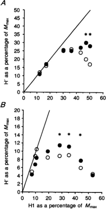
Figure 6 shows the variations of the H' reflex seen while increasing the amplitude of the Hlreflex in control conditions (open circles) and during scalp magnetic stimulation(filled circles). The results obtained in the wrist flexor motoneurones are from the same subject as the data shown in Fig. 4. The intensity of magnetic stimulation wasthe same (10% below the active threshold). The time interval between the magnetic shock and the
supramaximal stimulus for the H' reflex was kept
constant andcorresponded to thepeak of the facilitatory effect (+3ms in this case). Under control conditions, H'
equalled HI at low conditioning reflex amplitude. At
A 50 x 45 ? 40 o 35 a 30 Cu <x 25-0 I- 20' co 15-co 10' I 5 0 B x co E 0 0 0) CD a co U) co nr 20 18 16 14
/**
0 0higher conditioning Hi amplitudes, the size of the
H'reflex reached its maximum and remained constant
until the largest HI amplitudes possible were used at
which the H'reflex started rapidly to decrease. This pattern has already been described in detail in previous work (Rossi& Mazzocchio, 1992; Katz et al. 1993). Cortical stimulationeffectswererestrictedtothat partof the curve
where an increase in Hi reflex led to a fall in H'reflex. Within this range, the cortical action was such as to
increaseconsistently theamplitude of the H'reflex evoked by the same conditioning HI discharge. The results obtained from the soleus motoneurones are from another subject whose data are shown inFig.2A. The intensity of themagnetic stimuluswasthesame(10 % below the active threshold); the time interval between the magnetic shock
and the supramaximal stimulus for the H'reflex was
+4ms.Asobserved in thecaseof the wrist flexormuscles,
the size of the H' reflex was not significantly affected by the cortical stimulus within the ascending slope of the curve. On the other hand, the cortical facilitatory action
on the H' reflex became manifest at higher conditioning
Hi amplitudes and disappeared when the size of the Hlreflex was at its maximum. Similar patterns were
observed inthree othersubjects.
Control experiments
Facilitation ofan Hreflex by a weak stimulation of the homonymous nerve has been used to show that identical excitatoryinputs to soleus motoneurones cause asmaller increaseof thetestH'reflex than ofareference H reflex of similar size(seeHultborn& Pierrot-Deseilligny, 1979). We
Figure6. The effect ofmagneticbrain stimulation on the H' reflex of different size
ResultsfromA, wrist flexors and B, soleus. Control and conditioned H' reflexesare
expressed byopenand filledcircles, respectively.Theamplitudeof the H'reflexis
)10 20 30 40 50 60 plotted against that of theHIreflex, both expressed as a percentage of the maximal motorresponse(Mmax).The identity linerepresents the theoreticalcurve
which wouldbe obtained if H'equalledHI.Eachpoint is the meanoffive
measurements; standard deviationsranged from approximately 5 to15%of mean / * * * values. Asterisks indicate
significant
differences between control andconditionedH'reflexes (P <005).
o
-o S 0 S 0 10 20 30 40 50 60 Hi as apercentage of Mmax J.Phy8iol.481.2 494used the same experimental protocol to verify whether
there was a similardifference in the sensitivity of the two reflexes in the upperlimb. Figure7A shows the mean data obtained in four subjects. It can be seen thatthere was a
significant difference in the amount of facilitation of the tworeflexes, H' being smaller than thereference H reflex.
This finding further supports the notion that the motoneurones giving origin to the H'reflex may have
similar characteristics even though they belong to different motornuclei(seeMethods).
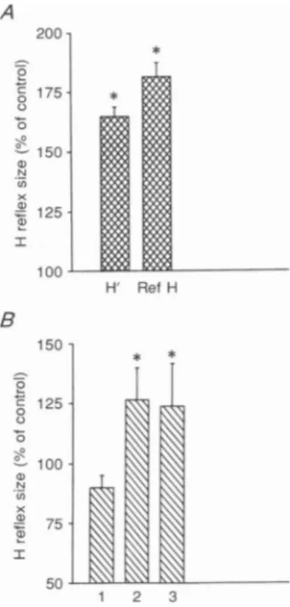
There is evidence that conditioning digit stimulation may reduce theamplitude ofacontrolH reflex fromwrist
flexor muscles at conditioning-test intervals of 9-10 ms (Sabatino, Ferraro, Caravaglios, Sardo, Delwaide & La Grutta, 1992). Since, in the upper limb, we used interstimulus intervals of 10-15 ms between the conditioning HIreflex and the test H'reflex, it could be argued that a reduction in cutaneous inhibition may be responsible for the corticospinal facilitation of the H'reflex. Figure 7B shows the results obtained from two subjects. As shown previously by Malmgren & Pierrot-Deseilligny (1988), a pure conditioning cutaneous stimulationapplied to the skin of the arm, mimicking the sensation evoked by the median nerve stimulus for the Hreflex, produced no significant change in this H reflex at
an interstimulus interval of 10 ms (column 1 in Fig. 7B). Column 2 shows the facilitatory effect of a cortical shock preceding thereference H reflex by 2 ms (conditioning-test intervalof +2 ms). Such an interstimulus interval produced
asignificantfacilitation of the H' reflexinboth subjects. The
effect of the conditioning cutaneous stimulation on the
amount of cortically induced facilitation of the reference H reflex is shown by column 3 in Fig. 7B. It can be seen
that there was no extra facilitation of the reference
H reflex when the cutaneous input was added. On the contrary,there was asmall depression of the facilitation of
a degree similar to the amount of inhibition evoked by
cutaneousstimulationalone.
DISCUSSION
We have inveAtigated how transcranial magnetic stimulation of the motor cortex changes the amount of recurrentinhibition elicited by a conditioning discharge of soleusand wristflexor motoneurones inhumans.
In ourexperiments a conditioning monosynaptic reflex (HI) was used to activate Renshaw cells orthodromically. The resulting recurrent inhibition was then estimated indirectly by asecond testH'reflex produced through a
collision techniqueby thesame motoneurones thathave
given rise to the conditioning discharge. It follows that
the relationship between the amplitudes of the HI conditioning and H' test reflex (as shown in Fig. 6) is
governednotonly by theamountof recurrent inhibition
setupby the conditioning reflex but also by the degreeof
after-hyperpolarization of the motoneurones (Bussel &
Pierrot-Deseilligny, 1977). There is evidence, however, that only recurrent inhibition is responsible for the increasingdepression of the H' reflex with increasing HI discharge, provided that the excitability of
motoneurones is constant (Bussel & Pierrot-Deseilligny, 1977; Mazzocchio& Rossi,1989).
A
Figure7. Effect ofdifferentconditioning stimuli on the wrist flexor
H reflex
A,comparisonof thefacilitationof the H' and reference H reflexes by a preceding (3 ms) weak stimulationof themediannerve(subliminal for evoking theHreflex).Mean(+1 S.E.M.) data from4subjects. Asterisksreferto
significant differences between control and conditionedHreflexes(P< 005). Thedifferenceinthedegreeof facilitation between the conditioned H' and referenceHreflexwasalsosignificant(P<005). B, conditioningstimuli were cutaneousstimulationmimicking thesensationproduced by mediannerve
stimulation(1;conditioning-test interval 10 ms), magnetic brainstimulation
(2; conditioning-test interval2ms; intensity10%below the thresholdfor the
activeevokedresponse), and combinedcutaneousand magneticbrain
stimulation(3). Asterisks refertosignificant differences between control and conditioned Hreflexes (P<005). :i_ 0 L-0 N U) I) I * H' Ref H B 150 -5 0 c 125 0 100 N (n x 4aJ 75 a) Ir 50 l .fr'K\\-4 1 2 3
R. Mazzocchio, A. Rossi and J C. Rothwell
Single electrical or magnetic stimuli applied to the
scalp, whichare below the threshold intensity needed to
produce direct muscle activation at rest, produce significant changes in spinal cord excitabilityasmonitored
by Hreflextesting. In thewristflexormuscles,the initial
effectisashort-latency facilitation, whereasinthe soleus, it is inhibition (Cowan et al. 1986; Iles & Pisini, 1992; Mazzocchio et al. 1994). When combined, asinthepresent
experiments, withtestsforrecurrentinhibition,this input willthereforebecapable of influencing thesizeof thetest H'reflex by way of its influence on a-motoneurones,
regardless of any additional effect it may have on
Renshaw cells. The direct effect on motoneuronal
excitability was quantified by studying the effect of cortical stimulation on the amplitude of a reference
Hreflex, i.e. an H reflex having the same size as the H'reflex. Apart from the effects elicited by the HI discharge on the H' reflex (see Methods), both reference
and H'reflexes should be subjected to the same type of
influences aftercortical stimulation.
Theprincipal findings of this study canbesummarized asfollows. Firstly, cortical magnetic stimulation produces
facilitationof theH' reflex inboth soleusand wristflexor
motor nuclei. In the former this facilitation is
superimposed on the background inhibition of the
reference H-reflex while in the latter itsummates witha
background facilitation. Secondly the threshold for producing facilitation of the H' reflex is lower than that needed to produce changes in the size of the reference
Hreflex in both soleus and wrist flexor motor nuclei.
Thirdly, theonset of H'facilitation follows the cortically induced changes in the reference Hreflex by 3-4ms in
bothmotor nuclei. The moststraightforward explanation
for these results is that the facilitation of the H'reflex
aftercorticalmagnetic stimulation is duetoadecrease in the amount of recurrent inhibition elicited by the
HIreflex discharge. In particular, the cortically induced
inhibitionof Renshaw cells wouldallowalarger number of motoneurones, in which collision has taken place, to be recruited by the test stimulus. However, there are two other possibilities. The first is thatthe magnetic stimulus might somehow reduce the amount of after-hyper-polarization of the motoneurones which have discharged
in the HIreflex. This would facilitate the H'reflex in the
absence ofanychanges in Renshawinhibition. Data from animal experiments have shown that the activation of descendingmonoaminergic pathwayscanreduce the after-hyperpolarization of target motoneurones (Hultborn & Toth, 1989; see also Kiehn, 1991). However, depression of motoneurone after-hyperpolarization occurred 50-100ms after repetitive electrical stimulation ofthe raphe nuclei (Hultborn & Toth, 1989). Moreover, careful inspection of
the curves in Fig. 6 makes it unlikely that cortical
stimulationinthe present experiments could have reduced the after-hyperpolarization of soleus and wrist flexor motoneuronesvia amuch shorter route. It is reasonableto
assumeon thebasisofprevious studies (Bussel & Pierrot-Deseilligny, 1977; Mazzocchio & Rossi 1989) that after-hyperpolarization of motoneurones is the main factor which prevents the H'reflex from continuing to grow in
parallel with the Hlreflex along the identity line (ascending slope ofthecurve). When the Hi reflexissmall, the amountofafter-hyperpolarization whichis setupcan
be overcome by the supramaximal stimulus for the H'reflex. As HI becomes larger, this is no longerpossible
and H' is smaller than HI (see Methods). If cortical stimulation induced a reduction in the after-hyper-polarization of the motoneurones, one would expect the H'reflex to follow the conditioning Hi reflex at high
amplitudes since more motoneurones would become available for H'. This behaviour was not observed. The cortical shock only facilitated the H' reflex (filled circles) on the descending limb of the Hi-H' curve. The second possibility as suggested by Burke, Gandevia & McKeon (1984) is that the amplitude of H reflexes may be affected by grouplb activity elicited by theteststimulusitself. It could therefore be argued that the H' facilitation may be dueto acorticospinalreduction ofIb inhibition.However, ifthiswere soonewould expect thereference H reflex and
the H'reflex tobeaffected inthesame way. This did not occur. Significant quantitative and qualitative differences
(i.e. opposite changes) were observed between the two
reflexesinthe upperand lowerlimb,respectively.
Inhibitory influences on Renshaw cells have been elicited in the cat by electrical stimulation of several
supraspinal loci (for references see Fung, Pompeiano & Barnes, 1987). In particular, activation ofthe pyramidal tract either by stimulation of the pericruciate cortex
(MacLean & Leffman, 1967) or of the capsula interna (Koehler, Windhorst, Schmidt, Meyer-Lohmann & Henatsch, 1978) can reduce Renshaw cell discharge
produced by an antidromic motor nerve volley. In both studies, depression of Renshaw cell activity lasts for a
considerable time after electrical stimulation (between 25 and 35ms). We observed a similar long-lasting change
after magnetic stimulation (see Fig. 2B). Such depression
may be caused either by direct supraspinal inhibition of Renshaw cells or by suppression of a tonic facilitatory
drive. The existence of tonic supraspinal facilitation of Renshaw cells in humans has been recently hypothesized (Mazzocchio&Rossi,1992).This could beoneof thereasons
for the threshold for the cortically induced recurrent
effectsbeing lower than the thresholdfor the
corticospinal
effects on the Hreflex. Whatever the mechanism, the 3-4ms difference in the onset latency of the recurrent effects with respect to that of the earlier motoneuronal J.Phy8iol.481.2
changes suggests that a short interneuronal chain is responsible for the observed cortically induced inhibition
ofRenshawcells.
Functional considerations
The responses elicited by low intensity cortical magnetic stimulation probably are transmitted viafast-conducting
corticospinal fibres (Brouwer & Ashby, 1992; Palmer & Ashby, 1992). These fibres are likely to be responsible
primarilyfor the phasic element of pyramidal control(see
Clough, Kernell & Phillips, 1968; Johansson, Lemon & Westling, 1993). We have found that in humans at rest activationof sucha systemisaccompanied by inhibitionof
Renshaw cell activity in two functionally different
motoneuronepools such as the soleusand theflexors of the
wrist. Similarly, experiments designed to study the
control of Renshaw cells during various natural
movements (Hultborn & Pierrot-Deseilligny, 1979) have
shown that Renshaw activity is strongly diminished
duringphasic ascompared with tonicmusclecontraction. It could, therefore, be suggested that recurrent inhibition is reduced during movements mediated by large
corticospinal neurones. Thisisconsistentwith the absence
or weakness of recurrent inhibition in motor nuclei
subserving the more distal muscles of the human limbs (Rossi &Mazzocchio, 1991, 1992; Katz et al. 1993) which are known to receive thelargerand faster corticospinal fibres and to be under a high degree of pyramidal control (Phillips & Porter, 1964; Palmer & Ashby, 1992). The implication is that Renshaw cell activity may be more
importantintonicrather than phasic muscle contraction. Interestingly, an increase in recurrentinhibitionofsoleus motoneurones has been observed duringthe maintenance
of unsupported upright posture(Pierrot-Deseilligny et al. 1977) and during tonic backward tilt (Rossi et al. 1987).
Also,enhancementof Renshawcell activityduringaweak tonic voluntary effort (Hultborn & Pierrot-Deseilligny, 1979) appears to be a general strategy in the lower limb
(Rossi & Mazzocchio, 1991). Although the frontier between
postureand movement is not quiteclear, it istemptingto suggest that recurrent inhibition may be mainly
concerned with the organizational processes underlying adaptation ofpostural responsestovoluntarymovements
(Rossi,Decchi&Vecchione, 1992).
REFERENCES
Awiszus, F. & FEISTNER, H. (1993). The relationship between estimates ofIa-EPSP amplitude and conduction velocity in human soleusmotoneurones. Experimental Brain Research 95, 365-370.
BALDISSERA, F., HULTBORN, H. & ILLERT, Al. (1981). Integrationin
spinal neuronal systems. In Handbook of Physiology, section 1, TheNervousSystem,vol. 2, MotorControl,ed.BROOKS, V.B.,pp. 509-595. American Physiological Society, Bethesda, AID,
USA.
BROUWER, B. & ASHBY, P. (1992). Corticospinal projections to
lower limb motoneurons in man. Experimental Brain Research 89, 649-654.
BURKE, D.,GANDEVIA, S. C. & MCKEON, B. (1984). Monosynaptic
and oligosynaptic contributions to human ankle jerk and Hreflex.Journal ofNeurophysiology 52, 435-448.
BUSSEL, B. & PIERROT-DESEILLIGNY, E. (1977). Inhibition of human motoneurones, probably of Renshaw origin, elicited by an orthodromic motor discharge. Journal of Physiology 269, 319-339.
CLOUGH, J. F. M., KERNELL, D. & PHILLIPS, C. G. (1968). The distribution of monosynaptic excitation from the pyramidal tract and from primary spindle afferents to motoneurones of the baboon's hand and forearm. Journal of Physiology 198, 145-166.
COWAN, J. M. A., DAY, B. L., MARSDEN, C. D. & ROTHWELL, J. C. (1986). The effect of percutaneous motor cortex stimulation on
Hreflexes in the muscles of the arm and leg in man. Journal of Physiology 377, 333-347.
DAY, B. L., DRESSLER, D., MAERTENS DE NOORDHOUT, A., MARSDEN, C. D., NAKASHIMA, K., ROTHWELL, J. C. & THOMPSON, P. D. (1989). Electric and magnetic stimulationof the human motorcortex: surface EMG and single motor unit responses. Journal of Physiology 412, 449-473.
FUNG,S. J., POMPEIANO,0.& BARNES, C. D. (1987). Suppression of the recurrent inhibitory pathway in lumbar cord segments during locus coeruleus stimulation in cats. Brain Research 402, 351-354.
HULTBORN, H. & PIERROT-DESEILLIGNY, E. (1979). Changes in
recurrent inhibition during voluntary soleus contractions in
man studied by an H reflex technique. Journal of Physiology 297, 229-251.
HULTBORN, H. & TOTH, T. (1989). Raphe-spinal depression of motoneurone after-hyperpolarization. Acta Physiologica Scandinavica 36,35.
ILES, J. F. & PISINI, J. V. (1992). Cortical modulation of
transmission in spinal reflex pathways of man. Journal of
Physiology455, 425-446.
JOHANSSON, R.S., LEAION, R. N. & WXESTLING, G. (1993).Cortical influence over precision grip in man is strongly modulated during differentphases of the task. Journal ofPhysiology459, 469P.
KATZ, R., MAZZOCCHIO, R., PENICAUD, A. & Rossi, A. (1993). Distribution of homonymous and heteronymous recurrent
inhibition in the human upper limb. Acta Physiologica
Scandinavica 149,183-198.
KATZ, R. & PIERROT-DESEILLIGNY, E. (1984). Facilitation of
soleus-coupled Renshaw cellsduring voluntary contraction of
pretibial flexor muscles in man. Journal of Physiology 355, 587-603.
KATZ, R., PIERROT-DESEILLIGNY, E. & HULTBORN, H. (1982). Recurrent inhibition of motoneurones prior to and during ramp and ballistic movements. Neuroscience Letters 31, 141-145.
KIEHN,0.(1991). Plateau potentials andactiveintegrationin the 'final common pathway' for motor behaviour. Trends in
Neurosciences 14, 68-73.
KOEHLER,W.,WINDHORST, U.,SCHMIDT, J., AIEYER-LOHMANN,J. & HENATSCH, H.-D. (1978). Diverging influences on Renshaw cells responses and monosynaptic reflexes from stimulation of
capsulainterna.Neuroscience Letters 8,35-39.
MACLEAN, J. B. & LEFFMAN, H. (1967). Supraspinal control of Renshawcells.Experimental Neurology 18, 94-104.
MALMGREN, K. & PIERROT-DESEILLIGNY, E. (1988). Evidence for non-monosynaptic Ia excitation of human wrist flexor motoneurones, possiblyviapropriospinal neurones. Journal of
498
MIAZZOCCHIO, R.& Rossi,A.(1989).Furtherevidence forRenshaw inhibition in man: a combined electrophysiological and
pharmacologicalapproach.Neurosciecce Letters 106,131-136.
i\IAZZOCCHIO, R. &Rossi, A. (1992). Are Renshaw cells tonically
active inhumans? Jourtnal ofPhysiology 452,IIOP
AIAZZOCCHIO, R., ROTHWELL, J. C., DAY, B. L. & THOMPSON, P. D. (1994). Effect oftonic voluntary activityontheexcitability of
humanmotor cortex.Journal of Physiology 474, 261-267. PALMER,E.& ASHBY,P. (1992).Corticospinalprojectionstoupper
limb motoneurones in humans. Journal of Physiology 448,
397-412.
PHILLIPS, C.G. & PORTER, RI. (1964).Thepyramidalprojectionto motoneuronesofsomemusclegroupsof thebaboon'sforelimb. Progress iniBrain Research 12,222-242.
PIERROT-DESEILLIGNY, E. & BUSSEL, B. (1975). Evidence for
recurrentinhibition bymotoneuronesinhuman subjects.Brain Research 88, 105-108.
PIERROT-DESEILLIGCNY, E., BUSSEL, B., HELD, J. P. & KATZ, R.
(1976). Excitability of humanmotoneuronesafterdischargeina
conditioning reflex. Electroencephalography and Clinical
Neurophysiology 40,279-287.
PIERROT-DESEILLIGNY,E., KATZ, R. & AIORIN, C. (1979). Evidence for lb inhibition in human subjects. Brain Research 166,
176-179.
PIERROT-DESEILLIGNY, E., AIORIN, C., KATZ, R. & BUSSEL, B. (1977). Influence of voluntary movement and posture on
recurrent inhibition in human subjects. Brain Research 124, 427-436.
Rossi, A., DECCHI, B. & VECCHIONE, V. (1992). Supraspinal influences on recurrent inhibition in humans. Paralysis of
descending control of Renshaw cells in patients with mental
retardation. Electroenicephalography an1d Clintical NIeurophysiology 85,419-424.
Rossi, A. & M1AZZOCCHiO, R. (1991). Presence of homonymous
recurrentinhibition inmotoneuronessupplying different lower limb muscles in humans. Experimetntal Brain Research 84,
367-373.
Rossi, A.& AIAZZOCCHiO, R.(1992). Renshawrecurrentinhibition
to motoneurones innervating proximal and distal muscles of the human upper and lower limbs. In MIuscle Afferents and
Spinal Conitrol ofMovemnent, IBROseries,ed.JAMI,L.,
PIERROT-DESEILLIGNY-,E. &ZYTNICKI, D., pp313-319.PergamonPress,
Oxford, UK.
Rossi, A., AIAZZOCCHIO, R. & SCARPINI, C. (1987). Evidence for Renshaw cell-motoneuron decoupling duringtonic vestibular
stimulationinman.Experimental Neurology98,1-12.
RYALL, R.NV., PIERCEY, Ml. F., POLOSA,C. &GOLDFARB, J. (1972).
Excitation ofRenshaw cells in relation to orthodromic and
antidromic excitation of motoneurons. Journal of Neurophysiology 35,137-148.
.A.BATINO, M., FERRARO, G., CARAVAGLIOS, G., SARDO, P.,
DELWVAIDE, P. G. & LA GRUTTA, V. (1992). Evidence of a
controlateral motorinfluence on reciprocal inhibition in man. Journal of Neural Transmission 4, 257-266.
Acknowledgements
WVeshould like to thank all the volunteerswhoparticipatedin
thisstudy. Dr R. Alazzocchio was supportedby grants from
the Human Frontier Science Programme and the Consiglio
NazionaledelleRicerche/BritishCouncil.
Received 11June 1993; accepted 14 MIay 1994.