1 Department of Molecular and Developmental Medicine, Area of Public Health, University of Siena, Italy 2 Health Science Department, University of Florence, Italy
3 Hygiene and Health Organization, Hospital “Careggi”, Florence, Italy
Microbiological surveillance of flexible bronchoscopes
after a high-level disinfection with peracetic acid:
preliminary results from an Italian teaching hospital
G. Troiano
1, A. Lo Nostro
2, C. Calonico
2, N. Nante
1, L. Magistri
3,
M.B. Pulci
3, F. Niccolini
3Key words: Bronchoscopes, disinfection, healthcare-associated infections Parole chiave: Broncoscopi, disinfezione, infezioni correlate all’assistenza
Abstract
Background. Flexible bronchoscopes are heat labile, complex and difficult to clean, and some nosocomial outbreaks related to bronchoscopy have been reported in literature. The aim of our study was to determine, through a systematic monitoring, whether bronchoscopes’ cleaning and disinfection procedures have been correctly adopted by health operators.
Methods. We conducted a 19 months-long prospective study in the Unit of Pulmonology at Careggi Teaching Hospital (Florence, Italy), analyzing endoscopes that were reprocessed through a high-level disinfection procedure. Samples collection was performed weekly by two trained operators. Results were organized in a database and then exported for descriptive and inferential statistical analysis.
Results. From February 2016 to September 2017 we collected 218 samples from bronchoscopes’ valves (N=109) and from their inner channels (N=109). Staphylococci were found in 34 samples (15.69% of all samples). Pseudomonas was found in 11 samples (5.04% of all samples). Pseudomonas aeruginosa wasn’t found in any sample.
Conclusions. Our results came out to be better than similar studies in literature and demonstrated that a correct endoscopes’ hygiene should be part of a more complex strategy of surveillance and control of healthcare-associated infections. However, a continuous monitoring of endoscopes could provide a wider view about this problem, and more reliable results.
Introduction
F l ex i b l e b r o n c h o s c o py ( F B ) i s one of the most common procedures performed in pulmonary medicine to obtain cytopathological or microbiological samples
(1). It is usually employed for the visual inspection of the airways, for laser therapy, for electrocautery and for the placement of stents (2).
Flexible bronchoscopes are heat labile, complex and difficult to clean (3). Although
infectious complications due to flexible bronchoscopy are uncommon, nosocomial outbreaks related to bronchoscopy have been reported, and endoscopes (including bronchoscopes) are the most common devices linked to outbreaks (4). Several bronchoscope-related outbreaks and pseudo-outbreaks of Pseudomonas aeruginosa and
Serratia marcescens infection have been reported (5-7), usually due to some breaches of the cleaning and disinfection guidelines (8, 9).
One of the most important limitations connected to the use of bronchoscopes is that heath sterilization is not practicable, because autoclaving damages the instrument and ethylene oxide reprocessing requires an often unacceptable amount of time (10-12).
Therefore, cleaning and high-level disinfection are routinely performed (13, 14).
Bronchoscopes’ washing is not sufficient for decontamination and an additional disinfection is always recommended (15). Microorganisms could be transmitted from previous patients or from contaminated devices used for reprocessing procedures. The analysis of the suspected and confirmed causes of contamination suggested that a better adherence to the guidelines could prevent more than 90% of the exogenous endoscopy-related outbreaks (16). Nevertheless, variations in bronchoscope disinfection practices (17) and inconsistencies among published reprocessing guidelines have been described (18).
T h e a i m o f o u r s t u d y w a s t o determine whether the procedures of bronchoscopes’cleaning and disinfection would have been correctly adopted by health operators. Through a systematic monitoring, we tried to understand whether an adequate decontamination of bronchoscopes would have been achieved.
Methods
We conducted a 19 months-long prospective study in the Unit of Pulmonology at Careggi Teaching Hospital (Florence, Italy), analyzing endoscopes that were reprocessed through a high level disinfection procedure. Samples collection was performed weekly by two trained operators.
For the analysis of the inner lumen, 5 mL of saline solution (through a sterile syringe) were injected and collected in a sterile box. For the analysis of the valves, a sterile cotton swab was used. Samples were marked with an identification code and immediately transported to the Applied Microbiology Laboratory of the Health Science Department, University of Florence.
Microbiological analysis
For the microbiological analysis a technique for semi-quantitative analysis was used, that included an enrichment phase in order to restore damaged cells. Culture media were furnished by Thermo Scientific (Thermo Scientific Oxoid Ltd., Hampshire, UK).
Enrichment procedures are used to improve the sensitivity of detection (19) because, although viable, microorganisms on environmental surfaces may be more difficult to culture (20).
The analysis of samples collected from the inner lumen was performed in several steps: 1 ml was immersed in Thioglycollate medium USP, incubated in anaerobic conditions at 37±1°C for 48 h and then streaked on Sulphite Polymyxin Sulphadiazine Agar for the detection of
Clostridium species and 1 ml was immersed in 10 ml of Letheen Broth, incubated at 37±1°C for 24 h and then streaked on the appropriate culture media for the detection of the other microorganisms.
Each sample derived from the valves was immersed in 5.5 ml of Letheen Broth, incubated at 37±1°C for 24 h and
then streaked on the appropriate culture media.
Identification of Clostridium spp suspected colonies was performed through Api 20A (bioMérieux Italia Spa, Florence, Italy). Furthermore, 1 ml of the same sample was immersed in 9 ml of Letheen Broth, incubated at 37±1°C for 24 h and then streaked on appropriate culture media for the detection of the other microorganisms.
Each sample derived from the valves was immersed in 5.5 ml of Letheen Broth, incubated at 37±1°C for 24 h and then streaked on the appropriate culture media.
Staphylococcus spp. detection was performed using Mannitol Salt agar incubated at 37°C for 24 h. Identification of S. aureus suspected colonies was performed through Api Staph (bioMérieux Italia Spa, Florence, Italy). Enterobacteriaceae detection was performed using Desoxycholate agar incubated at 37°C for 24 h. Detection and identification of Enterococcus spp. were performed through Slanetz and Bartley agar incubated for 48 h at 44°C. After incubation, suspected colonies of Enterococcus spp. were transferred to tryptone soy agar and incubated for 24 h at 37°C and identified through Gram stain, catalase production, and rapID STR (Thermo Scientific). Pseudomonas spp. detection was performed through Cetrimide agar incubated for 48 h at 28°C. Suspected colonies of P. aeruginosa were identified through Microbact 24E (Thermo Scientific). Yeast and molds detection was performed using Rose Bengal agar incubated for 48 h at 28°C and through optical microscopy observations.
The presence of these specific microorganisms is related to environmental and human contamination, so they can be used as indicators, to evaluate the overall hygienic conditions, and in particular S.
aureus and E. coli are index of the possible occurrence of pathogens. Furthermore, we focused on the detection of Gram-positive cocci, such as Staphylococcus or
Enterococcus spp., as they survive in dry environments for longer periods than Gram-negative bacteria (21).
The semi-quantitative scoring was determined by observing the growth in the four quadrants, which suggested the approximate number of colony forming units per ml (CFU/ml) of the bacteria in the specimen, and classified as follows: - no growth; + colonies present in the first quadrant; ++ colonies present in the second and third quadrant; +++ colonies present in the fourth quadrant.
Reprocessing procedure
The reprocessing procedure approved by the Health Direction consisted of multiple steps, according to ANOTE-ANIGEA Guidelines (22). Before the disinfection, the instruments should be cleaned and dried to remove the coarse residues. The high level disinfection procedure should be performed using peracetic acid (1:12.5 or 1:3) inserted into the inner lumen through a syringe. After this stage, the entire endoscope should be dipped in the peracetic acid for at least 10 minutes. Then the instrument should be washed with sterile distilled water in each part. To avoid the bacterial and fungal growth in the residual water, alcohol 70% should be used in the external part and in the inner lumen and dried using medical air (0.5 bar).
Statistics
The results were organized in a database and then exported for descriptive and inferential statistical analysis. In order to identify factors related to microbiological contamination of bronchoscopes (i.e. the site of sampling and the year of sampling), bivariate analysis was performed using the Chi square test and the calculation of Odds Ratios (Stata 12.0 statistical software, StataCorp, College Station, Texas USA). The level of significance was set at p<0.05.
Results
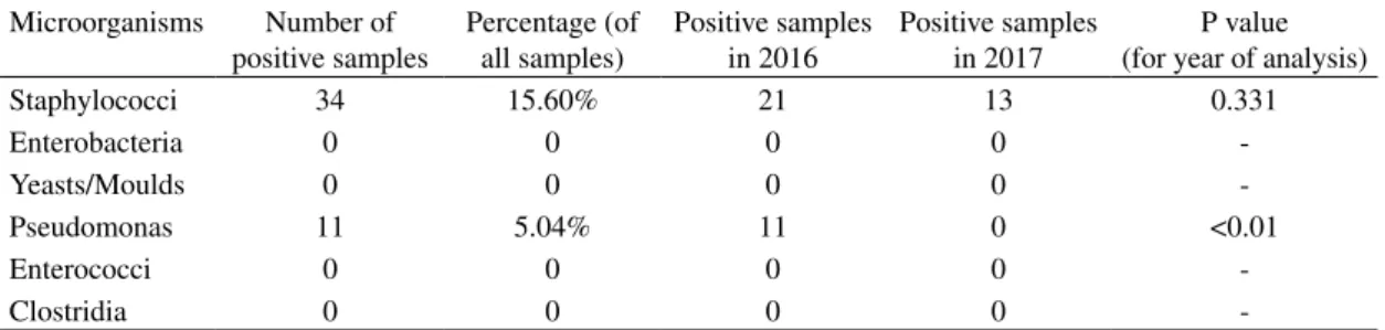
From February 2016 to September 2017 we collected 218 samples from bronchoscopes’ valves (N=109) and from their inner channels (N=109): 118 (54.13%) in 2016; 100 (45.87%) in 2017.
As described in Table 1, yeasts/moulds, enterobacteria, enterococci and clostridia were not detected.
Staphylococci were found in 34 samples (15.69% of all samples): 14 (6.42%) samples
had a low contamination; 14 (6.42%) a medium contamination; 6 (2.75%) a high contamination (see Figure 1). There wasn’t a statistically significant difference in the contamination due to the site of sampling (p=0.263, number of positive samples col-lected from the inner channels=14, number of positive samples collected from the valves=20) and to the year (p=0.331).
Pseudomonas was found in 11 samples (5.04% of all samples): 5 (2.29%) sam-ples had a low contamination; 2 (0.92%) a
Figure 1 – Positive samples grouped by level of contamination Table 1 – Microorganisms found in the collected samples
Microorganisms Number of positive samples Percentage (of all samples) Positive samples in 2016 Positive samples in 2017 P value (for year of analysis)
Staphylococci 34 15.60% 21 13 0.331 Enterobacteria 0 0 0 0 -Yeasts/Moulds 0 0 0 0 -Pseudomonas 11 5.04% 11 0 <0.01 Enterococci 0 0 0 0 -Clostridia 0 0 0 0
-medium contamination; 4 (1.83%) a high contamination (see Figure 1). Pseudomonas aeruginosa wasn’t found in any sample. Positive samples were found only in 2016. There wasn’t a statistically significant diffe-rence in the contamination due to the site of sampling (p=0.51, number of positive sam-ples collected from the inner channels=4, number of positive samples collected from the valves=7).
Discussion and conclusions
A better bronchoscopists’ knowledge about the national and international recommendations for bronchoscope reprocessing practices, is reported in literature as the first and most important recommendation for a correct endoscopes’ hygiene. However the extent of guidelines awareness is unclear. Many experienced bronchoscopists are unfamiliar with these guidelines and with the local practices related to bronchoscopes reprocessing. Publication of bronchoscope-specific, comprehensive reprocessing guidelines in the pulmonary literature could help to increase familiarity with this crucial process (23).
As already published by Cetre et al., microbiological results highlighted the necessity to regularly test bronchoscopic samples, in order to improve the design of bronchoscopes, and to implement the surveillance. These strategies are fundamental to improve both the quality of care and patient safety (3).
To our knowledge, the first study describing the results of a weekly microbiological surveillance program specifically designed for bronchoscopes was published in 2015. It demonstrated that an high-level disinfection wasn’t able to avoid a 3% risk of bronchoscope contamination by potentially pathogenic microorganisms and that 70% ethyl alcohol effectively could reduce it (1).
However there is a controversial issue because significant differences and inconsistencies regarding endoscopes drying procedures have been identified in current guidelines (18).
Some guidelines, in fact, recommend flushing channels with alcohol before using the forced air (as we have done in our study), while other guidelines consider the forced air alone as the preferred method (24).
In a study published in 2015, Gavaldà et al. (1) used an interesting reprocessing procedure: after each endoscopic examination, a manual procedure was used to clean the external and internal surfaces, including brushing and flushing the working channel with a solution of water and enzymatic detergent. Automated flexible endoscope reprocessors were used in the bronchoscopy suite whereas manual disinfection was performed in the intensive care area and in the operating room. The manual method consisted of the totally bronchoscope’s immersion for at least 12 min in Phthalaldehyde (Cydex OPA ortho-Phthalaldehyde, division of Ethicon, Inc. a Johnson & Johnson Co, Irvine, Canada). Inner channels were manually irrigated with the disinfectant. Once disinfected, the equipment was rinsed with sterile water and medical air was forced through to dry the endoscopes without alcohol rinsing. In the bronchoscopy suite, automatic disinfection was performed through an endo-thermo-disinfector using peracetic acid (Olympus miniETD2, Olympus Europa Holding MBH, Hamburg, Germany) (1).
In our study we followed an analogous procedure reaching quite similar results: we found Pseudomonas in 11 samples (5.04% of all samples) and staphylococci in 34 samples (15.69% of all samples); in their study they found staphylococci in 28 samples (4.5%), Pseudomonas aeruginosa in 3 (0.4%). However they found also yeasts/ moulds (12 samples) and enterobacteria (2 samples).
The presence of Pseudomonas and
Serratia could represent an important problem: several studies have been reported in literature about the isolation of Pseudomonas aeruginosa and Serratia
marcescens from bronchoalveolar lavage (BAL) samples. Pseudomonas is able to form biofilms, and these biofilms are extremely difficult to remove from the endoscope channels (16). The prevention of pseudo-outbreaks requires meticulous use of preventive measures for infection-prone medical procedures (25).
However, as Gavaldà et al. reported, it is probable that the origin of some dangerous microorganisms could be linked to defective or incomplete manual cleaning before the disinfection process. Regarding some microorganisms (i.e. Acinetobacter
baumannii, Pseudomonas aeruginosa and Aspergillus fumigatus) it should be said that they can survive for prolonged periods on unanimated surfaces. So the contamination of the equipment could be due to a defective cleaning or disinfection process or to an exogenous acquisition from the environment (1). One of the most important limits of our work could be considered the short period of study. However our purpose is to go on monitoring the endoscopes for a longer period of time in order to have a wider view of this problem. Another limit could be represented by the frequency of sampling: we decided to perform it weekly, but in some months (e.g. August and December) the collection and analysis was not performed because of organizational problems. However, several authors reported that there is neither consensus on the frequencies and methods of sampling nor on the interpretation of the results (1). So we invite other colleagues to perform further studies in order to have a unique point of view about this issue.
Concluding, the implementation and compliance with decontamination guidelines should be improved further, and it is also
necessary to improve a proper communication between physicians, bronchoscopists, and laboratory technicians not only to promptly recognize pseudo-outbreaks, but also to establish a coordinated approach to handle and implement correct disinfecting procedures (26). Our results highlighted that endoscopes’ hygiene must be part of a more complex and multidisciplinary strategy of surveillance and control of healthcare-associated infections.
Riassunto
Sorveglianza microbiologica di broncoscopi flessibili dopo disinfezione di alto livello con acido peraceti-co: risultati preliminari da un’azienda ospedaliero universitaria italiana
Introduzione. I broncoscopi flessibili sono termolabili e molto difficili da pulire, e in letteratura sono stati ripor-tati alcuni casi di infezioni correlate alla broncoscopia. Lo scopo del nostro studio è stato determinare se le procedure di pulizia e disinfezione dei broncoscopi fos-sero state correttamente adottate dagli operatori sanitari, attraverso un monitoraggio sistematico che ci ha con-sentito di valutare se fosse stata raggiunta un’adeguata decontaminazione degli stessi.
Metodi. Abbiamo condotto uno studio prospettico della durata di 19 mesi presso l’Unità di Pneumologia dell’Azienda Ospedaliero-Universitaria Careggi (Firen-ze), analizzando endoscopi che sono stati riprocessati attraverso una procedura di disinfezione di alto livello. Il prelievo dei campioni è stato eseguito settimanalmente da due operatori addestrati. I risultati sono stati organizzati in un database e quindi esportati per analisi statistiche descrittive e inferenziali.
Risultati. Da febbraio 2016 a settembre 2017 abbiamo raccolto 218 campioni dalle valvole (N = 109) e dai lumi interni (N = 109) dei broncoscopi. Stafilococchi sono stati trovati in 34 campioni (15,69% di tutti i campioni).
Pseudomonas è stato trovato in 11 campioni (5,04% di tutti i campioni). Nessuno dei campioni è risultato positivo per Pseudomonas aeruginosa.
Conclusioni. I nostri risultati sono risultati migliori rispetto a studi simili in letteratura e hanno dimostrato che un’igiene corretta degli endoscopi deve far parte di una strategia più complessa di sorveglianza e controllo delle infezioni correlate all’assistenza. Tuttavia un mo-nitoraggio continuo degli endoscopi potrebbe fornire una visione più ampia di questo problema e risultati più affidabili.
References
1. Gavaldà L, Olmo AR, Hernández R, Domínguez MA, Salamonsen MR. Microbiological monitor-ing of flexible bronchoscopes after high-level disinfection and flushing channels with alcohol: Results and costs. Respir Med 2015; 109(8): 1079-85. doi:10.1016/j.rmed.2015.04.015. 2. Seijo LM, Sterman DH. Interventional
pulmo-nology. N Engl J Med 2001; 344(10): 740-9. 3. Cêtre JC, Nicolle MC, Salord H, et al. Outbreaks
of contaminated broncho-alveolar lavage related to intrinsically defective bronchoscopes. J Hosp Infect 2005; 61(1): 39-45.
4. Srinivasan A, Wolfenden LL, Song X, et al. An outbreak of Pseudomonas aeruginosa infections associated with flexible bronchoscopes. N Engl J Med 2003; 348(3): 221-7.
5. Sorin M, Segal-Maurer S, Mariano N, Urban C, Combest A, Rahal JJ. Nosocomial trans-mission of imipenem-resistant Pseudomonas
aeruginosa following bronchoscopy associated with improper connection to the Steris System 1 processor. Infect Control Hosp Epidemiol 2001; 22(7): 409-13.
6. Sammartino MT, Israel RH, Magnussen CR.
Pseudomonas aeruginosa contamination of fibreoptic bronchoscopes. J Hosp Infect 1982; 3(1): 65-71.
7. Vandenbroucke-Grauls CM, Baars AC, Visser MR, Hulstaert PF, Verhoef J. An outbreak of
Serratia marcescens traced to a contaminated bronchoscope. J Hosp Infect 1993; 23(4): 263-70.
8. Ostrowsky B. Endoscopes -- current practices and controversies in infection control. Semin Infect Control 2001; 1: 267-79.
9. Weber DJ, Rutala WA. Lessons from outbreaks associated with bronchoscopy. Infect Control Hosp Epidemiol 2001; 22(7): 403-8.
10. Avero MS, Bond WW. Chemical disinfection of medical and surgical materials. In: Block SS, ed. Disinfection, sterilization, and preservation. 4th ed. Philadelphia: Lea & Febiger, 1991: 617-40.
11. Stilo A, Troiano G, Melcarne L, et al. Hand washing in operating room: A procedural com-parison EBPH– 2016; 13(2): e11734-1-7.doi: 10.2427/11734.
12. Serafini A, Troiano G, Franceschini E, Calzoni P, Nante N, Scapellato C. Use of a systematic risk analysis method (FMECA) to improve quality
in a clinical laboratory procedure Ann Ig 2016; 28: 288-95.
13. Rutala WA, Weber DJ. Disinfection of endo-scopes: review of new chemical sterilants used for high-level disinfection. Infect Control Hosp Epidemiol 1999; 20(1): 69-76.
14. Muscarella LF. High-level disinfection or “steri-lization” of endoscopes? Infect Control Hosp Epidemiol 1996; 17(3): 183-7.
15. Bär W, Márquez de Bär G, Naumann A, Rüsch-Gerdes S. Contamination of bronchoscopes with
Mycobacterium tuberculosis and successful sterilization by low-temperature hydrogen per-oxide plasma sterilization. Am J Infect Control 2001; 29(5): 306-11.
16. Kovaleva J, Peters FT, van der Mei HC, Dege-ner JE. Transmission of infection by flexible gastrointestinal endoscopy and bronchoscopy. Clin Microbiol Rev 2013; 26(2): 231-54. 17. Ruddy M, Kibbler CC. Endoscopic
decon-tamination: an audit and practical review. J Hosp Infect 2002; 50: 261-8.
18. Muscarella LF. Inconsistencies in endoscope-reprocessing and infection-control guidelines: the importance of endoscope drying. Am J Gastroenterol 2006; 101: 2147-54.
19. Landers T, Hoet A, Wittum TE. Swab type, moistening, and preenrichment for
Staphylococ-cus aureus on environmental surfaces. J Clin Microbiol 2010; 48 (6): 2235-36.
20. Oliver JD. Recent findings on the viable but nonculturable state in pathogenic bacteria. FEMS MIcrobiol Rev 2010; 34(4): 415-25. 21. Hirai Y. Survival of bacteria under dry
condi-tions; from a viewpoint of nosocomial infection. J Hosp Infect 1991; 19(3): 191-200.
22. ANOTE-ANIGEA. Linee Guida Pulizia e Disin-fezione in Endoscopia. Update 2011. Available on: https://www.anoteanigea.it/linee-guida- public/linee-guida-pulizia-e-disinfezione-in-endoscopia-update-2011/ [Last Accessed: 2018, February 5].
23. Srinivasan A, Wolfenden LL, Song X, Perl TM, Haponik EF. Bronchoscope reprocessing and infection prevention and control: bronchoscopy-specific guidelines are needed. Chest 2004; 125(1): 307-14.
24. Mehta AC, Prakash U, Garland R, et al. American College of Chest Physicians and American As-sociation for Bronchology Consensus Statement. Prevention of flexible bronchoscopy-associated infection. Chest 2005; 128: 1742-55.
25. Silva CV, Magalhães VD, Pereira CR, Kawagoe JY, Ikura C, Ganc AJ. Pseudo-outbreak of
Pseu-domonas aeruginosa and Serratia marcescens related to bronchoscopes. Infect Control Hosp Epidemiol 2003; 24(3): 195-7.
26. Saeed DK, Shakoor S, Irfan S, Hasan R. My-cobacterial contamination of bronchoscopes: Challenges and possible solutions in low re-source settings. Int J Mycobacteriol 2016; 5(4): 408-11.
Corresponding author: Gianmarco Troiano, MD, Department of Molecular and Developmental Medicine, Area of Public Health, University of Siena, Via A. Moro 2, 53100 Siena, Italy