Università degli Studi di Ferrara
DOTTORATO DI RICERCA IN
SCIENZE BIOMEDICHE
CICLO XXV
COORDINATORE Prof. Capitani Silvano
Cochlear implant outcomes and genetic mutations
in children with ear and brain anomalies
Settore Scientifico Disciplinare MED/32
Dottorando Tutore
Dott. BUSI MICOL Prof. GRANIERI ENRICO
Cotutore
______________ Prof. MARTINI ALESSANDRO
_________________
_____________________________
(firma) (firma)
INDICE
ABSTRACT ... 3
1. INTRODUCTION ... 5
1.1 Classification of inner ear abnormalities ... 13
1.2 Brain anomalies and hearing loss ... 18
1.3 Objectives ... 18
2. MATERIALS AND METHODS ... 19
2.1. Audiological assessment. ... 20
2.2. Imaging data ... 21
2.3. Genetic and molecular analysis ... 23
2.4. Speech perception (pre-operative assessment and post-operative outcomes): ... 23
2.4.1. Listening Progress Profile (Lip) (developed by Archbold 1994) ... 26
2.4.2. Early Speech Perception Test (ESP) (Moog & Geers, 1990) ... 28
2.4.3. GASP (Glendonald Auditory Screening Procedure) (Erber, 1982) ... 29
2.4.4. Northwestern University – Children’s Perception of Speech (NU-CHIPS) - (Elliot and Katz, 1980) ... 30
2.4.5. Word Intelligibility by Picture Identification (WIPI) (Ross and Lerman, 1979) . 31 3. RESULTS ... 33
4. DISCUSSION ... 47
5. CONCLUSIONS ... 59
NEUROIMAGING ATLAS... 61
ABSTRACT
Introduction. Cochlear implantation (CI) was a significant surgical innovation in the 20th century and represented the first artificial sensory organ that was applied in clinical medicine. Currently, CI is still one of the most effective medical procedures. Nonetheless, cochlear implantation in adults and children represents a controversial issue from an economic, clinical and ethical point of view, especially in specific clinical conditions that could compromise the CI outcome and drastically reduce the chance of an acceptable development of perceptual and linguistic capabilities. These conditions should certainly include the presence of inner ear malformations or brain abnormalities.
Objectives. The aims of this work were to study the diagnostic value of high resolution computed tomography (HRCT) and magnetic resonance imaging (MRI) in children with sensorineural hearing loss who were candidates for cochlear implants and to analyse the anatomic abnormalities of the ear and brain in patients who underwent cochlear implantation. We analysed the effects of ear malformations and brain anomalies on the CI outcomes. Finally, we described the genetic mutations that we found in the study group. A control study group of implanted patients without ear and brain anomalies was obtained (virtually) from clinical and literature data for statistical purposes.
Materials and methods. The present study is a retrospective observational review of cochlear implant outcomes among hearing-impaired children who presented ear and/or brain anomalies at neuroimaging investigations with MRI and HRCT. Furthermore, genetic results from molecular genetic investigations (GJB2/GJB6 and, additionally, in selected cases,
SLC26A4 or mitochondrial-DNA mutations) on this study group were herein described.
Longitudinal and cross-sectional analysis was conducted using statistical tests.
Results. Between 1 January 1996 and 1 April 2012 at the ENT-Audiology Department of the University Hospital of Ferrara, 620 cochlear implantations were performed. There were 426
implanted children at the time of the present study (who were <18 years). Among these, 143 patients (64 females and 79 males) presented ear and/or brain anomalies/lesions/malformations at neuroimaging investigations with MRI and HRCT. The age of the main study group (143 implanted children) ranged from 9 months and 16 years (average = 4.4; median = 3.0). The most common inner ear malformation is represented by an enlarged vestibular aqueduct; brain lesions are usually represented by white matter disorders. The 35delG in the GJB2 gene remain the most common mutation.
Discussion and Conclusions. Good outcomes with cochlear implants are possible in patients who present with inner ear or brain abnormalities, even if central nervous system anomalies represent a negative prognostic factor that is made worse by the concomitant presence of cochlear malformations. Common cavity and stenosis of the internal auditory canal (less than 2 mm) are negative prognostic factors even if brain lesions are absent.
Because the cochlear implantation is an invasive and expensive surgical procedure, the identification of predictive factors, even in hearing-impaired patients with cochlear and brain anomalies, is one of the most important goals, because it can help to guide rehabilitation programs that are tailored to meet the expectations of clinicians, teachers and parents. Our findings suggest that cochlear implantation (CI) is a safe and effective procedure even for patients with brain and inner ear abnormalities. Nonetheless, specific conditions, such as a common cavity, or in general, the absence of modiolus and the stenosis of the internal auditory canal, can increase the risk of post-operative complications and prevent the achievement of acceptable perceptual categories. For the aforementioned conditions, it is strictly recommended that cochlear implant indications, neuroimaging and surgery are performed in experienced hospitals.
Keywords: inner ear malformations, brain abnormalities, cochlear implantation, neuroimaging, GJB2 mutations.
1. INTRODUCTION
Cochlear implant (CI) was a significant surgical innovation in the 20th century and represented the first artificial sensory organ applied in clinical medicine. Currently, CI is still one of the most effective medical procedures. It can evoke acoustic sensations by electrically stimulating the inner ear [1]. Nonetheless, cochlear implantation in adults and children represent a controversial issue from an economic, clinical and ethical point of view. The U.S. Food and Drug Administration (FDA) approved the use of cochlear implants as an auditory rehabilitation procedure in adults in 1984. Six years later, in 1990, the FDA approved cochlear implants also for children who were affected by severe-to-profound bilateral sensorineural hearing loss (with a tonal hearing threshold that is major, or equal to 90 dB nHL decibel normal hearing level, in the better ear), who were at least 1 year of age and who also did not have the benefit of adequate auditory training (typically from 4 to 6 months) with hearing-aid amplification (http://www.fda.gov/cdrh/cochlear/). A similar position was taken in 2007 in a statement of the Joint Committee on Infant Hearing, which noted that
“Cochlear implantation should be given careful consideration for any child who appears to receive a limited benefit from a trial with appropriately fitted hearing aids. Infants with
profound bilateral hearing loss are candidates for cochlear implantation at 12 months of
age, and children with bilateral severe hearing loss are eligible at 24 months of age. The
presence of developmental conditions (e.g., developmental delay, autism) in addition to
hearing loss should not, as a rule, preclude the consideration of cochlear implantation for
an infant or a child who is deaf” [2]. According to the U.S. Food and Drug Administration
(FDA), as of December 2010, approximately 219,000 people worldwide have received cochlear implants [3]. Currently, there are three major FDA and EC (European Community) approved cochlear implant providers that are commonly used: Cochlear Ltd (Australia), Advanced Bionics (USA) and Med-El (Austria). The Neurelec’s devices (MXM – Neurelec
Corp, Vallauris, France) have not yet been approved by the FDA, but they have a CE registration and are currently used in Europe. All of the product devices comprise similar component parts: 1) an external unit, called a transmitter or processor, which constitutes a microphone, speech processor and batteries to drive the system; 2) an implanted device, called a receiver or stimulator, which can electrically stimulate the inner ear through an electrode array inserted into the cochlea. “The implant converts acoustic sound to electrical
pulses that stimulate the auditory nerve. Acoustic input enters the microphone, which is
worn on the ear, and is sent to the speech processor for analysis of intensity in a number of
set frequency bands. The resulting information is sent from the externally worn transmitting
coil to the subcutaneous receiver–stimulator through FM waves. These components are held
together by a pair of magnets so that they are separated only by the thickness of the skin
flap. Each frequency band is assigned to a particular electrode along the implanted array
(mimicking the normal basal-to-apical organization of high to low frequencies in the
cochlea). If instructed, this array will provide a biphasic electrical pulse to stimulate the
auditory nerve. The magnitude of the pulse provided by any one electrode will depend on the
acoustic intensity within the assigned frequency band and the dynamic range of current
(minimum to maximum) programmed for that electrode” [4] (Fig. 1). The design of the
electrode array must incorporate biocompatibility, mechanical stability and practical fabrication and must minimise insertion trauma. From a surgical point of view, efforts to reduce the insertion trauma must be accomplished at the materials and design levels as well as through the surgical technique. The CI has been devised to allow full access to verbal communication, through the perception of phonetic hallmarks. The success of this method is then given in general by the achievement of verbal communication performance by improving the skills of verbal perception, to become comparable to people with normal hearing. In the paediatric population, in children with profound hearing loss, which is
unsuitable for obtaining significant results with traditional hearing aids, CI (if performed early) allows the optimal development of auditory and linguistic abilities, which drives toward adequate communication and intellectual development.
Fig. 1 - The internal (receiver) and external (transmitter) components of a cochlear implant. “The cross section of the
cochlea shows the electrode array surgically placed in the scala tympani. The implant converts acoustic sound to electrical pulses that stimulate the auditory nerve. Acoustic input enters the microphone, which is worn on the ear, and is sent to the speech processor for analysis of intensity in a number of set frequency bands. The resulting information is sent from the externally worn transmitting coil to the subcutaneous receiver–stimulator through FM waves. These components are held together by a pair of magnets so that they are separated only by the thickness of the skin flap. Each frequency band is assigned to a particular electrode along the implanted array (mimicking the normal basal-to-apical organisation of high to low frequencies in the cochlea). If instructed, this array will provide a biphasic electrical pulse to stimulate the auditory nerve. The magnitude of the pulse provided by any one electrode will depend on the acoustic intensity within the assigned frequency band and the dynamic range of current (minimum to maximum) programmed for that electrode” [4].
On the other hand, the overall results of the cochlear implantation should not ignore the specific or unique conditions of the patients. It is known that auditory deprivation genres, first, have disadvantages in communication and reduction of daily activities that are closely
Papsin BC, Gordon KA. Cochlear implants for children with severe-to-profound hearing loss. N Engl J Med. 2007;357(23):2380-7.
related to hearing impairment, and second, over time, involves other areas of personal life, leading in some cases to permanent deficits. For example, children with congenital profound hearing loss accumulate disadvantages over time in language skills and certain learning areas, which can lead to permanent limitations of personal skills at a later age. Because of the auditory habilitation/rehabilitation training with a cochlear implant, most of these patients can reach a complete disability "compensation". The effectiveness of CIs in adults and children is demonstrated by many contributions in the medical literature. Specifically, several studies have reported the results of the development of auditory perceptual and expressive verbal abilities in children with pre-, peri- and post-lingual deafness. Clinical experiences across the world have also shown that, among children with pre-verbal onset hearing loss, there is a critical period for the development of language skills, presumably due to the underlying neuronal plasticity, and learning would be strictly dependent on the presence of an adequate auditory input, which explains the need for early intervention to prevent the occurrence of a delay in language development and perceptive or expressive skills. A multi-centre study that was conducted on a large number of cases [5] showed that the majority of patients who were implanted reached the performance of verbal recognition more or less quickly depending on the age at the time of the implant surgery.
The performance of the patients who received CIs varies significantly as a consequence of a substantial number of audiological and extra-audiological factors (age of hearing loss onset, duration of auditory deprivation, auditory function residuals, presence of associated disability and comorbidity, language skills at the time of the CI, duration of CI use, the presence of certain malformations of the inner ear, socio-economic status and familial environment) [6-13]. Even if a unique prediction of the results after CI is not yet available, mostly because of the extreme heterogeneity in the aetiology among profound hearing-impaired patients, there are many prognostic factors that can contribute to the audiological
assessment. Among these, we emphasise the importance of inner malformations and brain anomalies, which are a part of the present study.
Because cochlear implantation is an invasive and expensive surgical procedure, the identification of predictive factors is one of the most important goals; knowledge of the predictive factors can help to guide rehabilitation programs that are tailored to meet the expectations of clinicians, teachers and parents. The perceptual performance improves rather quickly if the selection criteria are correct. In agreement with research in the literature, the majority of patients who received a CI at our clinic have shown a rapid advancement in their perceptual abilities, reaching the 6th perceptive category according to the Geers and Moog classification one year after surgery [14]. The perception of verbal sounds is an important starting point to activate the processes of linguistic acquisition. The development of language depends on auditory skills and maturation of cortical functions (memory, attention, intellectual abilities). The linguistic processes typically follow the perceptive processes with a variable latency, which appears to be related to the age of the patients at the time of surgery [15,16]. Specifically, children implanted in a very early age (8-12 months) experience linguistic evolution with a speed that is higher than that of normal hearing children of the same age, probably because of using lines of development in various linguistic domains that are different from the usual capabilities of normal hearing children. In other words, congenitally deaf patients develop many abilities to reach an adequate communication condition (lip-reading, visual reinforcement) that, when auditory function has recovered, work in synergy with auditory inputs, enhancing perceptual and visual skills; an analogous process is similar in visually impaired patients who develop, more than normal, auditory and olfactory skills. On the other hand, it cannot be excluded that the normal auditory input is more detailed and complex and, thus, that it takes more time to develop and
integrate superior central functions. In contrast, cochlear implant stimuli are simpler; thus, they do not need complex integration in the corpus callosum or cortical areas.
With the advances in molecular genetics over the past 20 years, our understanding of the pathogenesis of sensorineural hearing loss has greatly increased. To date, more than 400 syndromes have been described in which hearing loss is a regular or occasional feature [17] and, after more than two decades of molecular biological analysis, approximately 95 non-syndromic recessive and 64 non-non-syndromic dominant loci have been identified [18], even if not all of them have led to the identification of specific genes. The most common mutations that are responsible for hearing loss involve the GJB2 gene; SLC26A4 mutations are the second cause of genetic hearing loss and the first among syndromic deafness. The SLC26A4 (PDS) gene mutations result in abnormalities of the endolymphatic system, which lead to the dilation of the vestibular aqueduct as seen in Pendred syndrome. Nonetheless, other genes can be involved: normal expression of the PAX2 and PAX3 genes is necessary for the normal development of the cochlea. The FGF3 gene appears to be necessary for differentiation within the otic vesicle. The EYA1 gene has an important role in encoding transcription factors and mutations of EYA1 are responsible for EYA1-related disorders that include BOR (branchio-oto-renal) syndrome, BO (branchio-otic) syndrome and OFC (oto-facio-cervical) syndrome [19]. Several studies have shown that patients who have mutations of GJB2 (or Cx26) (OMIM * 121011) usually have excellent perception of speech and an optimal language development after the cochlear implant [20,21]. Additionally, it has been reported that GJB2 mutations are not usually accompanied by macroscopic inner ear malformations. Nevertheless, there is no evidence that genetic mutations or the interaction of a genetic diagnosis with other prognostic factors (such as abnormalities of the ear and brain) can predict CI outcomes.
Therefore, pre-operative neuroimaging is mandatory in cochlear implant candidates for diagnostic and surgical purposes. This step usually includes an MRI as well a high-resolution computed tomography (HRCT) of the temporal bone. MRI should be performed with contrast (gadolinium), unless otherwise noted or unless the test is in children who are not believed to have lesions that require contrast to be diagnosed. HRCT of the temporal bone does not require iodine contrast. Note that Magnetic Resonance Imaging (MRI) is relatively contraindicated after cochlear implantation or it is arguably possible. Various experimental studies have shown that MRI scans can safely be performed with the CI in place [22,23]. This arrangement does not imply that it is generally safe to perform MRI in CI patients, because the type of implant, fixation method and MRI units and sequences could vary. Even if it can be performed safely, the distortion that is caused by the implanted magnet will cause sub-optimal interpretations [22]. For the aforementioned reasons, cochlear implantation can be contraindicated in patients who need periodic follow-up with MRI.
At our clinic, pre-operative radiological imaging of cochlear implant candidates includes both HRCT and MRI of the temporal bone during the same session, during anaesthesia, if required, which usually occurs in children. In addition to the MRI of the inner ear, we also perform brain and brainstem MRI scans. These scans enable us to exclude any incidental brain abnormalities that can contraindicate CI surgery. MRI is the best diagnostic tool for detecting malformations such as cochlear nerve hypoplasia or aplasia, and it is the best screening tool for early cochlear ossification following bacterial meningitis [24-26]. HRCT provides better images and definition of the facial nerve canal, middle ear and otic capsule [27]. Central nervous system findings have been reported in 20–40% of the patients [25-29]. Some of these findings could result in neurodevelopmental delay and could negatively impact the outcome of cochlear implantation [30]. Nonetheless, increased experience in cochlear implantation has led to more children with abnormal cochleovestibular anatomy
being considered as candidates [12]. According to the literature, approximately 20% of the children who have sensorineural hearing loss have associated radiological anomalies of the temporal bone [31-33]. These temporal bone anomalies are accompanied by a wide range of hearing acuity, varying degrees of progression of hearing loss, and the presence or absence of related non-otological anomalies [33].
The first cochlear implants in children with inner ear malformations were performed in the late 1980’s [33]. Since this time, cochlear implants have been fully described and surgical techniques have been tested; precautions have been identified that are necessary to prevent or remedy possible complications. Additionally, different electrode arrays have been developed that allow a choice of the most suitable array for the different types of malformations [13,34-39]. In general, “cochlear implantation is a relatively safe procedure
with a low complication rate that ranges from 6% to 20%. Major complications are those
that are life-threatening or require surgery, whereas minor complications are those that can
be medically treated. The inner ear malformations can increase the risk of meningitis,
cerebro-spinal fluid leakage and facial nerve palsy” [40,41]. We should note that “the rate
of postoperative complications was higher in patients with anomalous inner ears than in
patients with normal inner ears, most were minor and could be managed conservatively.
These findings suggest that cochlear implantation (CI) is safe even for patients with
anomalous inner ears in experienced hospitals” [41]. Nevertheless, the functional results
reached by these children (perceptual and linguistic performance) are still poorly described and have not been predictable. Case studies have limited conclusions because of the high inter-individual variability. For these reasons, it is not yet possible to draw clear guidance from the literature on which to base the selection of candidates [12,13,41]. The malformations in fact allow the correct insertion of a number of electrodes that are usually sufficient, and the patterns of neural responses are adequate to accomplish the recognition of
an open set words. However, specific conditions that prevent a correct coupling between the electrode array and the cochlear nerve, even if the latter is present, such as a common cavity, are usually characterised by a poor outcome, unless very specific surgical strategies are enacted.
1.1 Classification of inner ear abnormalities
Approximately 80% of the children who have a congenital hearing loss have no macroscopic abnormalities of the ear, and their hearing loss is assumed to be the result of dysfunctions at a cellular level in the membranous inner ear. The remaining 20% can present inner ear dysplasia, which can be demonstrated on high-quality neuroimaging (HRCT without contrast and MRI, with contrast in adults or in specific cases). The inner ear abnormalities, whether dysplastic or nondysplastic, can be isolated or can be part of a multi-organ syndrome [19]. In considering the problems specific to implanting children with ear malformations, it is valuable to consider the normal development of the inner ear. “During
the third week after conception, the otic placode appears on the surface ectoderm. This
becomes invaginated to form the otic pit and, in turn, the otic vesicle or otocyst by the end of
the fourth week. The vesicle divides into a ventral component, which gives rise to the saccule
and the cochlear duct, and a dorsal component, which forms the utricle, semicircular canals,
and endolymphatic duct. In the sixth week, the saccule forms a tubular outpocketing at its
lower pole, the cochlear duct. This penetrates the surrounding mesenchyme and by the end
of the eighth week has completed 21.2 turns. In the 10th week, vacuolisation in the
surrounding mesenchyme around the cochlear duct forms the scala tympani and vestibuli,
and following that membranous structures such as the organ of Corti begin to develop in the
cochlea. The semicircular canals appear as outpocketings of the utricle about the sixth week.
disappear giving rise to the three semicircular canals. The endolymphatic sac and duct is
initially a wide structure, but the proximal portion, the duct narrows about the seventh week.
If there is no developmental arrest before the eighth week, a normal cochlea is formed [24].
The statoacoustic ganglion forms from neural crest cells and cells derived from the otic
vesicle. It subsequently splits into cochlear and vestibular components, the spiral ganglion
and Scarpa’s ganglion. There is increasing interest in the genetic and molecular factors that drive this complicated process [19].
In considering the specific problems for the implant of children with deafness associated with inner ear malformations, it is worthwhile to consider the normal anatomy of the inner ear (Fig. 2). The cochlea has a discrete anatomical variability in its shape and dimensions, as reported by several authors [42-48]. In humans, “cochleae are mirror-shaped, fluidfilled,
coiled, fairly symmetrical bony tubes (3.2–4.2 cm long) situated in the petrous pyramids of
the temporal bones. Cochlea is composed of ~2 and three-fourth turns. The outer cochlear
wall had a mean length of 42.0 mm while the first turn was 22.6 mm (range, 20.3–24.3 mm)
representing 53% of the total length. The total height of the cochlea can vary from 4.4 to 5.9
mm (mean 5.1 mm). The large variations in cochlear lengths, angles between turns, and
position in the skull base can influence the straightforwardness for the insertion of a CI
electrode particularly passing the first turn. Most frequently damaged are the spiral
ligament at the junction of the first and second half of the first turn, basilar membrane (BM), and osseous spiral lamina” [48]. “The average LSCC (lateral semicircular canal) bony island width is 3.7 mm (normal range, 2.6-4.8 mm). Cochlear hypoplasia (< 4.4 mm) had a
positive predictive value of 100% for SNHL, whereas cochlear hyperplasia and bony island
dysplasia were less predictive” [44].
Developmental malformations that affect the otic capsule result in anomalies of both the membranous and bony labyrinth. The specific timing of the insult during otic capsule
development determines the resulting type of malformation along a spectrum of congenital inner ear malformations that can occur when the normal process of development is impacted, even if it is not necessarily understood why this result occurs.
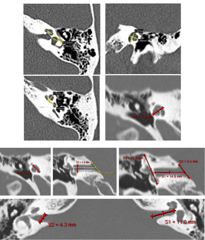
Fig. 2 - MRI and CT scans showing normal inner ear anatomy. The total length of the cochlear partition ranges from
38.6 to 45.6 mm (average 42.0 mm.). The length of the basal turn (53% of the total length) ranges from 20.3 to 24.3 mm. The width of the internal auditory canal is between 4.85 and 5.02 mm. The height ranges from 4.39 to 4.62 mm and the length from 11.22 to 11.44 mm [48].
The best review of these developmental anomalies is given by Sennaroglu and Saatci [24-25], which is an update on the valuable original work by Jackler et al. [31], and the present study is essentially based on their classification (Tab. I) [45]. Table II shows the recurrence rate of different temporal bone malformations [46]. No cases of variations in the dimension of the facial recess have been reported.
COCHLEAR MALFORMATIONS
DESCRIPTION %
Michel deformity Complete aplasia of all of the inner ear structures. This condition can be associated with aplasia of the internal auditory canal.
6
Cochlear aplasia Absent cochlea in the presence of a normal, dilated, or hypoplastic vestibule and semicircular canal system. The internal meatus might be normal.
5
Common cavity Round or ovoid structure that represents the cochlea and vestibule. The cochlear nerve might be absent.
8
Cochleovestibular hypoplasia
Cochlear and vestibular structures are clearly seen to be separate from each other. The cochlea is smaller than normal but might have a normal internal architecture (cochlea height < 4.4 mm and less than 2.5 turns). The cochlear aperture could be aplastic, and the cochlear nerve might be absent.
12
Incomplete partition type I (IP1): cystic cochleovestibular malformation
Cystic cochlea (the absence of partitions and cochlea has cystic aspects; furthermore, the modiolus and cribriform areas are absent) and dilated vestibule. Most cases have an abnormal internal auditory canal (IAC) with an absent lateral wall of the IAC.
20
Incomplete partition type II (IP2): Mondini malformation
Cystic cochlear apex (the cochlea has a normal basal turn but the medial and apical turns are fused together, leading to a one and and a half turn cochlea)
Minimally dilated vestibule Large vestibular aqueduct
19
Incomplete partition type III (IP3); X-linked deformity/deafness.
This condition is similar to IP1, except for the presence of the interscalar septa; the modiolus is absent. It could be misdiagnosed as a mixed type hearing loss.
2
Enlarged vestibular aqueduct (EVA) is the most common radiologically detectable inner ear malformation that is associated with hearing loss. Approximately 80% of hearing impaired patients with at least one mutation in the SLC26A4 gene presented EVA at neuroimaging. Nevertheless, EVA can be associated with other conditions, such as Waardenburg syndrome or BOR (branchio-oto-renal) syndrome.
MALFORMATIONS FREQUENCY
Malformed ossicles 67%
Abnormal oval window 57%
Facial Nerve Anomalies 57%
Enlarged Vestibular Aqueduct (EVA) 52% Enlarged Cochlear Aqueduct (ECA) 43%
Abnormal round window 29%
Middle ear aplasia 19%
Tab. II – Recurrence rates of the temporal bone malformations [46].
Malformations of the inner ear that represent an absolute contraindication to the IC are the bilateral aplasia of the cochlea or cochlear nerve. Aplasia of the cochlear nerve is frequently in the presence of a common cavity, CHARGE association and when the internal auditory canal has a diameter of less than 2 mm [25,47]. Other causes are rare syndromes such as LAMM [49] and Möbius. Hypoplasia of the cochlear nerve is not an absolute contraindication to the CI, but it could lead to reduced performances [12,13].
Anomalies of the facial nerve, malformations that increase the risk of gusher (such as enlarged vestibular aqueduct, absence or hypoplasia of the modiolus, dilation of the vestibule or semicircular canals and stenosis of the round window), common cavities, and abnormal vessel paths can complicate the surgical approach.
Finally, some malformations of the inner ear, such as common cavity, absence or hypoplasia of the modiolus, can complicate the fitting and can require repeated control mapping due to the instability of the electrode array inside the malformed cavity.
In summary, the various types of inner ear malformations could have quite different prognoses for auditory performance after a CI depending on the degree of the dysplasia. It should be emphasised that no single centre has, as yet, assembled a very large series of cochlear implants in dysplastic ears or brain abnormalities; thus, a clear picture of the expected outcomes in different groups is not possible at this stage.
1.2 Brain anomalies and hearing loss
A comprehensive review of brain anomalies is not an objective of the present study. Even if updating revisions of central nervous system (CNS) abnormalities are available in the literature, they were not discussed in this research. In theory, every CNS lesion can be responsible for hearing loss. In clinical practice, CNS lesions are usually represented by: neoplasms/neoformations, malformations, vascular/ischemic and gliotic lesions, white-matter disorders, demyelinating disorders, and viral/bacterial infections (meningitis and cytomegalovirus infections).
1.3 Objectives
The aims of this work were to study the diagnostic value of HRCT and MRI in children who have sensorineural hearing loss and who were candidates for cochlear implants; we also aim to analyse anatomic abnormalities of the ear and brain in patients who underwent CI. We analysed the effects of ear malformations and brain anomalies on the CI outcomes. Finally, we described the genetic mutations that we found in the study group. A control study group of implanted patients without ear and brain anomalies was obtained (virtually) from clinical data and literature data for statistical purposes.
MATERIALS AND METHODS
This study is a retrospective observational review of cochlear implant outcomes among hearing-impaired children who presented ear and/or brain anomalies at neuroimaging investigations with MRI and HRCT. Furthermore, genetic results from molecular genetic investigations (GJB2/GJB6 and, additionally, in selected cases, SLC26A4 or
mitochondrial-DNA mutations) on this study group were herein described. Longitudinal and cross-sectional
analyses were conducted using statistical tests. To create more homogenic groups and more study-specific findings (e.g., EVA) and to obtain more significant analysis, the main study group was divided into different subgroups, which were named with alphabetic letters, as follows (Tab. III):
STUDY GROUPS INCLUSION CRITERIA
MAIN STUDY GROUP Cochlear implant recipients who had less than 18 years of age at the time of surgery and
who presented neuroradiological findings at pre-operative neuroimaging investigations.
SUBGROUP A Patients with inner ear malformations (with or without brain anomalies)
SUBGROUP B Patients with internal auditory canal stenosis
SUBGROUP C Patients with only inner ear malformations (without brain anomalies)
SUBGROUP D Patients with only brain anomalies (without inner ear malformations)
SUBGROUP E Patients with inner ear malformations AND brain lesions or abnormalities (with brain
anomalies)
SUBGROUP F Monolateral CI
SUBGROUP G Bilateral CI
SUBGROUP H < 3 years of age at the time of surgery
SUBGROUP I > 3 years of age at the time of surgery
SUBGROUP J Patients with genetic mutations
SUBGROUP K Cytomegalovirus
SUBGROUP L Meningitis (as a cause of hearing loss)
SUBGROUP M Patients who presented EVA
SUBGROUP N CHARGE association
SUBGROUP O Demyelination
SUBGROUP P Gliosis
SUBGROUP Q Leukomalacia
SUBGROUP R Only cochlear malformations
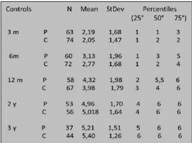
A control study group was created starting from literature data and randomised selected cases (from our casuistry) of implanted children without neuroradiological findings. A long-term follow-up was performed, which reported that the Geers and Moore score was achieved at 3, 6, 12, 24, and 36 months. Each of the subgroups was compared with the control study group. Furthermore, the subgroups were compared with each other only if the same patients were not present in either. Specific findings were reported as singular cases. A non-parametric test was used for statistical analysis: the Mann-Whitney U test. Furthermore, we used the ANOVA test (analysis of variance) for the comparison between patients with monolateral CI and patients with bilateral CI.
2.1. Audiological assessment.
Before implantation, all of the children had documented severe to profound or profound sensorineural HL (hearing loss) and failed an appropriate hearing-aid trial. Each patient has been investigated from an audiological point of view using objective tests, such as OAEs (otoacoustic emissions), ABR (auditory brainstem response), and ASSR (auditory steady state response), to estimate the pure-tone threshold and, in selected cases, to perform ECochG (electrocochleography). When possible, the audiological assessment was completed using behavioural and tonal audiometry with and without previously described hearing aids. Children of 6-36 months of age underwent conditioned orientation reflex (COR) and visual reinforcement audiometry (VRA) tests to investigate the tonal threshold at low frequencies and to assess the effectiveness of the hearing aids. From the age of three to four years, the pure-tone hearing thresholds can be obtained by motivational games that range from peep shows to finger-raising techniques, and at an age of six years, most children can perform formal audiometry the same as that used in adults. The testing is dependent only on the degree of cooperation of the child and the experience of the tester. Micro-otoscopy,
tympanometry and recording of stapedius reflex thresholds were part of the test procedure. The interpretation and diagnostic validity of stapedius-reflex-threshold testing in children are similar to the testing of adults, but the test might be difficult to perform in very young children.
2.2. Imaging data
High resolution HRCT (High Resolution Computed Tomography) and MRI (Magnetic Resonance Imaging) were conducted in all patients to obtain a radiological examination of the temporal bone and brain. If necessary, children underwent neuroradiological scans during general anaesthesia. HRCT scanning with contiguous 0.3 - 1 mm thick images through the petrous temporal bone in the axial and direct coronal planes was performed. Ear, brainstem and encephalon MRI scanning was acquired at 1.5 T and included high resolution axial and coronal T2-weighted imaging axial and coronal T1-weighted imaging, using CISS (Constructive Interference in Steady State) and FIESTA (Fast Imaging Employing Steady-state Acquisition). If contrast was required, then post-contrast T1-weighted images were acquired in all three planes. CISS and FIESTA are a gradient-echo MRI sequence that are used to investigate a wide range of pathologies when routine MRI sequences do not provide the desired anatomic information. MRI brain scanning was also acquired at 1.5 T and included axial T2-weighted imaging, axial fast fluid-inversion recovery sequence (FLAIR) imaging, axial T1-weighted inversion recovery imaging and, if contrast was required, axial T1-weighted imaging.
The neuroimaging findings of the temporal bone were categorised as: 1) Cochlear malformations
2) Vestibular and semicircular canal malformations 3) IAC (internal auditory canal) anomalies
The vestibular aqueduct is defined as enlarged if its diameter is greater than 1.5 mm at the midpoint. Vestibular and labyrinthine abnormalities included partial SCC aplasia and total SCC aplasia. Cochlear malformations were subsequently divided as follows:
1) Cochlear malformations
a. Common cavity deformity. b. Cochlear hypoplasia.
c. Incomplete partition type I (IP-I).
d. Incomplete partition type II (IP-II) (Mondini deformity). e. Incomplete partition type III (IP-III)
f. Basal turn dysplasia.
Mondini malformation is a cochlear anomaly that is characterised by a fusion of the apical and middle turn (only one and a half turns is present out of the normal two and a half turns). The brain MRI scans of all of the patients were reviewed, and all of the abnormal findings were identified and classified as follows:
1) Malformations
a. Aplasia, dysplasia or hypoplasia b. Dilatations
c. Arnold-Chiari malformations 2) Neoformations
a. Neoplasms (benign or malignant) b. Cystic lesions
3) White matter disorders a. Leukomalacia b. Leukodystrophy c. Demyelination
4) Gliotic lesions (including cytomegalovirus infections and ischemic lesions) 5) Other abnormalities
2.3. Genetic and molecular analysis
Informed consent was obtained from patients and parents according to current national rules and laws. Molecular genetic studies of the GJB2, GJB6, SLC26A4 genes and mitochondrial DNA (mit-DNA) were performed in 77 patients. Genomic DNA was extracted by standard protocols from peripheral blood leukocytes of patients. Direct DNA sequencing of the GJB2 gene (including analysis of the entire coding region) was performed. PCR amplification of the coding 21 exons, the flanking and promoter regions of the SLC26A4 gene was performed using specific primers. Amplification reactions were performed in a final volume of 25 ml containing 100 ng of genomic DNA, 200 mmol/l dNTPs, 10 mmol/l each primer 1.5 mmol/l MgCl2, and 1 U of Taq polymerase. After 5 min of denaturation at 94°C, 35 PCR cycles were carried out, each cycle comprising 45s of denaturation at 94 °C, 45s of annealing at 60 °C and 80s of extension at 72°C. Direct sequencing of the PCR products on both strands was performed on an ABI PRISM 3130xl sequencer, using the ABI BigDye Terminator v3.1 Cycle Sequencing Kit (Applied Biosystems by Life Technologies).
2.4. Speech perception (pre-operative assessment and post-operative outcomes):
Behavioural measures of speech perception scores [50-54] are routinely completed in all children in our study at follow-up visits and a database of these outcomes is maintained. The database also includes patient demographics (age at implant, gender, duration of implant use), audiological characteristics (congenital vs. progressive loss) and relevant medical history (other medical conditions, such as craniofacial syndromes).
Perceptive abilities are usually classified into 4 types of increasing complexity performances [52]:
1) Detection: ability to respond to the presence or absence of a signal;
2) Discrimination: ability to distinguish differences or similarities between two stimuli; 3) Identification: ability to choose an item from a known set;
4) Recognition: ability to repeat or imitate spoken stimuli.
The achieved performance enabled us to include each patient in a specific perceptual category. Geers and Moog propound perceptive classification with six categories, which are based on performances that have been analysed with sets of specific tests (Tab. IV) [14]:
Category 0 no detection of verbal sounds Category 1 detection of verbal sounds Category 2 discrimination of verbal sounds Category 3 identification of verbal sounds Category 4 closed set vowel recognition Category 5 closed set consonant recognition Category 6 open set recognition
Tab IV- Geers and Moog perceptive categories classification.
Geers & Moog perception was used for the present study. A comparison of specific speech perception tests was conducted between hearing impaired children with normal anatomy (called “well babies”) and those who were affected by cochleovestibular and brain abnormalities. An excellent tool for monitoring progress in young children is the Clinical Red Flag Procedure [50], which is a matrix of auditory benchmarks that has been established for identifying children who are progressing at a slower-than-expected rate. These benchmarks are based on research and clinical findings that document the listening skills that are achieved by the average CI child during the first year of device use (fig. 3). Three
different groups of CI children reflect different pre-implant characteristics and show different patterns of skill achievement [50].
Fig. 3 - Clinical red flags for slow progress in children with cochlear implants. Red flag Matrix for monitoring listening
progress in the first year of CI use. Robbins (2005). *Note that full-time implant use is an unconditional prerequisite to auditory development. If a child is not wearing the implant during all waking hours—at home, school, and other activities, then these benchmarks are not applicable. Children who fail to bond to their device and to wear it full-time within a few weeks of initial stimulation may exhibit insufficient progress and are at high risk of becoming non-users of their implants.
It appears evident that in the "well babies", the perceptual expected results after 3 months of use of the CI essentially comprise the detection of voice and first discrimination abilities until the recognition of words and phrases without the help of lip reading at 1 year of CI use. In summary, the expected perceptual results were the achievement of perceptual category 2 at 3 months from the CI activation, perceptual category 4 at 6 months and perceptual category 6 at 12 months. The follow-up initially should be very tightly controlled and should be performed in the first year after surgery, at 3, 6, 9 and 12 months and then yearly. The evaluation of perceptual skills and communication has been made by the administration of different tests according to the stage of language development of the child (pre-verbal stage, transitional stage and functional stage).
2.4.1. Listening Progress Profile (Lip) (developed by Archbold 1994)
The LIP is a profile that is designed to monitor changes in early auditory perception of young children and is most commonly used with children who use cochlear implants. Two types of nonlinguistic sound perception abilities are monitored using this profile: environmental sound awareness and environmental sound discrimination. LIP also monitors speech perception abilities [51]. The LIP identifies three different skills: response, discrimination, and identification. The response skill is used to describe the detection of a sound. Discrimination is used to describe the ability for the child to choose correctly between two different sounds. Identification is used to describe the ability to correctly choose the target sound from an open set of sounds. The child is then scored on each of these skills in the following way: N (Never/not known), S (Sometimes), and A (Always). These responses can be elicited or observed. The profile is structured as a list of behaviours or skills such as “Response to Environmental Sounds”.
There are six skills that pertain to nonlinguistic sounds and ten skills that relate to linguistic sounds:
1. Response to a drum
2. Response to a musical instrument
3. Discrimination between 2 different instruments 4. Discrimination between a loud and quiet drum 5. Discrimination between a single and repeated drum 6. Identification of environmental sounds
7. Speech detection (elicited) 8. Speech detection (spontaneous) 9. Linguistic sound detection
10. Short/Long discrimination of verbal material 11. Single/Repeated discrimination of verbal material 12. Soft/Loud discrimination (intensity discrimination) 13. Discrimination of at least 2 linguistic sounds 14. Discrimination of at least 5 linguistic sounds
15. Discrimination between 2 words of different lengths 16. Identification of own name
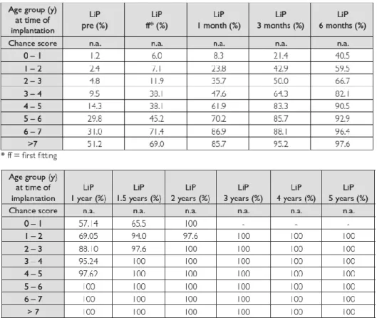
Additionally, little information is offered on the types of sounds that the child can identify or comprehend. Using the LiP test, we can see that the median percentage is achieved by the single age groups at different testing times after implantation. Children who are older than 5 years at the time of their implantation reached the maximum score on the LiP 1 year after implantation. Children between 3 and 5 years at the time of their implantation scored highest 1.5 years after implantation. The children who received implants at the youngest ages (<3 years) reached their highest possible value approximately 2 years after implantation.
Fig. 4. Evaluation of Auditory Responses to Speech - Normative Data Med-El, Innsbruck (1998)
2.4.2. Early Speech Perception Test (ESP) (Moog & Geers, 1990)
Italian version: PCAP (Test di misurazione delle Prime Categorie Percettive) (Arslan et al. 1997). This test measures the child’s ability to discriminate different words based on specific perceptual features. Therefore, it is possible to include each child in one of four perceptual categories [14]. The standard version was designed to obtain accurate information about the progression of speech discrimination skills in children who have profound hearing impairments as they develop. The standard verbal version was designed for children who are at least 6 years. The stimuli are composed of 36 words that are presented in an auditory-only condition as three subtests of 12. The format is closed-set. Each subtest is administered using both visual and auditory input to differentiate speech perception skills from language ability. The low verbal version was designed to estimate speech perception abilities in very young children (a minimum of 2 years) who have limited verbal abilities. Stimuli comprise words
that vary in pattern as well as spondees and monosyllabic words presented in sets of four. The format is a four-item closed-set format and can be presented via live voice or a recording. Test materials constitute objects (toys) instead of pictures.
2.4.3. GASP (Glendonald Auditory Screening Procedure) (Erber, 1982)
Italian version: TAP (Test delle Abilità Percettive) (Arslan et al. 1997). This test measures the ability to understand simple sentences, and it also uses only one type of sentence structure (questions). This arrangement is useful because children can become confused when faced with different sentence structures (questions, commands or statements) in the same test. Children are allowed to either repeat the question or to answer it. This test comprises 3 subtests [52]: 1) detection, 2) identification and 3) comprehension.
1) DETECTION: a. purpose
i. to determine the device settings and to check whether the CI is working;
ii. to attract a child's attention and to orient him or her to the listening task;
iii. to determine which sounds are available to the child; b. response
i. repeatedly says a new word in a meaningful context.
There is rarely any need to spend much time at this level, although it could be worthwhile to help the child to detect those sounds, such as fricative consonants, which are low in acoustic energy.
2) IDENTIFICATION: a. purpose
ii. to learn speech patterns; b. response
i. point to an item named by the speaker, repeat stimulus, write stimulus.
When writing sentences, it is a good idea to construct sentences that have some relevance to the children. It is best to attempt to make sentences that are sufficiently different in length and/or pattern to enable all children in the group to achieve some success in identification. When presenting an identification task, include "no sound" (the absence of sound) as a response option. This option helps the child to become aware of the possibility that he or she might hear nothing, and thus, the child can learn to indicate that the IC is off, has a dead battery, or is faulty. A response of "no sound" could also indicate that the talker is too far away from the child.
3) COMPREHENSION: a. purpose
i. To understand the meaning of a spoken stimulus
ii. To make complex associations between sounds and events/objects or between the sounds themselves
b. response
i. The child performs the required task or answers questions.
2.4.4. Northwestern University – Children’s Perception of Speech (NU-CHIPS) - (Elliot and Katz, 1980)
Italian version: T.I.P.I.1 (Test di Identificazione Parole Infantili 1) (Arslan et al.1997). NU-CHIPS is a closed-set picture-pointing word recognition test for children. This test is composed of 50 words that are familiar to three-year-old children in four randomisations called forms. The test includes one CD-Rom, which contains two picture books with 50
monochrome plates, with four pictures per plate. There are two recordings, with one male speaker and one female speaker. [53]
2.4.5. Word Intelligibility by Picture Identification (WIPI) (Ross and Lerman, 1979)
Italian version: T.I.P.I.2 (Test di Identificazione Parole Infantili 2) (Arslan et al. 1997). This test is a closed-set discrimination task in which the child must identify the stimulus word from a set of six pictures. The recommended age is a minimum of 4 years. The stimuli are composed of four lists of 25 single-syllable words. Chance performance amounts to a score of approximately 18%. This discrimination task is more difficult than the majority of other closed-set discrimination tasks in that the vocabulary level is higher and the response choices differ in fine segmental information [54].
3. RESULTS
Between 1st January 1996 and 1st April 2012 at the ENT-Audiology Department of the University Hospital of Ferrara, 620 cochlear implantations were performed. There were 426 implanted children at the time of the present study (who were <18 years).
Reviewing the neuroradiological findings of the 426 implanted children revealed no abnormalities in 283 cases and ear and / or brain anomalies in 143 cases (33.6% of 426). Among these 143 patients (64 females and 79 males), 123 children had unilateral cochlear implantation (68 in the right ear; 55 in the left ear), and 20 underwent bilateral cochlear implantations (3 simultaneously, 17 sequentially). The age of the main study group (143 implanted children) ranged from 9 months and 16 years (mean = 4.4; median = 3.0). These patients showed an average period of cochlear implant use of 74 months.
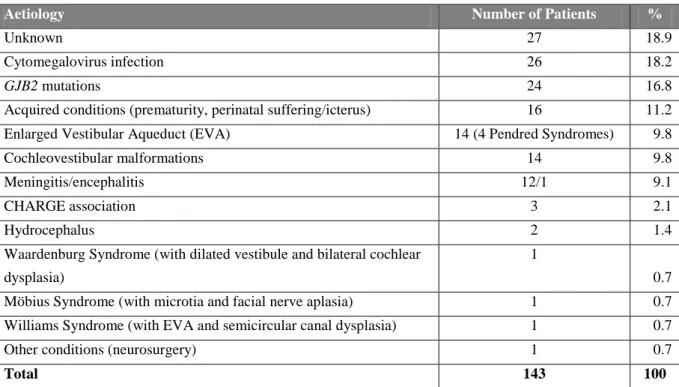
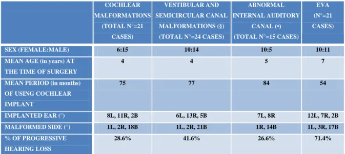
The CT and MRI scans of 143 children included in the present study were re-evaluated, and the following abnormalities were detected: in 69 cases (48.2% of 143), ear malformations were present, of which 55 had bilateral ear involvement; therefore the implanted ear was necessarily the malformed ear; in 11 children, the malformed ear was the right ear (of which only in one case the malformed side was the implanted side), and in 3 cases, the malformation was detected in the left side (also in this series only one child underwent cochlear implantation in the malformed ear); 74 cases (51.7% of 143) presented only brain anomalies. A total of 45 patients (31.5% of 143) presented either ear or brain abnormalities. Table V shows different aetiologies of hearing loss that we found in our series. Details of the identified cochleovestibular (inner ear) malformations are presented in Table VII and VIII.
Aetiology Number of Patients %
Unknown 27 18.9
Cytomegalovirus infection 26 18.2
GJB2 mutations 24 16.8
Acquired conditions (prematurity, perinatal suffering/icterus) 16 11.2 Enlarged Vestibular Aqueduct (EVA) 14 (4 Pendred Syndromes) 9.8
Cochleovestibular malformations 14 9.8
Meningitis/encephalitis 12/1 9.1
CHARGE association 3 2.1
Hydrocephalus 2 1.4
Waardenburg Syndrome (with dilated vestibule and bilateral cochlear dysplasia)
1
0.7 Möbius Syndrome (with microtia and facial nerve aplasia) 1 0.7 Williams Syndrome (with EVA and semicircular canal dysplasia) 1 0.7
Other conditions (neurosurgery) 1 0.7
Total 143 100
TAB V - Suspected main aetiology of hearing loss among 143 children who underwent cochlear implantation and presented
ear or brain anomalies.
Demographic groups and audiological features are resumed in Table VI.
EXTERNAL EAR MALFORMATI ONS (TOTAL N°= 3 CASES) MIDDLE EAR MALFORMATIO NS (TOTAL N°= 18 CASES) INNER EAR MALFORMATIO NS (TOTAL N°= 30 CASES) BRAIN ANOMALIES (TOTAL N°= 119 CASES) WITHOUT BRAIN ANOMALIES (TOTAL N°= 24 CASES) SEX (FEMALE:MALE) 1:2 9:9 11:19 55:64 9:15
MEAN AGE (in years) AT THE TIME OF SURGERY
3.0 3.0 4.4 4.0 7.0
MEAN PERIOD (in months) OF USING COCHLEAR IMPLANT 78 73 74 75 71 IMPLANTED EAR (°) 2L, 1R 8L, 7R, 3B 11L, 15R, 4B 42L, 61R, 16B 13L, 7R, 4B % OF PROGRESSIVE HEARING LOSS - 22.2% 30.0% 45.3% 54.1% CYTOMEGALOVIRUS INFECTIONS - 3 2 26 - MENINGITIS - - 1 11 1 GENETIC MUTATIONS - 2 5 21 3 SYNDROMES 3 3 5 6 4 UKNOWN AETIOLOGY - 3 - 26 1 OTHER CONDITIONS (*) - 2 2 19 1
Tab. VI – Demographic, clinical and audiological data and aetiologies of hearing loss; ° (R=RIGHT; L=LEFT;
COCHLEAR MALFORMATIONS (TOTAL N°=21 CASES) VESTIBULAR AND SEMICIRCULAR CANAL MALFORMATIONS (§) (TOTAL N°=24 CASES) ABNORMAL INTERNAL AUDITORY CANAL (•) (TOTAL N°=15 CASES) EVA (N°=21 CASES) SEX (FEMALE:MALE) 6:15 10:14 10:5 10:11
MEAN AGE (in years) AT THE TIME OF SURGERY
4 4 5 7
MEAN PERIOD (in months) OF USING COCHLEAR IMPLANT 75 77 84 54 IMPLANTED EAR (°) 8L, 11R, 2B 6L, 13R, 5B 7L, 8R 12L, 7R, 2B MALFORMED SIDE (°) 1L, 2R, 18B 1L, 2R, 21B 1R, 14B 1L, 3R, 17B % OF PROGRESSIVE HEARING LOSS 28.6% 41.6% 26.6% 71.4%
Tab. VII – Anatomic distribution of inner ear malformations; ° (R=RIGHT; L=LEFT; B=BILATERAL); §(6
HYPOPLASIAS; 7 DILATATIONS; 3 APLASIAS); •(7 STENOSIS; 8 DILATATIONS).
COMMON CAVITY COCHLEAR HYPOPLASIA (TOTAL N°= 6 CASES) INCOMPLETE PARTITION TYPE 1 (TOTAL N°= 2 CASES) INCOMPLETE PARTITION TYPE 2 (TOTAL N°= 7 CASES) INCOMPLETE PARTITION TYPE 3 (TOTAL N°=2 CASES) COCHLEAR BASAL TURN DYSPLASIA (TOTAL N°= 4 CASES) SEX (FEMALE:MALE) - 2:4 0:2 3:4 1:1 0:4
MEAN AGE (in years) AT THE TIME OF
SURGERY
- 5 3 5 7 2
MEAN PERIOD (in months) OF USING COCHLEAR IMPLANT - 59 82 85 77 73 IMPLANTED EAR (°) - 2L, 4R 1L, 1R 2L, 4R, 1B 1L, 1R 2L, 1R, 2B MALFORMED SIDE (°) - 1L, 1R, 4B 2B 1R, 6B 2B 4B % OF PROGRESSIVE HEARING LOSS - 40% - 28.6% 50% 25%
Tab. VIII – Types of cochlear malformations; ° (R=RIGHT; L=LEFT; B=BILATERAL).
Finally, Table IX reports in detail the brain anomalies that were found, with the total number of cases for each type.
Type of lesion/malformation Total number of cases among 143 implanted children
Gliosis 32
Dismyelination/demyelination 25
Leukomalacia 24
Pineal cyst 9
Arnold-Chiari malformation (type 1) 7
Cerebellar hypoplasia 6
Cortical dysplasia 5
Calcifications 3
Arachnoid cyst 3
(External) Hydrocephalus 3
Dilated lateral ventricles 2
Corpus callosum hypoplasia 2
Dilated fourth ventricle 2
Trigonocephaly 2
Facial nerve aplasia 1
Pinealoma 1
Hamartoma 1
Hydrocephalus 1
Bulbar atrophy 1
Cisternal dilatation 1
Malignant neoplasm of encephalon (after surgery)
1
Pellucid septum cyst 1
Dilated subarachnoid space 1
Focal ischemic lesions 1
Microcephaly 1
Lipoma 1
(Occipital) Myelomeningocele 1
Leukodystrophy 1
Pachygyria 1
Temporal lobe hypoplasia 1
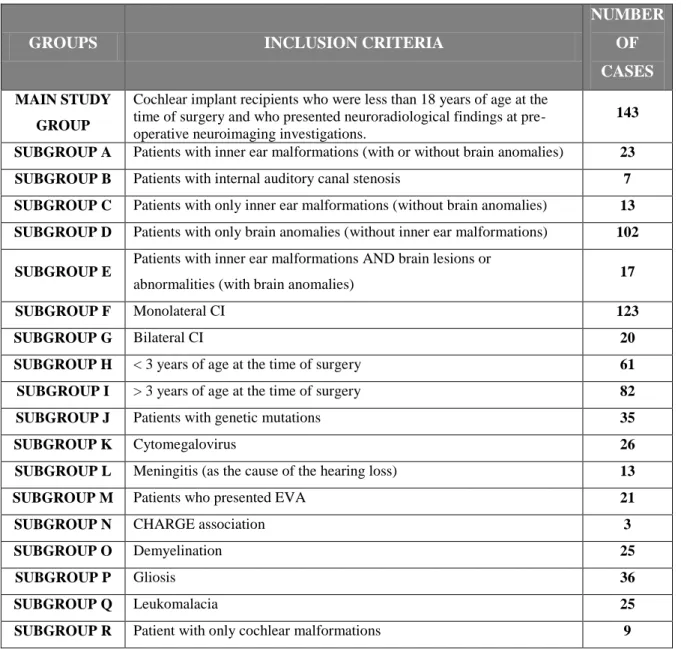
For statistical purposes, the main group was divided into subgroups, as follows (table X):
GROUPS INCLUSION CRITERIA
NUMBER OF CASES
MAIN STUDY GROUP
Cochlear implant recipients who were less than 18 years of age at the time of surgery and who presented neuroradiological findings at pre-operative neuroimaging investigations.
143 SUBGROUP A Patients with inner ear malformations (with or without brain anomalies) 23 SUBGROUP B Patients with internal auditory canal stenosis 7 SUBGROUP C Patients with only inner ear malformations (without brain anomalies) 13 SUBGROUP D Patients with only brain anomalies (without inner ear malformations) 102 SUBGROUP E
Patients with inner ear malformations AND brain lesions or
abnormalities (with brain anomalies) 17
SUBGROUP F Monolateral CI 123
SUBGROUP G Bilateral CI 20
SUBGROUP H < 3 years of age at the time of surgery 61 SUBGROUP I > 3 years of age at the time of surgery 82
SUBGROUP J Patients with genetic mutations 35
SUBGROUP K Cytomegalovirus 26
SUBGROUP L Meningitis (as the cause of the hearing loss) 13
SUBGROUP M Patients who presented EVA 21
SUBGROUP N CHARGE association 3
SUBGROUP O Demyelination 25
SUBGROUP P Gliosis 36
SUBGROUP Q Leukomalacia 25
SUBGROUP R Patient with only cochlear malformations 9
Table X – Different subgroups were defined to implement the statistical analysis.
After 3 months of using the cochlear implant, more than half of the patients in the main study group did not achieve the 3th category of perception at the Geers & Moog scale; in the same group, the 50% of the children did not reach the 4th category at the 6-month follow-up, nevertheless they achieved the 5th category 6 months after (1-year follow-up). Only 2 years after cochlear implant activation, the majority of the patients attain a 6th perceptual category at the Geers & Moog scale (fig. 5).
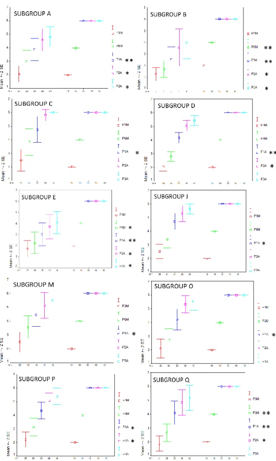
Statistical results are reported in full as the following graphs (fig. 5-15). These graphs show the comparison between the control group and each of the subgroups; nonetheless, different subgroups were compared.
Graphs are structured as follows: on the abscissa axis are reported the number of cases, and they are distributed over the time of the 3, 6, 12, 24 and 36 month follow-ups, using a colour code for identification (red for the 3-month control, green for the 6-month control, blue for the 12-month control, violet for the 24-month control and azure for the 36-month control).
There was a statistically significant difference (p ≤ 0.01) between the control group and the subgroup B (patients with internal auditory canal stenosis) at the 6-months follow-up. Similar results were obtained comparing control group and subgroup Q (patients affected by leukomalacia).
At the 1-year and long-term (2-3 years) follow-ups, statistically significant differences (p ≤ 0.05) were also found comparing control group and subgroups A,B, D and E, respectively.
Comparing subgroups C and P we found that the first one achieved the "identification of verbal sounds" 6 months after device activation and the “vowel recognition” in closed set tests 6 months later; while the subgroup P more slowly reached the same perceptual skills. Nevertheless, at the long-term follow-up (2 years later) the results achieved by the two groups are optimal and similar (Fig. 8).
Fig 6. – In the figure are reported all of the statistical graphs that were obtained from the comparison between the subgroups
A, B, C, D, E, J, M, O, P, and Q and the control group. Colour code for identification (red for the 3-month control, green for the 6-month control, blue for the 12-month control, violet for the 24-month control and azure for the 36-month control). * P≤0.05; ** P≤0.01.
Fig. 7 – Statistical data of the subgroups A, B, C, D, E, J, K, L, M, O, P, Q, and R.
Fig. 8 - Comparison of the perceptual outcomes at the 3-month follow up on congenital hearing loss without progression
(C) and progressive hearing loss (P).
Figure 9 shows the statistical results obtained from patients with or without genetic mutations.
As reported in table XI, the most common mutation was the 35delG in the GJB2 gene. All patients with SLC26A4 mutations presented bilateral EVA. Note that they had mutations on both alleles. Among these mutations, to our knowledge, 2 have never before described
(Q235R e G557D) and 1 was recently reported in one of our scientific publications entitled "Novel mutations in the SLC26A4 gene” [55].
Fig. 9 – Perceptual outcomes at the 3-month follow up on the children who underwent genetic investigation; differences
between children with genetic mutations (+= mut) and children without mutations (─ = no mut) are shown in the graphs.
GENE MUTATION N° GJB2 35delG/35delG 15 GJB2 35delG/R184P 4 GJB2 35delG/167delT 1 GJB2 35delG/R143V 1 GJB2 V27I/E114G 1 GJB2 VS1+1G>A/delE120 1 SLC26A4 G209V/Q235R 1 SLC26A4 L445W/G557D 1 SLC26A4 R409H/IVS2+1delG 1 SLC26A4 R409H/Q235R 1
Fig 10 - Perceptual outcomes of subgroup M (EVA) at the 3-year follow-up
After 3 months of using the cochlear implant, more than half of the patients belonging to the subgroup M (patients who presented EVA) did not achieve the 3th category of perception at the Geers & Moog scale; in the same group, the 50% of the children did not reach the 4th category at the 6-month follow-up, nevertheless they achieved the 5th category 6 months after (1-year follow-up). Only 2 years after cochlear implant activation, the majority of these patients attained a 6th perceptual category at the Geers & Moog scale (fig. 10). There was a statistically significant difference (p ≤ 0.05) between the control group and the subgroup M (patients who presented EVA) at the 1-year follow-up (Fig. 6).
Fig 11 - Comparison between the perceptual outcomes at the 3-year follow-up of children who were younger than 3 years

![Tab. II – Recurrence rates of the temporal bone malformations [46].](https://thumb-eu.123doks.com/thumbv2/123dokorg/4701773.44834/18.892.257.638.345.575/tab-ii-recurrence-rates-temporal-bone-malformations.webp)