ORIGINAL ARTICLE
Irrelevance of Panton-Valentine leukocidin in hidradenitis
suppurativa: results from a pilot, observational study
Monica Corazza1&Alessandro Borghi1 &Vincenzo Bettoli1&Roberto Pora2&Ilaria Bononi3&Elisa Mazzoni3& Elisa Mazzola4&Silva Saraceni1&Martina Maritati1&Carlo Contini1
Received: 6 July 2020 / Accepted: 30 July 2020 # The Author(s) 2020
Abstract
Panton-Valentine leukocidin (PVL) appears to be a virulence factor which, among others, can exacerbate the pathogenicity of Staphylococcus aureus infections, especially inducing severe necrotic, deep-seated skin infections, abscesses, and recurrences. These peculiarities have some overlaps with hidradenitis suppurativa (HS). Our main aim was to assess if S. aureus producing PVL could have some role in influencing clinical features and/or course of HS, specifically in the suppuration and recurrence of lesions. This pilot, mono-centric, observational study included all adult subjects affected with HS consecutively referring to our HS clinic over a 3-month period. Clinically evident suppuration and at least 2 weeks wash out from any antibiotic were the main inclusion criteria. Purulent material from HS skin lesions was collected with swabs in order to isolate micro-organisms, with specific regard to S. aureus. Detection of PVL was performed by real-time quantitative PCR (RT-qPCR). We also analyzed purulent material from suppurative skin lesions other than HS, as a control. Thirty HS patients were included; 29 purulent lesions (96.7%) harbored at least one bacterial species. Five (16.7%) swab samples were positive for S. aureus, none of which was positive for PVL genes. Among the 30 purulent disorders included as controls, 8 (26.3%) were positive for S. aureus; of these, 4 strains (50%) expressed LPV. The study results seem to exclude the pathogenetic involvement of S. aureus producing PVL in HS; as a result, PVL does not seem to represent a potential target in the future development of HS treatments.
Keywords Hidradenitis suppurativa . Panton-Valentine leukocidin . Staphylococcus aureus . Infections . RT-qPCR
Introduction
Hidradenitis suppurativa (HS), also known as acne inversa, is a chronic-relapsing, debilitating inflammatory disease of the hair follicle, which affects apocrine gland-bearing skin, most
commonly the axillae, inguinal regions, anogenital area, and infra- and inter-mammary folds [1]. It is clinically characterized by recurrent, painful, deep-seated nodules commonly ending in abscesses and sinus tracts with persistent suppuration and hyper-trophic, bridged scarring. HS usually presents after puberty and its estimated prevalence ranges from less than 0.1 to 4% [1,2]. HS has a huge negative influence on patients’ lives due to its chronic course, pain and other disease-related symptoms, persis-tent malodor, peculiar anatomical localization, disfiguring se-quelae, and lack of a definitive cure [3,4].
The pathogenesis of HS is not fully recognized and appears to involve a number of interacting factors, including a suscep-tible genetic background, a dysregulated immune response to various heterogeneous stimuli, and follicular occlusion [1,2,
5]. The role of bacterial infection in the initiation or propaga-tion of HS remains under investigapropaga-tion [6,7]. Most frequently bacteriological studies have shown a mixed growth of com-mensal microbes, which is more consistent with the view that HS is an immune-mediated condition rather than a disease of primary infectious etiology [7,8]. Bacterial colonization of
Monica Corazza, Alessandro Borghi, and Vincenzo Bettoli contributed equally to this study and share first authorship.
* Alessandro Borghi [email protected]
1
Section of Dermatology and Infectious Diseases, Department of Medical Sciences, University of Ferrara, Via L. Ariosto 35, 44121 Ferrara, Italy
2
Analysis Laboratory, S. Anna University Hospital, Ferrara, Italy
3 Laboratories of Cell Biology and Molecular Genetics, Section of
Pathology, Oncology and Experimental Biology, Department of Morphology, Surgery and Experimental Medicine, University of Ferrara, Ferrara, Italy
4
Palermo, Italy
HS lesions may act synergistically with a dysfunctional mune response, involving both the innate and adaptive im-mune systems, which leads to a chronic, relapsing inflamma-tory scenario. On the other hand, HS may predispose to an increased risk of skin infections [9]. Panton-Valentine leukocidin (PVL) is a two-component pore-forming cytotoxin produced by Staphylococcus aureus inducing leukocyte de-struction and tissue necrosis [10]. In addition, PVL is a poly-morphonuclear neutrophils priming agent [11]. In keeping with this, it appears to be a highly relevant virulence factor which, among others, can exacerbate the pathogenicity of S. aureus infections [12]. In particular, it is related to deep-seated skin and soft tissue infections and causes furuncles, cutaneous abscesses, and severe necrotic skin infections [13,
14]. Moreover, PVL seems to be a marker for severity and recurrence [15]. Based on these assumptions, we were inter-ested in assessing if S. aureus producing PVL could have some role in influencing clinical features and/or the course of HS, especially in the suppuration and recurrence of lesions.
Materials and methods
Study design and patients
In the present pilot, mono-centric, observational study, we included all adult subjects affected by HS consecutively refer-ring to our HS clinic over a 3-month period. Patients were excluded in the presence of the following: (i) lack of reliable diagnosis, (ii) absence of clinically evident suppuration, (iii) topical or systemic antibiotics during the 2 weeks before in-clusion. Purulent material from HS skin lesions was collected with swabs in order to isolate micro-organisms, with specific regard to S. aureus releasing PVL. We also collected purulent material from suppurative skin lesions other than HS, over a 3-month period, as a control. Controls were excluded if they were treated with topical and/or systemic antibiotics or if they had a diagnosis of HS or clinical features resembling HS. The eligible subjects who were visited more than once over the study period underwent skin swabs only at the first visit.
This was a spontaneous survey, with no funding from ex-ternal sources. The study was approved by the University-Hospital of Ferrara institutional review board. Both patients and controls provided their written informed consent.
Data collection
The following data from HS patients were recorded by using a standardized data collection form: (1) age at inclusion; (2) gender; (3) disease duration, taken as the time between the patient-reported onset of symptoms and/or signs and study inclusion; (4) previous treatments, defined as documented courses with any pharmacological active or surgical
interventions prior to entering this study; (5) current treatment; (6) Hurley stage; (7) sites involved by the disease; (8) number of sites with active suppuration at study inclusion; (9) number of suppurative episodes during the previous 6 months; (10) isolation of microbes, in particular S. aureus; (11) Panton-Valentine leukocidin expression by S. aureus strains.
From controls, we recorded the following information: (1) diagnosis of purulent skin disorders they were affected by; (2) isolation of S. aureus; (3) Panton-Valentine leukocidin ex-pression. These purulent skin disorders were categorized as follows: (i) primary pyodermitis versus secondary pyodermitis/impetiginization of another primary skin disor-der; (ii) primarily follicular versus non-follicular purulent dis-orders; (iii) deep versus superficial inflammatory process.
Microbiological assessments
Samples were collected from the purulent material from HS draining lesions as well as from control suppurative disorders. One sample was collected from a single purulent lesion of each included subject. Content was drained with gentle pres-sure exercised on previously sterilized skin surface, using ster-ile gloves and swabs. A suitable transport media for aerobes and anaerobes was used (ESwab Liquid Amies Collection and Preservation System, Catalog No. 490CE.A, Copan Diagnostic, Murrieta, CA, USA). Specimens were stored and delivered to the laboratory according to the manufac-turer’s instructions.
Swabs from clinical specimens were grown on sever-al selective agar plates to isolate specific organisms, as described above [16]. The S. aureus culture suspensions were then stored at 4 °C prior to nucleic acid extraction and PCR analysis. The commercial kit RIDA®GENE PVL assay was used for the extraction of bacterial DNA and for the real-time quantitative PCR (RT-qPCR) analysis through the amplification of a PVL-specific fragment (Panton Valentine Leukocidine lukF-PV). To verify whether cross-contamination had oc-curred during DNA extraction, purification, and PCR procedures, each sample was processed simultaneously with negative controls, represented by sample lacking DNA (real-time PCR mix). Samples and controls were analyzed in duplicate replica experiments. Samples were run in CFX96 Touch Real-Time PCR Detection System (Applied Biosystems) instrument and sample analysis was performed by Bio-Rad CFX Manager software, fol-lowing the manufacturer’s instructions. Positive and negative controls must show correct results. The posi-tive control had a concentration of 103 copies/μl. A total of 5 × 10³ copies were used in each RT-qPCR run. The RIDA®GENE PVL detection limit was ≤ 5 copies of DNA for the reaction.
Statistics
Continuous variables were presented as mean ± standard de-viation (SD) and categorical variables as frequencies and per-centages. Comparisons between groups were made using t test for quantitative variables, while Pearson’s chi-squared test or the Fisher exact test was used for qualitative variables. For categorical data, odds ratios (OR) with 95% confidence inter-vals (95% CI) were calculated. All statistical analyses were performed using Stata version 13, setting the significant level α to 0.05.
Results
HS patients
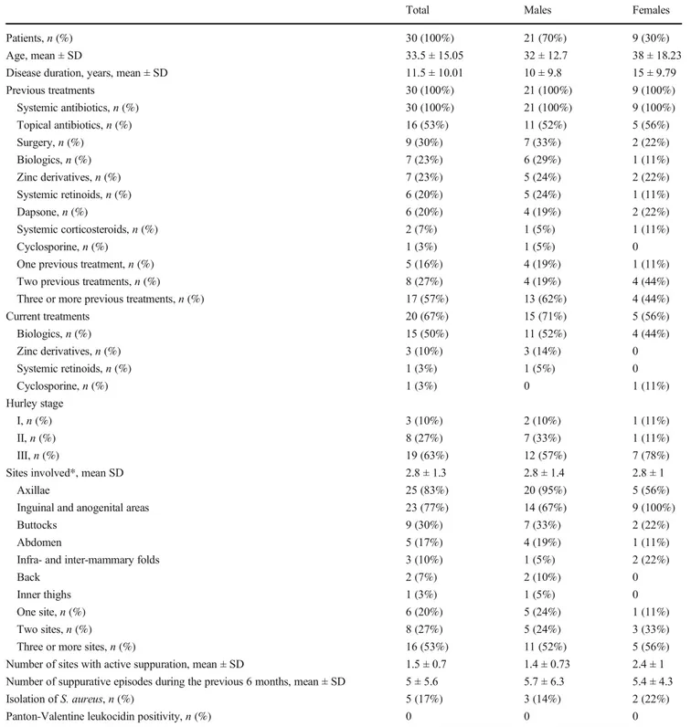
Thirty patients affected by HS were included. Table1reports the main HS patients’ characteristics. The majority of patients (19 of 30, 63.3%) had a Hurley stage III, 16 (53.3%) had at least 3 different anatomical sites involved by HS, and 9 (30%) reported≥ 6 episodes of suppuration during the previous 6 months. Seventeen patients (56.7%) had undergone at least 3 previous different treatments and 20 (66.7%) were in treat-ment with drugs other than antibiotics.
All HS samples but one harbored at least one bacterial species (detailed in Table2). Five (16.7%) swab samples were positive for S. aureus. Isolation of S. aureus did not significantly correlate with disease Hurley stage (Hurley stage III had an OR 2.6667, 95% CI: 0.2587 to 27.4865, p = 0.409), gender (males had an OR 0.5833, 95% CI: 0.0797 to 4.2711, p = 0.5957), previous systemic antibiotics (OR 0.2157, 95% CI: 0.0038 to 12.0965, p = 0.4553), patient age (p = 0.11), number of involved sites (p = 0.78), number of current suppurative lesions (p = 0.44), and number of suppurative recurrences during the previous 6 months (p = 0.15). Disease duration was higher (18.6 years, SD ± 11.80) among patients harboring S. aureus than among the others (10.1, SD ± 9.28) (p = 0.04). None of the S. aureus strains isolated was positive for PVL genes.
Controls
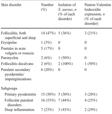
Table3shows clinical diagnosis and swab sample data of the 30 purulent disorders included as controls (17 males and 13 females, mean age 39 ± 18.59). Among these samples, 8 (26.3%) were positive for S. aureus. Four S. aureus strains (50%) expressed PVL. Expression of PVL was strictly related with primary pyodermitis (OR 3.5000, 95% CI: 0.3204 to 38.2337, p = 0.3044), primarily follicular inflammations (OR 10.4400, 95% CI: 0.5104 to 213.5310, p = 0.127), and deep infectious processes (OR 4.2000, 95% CI: 0.4704 to 37.5001, p = 0.198). None of these associations reached the significance threshold, probably due to the small sample sizes.
Discussion
Bacteria have long been considered in the pathogenesis of HS. It is generally acknowledged that bacteria do not play a direct role in the etiology of HS; however, they may be involved in the pathogenesis of HS via follicular dysbiosis and biofilm as well as eliciting an inflammatory response in a genetically predisposing background [7]. Interestingly, it had previously been shown that the microbiological population of HS chang-es considerably according to the clinical severity of the disease [17]. Our findings support the high prevalence of positive culturing samples from HS lesions [7], since bacteria speci-mens were isolated from almost all the study samples (29 of 30, 96.7%).
With special reference to S. aureus, this has often been de-scribed in association with HS and has even been proposed as a potential causative organism [18,19]. However, the literature includes some controversial data [20] and its action in the path-ogenesis of the disease remains unknown. Based on the recog-nized virulent role of PVL in S. aureus infections, especially in those by community-associated methicillin-resistant S. aureus (CA-MRSA), also at skin level [21,22], we wanted to specifi-cally seek its presence in HS lesions. In particular, we were interested in deepening a possible link between S. aureus pro-ducing PVL and degree of suppuration, skin involvement, dis-ease chronicity, and recurrence. We focused on PVL because it exerts peculiar mechanisms of action that may be in some way compatible with HS. In fact, it induces the release of pro-inflammatory cytokines and nuclear factor kappa B (NF-κB) in neutrophils, leading to skin infections which tend to be invasive, necrotizing, and recurrent [22,23]. Analyzing the findings ob-tained from the purulent skin disorders included as controls, these were fairly consistent with this pathogenic profile (Table 3). Prevalence of PVL-positive S. aureus was highly correlated with primary pyodermitis when compared with impetiginized lesions, primarily follicular inflammation and deep inflammatory processes.
The rate of S. aureus strains isolated from purulent material drained from abscesses and sinuses of HS patients was quite in line with previous reports, being about 17% [7,19,24]. The isolation of S. aureus did not correlate with patient or disease characteristics, including severity of HS in terms of Hurley’s score, number of sites affected, and frequency of recurrence, unlike what other researchers have found [25]. The most note-worthy finding of our study was that none of the strains of S. aureus isolated from HS purulent lesions expressed PVL. This finding strongly suggests an irrelevant role of PVL, not only in HS etiopathogenesis but also in its clinical features and course. Therefore, in speculating about the relevance of S. aureus in HS, our results seem to exclude the involvement of PVL. This can also be seen from a therapeutic perspective, since PVL does not seem to represent a potential target in the development of a treatment for HS.
The role of toxins, especially necrotizing toxins, in HS is uncertain and has been poorly investigated. Diphtheria by the toxigenic zoonotic pathogen Corynebacterium ulcerans is increasingly occurring in children, adolescents, and adults. In addition to diphtheria toxin (DT), the exotoxin
phospholipase D (PLD) is considered an important viru-lence factor of C. ulcerans and C. pseudotuberculosis [26]. PLD has been found to induce dermonecrotic le-sions, increased vascular permeability in vivo, and syner-gistic hemolysis of sheep blood cells.
Table 1 Main characteristics of the patients affected with hidradenitis suppurativa
Total Males Females
Patients, n (%) 30 (100%) 21 (70%) 9 (30%)
Age, mean ± SD 33.5 ± 15.05 32 ± 12.7 38 ± 18.23
Disease duration, years, mean ± SD 11.5 ± 10.01 10 ± 9.8 15 ± 9.79
Previous treatments 30 (100%) 21 (100%) 9 (100%) Systemic antibiotics, n (%) 30 (100%) 21 (100%) 9 (100%) Topical antibiotics, n (%) 16 (53%) 11 (52%) 5 (56%) Surgery, n (%) 9 (30%) 7 (33%) 2 (22%) Biologics, n (%) 7 (23%) 6 (29%) 1 (11%) Zinc derivatives, n (%) 7 (23%) 5 (24%) 2 (22%) Systemic retinoids, n (%) 6 (20%) 5 (24%) 1 (11%) Dapsone, n (%) 6 (20%) 4 (19%) 2 (22%) Systemic corticosteroids, n (%) 2 (7%) 1 (5%) 1 (11%) Cyclosporine, n (%) 1 (3%) 1 (5%) 0
One previous treatment, n (%) 5 (16%) 4 (19%) 1 (11%) Two previous treatments, n (%) 8 (27%) 4 (19%) 4 (44%) Three or more previous treatments, n (%) 17 (57%) 13 (62%) 4 (44%)
Current treatments 20 (67%) 15 (71%) 5 (56%) Biologics, n (%) 15 (50%) 11 (52%) 4 (44%) Zinc derivatives, n (%) 3 (10%) 3 (14%) 0 Systemic retinoids, n (%) 1 (3%) 1 (5%) 0 Cyclosporine, n (%) 1 (3%) 0 1 (11%) Hurley stage I, n (%) 3 (10%) 2 (10%) 1 (11%) II, n (%) 8 (27%) 7 (33%) 1 (11%) III, n (%) 19 (63%) 12 (57%) 7 (78%)
Sites involved*, mean SD 2.8 ± 1.3 2.8 ± 1.4 2.8 ± 1
Axillae 25 (83%) 20 (95%) 5 (56%)
Inguinal and anogenital areas 23 (77%) 14 (67%) 9 (100%)
Buttocks 9 (30%) 7 (33%) 2 (22%)
Abdomen 5 (17%) 4 (19%) 1 (11%)
Infra- and inter-mammary folds 3 (10%) 1 (5%) 2 (22%)
Back 2 (7%) 2 (10%) 0
Inner thighs 1 (3%) 1 (5%) 0
One site, n (%) 6 (20%) 5 (24%) 1 (11%)
Two sites, n (%) 8 (27%) 5 (24%) 3 (33%)
Three or more sites, n (%) 16 (53%) 11 (52%) 5 (56%) Number of sites with active suppuration, mean ± SD 1.5 ± 0.7 1.4 ± 0.73 2.4 ± 1 Number of suppurative episodes during the previous 6 months, mean ± SD 5 ± 5.6 5.7 ± 6.3 5.4 ± 4.3 Isolation of S. aureus, n (%) 5 (17%) 3 (14%) 2 (22%) Panton-Valentine leukocidin positivity, n (%) 0 0 0 *Patient could have simultaneously more than one anatomical site involved by HS
Pseudomonas aeruginosa can cause severe human oppor-tunistic infections, including many hospital-acquired infec-tions. However, there is scarce evidence about its clinical rel-evance in patients with HS. Among its toxins, exotoxin A is
the main virulence factor [27]. Similarly but less effectively than DT, exotoxin A interrupts protein synthesis in the host cell and has an immunosuppressive action. Pigments, such as pyocyanin, which catalyzes reactive oxygen species (ROS) production and attracts neutrophils, and pioverdin (siderophore), exert multiple detrimental effects on the host too. The blue-green phenazine, pyocyanin, is a key virulence factor that can kill competing organisms as well as host cells and can inactivate catalases to protect against ROS generated by host tissues [28]. Pyocyanin in particular can serve as a redox cycler similar to methyl viologen, resulting in superox-ide radical production and oxidative stress [29]. The majority of these virulence factors are under the control of two regula-tory systems: the two-component system and the sensing quo-rum, which allow the survival and multiplication of this mi-croorganism in the host [30].
Despite the inherent potential to cause harmful effects in humans, little is known about the role of these toxins in HS and further investigation could be conducted. Furthermore, the methods used for their detection will still require time for their optimal development and application in this specific context.
This study has some limitations, especially the relatively small number of patients affected with HS included. However, it was designed as pilot in nature and with the possibility of an extension being planned in the event of positive results. The patients attended a tertiary clinic, specifically dedicated to HS, so the study population may be not representative of the entire population affected by this disease and a selection bias cannot be excluded. Other variables, such as comorbidities, body mass index, and lifestyle habits like smoking, potentially rel-evant for the study assessments, were not considered.
In spite of these limitations, this is the first study to address the potential role of an S. aureus virulence factor, like PVL, in HS clinical expression. The results of our study seem to ex-clude its pathogenetic involvement as well as its relevance in the management of patients.
Author contributions M. Corazza: design of the study, interpretation of data, critical revision of the manuscript. A. Borghi: design of the study, analysis and interpretation of data, drafting of the manuscript, critical revision of the manuscript. V. Bettoli: design of the study, acquisition of data, critical revision of the manuscript. R. Pora: acquisition of data. I. Bononi, E. Mazzoni, S. Saraceni, and M. Maritati performed the experiments. E. Mazzola: acquisition of data. C. Contini: design of the study, analysis and interpretation of data, drafting of the manuscript, critical revision of the man-uscript. All authors read and approved the final manman-uscript.
Funding information Open access funding provided by Università degli Studi di Ferrara within the CRUI-CARE Agreement. This study was supported by the Fondo per l’Incentivazione alla Ricerca (FIR) 2018. Data availability All data generated or analyzed during this study are included in this manuscript. Detailed data are available from the corre-sponding author on request.
Table 2 Bacterial isolates from purulent material drained from hidradenitis suppurativa lesions, other than S. aureus
Bacteria Number (%) Coagulase-negative staphylococci 16 (53%) S. epidermidis 7 (23%) S. haemoliticus 7 (23%) S. lugdunensis 2 (7%) Peptostreptococcaceae 8 (27%) P. anaerobius 5 (17%) P. asaccharolyticus 1 (3%) F. magna 2 (7%) Enterobacteriaceae 9 (30%) P. mirabilis 6 (20%) E. coli 2 (7%) C. diversus 1 (3%) S. anginosus 2 (7%) P. bivia 2 (7%) E. faecalis 1 (3%) C. striatum 1 (3%) P. aeruginosa 1 (3%) A. turicensis 1 (3%)
Table 3 Purulent skin disorders included as controls Skin disorder Number
(%) Isolation of S. aureus, n (% of each disorder) Panton-Valentine leukocidin expression, n (% of each disorder) Folliculitis, both
superficial and deep
14 (47%) 5 (36%) 3 (21%) Erysipelas 1 (3%) 0 0 Pustules in acne vulgaris or rosacea 5 (17%) 0 0 Paronychia 2 (6%) 1 (50%) 0 Folliculitis decalvans 2 (6%) 2 (100%) 1 (50%) Purulent secondary pyodermitis/ impetiginizations 6 (20%) 0 0 Subgroups Primary pyodermitis 15 (50%) 5 (30%) 3 (20%) Follicular purulent disorders 16 (53%) 7 (44%) 4 (25%) Deep inflammation 7 (23%) 3 (43%) 2 (29%)
Compliance with ethical standards
Conflict of interest The authors declare that they have no conflicts of interest.
Ethics approval This study received approval from the University-Hospital of Ferrara institutional review board. Approval Number 171174. Approval Date 14/12/2017.
Consent to participate Informed consent was obtained prior to survey participation.
Consent for publication Informed consent was obtained prior to survey participation.
Open Access This article is licensed under a Creative Commons Attribution 4.0 International License, which permits use, sharing, adap-tation, distribution and reproduction in any medium or format, as long as you give appropriate credit to the original author(s) and the source, pro-vide a link to the Creative Commons licence, and indicate if changes were made. The images or other third party material in this article are included in the article's Creative Commons licence, unless indicated otherwise in a credit line to the material. If material is not included in the article's Creative Commons licence and your intended use is not permitted by statutory regulation or exceeds the permitted use, you will need to obtain permission directly from the copyright holder. To view a copy of this licence, visithttp://creativecommons.org/licenses/by/4.0/.
References
1. Zouboulis CC, Bechara FG, Dickinson-Blok JL, Gulliver W, Horváth B, Hughes R, Kimball AB, Kirby B, Martorell A, Podda M, Prens EP, Ring HC, Tzellos T, van der Zee HH, van Straalen KR, Vossen ARJV, Jemec GBE (2019) Hidradenitis suppurativa/ acne inversa: a practical framework for treatment optimization -systematic review and recommendations from the HS ALLIANCE working group. J Eur Acad Dermatol Venereol 33: 19–31.https://doi.org/10.1111/jdv.15233
2. Goldburg SR, Strober BE, Payette MJ (2020) Part I. Hidradenitis suppurativa: epidemiology, clinical presentation, and pathogenesis. J Am Acad Dermatol 82:1045–1058.https://doi.org/10.1016/j.jaad. 2019.08.090
3. Jørgensen AR, Holm JG, Ghazanfar MN, Yao Y, Ring HC, Thomsen SF (2019) Factors affecting quality of life in patients with hidradenitis suppurativa. Arch Dermatol Res.https://doi.org/10. 1007/s00403-019-02025-5
4. MatusiakŁ (2018) Profound consequences of hidradenitis suppurativa: a review. Br J Dermatol.https://doi.org/10.1111/bjd. 16603
5. Tricarico PM, Boniotto M, Genovese G, Zouboulis CC, Marzano AV, Crovella S (2019) An integrated approach to unravel hidradenitis suppurativa etiopathogenesis. Front Immunol 10:892.
https://doi.org/10.3389/fimmu.2019.00892
6. Satoh TK, Mellett M, Contassot E, French LE (2018) Are neutro-philic dermatoses autoinflammatory disorders? Br J Dermatol 178: 603–613.https://doi.org/10.1111/bjd.15105
7. Ring HC, Riis Mikkelsen P, Miller IM, Jenssen H, Fuursted K, Saunte DM, Jemec GB (2015) The bacteriology of hidradenitis suppurativa: a systematic review. Exp Dermatol 24:727–731.
https://doi.org/10.1111/exd.12793
8. Nikolakis G, Join-Lambert O, Karagiannidis I, Guet-Revillet H, Zouboulis CC, Nassif A (2015) Bacteriology of hidradenitis suppurativa/acne inversa: a review. J Am Acad Dermatol 73(5 Suppl 1):S12–S18.https://doi.org/10.1016/j.jaad.2015.07.041
9. Lee HH, Patel KR, Singam V, Rastogi S, Silverberg JI (2020) Associations of cutaneous and extracutaneous infections with hidradenitis suppurativa in U.S. children and adults. Br J Dermatol 182:327–334.https://doi.org/10.1111/bjd.18093
10. Genestier AL, Michalete MC, Prévoset G, Bellot G, Chalabreysse L, Peyrol S, Thivolet F, Etienne J, Lina G, Vallette FM, Vandenesch F, Genestier L (2005) Staphylococcus aureus Panton-Valentine leucocidin directly targets mitochondria and induces Bax-independent apoptosis of buman neutrophils. J Clin Invest 115:3117–3127.https://doi.org/10.1172/JCI22684
11. Colin DA, Monteil H (2003) Control of the oxidative burst of hu-man neutrophils by staphylococcal leukotoxins. Infect Immun 71: 3724–3729.https://doi.org/10.1128/iai.71.7.3724-3729.2003
12. Vandenesch F, Naimi T, Enright MC, Lina G, Nimmo GR, Heffernan H, Liassine N, Bes M, Greenland T, Reverdy ME, Etienne J (2003) Community acquired methicillin-resistant Staphylococcus aureus carrying Panton-Valentine leukocidin genes: worldwide emergence. Emerg Infect Dis 9:978–984.
https://doi.org/10.3201/eid0908.030089
13. Varshney AK, Martinez LR, Hamilton SM, Bryant AE, Levi MH, Gialanella P, Stevens DL, Fries BC (2010) Augmented production of Panton-Valentine leukocidin toxin in methicillin-resistant and methicillin-susceptible Staphylococcus aureus is associated with worse outcome in a murine skin infection model. J Infect Dis 201: 92–96.https://doi.org/10.1086/648613
14. Klein S, Menz MD, Zanger P, Heeg K, Nurjadi D (2019) Increase in the prevalence of Panton-Valentine leukocidin and clonal shift in community-onset methicillin-resistant Staphylococcus aureus caus-ing skin and soft-tissue infections in the Rhine-Neckar Region, Germany, 2012-2016. Int J Antimicrob Agents 53:261–267.
https://doi.org/10.1016/j.ijantimicag.2018.10.026
15. Zetola N, Francis JS, Nuermberger EL, González MPM, Limon E, Hornero A, Martín R, Gudiol F, Ariza J (2005) Community-acquired methicillin-resistant Staphylococcus aureus: an emerging threat. Lancet Infect Dis 5:275–286
16. Bettoli V, Manfredini M, Massoli L, Carillo C, Barozzi A, Amendolagine G, Ruina G, Musmeci D, Libanore M, Curtolo A, Mantovani L, Contini C, Pellacani G, Corazza M (2019) Rates of antibiotic resistance/sensitivity in bacterial cultures of hidradenitis suppurativa patients. J Eur Acad Dermatol Venereol 33:930–936.
https://doi.org/10.1111/jdv.15332
17. Guet-Revillet H, Jais J-P, Ungeheuer M-N, Coignard-Biehler H, Duchatelet S, Delage M, Lam T, Hovnanian A, Lortholary O, Nassif X, Nassif A, Join-Lambert O (2017) The microbiological landscape of anaerobic infections in hidradenitis suppurativa: a pro-spective metagenomic study. Clin Infect Dis 65:282–291.https:// doi.org/10.1093/cid/cix285
18. Lapins J, Jarstrand C, Emtestam L (1999) Coagulase-negative staphylococci are the most common bacteria found in cultures from the deep portions of hidradenitis suppurativa lesions, as obtained by carbon dioxide laser surgery. Br J Dermatol 140:90–95.https://doi. org/10.1046/j.1365-2133.1999.02613.x
19. Jemec GB, Faber M, Gutschik E, Wendelboe P (1996) The bacte-riology of hidradenitis suppurativa. Dermatology 193:203–206.
https://doi.org/10.1159/000246246
20. Sartorius K, Killasli H, Oprica C, Sullivan A, Lapins J (2012) Bacteriology of hidradenitis suppurativa exacerbations and deep tissue cultures obtained during carbon dioxide laser treatment. Br J Dermatol 166:879–883.https://doi.org/10.1111/j.1365-2133. 2011.10747.x
21. Hanawa T, Shimoda-Komatsu Y, Araki K, Ohyama M, Ohnishi H, Kamiya S, Matsuda T (2020) Skin and soft tissue infections caused
by different genotypes of PVL-positive community-acquired methicillin-resistant Staphylococcus aureus strains. Jpn J Infect Dis 73:72–75.https://doi.org/10.7883/yoken.JJID.2019.162
22. Saeed K, Gould I, Esposito S, Ahmad-Saeed N, Ahmed SS, Alp E, Bal AM, Bassetti M, Bonnet E, Chan M, Coombs G, Dancer SJ, David MZ, De Simone G, Dryden M, Guardabassi L, Hanitsch LG, Hijazi K, Krüger R, Lee A, Leistner R, Pagliano P, Righi E, Schneider-Burrus S, Skov RL, Tattevin P, Van Wamel W, Vos MC, Voss A, International Society of Chemotherapy (2018) Panton-Valentine leukocidin-positive Staphylococcus aureus: a po-sition statement from the International Society of Chemotherapy. Int J Antimicrob Agents 51:16–25. https://doi.org/10.1016/j. ijantimicag.2017.11.002
23. Shallcross LJ, Fragaszy E, Johnson AM, Hayward AC (2013) The role of the Panton-Valentine leucocidin toxin in staphylococcal dis-ease: a systematic review and meta-analysis. Lancet Infect Dis 13: 43–54.https://doi.org/10.1016/S1473-3099(12)70238-4
24. Guet-Revillet H, Coignard-Biehler H, Jais JP, Quesne G, Frapy R, Poirée S, Le Guern AS, Le Flèche-Matéos A, Hovnanian A, Consigny PH, Lortholary O, Nassif X, Nassif A, Join-Lambert O (2014) Bacterial pathogens associated with hidradenitis suppurativa, France. Emerg Infect Dis 20:1990–1998.https://doi. org/10.3201/eid2012.140064
25. Katoulis A, Koumaki V, Efthymiou O, Koumaki D, Dimitroulia E, Voudouri M , Voudouri A , Boz i E, T sa kris A (2020) Staphylococcus aureus carriage status in patients with hidradenitis suppurativa: an observational cohort study in a tertiary referral hos-pital in Athens, Greece. Dermatology 236:31–36.https://doi.org/ 10.1159/000504537
26. Simpson-Lourêdo L, Silva CMF, Hacker E, Souza NF, Santana MM, Antunes CA, Nagao PE, Hirata R Jr, Burkovski A, Villas Bôas MHS, Mattos-Guaraldi AL (2019) Detection and virulence potential of a phospholipase D-negative Corynebacterium ulcerans from a concurrent diphtheria and infectious mononucleosis case. Antonie Van Leeuwenhoek 112:1055–1065.https://doi.org/10. 1007/s10482-019-01240-4
27. Crousilles A, Maunders E, Bartlett S, Fan C, Ukor EF, Abdelhamid Y, Baker Y, Floto A, Spring DR, Welch M (2015) Which microbial factors really are important in Pseudomonas aeruginosa infections? Future Microbiol 10:1825–1836.https://doi.org/10.2217/fmb.15. 100
28. Wilson R, Sykes DA, Watson D, Rutman A, Taylor GW, Cole PJ (1988) Measurement of Pseudomonas aeruginosa phenazine pig-ments in sputum and assessment of their contribution to sputum sol toxicity for respiratory epithelium. Infect Immun 56:2515–2517 29. Van Laar TA, Esani S, Birges TJ, Hazen B, Thomas JM, Rawat M (2018) Pseudomonas aeruginosa gshA mutant is defective in bio-film formation, swarming, and pyocyanin production. mSphere 3: e00155–e00118.https://doi.org/10.1128/mSphere.00155-18
30. Ben Haj Khalifa A, Moissenet D, Vu Thien H, Khedher M (2011) Virulence factors in Pseudomonas aeruginosa: mechanisms and modes of regulation. Ann Biol Clin (Paris) 69:393–403.https:// doi.org/10.1684/abc.2011.0589
Publisher’s note Springer Nature remains neutral with regard to jurisdic-tional claims in published maps and institujurisdic-tional affiliations.