1
UNIVERSITÀ DEGLI STUDI DI GENOVA
SCUOLA DI SCIENZE MEDICHE E FARMACEUTICHE
CORSO DI LAUREA MAGISTRALE IN MEDICINA E CHIRURGIA
EARLY
HEPATIC
RECURRENCE
AFTER
COLORECTAL
CANCER
LIVER
METASTASIS
SURGERY:
A
SINGLE
PROSPECTIVE
CENTRE
STUDY
Relatore: Chiar.mo Prof. Alberto Ballestrero
Relatore: Chiar.mo Prof.Antonio Iannelli
Correlatore: Dott. Andrea Massobrio
Candidato:
Lorenzo Epis
ANNO ACCADEMICO 2019-2020
2
CONTENTS
1. INTRODUCTION 1.1 Anatomy of the liver
1.1.1 Gross anatomy 1.1.2 Functional anatomy 1.1.3 Portal vein 1.1.4 Hepatic artery 1.1.5 Hepatic veins 1.1.6 Biliary system 1.1.7 Lymphatics
1.2 Colorectal liver metastasis and the evolution of surgical resectability criteria 1.3 The paradigm shift from large to parenchymal sparing resections and the importance of the surgical margin
1.4 Recurrence after liver resection for colorectal liver metastasis 2. THE STUDY
2.1 Aims of the study 2.2 Materials and methods
2.2.1 Study design and patients’ selection 2.2.2 Preoperative evaluation
2.2.3 Surgical procedures
2.2.4 Postoperative period and follow-up 2.2.5 Statistical analysis
2.3 Results
2.3.1. Patients’ demographics and clinical characteristics
2.3.2 Clinicopathologic characteristics of the primary and metastatic tumors 2.3.3 Surgical resections and perioperative results
3
2.5 Conclusion
3. APPENDIX 4. REFERENCES
4
1. INTRODUCTION
1.1. ANATOMY OF THE LIVER
GROSS ANATOMY
The liver is a solid gastrointestinal organ, whose mass (1.2-1.6 kg) largely occupies the right upper quadrant of the abdomen.
The costal margin coincides with the lower margin of the liver, and the diaphragm drapes over the superior surface of the liver. The large majority of the right liver and the most of the left liver are covered by the thoracic cage.
The posterior surface straddles the inferior cava. A wedge of the liver extends to the left side of the abdomen. The liver is covered by peritoneum except for the gallbladder fossa, porta hepatis, and posterior aspect of the liver on either side of the IVC in two wedge-shaped areas. The part of the liver on the right of the IVC is called the bare area of the liver.
The peritoneal duplications are referred to as ligaments, the diaphragmatic peritoneal duplications are referred to as the coronary ligaments, whose lateral margins on either side are the right and left triangular ligaments. From the center of the coronary ligament emerges the falciform ligament, which extends anteriorly as a thin membrane connecting the liver surface to the diaphragm, abdominal wall and umbilicus.
The ligamentum teres (the obliterated umbilical vein) runs along the inferior edge of the falciform ligament from the umbilicus to the umbilical fissure, which is on the inferior surface of the left liver and contains the left portal pedicle. On the posterior surface of the left liver, running from the left portal vein in the porta hepatis toward the left hepatic vein and the IVC is the ligamentum venosum (obliterated sinus venosus) that also runs in a fissure. Hepatic arterial blood and portal venous blood enter the liver at the hilum and branch throughout the liver as a single portal pedicle unit, which also includes a bile duct. These portal triads are invested in a peritoneal sheath that invaginates at the hepatic hilum. Venous drainage is through the right, middle, and left hepatic veins that empty directly into the suprahepatic IVC.
5
FUNCTIONAL ANATOMY
The anatomy of the liver is very complex, rich in anatomical variants.
It is composed of eight segments which represents the numbering of the neighborhoods of Paris made by Queirot, each supplied by a single portal triad (also called a pedicle) composed of a portal vein, hepatic artery, and bile duct. These segments are further organized into four sectors separated by scissurae containing the three main hepatic veins.
It is divided through sagittal and transverse lines. There are three sagittal scissurae (right, median, left) that correspond to the three hepatic vein. The median one correspond to Cantlie’s line which divides the liver in a right hemiliver and in a left hemiliver.
The right liver is divided into anterior (segments V and VIII) and posterior (segments VI and VII) sectors by the right scissurae, which contains the right hepatic vein. The right pedicle is composed of the right hepatic artery, portal vein, and bile duct. It is divided into right anterior and right posterior pedicles, which supplies the segments of the anterior and posterior sectors.
The left liver has a visible fissure along its inferior surface called the umbilical fissure. The ligamentum teres, containing the remnant part of umbilical vein, run into the fissure. The umbilical fissure is not a scissurae and does not contain an hepatic vein;
6
it contains the left portal pedicle which contains the left portal vein, hepatic artery, and bile duct. The left liver is split into anterior (segments III and IV) and posterior (segment II, the only sector composed of a single segment) sectors by the left scissurae. It runs posterior to the ligamentum teres and contains the left hepatic vein. At the hilum of the liver, the right portal triad has a short extrahepatic course of approximately 1 to 1.5 cm before entering the substance of the liver and branching into anterior and posterior sectoral branches.
The left portal triad, however, has a long extrahepatic course of up to 3 to 4 cm and runs transversely along the base of segment IV in a peritoneal sheath, which is the upper end of the lesser omentum. This connective tissue is also defined as the hilar plate, and it is the overturning of the peritoneum on the Glissoniana. The continuation of the left portal triad runs anteriorly and caudally in the umbilical fissure and gives branches to segment II and III and recurrent branches to segment IV.
The caudate lobe (segment I) is the dorsal portion of the liver. It embraces the IVC on its posterior surface and lies posterior to the left portal triad inferiorly and the left and middle hepatic veins superiorly. The main bulk of the caudate lobe is to the left of the IVC, but inferiorly it traverses between the IVC and left portal triad, where it fuses to the right liver (segments VI and VII). This part of the caudate lobe is known as the right portion or the caudate process or paracaval. The left portion of the caudate lobe defined also Spiegel lobe lies in the lesser omental bursa and is covered anteriorly by the gastrohepatic ligament (lesser omentum) that separates it forms segments II and III anteriorly. The gastrohepatic ligament attaches to the ligamentum venosum along the left side of the left portal triad.
The vascular inflow and biliary drainage to the caudate lobe come from both the right and left pedicles. The right side of the caudate largely derives its portal venous supply from the right portal vein or the bifurcation of the main portal vein. The left portion derives its portal venous inflow from the left main portal vein. The arterial supply and biliary drainage are generally through the right posterior pedicle system for the right portion and through the left main pedicle for the left portion. The hepatic venous drainage of the caudate is unique because a number of posterior small veins drain directly into the IVC.
7
The posterior edge of the left side of the caudate terminates as a fibrous component that attaches to the crura of the diaphragm and also runs posteriorly, wrapping behind the IVC and attaching to segment VII of the right liver.
In up to 50% of people, this fibrous component is composed partially or completely of liver parenchyma, and liver tissue may completely encircle the IVC. This structure is known as the caval ligament and it is important to recognize in mobilizing the right liver or the caudate lobe off the vena cava.
PORTAL VEIN
It provides approximately 75% of the hepatic blood inflow. Despite being postcapillary and largely deoxygenated, its high flow rates provides 50% to 70% of the liver’s oxygen requirement. The lack of valves n the portal venous system provides a system that can accommodate high flow at low pressure: so it is possible to calculate portal venous pressure at any point along the system.
The portal vein forms behind the neck of the pancreas at the confluence of the superior mesenteric vein and the splenic vein. The length of the main portal vein ranges from 5.5 to 8 cm, and its diameter is approximately 1 cm. cephalad to its formation behind
8
the neck of the pancreas, the portal vein runs behind the first portion of the duodenum and into the hepatoduodenal ligament, where it runs along the right border of the lesser omentum, usually posterior to the common bile duct and proper hepatic artery. It divides into main right and left branches at the hilum of the liver. The left branch of the portal vein runs transversely along the base of segment IV and into the umbilical fissure, where it gives off the branches to segments II and III and feedback branches to segment IV. It also gives posterior branches to the left side of the caudate lobe.
The right portal vein has a short extrahepatic course; it usually enters the substance of the liver, where it splits into anterior and posterior sectoral branches. These ones can occasionally be seen extrahepatically and can come off the main portal vein before its bifurcation. There is usually a small caudate process branch off the main right portal vein or at the right portal vein bifurcation that comes off posteriorly to supply this portion of liver.
There are a number of connection between the portal and systemic venous system and the most significant portosystemic collateral locations are the submucosal veins of the proximal stomach and distal esophagus receive portal flow from the short gastric veins and the left gastric vein and can result in varices, with the potential for hemorrhage; the umbilical vein in the ligamentum teres, resulting in caput medusae; the superior hemorrhoidal plexus receives portal flow from inferior mesenteric vein tributaries and can form large hemorrhoids.
The anatomy of the portal vein and its branches is relatively constant and has much less variation than the biliary ductal and hepatic arterial systems. It is rarely found anteriorly to the neck of the pancreas and duodenum. Very rarely a pulmonary vein may enter the portal vein. Finally, there may be a congenital absence of the left branch of the portal vein. In this situation, the right branch courses around peripherally to supply the left liver, or the right anterior sectoral vein can arise for the left portal vein.
9
HEPATIC ARTERY
The hepatic artery, representing high-volume oxygenated systemic arterial flow, provides approximately 25% of the hepatic blood flow and 30% to 50% of its oxygenation. Only in 60% of the time the common description of the arterial supply to the liver and biliary tree.
The celiac trunk originates directly off the aorta, just below the aortic diaphragmatic hiatus, and trifurcates into splenic artery, left gastric artery, and common hepatic artery. The common hepatic artery passes forward and to the right along the superior border of the pancreas and runs along the right side of the lesser omentum, where it ascends toward the hepatic hilum, lying anterior to the portal vein and to the left of the bile duct. At the point where the common hepatic artery begins to head superiorly toward the hepatic hilum, it gives off the gastroduodenal artery, followed by the supraduodenal artery and right gastric artery.
The common hepatic artery beyond the origin of the gastroduodenal is called the proper hepatic artery it divdies into right and left hepatic arteries at the hilum. The left hepatic artery heads vertically toward the umbilical fissure to supply segments II, III, and IV. It usually also gives off a middle hepatic artery branch that heads toward the right side of the umbilical fissure and supplies segment IV.
10
The right hepatic artery usually runs posterior to the common hepatic bile duct and enters Calot triangle, bordered by the cystic duct, commo hepatic duct, and liver edge, where it gives off the cystic artery and then continues into the substance of the right liver.
Unlike portal vein anatomy, hepatic arterial anatomy is extraordinary variable. An accessory vessel is described as an aberrant origin of a branch that is in addition to the normal branching pattern. A replaced vessel is described as an aberrant origin of a branch that substitutes for the lack of the normal branch.
The hepatic artery usually originates from the celiac trunk. Sometimes branches or the entire hepatic arterial system can originate off the superior mesenteric artery (SMA). The right and left hepatic arteries can also arise separately off the celiac axis. Replaced or accessory right hepatic arteries come off the SMA ad are present approximately 11% to 21% of the time. Hepatic vessels replaced to the SMA run behind the head of the pancreas, posterior to the portal vein in the portacaval space. The right hepatic artery, in its usual branching pattern, can also course anterior to the common hepatic duct. A replaced or accessory left hepatic artery is present approximately 3.8% to 10% of the time, originates from the left gastric artery, and courses within the lesser omentum, heading toward the umbilical fissure.
Other important variations include the origin of the gastroduodenal artery, which has been found to originate from the right hepatic artery and is occasionally duplicated. The anatomy of the cystic artery is also variable; knowledge of this variations is important in the performance of cholecystectomy. An accessory cystic artery can originate from the proper hepatic artery or gastroduodenal artery or directly from the celiac axis. These variants cystic arteries can run anterior to the bile duct and are not necessarily present in the triangle of Calot.
11
HEPATIC VEINS
The three major hepatic veins drain from the superior-posterior surface of the liver directly into the IVC.
The right hepatic vein runs in the right scissurae between the anterior and posterior sectors of the right liver and drains most of the right liver after a short extrahepatic course into the right side of the IVC.
The left and middle hepatic veins usually join intrahepatically and enter the left side of the IVC as a single vessel, although they may drain separately. The left hepatic vein runs in the left scissurae between segment IV and the anterior sector of the right liver, composed of segments V and VIII, and drains segment IV and some of the anterior sector of the right liver.
The umbilical vein is an additional vein that runs under the falciform ligament, between the left and middle veins, and usually empties into the left hepatic vein. A number of small posterior venous branches from the right posterior sector and caudate lobe drain directly into the IVC. A substantial inferiorly located accessory right hepatic vein is commonly encountered. There is also often a venous tributary from the caudate lobe that drains superiorly into the left hepatic vein.
12
BILIARY SYSTEM
The intrahepatic bile ducts are the terminal branches of the right and left hepatic duct branches that invaginate Glisson capsule at the hilum, along with their corresponding portal vein and hepatic artery branches, forming the peritoneal covered portal triads also known as portal pedicles. The bile duct branches are usually superior to the portal vein, whereas the hepatic artery branches run inferiorly.
The left hepatic bile duct drains segments II, III, and IV, which constitute the left liver. The intrahepatic ductal branches of the left liver join to form the main left duct at the base of the umbilical fissure, where the left hepatic duct courses transversely across the base of the segment IV to join the right hepatic duct drains one to three small branches from segment IV.
13
The right hepatic duct drains the right liver and is formed by the joining of the anterior sectoral duct runs vertically. The main right hepatic duct bifurcates just above the right portal vein. Th short right hepatic duct meets the longer left hepatic duct to form the confluence anterior to the right portal vein, constituting the common hepatic duct. The caudate lobe (segment I) has its own biliary drainage, which is usually through right and left systems.
The common hepatic duct drains inferiorly. Below the takeoff of the cystic duct, it is referred to as the common bile duct. The common bile duct usually measures 10 to 15 cm in length and is typically 6 mm in diameter. It continues inferiorly behind the first portion of the duodenum and into the head of the pancreas in an inferior and slightly rightward direction.
The intrahepatic distal common bile duct then joins with the main pancreatic duct of Wirsung, with or without a common channel, and enters the second portion of the duodenum through the major papilla of Vater. At the choledochoduodenal junction, a muscular complex known as sphincter of Oddi regulates bile flow and prevents reflux of duodenal contents into the biliary tree.
The gallbladder is a biliary reservoir that lies against the inferior surface of the segments IV and V of the liver, usually making an impression against the liver. A peritoneal layer covers most of the gallbladder, except for the portion adherent to the liver. Here, it adheres to the liver by a layer of fibroconnective tissue known as the
14
cystic plate, an extension of the hilar plate. Variable in size but usually about 10 cm long and 3 to 5 cm wide, the gallbladder is composed of a fundus, body, infundibulum, and neck, which ultimately empty into the cystic duct.
The fundus usually projects just slightly beyond the liver edge anteriorly; when it is folded on itself, it is described as a phrygian cap. The body of the gallbladder is usually close to the second portion of the duodenum ad transverse colon.
The infundibulum (or Hartmann pouch) hangs forward along the free edge of the lesser omentum and can fold in front of the cystic duct. The portion of gallbladder between the infundibulum and cystic duct is referred to as the neck. The cystic duct is variable in its length, course, and insertion into the main biliary tree. The first portion of the cystic duct is usually tortuous and contains mucosal duplications, referred to as the folds of Heister, which regulate the filling and emptying of the gallbladder. The cystic duct usually joins the common hepatic duct to form the common bile duct.
Anomalies of the hepatic ductal confluence are common and are present approximately one third of the time. The most common anomalies of the biliary confluence involve variations in the insertion of the right sectoral ducts. The confluence can be a trifurcation of the right anterior sectoral, right posterior sectoral, and left hepatic ducts. Either of the right sectoral ducts can drain into the left hepatic duct, the common hepatic duct, the cystic duct or, rarely, the gallbladder.
Anomalies of the gallbladder itself are rare. The position and the entry of the cystic duct into the main ductal system are also variable:
¨ Trifurcation at the confluence
¨ Either of the right sectoral ducts drains into the common hepatic duct ¨ Either of the right sectoral ducts drains into the left hepatic duct ¨ Absence of a hepatic duct confluence
¨ Absence of the right hepatic duct and drainage of the right posterior sectoral duct The supraduodenal and infrahilar bile ducts are predominantly supplied by two axial vessels that run at 3- and 9- o’clock positions. These vessels are derived from the
15
superior pancreaticoduodenal, right hepatic, cystic, gastroduodenal, and retroduodenal arteries.
It has been estimated that only 2% of the arterial supply to this portion of the bile duct is segmental, arising directly off the proper hepatic artery. The bile duct and its bifurcation in the hilum derive their arterial blood supply from a rich network of multiple small branches from surrounding vessels. The retropancreatic bile duct derives its arterial supply from the retroduodenal artery, which provides a rich network of multiple small branches.
Venous drainage of the bile duct parallels the arterial supply and drains into the portal venous system. The venous drainage of the gallbladder empties into the veins that drain the bile duct and does not flow directly into the portal vein.
LYMPHATICS
Most lymph node drainage from the liver is to hepatoduodenal ligament. Form here, lymphatic drainage usually continues along the hepatic artery to celiacv lymph nodes and then to the cisterna chyli. The lymphatic drainage of the gallbladder and most of the extrahepatic biliary tract is generally into the lymph nodes of the hepatoduodenal ligament. This drainage may follow along the hepatic artery to the celiac lymph nodes, but it can also flow into lymph nodes behind the head of the pancreas or within the aortocaval groove.
16
1.2 COLORECTAL LIVER METASTASIS AND THE EVOLUTION OF SURGICAL RESECTABILITY CRITERIA
Colorectal cancer is the third most common cancer1. Up to 70% of patients develop distant
metastases during the progress of the disease, most commonly located in the liver; in 30– 40% of these patients, the metastatic spread is confined to the liver2. Without treatment, the
median survival of colorectal liver metastases (CRLM) is 6–8 months3.
Hepatic resection is the only treatment modality associated with long-term survival in patients with CRLM, with 5-year survival rates ranging from 40 to 58% in selected patients4.The surgical management of CRLM has changed dramatically during the past three
decades, leading to a marked improvement in overall survival, with a near doubling of the historical 5-year survival rate of 30% to 35%, in parallel with advances in surgical technique, better perioperative care, as well as more effective systemic chemotherapeutic agents5.
Historically, major hepatectomy represented the treatment of choice in patients with CRLM. This paradigm has changed with the diffusion of the parenchymal-sparing liver resections (PSLR). Therefore, there has been an expansion in the criteria of resectability for colorectal liver metastasis and specifically, the number of metastasis, size of tumor lesion, and a mandatory 1 cm margin of resection are no longer considered absolute criteria for a curative surgical approach. The current definition of resectability includes the potential for complete resection with tumor-free margins (R0 resection), with preservation of at least two disease-free liver segments with viable vascular in-flow, outflow, and biliary drainage and an adequate future liver remnant (FLR) volume6, that means at least 20% of the total estimated
liver volume for normal parenchyma, 30%–60% if the liver is injured by chemotherapy, steatosis, or hepatitis, or 40%–70% in the presence of cirrhosis, depending on the degree of underlying hepatic dysfunction7.
Nowadays, unresectable extrahepatic metastases or unresectable primary tumor, prohibitive anesthesiologic risk, and medical contraindications to hepatectomy still constitute contraindications for resection.
17
Indeed, resection of the hepatic lesion should only be considered, however, when the extra-hepatic metastasis is surgically resectable or controllable via adjuvant therapies8.
Nowadays, a greater number of parenchymal sparing strategies are being performed, which are considered by many the first-choice strategy because it preserves non-tumoral parenchyma, allows repeated resection in case of recurrence, and does not compromise oncological outcomes9 10. Indeed, parenchymal sparing resections might be particularly
beneficial for patients with a high operative risk for major resection, who would otherwise not be candidates for resection. Intraoperative ultrasound (IOUS) has a key role in the modern hepatic surgery not only to better stage the disease, but above all as guidance to resection, as it is able to confirm and extend previous findings. The extensive use of IOUS allows to maximize the parenchymal sparing of healthy liver tissue, becoming essential for intraoperative decision-making11.
However nearly 80% of patients with CRLM are not to be resectable at the time of diagnosis12 13.These patients were traditionally considered for palliative chemotherapy. The advent of
more effective chemotherapy and developments of surgical procedure and perioperative management have expanded the pool of resectable patients with CRLM, and a certain number of patients with initially unresectable CRLM can be converted to resectable1415. However,
even with effective chemotherapy with or without targeted therapy, conversion rate is reported to be only 20%15.
For patients with extensive bilateral multinodular CRLM, a single hepatectomy, even with specific procedures such as portal vein embolization (PVE) and local ablation therapy is sometimes not sufficient to remove all the tumors, even after significant downsizing by chemotherapy. In these cases, it is necessary to balance two conflicting objectives: (1) to achieve a complete tumor resection with curative intent (negative margins), and (2) to preserve as much liver parenchyma as possible to avoid liver failure. However, major hepatectomies are often required to achieve an R0 resection, and these are associated with substantial rates of morbidity and mortality(17). Post-hepatectomy liver failure (PHLF) is the main cause of death after major hepatectomy and it is strictly related to the volume and
18
quality of the future liver remnant (FLR)16. Several strategies have been developed in order
to minimize the risk of PHLF and expand resecability.
So, in 2000, Adam et al. reported the concept of two-stage hepatectomy (TSH), based on two sequential procedures to remove multiple bilateral tumors impossible to remove by a single hepatectomy, and using the liver regeneration obtained after the first procedure17.
Twelve years after the introduction of TSH, Schnizbauer et al.18 reported a technical
innovation to this important concept that undoubtedly represented a major breakthrough in surgery. This new approach, so-called associating liver partition and portal vein ligation for staged hepatectomy (ALPPS), considerably accelerates FLR hypertrophy and drastically reduces the time interval between stages, therefore increasing resecability rates. As originally described, the technique consists in right PVL combined with in-situ splitting of liver parenchyma during the first stage, followed 7–10 days after by a second stage resecting the diseased hemi-liver.
19
1.3 THE PARADIGM SHIFT FROM LARGE TO PARENCHYMAL SPARING RESECTIONS AND THE IMPORTANCE OF THE SURGICAL MARGIN
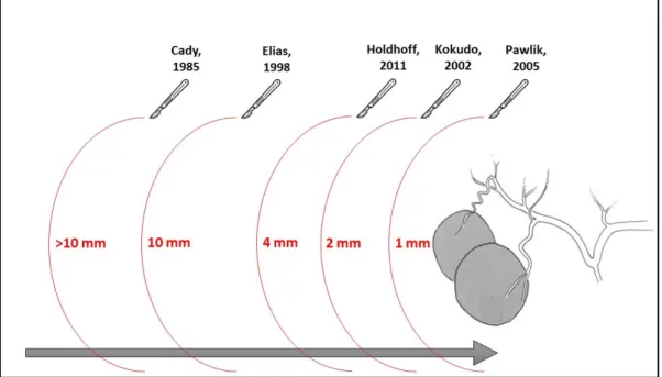
Surgeons have progressively moved from the “1-cm” rule to the “1-mm” rule in the treatment of CRLM and a negative (≥ 1-mm) surgical margin is the present standard (Figure 1). In the 1985, Cady et al. have reported that a surgical margin less than 1 cm was associated with a significantly shorter disease-free survival. As a result, major centers have adopted a 1-cm margin as a target during resection to minimize hepatic recurrence and improve survival after resection of CRLM. In fact, a 1-cm margin has been proposed as the minimally acceptable margin even for ablative techniques19.
Later on, considering that the use of 1-cm rule for resection could exclude a large number of patients from the only therapeutic interventions able to affect long term survival, not reaching this margin became not a contraindication but a strong recommendation20. In 1998, firstly
Elias stated that the “one-centimeter free margin” concept should not be rigidly adhered to. For the author, it is justifiable to undertake resection of liver metastases from colorectal cancer, provided it is curative and safe, even in the face of what would classically be considered poor prognostic factors.
In 2011, Holdhoff evaluated the resection margin through a combination of histopathologic and genetic analyses and found that tumors with a significant radiologic response to chemotherapy were not associated with any increase in mutant tumor DNA in beyond 4 mm of the main tumor, supporting the clinical evidence that a negative (R0) margin may be sufficient. Furthermore, authors did not find evidence of residual tumor DNA in the region in which the tumor likely existed prior to chemotherapy, suggesting that tumors which respond to chemotherapy likely do so in a concentric fashion21.
In the last 15 years, various authors have shown comparable results with narrower margins and even with positive microscopic margins (R1). In 2002, Kokudo et al. reported that micrometastases around liver tumors were mostly confined to the immediate vicinity of the tumor border and so hypothesized that the minimum surgical margin for successful liver resection without cut-end recurrence may lie somewhere between 0 mm and 10 mm. Indeed,
20
a surgical margin of 2 mm appears to be a clinically acceptable minimum requirement, carrying an approximately 6% risk of margin-related recurrence.Kokudo’s study represents the first multicenter report to examine the effect of surgical margin status after resection of hepatic CRM on both margin recurrence and survival22.
In 2005, for the first time in literature, according to Pawlik et al., a positive margin was considered to be a margin less than 1 mm, defining as the presence of exposed tumor along the line of transection or the presence of tumor cells at the line of transection detected by histologic examination.Although a positive surgical margin was associated with an increased risk of margin recurrence (11%), the width of the margin was not significant. Patients with a margin of 1 mm to 4 mm did have a slightly increased rate of margin recurrence compared with patients who had wider margins; this did not reach statistical significance. In conclusion, this study demonstrated that the width of a negative surgical margin does not affect survival, recurrence risk, or site of recurrence23.
21
Figure 1. The paradigm shift from large to parenchymal sparing resections.
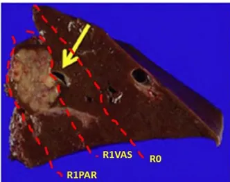
Similarly, detachment of CRLM from major intrahepatic vessels if they have not been infiltrated (R1 vascular resection) was recently shown to have an excellent outcome. In fact, in 2016, Viganò et al. tried to clarify the clinical relevance of R1 resection for CRLMs in a large, recent, single-center series, with a focus on the distinction between tumor exposure along the transection plane (standard R1) and CRLM detachment from intrahepatic vessels that is called R1 vascular (R1Vas) (Figure 2). Instead of R1 parenchymal (R1Par), representing an independent negative prognostic factor of overall survival, conversely R1vascular surgery achieves outcomes equivalent to R0 resection. So, CRLM detachment from intrahepatic vessels can be pursued to increase patient resectability and resection safety24.
22
R1PAR: R1 parenchymal; R1VAS: R1 vascular
Figure 2. Representation of the different types of section margins
In conclusion, after Pawlik’study, the paradigm shift from large to parenchymal sparing resections had really achieved, leading to a modification of the oncological concept of safe resection margins.
Because the current consensus is that the thickness of the margin does not modify survival, the aggressive indications for CRLM and complexity of surgical procedures corresponded to high R1 resection rates.In the most experienced hepatobiliary units, R1 resection occurs in 10 to 30%25 of patients, reaching 30 to 60% of patients with multiple bilobar CRLM or with
initially unresectable disease26.
However, the adequate width of the surgical margin is still a matter of debate, as the outcomes of R1 resection is associated with higher local recurrence rate and worse survival and several pathological data support the inadequacy of R1 surgery.
Actually, in 2008, Adam et al. first reported no negative prognostic impact of positive surgical margins. Some recent studies have shown that perioperative chemotherapy may reduce or even cancel the relevance of R1 surgery27.
Later on, subsequent several recent studies had denied any negative prognostic role of R1 resection in the era of aggressive and effective perioperative chemotherapy2829.
23
Finally, in 2015, the EGOSLIM (Expert Group on OncoSurgery management of Liver Metastases) group convened and published a brief but clear statement; ‘‘safe resection margins are still a goal of therapy; a minimal surgical clearance margin of 1 mm has been suggested as sufficient.’’ Nonetheless, the optimal surgical margin for CRLM remains unknown30.
Considering that several evidences is still in favor of R0 surgery, a reappraisal of R1 resection is needed.
1.4 RECURRENCE AFTER LIVER RESECTION FOR COLORECTAL LIVER METASTASIS
Advances in surgical and medical oncology have resulted in prolongation of survival for patients with colorectal liver metastasis. However, many patients still develop recurrent disease. Studies addressing overall recurrence have reported rates ranging from 60 to 85% at one year31 32. Specifically, hepatic recurrence occurs in 50% of patients during follow-up,
with 2.8% to 13.9% presenting with surgical margin recurrence3132.
However, data on rates and patterns of recurrence following curative intent surgery for colorectal liver metastasis are limited. In fact, most studies reporting on outcomes following surgical management of colorectal metastasis have exclusively focused on overall survival rather than recurrence31. Unfortunately, to date, most series on the topic of pattern of
recurrence for colorectal metastasis have been limited by small sample sizes and the few largely single-institution studies were published in an era prior to more effective systemic chemotherapy.
One of the most frequently used scoring systems is the clinical risk score (CRS) developed by Fong et al.31. This system categorizes patients into “low risk” and “high risk” groups for
disease recurrence, with low scoring patients having an overall median survival of 74 months and high-scoring patients having an overall median survival of 22 months.
24
CRS was calculated as 1 point per criterion met: node positive primary, >1 preoperative liver lesion, largest preoperative liver lesion >5 cm, preoperative CEA >200 pg/L, and time between removal of primary and appearance of liver metastases <12 months. Since a clinical risk score of this kind was important for the accurate care of colorectal cancer patients, after its publication in 1999, the CRS has seen longstanding and pervasive use in surgical practice. Nevertheless, the improvements in clinical care may have modified the accuracy of this scoring system and probably, today it would be necessary to reassess and modernize the score, adding some factors now relevant for the management of CRLM and that may improve the prognostic power of the CRS in the modern era of liver resection33.
For example, blood loss and need for a transfusion remain a significant concern that can impact both immediate and long-term outcomes. Allogeneic red blood cell transfusions and their transfusion-related immunomodulation effects have been recently suggested as a cause for early cancer recurrence and worse overall outcomes34.
Then, numerous intra-operative strategies have been developed to limit blood loss. Of these, portal pedicle clamping (PPC), first described by Hogarth Pringle for liver trauma, is one of the only strategies proven effective to reduce intra-operative blood loss in randomized controlled trials. Portal pedicle clamping has also been recently employed in regular hepatobiliary practice and its effects on survival and recurrence has been investigated. De Carlis et al. found that patients who received intermittent hepatic pedicle clamping, comparing with those who did not, had similar 5-year overall survival rate, but the 5-year recurrence-free rate was significantly higher. Noteworthy, the study was limited by the exclusion of patients with higher-risk disease according to CRS35. Conversely, other results
are consistent with the lack of a difference in overall survival and recurrence-free survival36.
Moreover, in the last decade numerous studies have investigated factors associated with recurrence after hepatectomy in CRLM (Table 1).
25
Table 1. Factors associated with recurrence after hepatic surgery for colorectal liver metastasis. Literature
between 2012 -2018.
Authors N Median
age [years]
Type of study Median FU [months]
Factors related to recurrence Type of recurrence Incid ence [%] Jung S.W. 201634 279 65.5 Perspective 6 (univariate)
Poorly differentiate CRC, synchronous metastasis, ≥5 cm of liver mass, preoperative CEA≥50 ng/mL, positive liver resection margin, and surgery alone without perioperative chemotherapy (multivariate)
poorly differentiated CRC, ≥5-cm metastatic tumor size, positive liver resection margin, and surgery alone without perioperative chemotherapy
ER 10.8
Akyuz M.
201537 206 62 Retrospective 29 (univariate) Tumor size CEA pre-op, margin status
(multivariate)
Positive margin status increased 3-6 folds risk of SMR
SMR 15.5
Hallet J.
201638 2320 63 Retrospective 27 Node-positive primary, No. of lesions>3, Size of largest lesion>4 cm Overall Intrahepatic
Extrahepatic Intra and extra hepatic 47 46.2 31.8 (54 lung) 22 Viganò L.
201339 6025 >70y 25,4% LiverMetSurvey 34.4 T3–4 primary tumor; synchronous CRLM; > 3 CRLM; 0 mm margin liver resection; associated intraoperative radiofrequency ablation
Protective factors: preoperative chemotherapy and response to pre-operative chemotherapy
Overall
ER 45.4 10.6
Ayez N.
201228 264 62 Retrospective 34 T stage primary tumor; positive lymph node in primary tumor; >4 CRLM; no neoadjuvant chemotherapy
No difference in DFS and OS between R0/R1
Overall Intrahepatic Extrahepatic Intra and extra hepatic 65 20 33 11 Bhogal, R.H. 201540
243 66 Retrospective 58 For liver recurrence: male sex and advanced primary tumors (Dukes C)
For any recurrence: Number of metastases, largest tumor size ER (18 months) Liver Overall 38 11 27 Angelsen J.H. 201441 253 66 Prospective and
retrospective 60 RMs do not impact hepatic recurrence, whereas extrahepatic recurrence was more frequent compared to no recurrence with RMs <5 mm SMR Intrahepatic Extrahepatic 16.5 21.5 32.6 Mao R. 201742
255 56 Retrospective 28.6 CEA ≥ 30 ng/ml, primary tumor lymph vascular invasion (LVI), number of metastases ≥ 4, R1 resection, initially unresectable disease
ER Overall 34 65 Imai K. 201643 846 61 Retrospective 24 (Univariate)
Age, primary tumor stage, bilobar distribution of liver metastases, preoperative chemotherapy cycles and lines, response to last-line chemotherapy, tumor number and size at hepatectomy, CEA and CA19-9 at hepatectomy, PVE, major hepatectomy, two-step approach, surgical margin status of liver metastases, and concomitant extrahepatic disease
(Multivariate)
Age ≤57 years, preoperative chemotherapy line, progression of disease during last-line chemotherapy, 3 tumors at hepatectomy, and CA19-9.60 U/mL at hepatectomy
ER* 43
Lin J.
201844 307 57.5 Retrospective 31.7 (Univariate) Node-positive primary tumor and metastatic diameter > 3 cm
(Multivariate)
Node-positive primary tumor and metastatic diameter > 3 cm
ER • intrahepatic • extraepatic • unknown 16,0 57,1 30.6 12.2 Angelsen J.H. 201545
311 66.1 Retrospective 4.2 Number and size of metastases, ASA score and synchronous disease. Perioperative chemotherapy
Overall (4yr) intraepatic extraepatic intra and extra hepatic 67.4 43.1 28.2 28,7 de Jong M.C. 200946 1669 61 Retrospective 30 (Univariate)
Node-positive primary tumor, synchronous hepatic metastasis, history of RFA, and receipt of chemotherapy, the clinical risk score, tumor size >5, preoperative CEA level, surgical margin status were not associated
(Multivariate)
rectal primary tumor site, disease-free interval >12 months, history of RFA, receipt of chemotherapy, the clinical risk score
Overall Intraepatic Extraepatic intra hepatic + lung intra hepatic + other extraepatic 56.7 43.2 35.8 11.6 9.4 Gomez D.
201447 259 ≥65y 68% Retrospective 28 Higher tumor number, presence of perineural invasion and R1 resection Overall 53.3
Kim Y.
201948 68 57.1 Retrospective 2 Resection margin of the metastatic tumor and ypN NA
Schierge T.S. 201534
106 64.5 Retrospective 29 blood loss; comorbidities; tumor load, and positive resection margins,
transfusion NA
Kuo I.M
201549 159 58.5 Retrospective 38.5 (Univariate) Centrally located metastasis, primary tumor in the transverse colon,
metastasis in regional lymph nodes, initial extrahepatic metastasis, synchronous liver metastasis, multiple lesions, poorly differentiated tumor, and resection margin <10 mm
(Multivariate)
inadequate resection margin and centrally located liver metastasis
Overall
26 Parau A. 2015 70 ≥ 53 y 49% Retrospective NA (Univariate)
Age >53 years, advanced T stage of primary tumor, moderately- poorly differentiated tumor, positive and narrow resection margin, preoperative CEA level >30 ng/ml, DFS <18 months (Multivariate)
Perioperative chemotherapy and achievement of resection margins beyond 1 mm
NA
Abbreviations: SMR Surgical margin recurrence; ER: early recurrence (within 6 months after liver resection; * within 8 months after liver resection); CEA carcinoembryonic antigen level; CRC colorectal cancer; CRLM colorectal liver metastasis; RFA radiofrequency ablation; NA not available.
Several clinicopathologic and morphological factors are now considered to be independent prognostic factors associated with recurrence and hepatic recurrence. Primary colorectal tumor stage, differentiation and lymph node metastasis of primary colorectal tumor, time interval to the appearance of metastasis, number and size of metastases, preoperative CEA level, neoadjuvant/adjuvant chemotherapy and the status of the resection margin have been established as important determinants of tumor recurrence in CRLM28323437404249.
Among these several prognostic factors for recurrence, the surgical margin status or resection margins (RMs) is a technical, operative variable that is directly dependent upon the surgeon’s technique and it has also been traditionally associated with long-term prognosis50. However,
as previously mentioned, the importance of the surgical margin achieved during liver resection, as prognostic factor to predict the development of local recurrence and long-term outcome, remains controversial.
Actually, the prolonged overall survival observed with submillimeter margins is likely a microscopic surrogate for the biologic behavior of a tumor rather than the result of surgical technique51. In fact, the potential aggressiveness of colorectal cancer is readily evident when
relapses occur early after resection of the primary tumor, when it recurs in the liver with large or bilobar metastases, and when there is little or no measurable response to chemotherapy52 53.
Indeed, in 2006 Takahashi et al. showed that time to recurrence after liver resection for CRLM strongly correlated with prognosis and especially patients with disease recurrence within 6 months after liver resection have the poorest outcome54. According to these data,
early recurrence was defined as any recurrence occurring within 6 months after liver resection and the same time interval was adopted by Malik et al. in their study about early recurrences after liver resection for CRLM55. Interestingly, in this study the author found that the
27
recurrence on multivariable analysis. In addition, the presence of numerous hepatic metastases was also a predictor of extra-hepatic recurrences and unresectable recurrent disease, suggesting that early recurrence is a marker of aggressive tumor biology55.
In 2004, Tanaka et al. had already reported that short tumor doubling time in CRLM is a poor prognostic factor for both overall and disease-free survival53. The authors demonstrated that
only doubling time was retained as independent predictive factors for remnant liver recurrence and a doubling time of 45 days or less was associated with multiple, early remnant liver recurrences, precluding repeat hepatectomy and resulting in a poor prognosis. Interestingly, only tumor size and the prognostic nutritional index (PNI) based on the peripheral blood lymphocyte count and serum albumin concentration were significantly related to tumor doubling time, suggesting how doubling time would be determined by the interplay of both tumor characteristics and the patients’ immune and nutritional status. Nowadays, indicators of tumor biology and how they might influence outcome are of increasing interest. Mutation of the KRAS gene may be an indicator of biological aggressiveness. In this perspective, Cucchetti1et al. stratified a cohort of patients who underwent resection for only metachronous disease in three subgroup according to a mathematical model to estimate CRLM doubling times: the fast-growing CRLMs, doubling time less than 48 days; the intermediate-growing CRLMs, doubling time 48–82 days and the slow-growing CRLMs, doubling time more than 82 days. The study demonstrated that the tumor doubling time was shorter in patients with more advanced primary tumor stages, with mutant KRAS and in those who did not receive chemotherapy. In addition, for the fast-growing group, the risk of recurrence was highest within the first postoperative year and was about 7 per cent per month52.
Several studies had showed that histopathologic factors of primary CRC were related to liver metastasis. Conversely, few studies, focusing on the histopathology of metastatic lesions as a predictive marker of tumor recurrence have been performed. Histopathological studies of liver metastases have resulted in the description of three histological growth pattern (GP). These are: desmoplastic GP, where a rim of collagen surrounds the tumor tissue and separates the liver parenchyma from the cancer cells; pushing GP, where tumor cells push the liver parenchyma aside, encompassing pressure on the hepatocytes at the tumor margin; and replacement GP, where tumor cells replace the hepatocytes hereby maintaining the trabecular
28
architecture of the liver parenchyma (Figure 3)56. CRLMs grow according to different GPs
with different angiogenic properties. In a recent study that enrolled 205 patients from 1995 to 2005, who were resected for liver metastasis and followed for 2 years, a pushing GP was the only independent predictor of poor survival, suggesting that this pattern is characterized by a more aggressive tumour biology in comparison to patients with desmoplastic or replacement GP57. Similarly, in a second prognostic study by Nielsen et al., survival was
related to GPs, and desmoplastic growth was associated with small tumor size, dense lymphocytic infiltration and a more favorable prognosis in term of overall survival58. A more
recent study considered also the effect of the therapeutic approach, comparing chemo-naive patients and patients receiving neo-adjuvant therapy56. Authors found that desmoplastic GP
in resected liver metastases predicts a reduced risk of recurrence in comparison to other GPs, while the patients resected for pushing metastases tended to have earlier recurrence. Interestingly, the prevalence and impact of desmoplastic GP was independent of whether or not neo-adjuvant chemotherapy had been given56.
29
Figure. 3 Illustration of growth patterns in colorectal liver metastases. The different growth patterns are
illustrated in a, b, g (desmoplastic growth pattern), b, e, h (pushing growth pattern) and c, f, i (replacement growth pattern). The mixed growth pattern is not shown, but is usually a mixture of two patterns, often including a pushing component
Finally, data on the prognostic implications of vascular, biliary, perineural and lymphatic invasion in patients with CRLM are limited. Gomez et al, identified three independent predictors of disease-free survival, mainly tumor number, perineural invasion, and resection margin. In addition, the presence of perineural invasion was the only independent predictor of poorer overall survival on multivariate analysis47. More recently, Park et al. found that
tumor infiltrating inflammation and presence of dedifferentiation of metastatic lesion were independent risk factors for tumor recurrence after hepatic resection in CRLM59.
In other words, the pathophysiological mechanisms behind overall and hepatic recurrence may not simplistically include inadequate margin resection, but rather they could represent the expression of cancer aggressiveness and the natural progression of micrometastatic disease from the primary tumor.
Further elucidation of the mechanisms and biological pathways involved in and responsible for the differences in GP between CRC liver metastases in different patients might lead to therapeutic agents and strategies and may contribute to a histology-based prognostic biomarker for patients with colorectal liver metastases.
Therefore, the relationship between the growth rate of CRLMs and biological features of colorectal cancer may provide additional information related to outcome pathologic prognostic markers of hepatic tumors predicting the prognosis of these patients have been identified.
30
2. THE STUDY
2.1 AIMS OF THE STUDY
The aim of this study was to investigate the impact of margin width resection on early liver recurrence and disease-free survival after hepatic resection for colorectal metastasis in a consecutive series of patients from a single institution. The hypothesis of the present study was that margin width resection (R0 or R1) does not influence oncological outcomes after resection for CRLM.
In addition, the study aimed to identify other clinicopathologic prognostic factors predictors for early recurrence (defined as recurrence within 6 months of CRLM resection) and for disease-free survival.
Moreover, the study sought to examine the pattern of early and late recurrence (intra- or extra-hepatic recurrence) of patients who were managed with curative intent resection.
2.2 MATERIALS AND METHODS
2.2.1 STUDY DESIGN AND PATIENTS’ SELECTION
This is a prospective observational study, performed at the Oncological Surgery, Hospital Policlinic San Martino, Genoa, Italy from 1st April 2014 to the 1st July 2020. The study was approved by the Local Ethical Committee and met the guidelines of the local Govern-mental Agency. Patients provided written informed consent before inclusion.
Patients undergoing primary hepatic resection for colorectal liver metastasis with curative intent and having a minimum follow-up period of 6 months were included.
The selection criteria for surgery in our center included a sufficient remaining tumor-free liver volume6 7 with adequate blood perfusion and bile drainage, and absence of: a)
non-31
resectable extrahepatic metastases, and/or b) no disseminated disease as evaluated pre-operatively.
Exclusion criteria for this study were patients who underwent repeat hepatic resections, who colorectal resection was not performed in our center, patients with R2 resections or Dindo Clavien V and patients who underwent combined resection with radiofrequency ablation (RFA).
2.2.2 PREOPERATIVE EVALUATION
Before surgery, all patients were evaluated with a baseline history and physical examination, serum laboratory tests, and appropriate imaging studies. Preoperative investigations included computed tomography (CT) scan of the chest and abdomen/pelvis, and tumour marker analysis (CEA: carcinoembryonal antigen). In cases with an inconclusive CT scan, magnetic resonance imaging (MRI) of the liver, contrast-enhanced ultrasound and 18 F-fluorodeoxyglucose 18(FDG)-positron emission tomography (PET)/CT scan were performed.
Each patient was discussed in a multidisciplinary team meeting with surgeons, oncologists and radiologists and also geriatric evaluation in patients 65-year-old and older.
Preoperative chemotherapy was administered for patients with initially unresectable CRLM in the conversion setting or patients with synchronous (diagnosed before, during, or within 3 months after colorectal resection) or marginally resectable CRLM in the neoadjuvant setting. Standard demographic and clinicopathologic data were collected for each patient including sex, age, ASA score, CEA level, comorbidity (with particular attention to chronic liver disease and cirrhosis, staging through MELD and Child-Pugh Score), as well as treatment related variables including history of preoperative chemotherapy, number of cycles.
Data were also collected on primary tumor characteristics, specifically on primary tumor location, date of resection (primary), TNM stage, genotype mutations (KRAS, NRAS, BRAF), microsatellite instability. Furthermore, the Lymph nodes ratio (LNR), defined as the ratio between positive lymph nodes and the total number of retrieved lymph nodes, was also
32
collected for each colorectal surgical procedure. Then, date of detection of CLM and presentation (synchronous vs. metachronous), number, size and location of CLM were recorded. In the present study, diagnosis of liver metastasis within three months were considered as metachronous, even though up-to-date there is no consensus on the defining time point for synchronous/metachronous disease60.
2.2.3 SURGICAL PROCEDURES
All patients underwent conventional open liver resection with curative intent, and to achieve complete resection (R0) while preserving as much normal functional liver parenchyma (with adequate vascular inflow, outflow, and biliary drainage) as possible. Resection of three or more segments was considered a major hepatic resection. The presence of extrahepatic tumors was not considered a contraindication to hepatic resection if the lesions were limited and resectable. Extrahepatic disease identified in the abdominal cavity was resected at the same time as hepatic resection. For extrahepatic disease located outside the abdomen, resection was performed 2–3 months after hepatectomy if the disease remained controlled with interval chemotherapy.
The operations were performed by two different surgeons. Intraoperative ultrasound of the liver was carried out in all patients. A central venous pressure less than 5 mm Hg was maintained during parenchymal transection and monitored by central venous access.
All patients received therapeutic liver resection and hepatic hilar lymph node dissection was not performed routinely. Anatomical resection was characterized as complete anatomical resection based on Couinaud’s classification (segmentectomy, sectionectomy, and hemi-hepatectomy or extended hemihemi-hepatectomy) in patients with an acceptable liver reserve61.
Non-anatomical resection (atypical resection) was the first-choice type of resection, according to the concept of parenchyma spearing, but if it is not suitable, a two-stage hemi-hepatectomy approach, as ALPPS, has been performed. As regard synchronous metastases, primary tumor resection was combined to metastases resection balancing patient's clinical conditions and fitness for surgery and the burden and extension of metastatic disease.
33
Pringle maneuver had always been carried out. Intermittent portal pedicle clamping was used at the discretion of the operating surgeon (no longer than 15 minutes clamped with 5 minutes unclamped).
In general, the hepatic parenchymal transection was performed through the clamp-crush technique. Once the parenchyma is crushed, the exposed vessels and bile ducts were divided through absorbable suture or non-absorbable suture ligation. Alternatively, vascular clips for larger caliber vessels and bipolar energy device (bipolar forceps or Aquamantys®) for smaller caliber vessels were used.
When hemostasis on the liver section area is not convincing, a flap of sealant matrix of human fibrinogen/human thrombin is applied.
As regarding resection margins (RM), RM <1 mm were defined as positive (R1), in accordance with Pawlik et al23. In addition, RM status was obtained from the microscopic
measurements in the histological reports, in which the closest distance was measured between the tumor edge and the transection surface of the liver parenchyma. Microscopically and in line with the histological reports, the widths were stratified as coincidental margins if the tumor was in contact with the surgical margin (0 mm); widths of less than, or equal to, 1 mm or greater than 1 mm.
2.2.4 POSTOPERATIVE PERIOD AND FOLLOW-UP
Postoperative complications were graded according to the validated classification criteria described by Dindo Clavien Classification62 and the Comprehensive Complication Index
(CCI®-Calculator)63 and major complications were defined as any complication of grade III
or higher.
Adjuvant chemotherapy was recommended routinely, using the same protocol as that applied before surgery.
All patients were followed up every three months for the first two years, with a physical examination, carcinoembryonic antigen (CEA) measurement, and abdominal ultrasonography. Every six months, patients underwent computed tomography scan of the
34
abdominal/thoracic/pelvic region (enhanced MRI could replace CT) to detect any intrahepatic or distant recurrence. In accordance with previous reports5455, early recurrence
was defined as any recurrence (liver recurrence (LR) or extrahepatic recurrence) occurring within 6 months after liver resection.
Patient characteristics and details of surgical treatment, as margin resection width, and perioperative chemotherapy were analyzed to identify predictive factors of early recurrence. At last date of follow-up date overall survival (OS) and disease-free survival (DFS) were also collected.
2.2.5 STATISTICAL ANALYSIS
Patients’ data were collected from a prospective computerized database. The descriptive analysis for quantitative variables was expressed as median or mean and standard deviation (SD).
The association between categorical data was performed with the two-tailed Pearson χ2, or Fisher’s exact test when appropriate. Student’s t-test was used to analyze any significant clinical pathological differences among patients who developed early recurrence when compared to the remaining cohort.
Recurrence and disease-free survival were estimated using the Kaplan-Meier method, and any significant difference between the sub-groups noted by univariate analysis was compared using the log-rank test.
All statistical analyses were performed using the SPSS for Windows version 20.0 (SPSS Inc., Chicago, IL, USA).
35
2.3 RESULTS
2.3.1. PATIENTS’ DEMOGRAPHICS AND CLINICAL CHARACTERISTICS During the study period, 66 patients underwent primary hepatic resection for CRLM. Two patients died postoperatively within 30 days. Nine patients were excluded due to uncompleted collection data. Thus, a total of 57 patients were ultimately included in the study.
There were 38 (67) men and 19 (33%) women, and the median age at the time of surgery was 69,15 (range 38 to 86); 20 patients (35% of the sample) were age 75 or older.
More than 82% of patients had one or more comorbidities and 50% of patients had multimorbidity (i.e. CRLM plus two or more comorbidities), with a median number of drugs of 3,40. The most common comorbidities were diabetes, hypertension, heart failure and COPD. So, that most of patients were classified as ASA 3 (42%) or ASA 2 (45%).
Patients’ clinical characteristics are illustrated in Table 1.
Table 1. Patients’ demographic and clinical characteristics.
VARIABLES N %
MEDIAN AGE (range) 69,15 (38–86 y)
GENDER: Female Male 19 38 33 67 ASA SCORE 1 2 3 10 25 22 18 44 39
NUMBER OF DRUGS Median (range) 3,4(0-11)
COMORBIDITY Diabetes Hypertension Heart failure Kidney disease COPD Thyroid pathologies HCV HBV 12 29 13 1 4 4 2 2 21 51 23 2 7 7 4 4 CHILD (N=43) A5 A6 38 5 88 12
MELD (N=45) Median (range) 7,55 (6-14) MARKERS CEA > 200 µg/L CA 19.9 > 33,0 7 18 13 33
36 PERI-CHEMOTERAPY • neo-adjuvant • Adjuvant • Pre + adjuvant 37 23 23 13 66 41 41 23
Overall, 23 patients (40%) underwent neoadjuvant chemotherapy before liver surgery, including 87% who received neo-adjuvant fluoropyrimidine-based cytotoxic chemotherapy. In detail, 9 patients received neo-adjuvant 5FU ± oxaliplatin; 5 patients neo-adjuvant 5FU ± oxaliplatin + bevacizumab or + panitumumab (n=3). Only a minority of the patients (n = 3) received fluoropyrimidine-based treatment ± irinotecan ± cetuximab (EGFR inhibitor). Then, biologic agents such as bevacizumab, cetuximab, and panitumumab were used in 9 patients (50%). Preoperative chemotherapy included ≥ 4 cycles in 22 patients (96 %) while ≥ 6 cycles in 15 (65%) patients.
2.3.2 CLINICOPATHOLOGIC CHARACTERISTICS OF THE PRIMARY AND METASTATIC TUMORS
Table 2 illustrates primary and metastatic tumor characters. With respect to primary colorectal cancer characteristics, almost 55% of the sample had a primary colon tumor, while 45% had a primary rectal tumor. In terms of pathologic stage, most patients had T3–T4 tumors (n = 55; 96%), lymph node metastasis (n = 32; 56%). During the primary resection, the median number of lymph nodes removed per patient was 20 (range: 5–55).
Thirty-eight patients (67%) had moderately differentiated colorectal tumors (G2), while 11 (19%) poorly differentiated tumors (G3 e 4).
In 36 patients were available the genotype analysis of KRAS and BRAF mutation. KRAS and BRAF mutations were detected in 30.5% and 0.5% of the cases, respectively. Indeed, all patients reported microsatellite stability (MSS).
In 38 patients (67%), the presentation of liver metastasis was synchronous with the primary while 19 patients (33%) had metachronous liver metastasis with a median disease-free time of 10 months.
37
The median number of liver metastasis was 2 (range 1 to 14). Sixty-one per cent of patients had a solitary liver tumor, while 22 patients (39%) had ≥ 2 tumors and 72% of patients had tumors measuring <5 cm.
Most patients had bilobar hepatic disease (25%) while six patients confined to only one hemi-liver (75%). Indeed, the most prevalent growth pattern distribution were the pushing GP (30%), the mixed GP (25%) and the replacement (14%).
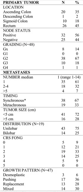
Table 2. Clinicopathologic characteristics of the primary and metastatic tumors
PRIMARY TUMOR N % LOCATION Ascending Colon Descending Colon Sigmoid Colon Rectum 20 1 10 26 35 2 18 45 NODE STATUS Positive Negative 32 25 56 44 GRADING (N=48) Gx G1 G2 G3 G4 8 0 38 10 1 14 0 67 18 1 METASTASES
NUMBER median 1 (range 1-14)
1 2-4 >5 35 18 4 61 32 7 TIMING Synchronous* Metachronous 38 19 67 33 TUMOR SIZE (cm) <5 cm >5 cm 41 16 72 28 DISTRIBUTION (N=19) Unilobar Bilobar 43 14 75 25 CRS FONG 0 1 2 3 4 5 5 12 19 14 5 0 9 21 33 25 9 0 GROWTH PATTERN (N=47) Desmoplastic Pushing Replacement Mixed 3 17 13 14 6 36 28 30 *diagnosis of liver metastasis within three months
38
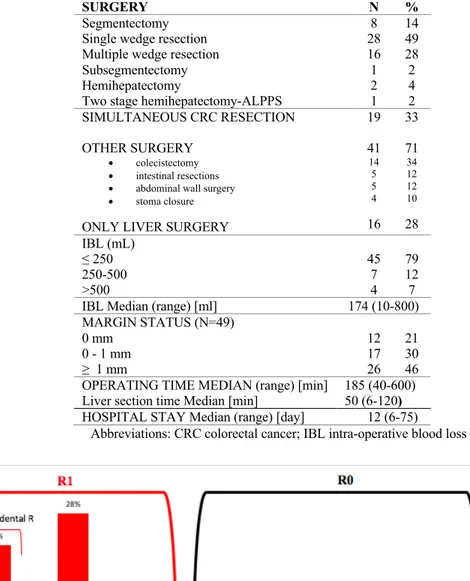
2.3.3 SURGICAL RESECTIONS AND POSTOPERATIVE RESULTS
Surgical treatment was conventional open liver resection with curative intent in all patient. The distribution of type of hepatic resections is shown in Table 3.
Patients undergoing combined surgery were 19 (37%), one patient was submitted to staged liver resections and underwent portal vein embolization. The most common surgical resections performed in this series were single and multiple wedge resection, counting for the almost 80% of the total procedures.
The blood transfusions were made only in two cases during surgery, while during the postoperative period seven patients were treated with antiplatelet agents.
The Pringle maneuver was used in all patients, and when needed, the median of time of application was 38 min (5-80 min).
The median length of hospital stay was 12 days (range 6 to 47). Intensive care admission after surgery was needed in 13 (26%) patients, but only for the first 24 hours.
As regarding resection margins, the mean resection margin in all patients was 1.4 mm ± 1.5 mm, which reflects a parenchyma-sparing operative approach. Margin status was unknown only in one patient. The distribution of RMs, defined as positive (R1) if ≤ 1 mm according to Pawlik et al.23 was represented in Figure 5. An R0 resection was achieved in 21 (32%)
patients and an R1 resection occurred in the remaining 36 (68%) patients. Histological reports categorized 21% patients as coincidental margins (0 mm); 30% with margins greater than 0.1 mm to 1 mm; 46% with margins greater than 1 mm; and no patients with margins greater than 1 cm.
39 Table 3. Types of surgery and resection margin status.
SURGERY N %
Segmentectomy Single wedge resection Multiple wedge resection Subsegmentectomy Hemihepatectomy
Two stage hemihepatectomy-ALPPS
8 28 16 1 2 1 14 49 28 2 4 2 SIMULTANEOUS CRC RESECTION OTHER SURGERY • colecistectomy • intestinal resections • abdominal wall surgery • stoma closure ONLY LIVER SURGERY
19 41 14 5 5 4 16 33 71 34 12 12 10 28 IBL (mL) ≤ 250 250-500 >500 45 7 4 79 12 7
IBL Median (range) [ml] 174 (10-800)
MARGIN STATUS (N=49) 0 mm 0 - 1 mm ≥ 1 mm 12 17 26 21 30 46 OPERATING TIME MEDIAN (range) [min]
Liver section time Median [min]
185 (40-600) 50 (6-120)
HOSPITAL STAY Median (range) [day] 12 (6-75)
Abbreviations: CRC colorectal cancer; IBL intra-operative blood loss
Abbreviations: R: resection; * according to Pawlik23.
40
Within 30 days of surgery, 42 patients (74%) developed ≥1 medical or surgical complication, either during the hospitalization or after discharge, without leading to readmission (Table 4). The most common complications were minor (50%), or even classified as I -II grade according to Dindo-Clavien classification62. Actually, although resection may be associated
with liver-related complications such as hemorrhage, bile leak and liver insufficiency, these complications were uncommon and only 26% were major complication (> III grade). Bile leak and abscess at the resection site were the most frequent (9% and 9% respectively) and they required to be treated with percutaneous drainage placement. Reoperation occurred in three cases but as complication related to colorectal surgery (2 patients who developed anastomotic leak and one patient whit obstruction bowel). Indeed, no bleeding occurred in this series and the median perioperative blood loss was 174 ml (10-800). Nevertheless, most complications in this series were pulmonary (14%) and renal (18%).
Although there seemed to be an increased length of hospital stay in parallel with the degree of severity of the complications according to Dindo Calvien classification (Figure 6), these difference did not reach the statistically significance (no complications 8.4 days; I-II grade 10.6 days and III-IV grade 13.3 days; p<0.2).
The distribution of complicated patients according to Comprehensive Complication Index (CCI) is shown in Figure 7, in which each complication grade is designated to prefixed scores (grade I = 8.7, grade II = 20.9, grade IIIa = 26.2, grade IIIb = 33.7, grade IVa = 42.4, grade IVb = 46.2). As the majority of grade I and grade II complicated patients showed a single complication, the median CCI was 20.0 (range 0-100). Indeed, high CCI scores were positively correlated with prolonged hospital stay (Pearson correlation t = 2.15, df = 36, p-value <0.03; 95%CI: 0.02 - 0.59; cor 0.33) (Figure 8).