Correspondence: Josep M. Campistol Servicio de Nefrología.
Hospital Clínic.
C/ Villarroel, 170. 08036 Barcelona. (Spain). [email protected]
Actualización en síndrome hemolítico urémico atípico: diagnóstico y tratamiento. Documento de consenso RESUMEN
El síndrome hemolítico urémico (SHU) es una entidad clínica definida por la tríada anemia hemolítica no inmune, trombo-citopenia e insuficiencia renal aguda, en la que las lesiones sub-yacentes están mediadas por un proceso de microangiopatía trombótica (MAT) sistémica. El SHU atípico (SHUa) es un subti-po de SHU en el que los fenómenos de MAT son consecuencia de la pérdida de regulación de la vía alternativa del comple-mento sobre las superficies celulares de causa genética. El SHUa es una enfermedad ultra-rara que, pese al tratamiento están-dar con terapia plasmática, frecuentemente evoluciona a la in-suficiencia renal crónica terminal, con elevada mortalidad. En los últimos años, se ha establecido el papel clave que desem-peña el sistema del complemento en la inducción de daño en-dotelial en los pacientes con SHUa, mediante la caracterización de múltiples mutaciones y polimorfismos en los genes que co-difican determinados factores del complemento. Eculizumab es un anticuerpo monoclonal que inhibe la fracción terminal del complemento bloqueando la formación del complejo de ataque de membrana. En estudios prospectivos en pacientes con SHUa su administración ha demostrado la interrupción rá-pida y sostenida del proceso de MAT, con mejorías significati-vas de la función renal a largo plazo y con una reducción im-portante de la necesidad de diálisis o terapia plasmática. En el presente documento se revisan y actualizan los diversos aspec-tos de interés de esta enfermedad, con especial atención a cómo los recientes avances diagnósticos y terapéuticos pueden modificar el tratamiento de los pacientes con SHUa.
Palabras clave: Síndrome hemolítico urémico atípico. Eculizumab. Complemento. Microangiopatía trombótica.
INTRODUCTION
Haemolytic uraemic syndrome (HUS) is a clinical entity defined by the triad of non-immune haemolytic
ABSTRACT
Haemolytic uraemic syndrome (HUS) is a clinical entity defined as the triad of nonimmune haemolytic anaemia, thrombocytopenia, and acute renal failure, in which the underlying lesions are mediated by systemic thrombotic microangiopathy (TMA). Atypical HUS (aHUS) is a sub-type of HUS in which the TMA phenomena are the consequence of decreased regulation of the alternative complement pathway on cell surfaces due to a genetic cause. aHUS is an extremely rare disease that, despite the administration of standard treatment with plasma therapy, often progresses to terminal chronic renal failure with a high associated rate of mortality. In recent years, research has established the key role that the complement system plays in the induction of endothelial damage in patients with aHUS, through the characterisation of multiple mutations and polymorphisms in the genes that code for certain complement factors. Eculizumab is a monoclonal antibody that inhibits the terminal fraction of the complement protein, blocking the formation of a cell membrane attack complex. In prospective studies in patients with aHUS, administering eculizumab produces a rapid and sustained interruption in the TMA process, with significant improvements in long-term renal function and an important decrease in the need for dialysis or plasma therapy. In this document, we review and bring up to date the important aspects of this disease, with special emphasis on how recent advancements in diagnostic and therapeutic processes can modify the treatment of patients with aHUS.
Keywords: Atypical haemolytic uraemic syndrome. Eculizumab. Complement. Thrombotic microangiopathy.
An update for atypical haemolytic uraemic syndrome:
diagnosis and treatment. A consensus document
Josep M. Campistol
1, Manuel Arias
2, Gema Ariceta
3, Miguel Blasco
1, Mario Espinosa
4,
Josep M. Grinyó
5, Manuel Praga
6, Roser Torra
7, Ramón Vilalta
3, Santiago Rodríguez de Córdoba
8 1 Servicio de Nefrología. Hospital Clínic. Barcelona (Spain)2Servicio de Nefrología. Hospital Universitario Marqués de Valdecilla. Santander (Spain)
3Servicio de Nefrología Pediátrica. Hospital Universitari Materno-Infantil Vall d'Hebrón. Barcelona (Spain) 4Servicio de Nefrología. Hospital Universitario Reina Sofía. Córdoba (Spain)
5Servicio de Nefrología. Hospital Universitari de Bellvitge. Barcelona (Spain) 6Servicio de Nefrología. Hospital Universitario 12 de Octubre. Madrid (Spain) 7Enfermedades Renales Hereditarias. Fundació Puigvert. Barcelona (Spain)
8Departamento de Medicina Celular y Molecular. Centro de Investigaciones Biológicas (CSIC). Madrid (Spain)
Nefrologia 2013;33(1):27-45
microangiopathic anaemia, thrombocytopenia, and acute renal failure.1 Histological damage from HUS is
characterised by the appearance of systemic thrombotic microangiopathy (TMA), which primarily affects the renal blood vessels, producing thickening of the vessel wall, thrombosis, and obstruction of the vascular lumen. The majority of cases of HUS are caused by an enteric infection by shiga toxin-producing Escherichia coli (STEC) or other micro-organisms that produce verotoxin (VTEC), giving way to the so-called typical HUS or STEC (VTEC)-HUS. In a similar manner, TMA lesions and HUS can be secondary to other underlying diseases, drugs, certain types of organ transplants, or pregnancy. Rarely, HUS is produced as the consequence of deregulation of the alternative pathway for the complement system due to genetic abnormalities, which leads to endothelial damage and convey systemic TMA.2
This type of HUS is known as atypical HUS (aHUS), constituting a severe disease associated with a poor prognosis and elevated mortality rates. The prognosis for aHUS is sombre despite recommendations for intensive treatment regimens with plasma therapy (PT) and life support measures. More than 50% of patients with aHUS die from this disease, require dialysis, or develop permanent kidney damage during the year following diagnosis.3
Recent advances in the characterisation of the genetic component of aHUS (including the identification of multiple mutations and polymorphisms in the genes that code for certain complement proteins) have led to the conclusion that the endothelial damage produced by the complement system is the critical factor in the pathophysiology of this disease, and the hypothesis that inhibition of the complement system is a viable treatment option in these patients. In 2011, the governing regulatory agencies of the United States and Europe approved the indication for eculizumab (Soliris®; Alexion
Pharmaceuticals, Connecticut, USA),4 a humanised
monoclonal antibody that inhibits the activation of C5 and blocks the generation of the proinflammatory anaphylotoxin C5a and the lytic pathway of the complement system (which produces cell lysis), for the treatment of aHUS.5In prospective
studies of patients with aHUS, eculizumab has been shown to effectively interrupt the process of TMA and its consequences, associated with significant long-term improvements in haematological parameters and renal function.6-9
With the objective of elaborating updated diagnostic and treatment protocols for aHUS, a consensus conference was held on February 2 and 3 of 2012 in Barcelona, which brought together clinical and research experts in the field of TMA. Using the platform of the available scientific evidence and clinical experience, this conference discussed various aspects of interest of the disease, such as the aetiological classification of TMA, the pathophysiology of aHUS, and differential diagnosis, and recommendations were made for treating patients with aHUS. This article summarises the primary conclusions derived from this meeting.
AETIOLOGICAL CLASSIFICATION OF THROMBOTIC MICROANGIOPATHY
The term TMA defines a histological lesion found in the arterioles and capillaries that is characterised by thickening and inflammation of the vascular wall, detachment of endothelial cells, subendothelial widening due to the accumulation of proteins and cellular debris, and platelet thrombi that occlude the vascular lumen (Figure 1).1Two different clinical entities are
characterised by primary TMA lesions, which have different causes and histopathological foundations: thrombocytopenic thrombotic purpura (TTP) and HUS.
Intravascular thrombosis in TTP is the consequence of a severe deficiency of metalloprotease activity of the ADAMTS13 (A Disintegrin And Metalloproteinase with a ThromboSpondin type 1 motif, member 13), a plasma enzyme responsible for fragmenting the ultra-long multimeres of the von Willebrand factor.10This deficiency can be caused by genetic abnormalities
or can be acquired through circulating IgG antibodies that block ADAMTS13 (especially in patients on treatment with anti-platelet medications).11
Approximately 90% of cases of HUS are caused by an enteric infection by STEC from contaminated foods (typical HUS/STEC[VTEC]-HUS).2 The shiga toxin exerts a direct
damaging effect on the vascular endothelium, triggering multiple cellular and vascular phenomena that lead to the
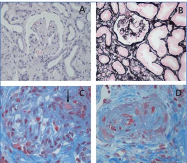
Figure 1. Renal histopathological lesions from haemolytic
uraemic syndrome.
A. Ischaemic and retracted glomeruli; B. Mesangiolysis; C. Thrombi in the glomerular capillaries (arrow); D. Artery occluded by platelet thrombi.
Photographs courtesy of Dr R. Ortega (Histopathology department of the Hospital Universitario Reina Sofía de Córdoba).
A
B
development of TMA.2The disease appears clinically in the
form of abdominal pain and diarrhoea, with acute renal failure appearing after 4-10 days. The prognosis tends to be good: mortality is <5% and a complete clinical recovery is achieved in 80% of patients.12,13
In contrast, aHUS is a disease in which the TMA phenomena are the consequence of a deregulation of the alternative complement pathway on cell surfaces. This alteration can be produced by mutations or polymorphisms that decrease the activity of complement regulating proteins, or that increase the function of activator proteins. In both cases, activation of the complement system (induced by a range of triggering factors) is not appropriately controlled, provoking endothelial damage and thrombosis. Of the more than 1000 patients with aHUS published in the medical literature, mutations have been detected in one or more complement proteins in 50%,14-21
although we cannot rule out that in the other cases there may have also been a genetic or environmental component involved that could have affected the abnormal complement system activity. In fact, it is notable that auto-antibodies against complement factor H (FH) are found in 5%-10% of patients with aHUS.22In contrast to STEC-HUS, which tends to occur as
a single event, aHUS is a chronic condition due to the genetic origins of the disease, and involves a poor prognosis. After the first episode of aHUS, mortality is 10%-15%, and 50% of patients will never recover renal function.3,14,15
In addition to infection by STEC or genetic deregulation of the complement system, there are many other clinical entities and triggering factors that can be associated with the development of HUS or TTP (secondary TMA). In children, some cases are associated with methylmalonic aciduria23or,
more frequently (5% of cases of HUS in children), with invasive infections by Streptococcus pneumoniae strains that produce the enzyme neuraminidase, which exposes the cryptoantigen T in the cell surface, thus producing TMA,24or
by H1N1 virus infection.25In adults, cases of TMA have been
described in association with human immunodeficiency virus (HIV) infection, certain types of cancer, drugs (chemotherapy agents, calcineurin inhibitors [cyclosporine and tacrolimus], mTOR [mammalian target of rapamycin; sirolimus, everolimus] inhibitors, vascular endothelial growth factor inhibitors, anti-platelet agents, and oral contraceptives, among others), malignant arterial hypertension, bone marrow and solid organ transplantation, pregnancy, and systemic diseases (systemic lupus erythematosus, scleroderma, and antiphospholipid syndrome).26
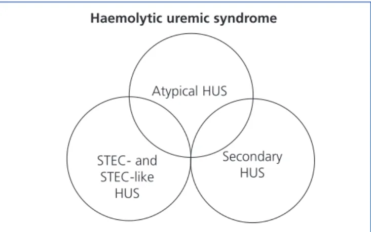
Table 1 displays the proposed aetiological classification of TMA. In certain patients more than one aetiological factor responsible for the TMA lesion may coexist, producing a variety of clinical presentations. Recently, it has been shown that as many as 25% of patients with STEC-HUS, and 86% of patients with HUS secondary to pregnancy, may have mutations in complement system gene sequences,27,28which
could signify that the underlying cause of these cases is in reality aHUS. In this context, Figure 2 represents the potential overlap that could occur between these two clinical entities. The primary motivation for discussion in this article is an update of aHUS mediated by altered complement regulation, and the following sections will refer exclusively to this entity.
ATYPICAL HAEMOLYTIC URAEMIC SYNDROME: CLINICAL ENTITY
Epidemiology
aHUS is considered to be an extremely rare disease. Very few sources of data are available regarding the incidence and prevalence of this condition, severely limiting our understanding of its true epidemiology. In the United States, aHUS is estimated to have an annual incidence rate of ~1-2 cases/million inhabitants.29 In Europe, a recent international,
multi-centre study reported an incidence of 0.11 cases/million inhabitants between the ages of 0 and 18 years. As regards prevalence, the EMA (European Medicines Agency) estimates approximately 3.3 patients/million inhabitants/year in individuals younger than 18 years of age, with lower rates in adults.
aHUS primarily affects children and young adults, although it can appear at any stage of life.14,15The start of the disease is
more common before the individual has reached 18 years of age (60% vs. 40%), and the sex distribution is similar (with a slight predominance in females when the disease appears later in life).14,16
Clinical manifestations
The initial onset of this disease can be abrupt, although it may occur progressively in approximately 20% of patients (a matter of weeks or months), with sub-clinical anaemia, fluctuating thrombocytopenia, and conserved renal function.14Symptoms
are characterised by the triad of non-immune microangiopathic haemolytic anaemia, thrombocytopenia, and acute renal failure.1
High levels of lactate dehydrogenase (LDH), undetectable haptoglobin levels, and schistocytes confirms the presence of intravascular haemolysis.3Patients also develop haematuria,
proteinuria, and/or acute renal failure (with or without oligoanuria). Arterial hypertension is a common finding, whether due to volume overload or vascular damage.1
Although aHUS predominantly affects the renal vessels, the diffuse character of TMA phenomena leads to involvement of the microvasculature of other organ systems (the brain, heart, intestines, pancreas, and lungs),1which explains the
common appearance of extra-renal symptoms.14,15The most
can include irritability, somnolence, confusion, convulsions, encephalopathy, cerebrovascular accidents, hemiparesia, hemiplexia, and coma.1,15,30,31Myocardial infarction has also
been described in as many as 3% of patients with aHUS, and can cause sudden death.15,32Cardiomyopathy, heart failure,
and peripheral ischaemic heart disease have also been described,22,30,33,34 in addition to diarrhoea (30%) and other
gastrointestinal symptoms (colitis, nausea and vomiting, and abdominal pain).15,22,31,35 The variability in symptoms
presented is an impediment to performing a differential diagnosis in comparison with other sources of TMA.
Pathophysiology
The complement system, which is composed of numerous proteins in plasma and in the cellular membranes, is essential in the defence against infection, the processing of immune complexes, the antibody responses, and the elimination of cell remnants from apoptosis. The activation of the complement system through any of the known pathways (classical/lectin and alternative pathways) leads to the formation of multi-protein complexes with C3-convertase activity that cleaves C3, generating C3b (Figure 3). This molecule can bind covalently to the cell surfaces responsible for complement activation, facilitating phagocytosis by polymorphonuclear leukocytes and macrophages, and initiating the cell membrane attack complex that induces cellular lysis. In addition, C3b exponentially amplifies the activation of the complement system, promoting the formation of more C3-convertase.36 In order to avoid
complete consumption by the activation of the complement system as well as damage to untargeted tissues (C3b binds indiscriminately to both pathogenic cells and native somatic cells), a number regulatory proteins such as complement factor H (FH), membrane cofactor protein (MCP), and complement factor 1 (F1) act to dissociate the C3-convertase
Table 1. Aetiological classification of thrombotic
microangiopathies
TTP associated with genetic or immune abnormalities of ADAMTS13 (activity <_ 5%)
- Genetic
- Antibodies (associated with ticlopidine and
clopidogrel)
HUS associated with infections (STEC and STEC-like) - HUS from STEC infection (VTEC) strain O157:H7 and
other non-O157:H7 strains, Shigella disenteriae type I
- HUS associated with Streptococcus pneumoniae
infection (neuraminidase)
Atypical HUS associated with genetic abnormalities or immune system alterations of the complement system - Mutations in CFH, MCP, CFI, THBD, CFB and C3 - Anti-CFH antibodies Secondary TMA - Pregnancy HELLP syndrome Postpartum - Diseases - Systemic SLE Anti-phospholipid syndrome Scleroderma - Other HIV infection Glomerulopathies
Malignant arterial hypertension H1N1 infection (influenza A) Cancer
Methylmalonic aciduria with homocysteinuria - Treatments - Quinine - Mitomycin - Gemcitabine - Cisplatin - Ionising radiation - Interferon
- VEGF and tyrosine kinase (sunitinib, imtinib, and dasatinib)
- Ticlopidine and clopidogrel
- Calcineurin inhibitors (cyclosporine, tacrolimus) - Sirolimus
- Valaciclovir - Oral contraceptives
- Solid organ and bone marrow transplants
ADAMTS13: a disintegrin and metalloproteinase with a thrombospondin type 1 motif, member 13; CFB: complement factor B gene; CFH: complement factor H gene; CFI:
complement factor I gene; HELLP: haemolysis, elevated liver enzymes, low platelet count; SLE: systemic lupus
erythematosus; TMA: thrombotic microangiopathy; MCP: membrane cofactor protein; STEC: shiga toxin producing Escherichia coli; THBD: thrombomodulin gene; VEGF: vascular endothelial growth factors; HIV: human immunodeficiency virus; VTEC: verotoxin producing Escherichia coli.
Figure 2. Representation of the clinical overlap of the
different types of haemolytic uraemic syndrome. HUS: haemolytic uraemic syndrome; STEC: shiga toxin producing Escherichia coli.
Haemolytic uremic syndrome
Atypical HUS Secondary HUS STEC- and STEC-like HUS
and induce the degradation of C3b. As a consequence, under normal conditions, C3b is maintained at low levels, and when the complement system is activated, its deposition is limited to those structures responsible for the activation.
Multiple studies have reported that 40%-60% of patients with aHUS carry of mutations in the complement system genes (complement factor H gene [CFH], membrane cofactor protein gene [MCP], complement factor B gene [CFB], and C3 gene [C3]),37-46 which are related to
deregulation of the alternative pathway (Table 2). FH acts in the plasma controlling homeostasis of the complement
system, as well as avoiding damage to self cell surfaces. Mutations in the FH gene associated with aHUS cluster at the C-terminal region, which results in a decreased FH-mediated protection of cell surfaces against accidental damage produced through activation of the complement system, but does not affect complement regulation in the plasma.47 One conclusion from the functional analyses
performed with these FH mutants is that aHUS is the consequence of damage caused by the complement system due to the dysregulation of the complement activation on self cell surfaces. Functional analyses of mutations associated with aHUS found in other complement genes,
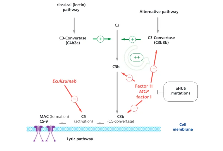
Figure 3. Complement dysregulation in atypical haemolytic uraemic syndrome.
Activation of the complement system by any of its three mechanisms (recognition of foreign antigens, alternative pathway, antibodies, classical pathway; or mannan polysaccharides (lectin pathway) leads to the deposition of large quantities of C3b on the activator cell membrane, which produces opsonisation and C5 activation (lytic pathway), which in turn produces the membrane attack complex and cell lysis. Activation of the complement system produces inflammation and recruitment of leukocytes. C3b production is the fundamental process in complement activation, and is also the best regulated. For the creation of C3b, unstable enzymatic complexes known as C3-convertases are needed, which catalyse the rupture of C3 to create C3b. C3b, in turn, is capable of forming more C3-convertase through the alternative pathway (C3bBb), thus amplifying the initial activation. The production of C3b is regulated at two levels: dissociation of C3-convertase, and proteolytic inactivation of C3b and C4b. Several regulatory proteins in the plasma and cell membrane carry out this regulatory activity. Among these, factor H, MCP, and factor I play key roles in the dissociation of alternative pathway C3-convertase (C3bBb) and the proteolytic degradation of C3b. Mutations in these genes observed in patients with aHUS interfere with this regulatory function of the activation of the alternative pathway.
MAC: membrane attack complex; MCP: membrane cofactor protein gene; aHUS: atypical haemolytic uraemic syndrome.
classical (lectin) pathway C3b C3 Alternative pathway C3-Convertase (C4b2a) Eculizumab Factor H MCP factor I C3-Convertase (C3bBb) Lytic pathway MAC (formation) C5-9 C5 (activation) C3b (C5-convertase) Cell membrane aHUS mutations
+
+
++
–
–
–
–
such as MCP, CFI, CFB, and C3, have confirmed the hypotheses anticipated in the studies with FH mutants, demonstrating, in addition, that dysregulation of complement on cell surfaces in aHUS may be due to decreased activity of the regulatory proteins or to an abnormally high activity of the C3-convertase. In this context, whereas mutations in FH, MCP, FI, and thrombomodulin (THBD) incapacitate these proteins from being able to carry out their regulatory function, mutations in complement factor B (FB) or in C3 result in a more active C3-convertase.
Anti-FH auto-antibodies directed towards the C-terminal region are found in 5%-10% of all patients with aHUS, with similar consequences to those produced by FH mutations.48,49
The role of these mutations in the pathogenesis of aHUS is not completely understood, but it appears to be associated with both the onset and the recurrences of disease. Antibody titres decrease with time, and should be measured at the first indication of aHUS.
Penetrance of aHUS in carriers of mutations in the complement genes is approximately 50%. It is common that in families with complement mutations only some carriers develop aHUS, with variable clinical presentations. There is also a wide range of clinical heterogeneity between unrelated patients with the same mutation. This suggests that additional genetic and environmental factors must exist that modulate the development and progression of this disease. The search for aditional mutations to genes in complement genes in patients with aHUS along with case-control association studies that using genetic polymorphisms in candidate genes or genetic markers distributed throughout the whole human genome have demonstrated that certain variants (polymorphisms) in the genes CFH and MCP modulate the presence and severity of this disease (Table 2).41,50,51These observations, along with the fact that as many
as 10% of patients with aHUS have mutations in more than one complement gene, indicates that the coincidence of different genetic risk factors is a determining factor in the development of aHUS (multiple hits theory).51
In addition to these genetic abnormalities, the onset of aHUS involves the participation of triggering environmental factors. The aforementioned mutations predispose the patient to disease, hampering adequate regulation of the complement system on cell surfaces in a situation that affects the microvasculature system. Infectious diseases can trigger aHUS in 50%-80% of patients,14,15,33 especially
diseases of the upper respiratory tract (H1N1 virus). Diarrhoea from gastroenteritis may precede aHUS in as many as 30% of cases (including STEC diarrhoea14,15,22). In
women, pregnancy can also be a triggering factor for aHUS.15,28
Prognosis
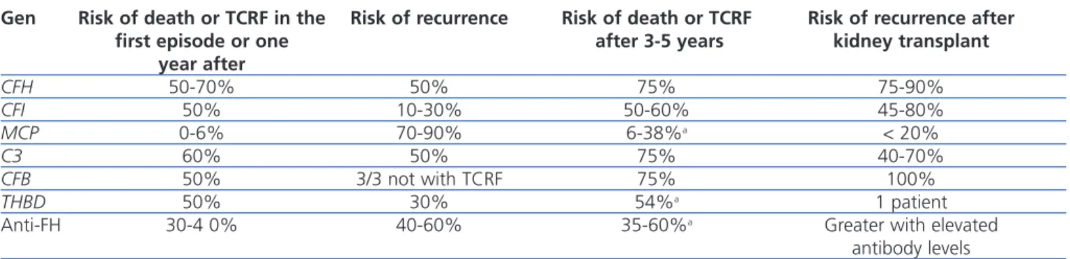
Table 3 displays the clinical evolution of patients with aHUS based on the type of mutation involved. In general, patients with FH and C3 mutations have a worse prognosis during the episode of aHUS and following months, with rates of mortality and terminal chronic renal failure (TCRF) or recurrence of 50%-70% and 50%, respectively. On the other hand, only 0.6% of patients with MCP mutations dies or develops TCRF, although the risk of recurrence is greater. Three out of four patients with FH, C3, or FB mutations die or develop TCRF.1
Table 2. Risk factors for atypical haemolytic uraemic
syndromea Mutations - Loss of function CFH (~13%) MCP (~11%) CFI (~10%) THBD (~4%) - Gain of function C3 (~4%) CFB (~3%) Polymorphisms - Increased risk CFH: c.-332C>T; c.2016A>G (p.Gln672Gln); c.2808G>T (p.Glu936Asp)
MCP: c.-652A>G; c.-366A>G; c.989-78G>A; a897T>C - Protection conferred CFH: c.184G>A (p.Val62Ile) Auto-antibodies - Anti-FH (~5%) Environmental factors - Infections - Immunosuppressants - Oral contraceptives - Anti-cancer drugs
Anti-FH: anti-complement factor H antibody; CFB: complement factor B gene; CFH: complement factor H gene; CFI:
complement factor I gene; MCP: membrane cofactor protein gene; aHUS: atypical haemolytic uraemic syndrome; THBD: thrombomodulin gene.
a Multiple hits theory. aHUS is a complex disease that normally
involves the combination of multiple genetic and environmental risk factors. It is not uncommon that patients will have more than one mutation in the genes coding for complement factors, or a combination of mutations with risk-associated
polymorphisms. The concurrence of one mutation with others, with risk-associated polymorphisms, with auto-antibodies, or with triggering environmental factors, explains the incomplete penetrance of aHUS, as well as the differences in its presentation and progression, in patients with mutations to complement genes.
recommended to be performed in patients with a suspected diagnosis of aHUS.2Due to the rapid and severe progression
of TMA, the diagnostic process must first involve an immediate phase (first 24 hours) that involves identifying the syndrome responsible and instating initial support measures, followed by a second phase of aetiological diagnosis. In patients with TMA, laboratory results indicate the presence of thrombocytopenia (platelets <150 000mm3or
decrease of >25% since initial level3) and microangiopathic
haemolysis (haemoglobin <10mg/dl with a negative direct Coombs test result [although some patients with HUS related to pneumococcal infection may have a positive direct Coombs test result],24elevated LDH, decreased haptoglobin
levels, reticulocytosis, and schystocytes3,56). Although it is
possible to detect schistocytes in the majority of patients with kidney disease, preeclampsia, and mechanical valves, a number of schistocytes >1% is diagnostic of TMA when in the absence of another known cause.58
Elevated serum creatinine levels, low glomerular filtration rates (GFR), proteinuria, or haematuria would be indicative of renal dysfunction.3,14,54 In paediatric patients, a renal
biopsy is not needed for diagnosis. A renal biopsy is usually indicated in adult patients in the case of acute renal failure in order to establish the aetiology of the disease, rule out other processes, and evaluate prognosis, although the indications for biopsy should be individually evaluated in patients suspected of TMA due to a risk of bleeding.
A complete clinical history is imperative, including family history, triggering factors, and an exhaustive physical examination. Contrary to medical opinion several years ago, it is currently held that the signs and symptoms for different types of TMA are not specific to each individual form, and so a differential diagnosis is not feasible.1Traditionally, the
Recurrence of atypical haemolytic uraemic syndrome after kidney transplantation
The results of kidney transplants (KT) in patients with TCRF caused by aHUS have been limited historically by the high percentage of post-transplant recurrence of disease (~50%; graft loss rate: 80%-90%52,53), although results vary based on
the type of mutation present. FH mutations are associated with a greater risk of recurrence or graft loss following KT (75%-90%), and high levels of risk are also associated with C3 and FI mutations (40%-80%; Table 3).15,35,40,54Until now,
very few transplants have been attempted in patients with FB mutations, but all cases that have been reported to date have involved recurrence of aHUS and graft loss.41,55 In general, the
plasma complement factors involved in aHUS are primarily synthesised by the liver, and so patients with mutations to complement genes that code for these factors continue to be susceptible to aHUS after KT, since the dysfunctional factors continue to be produced. On the other hand, MCP is a trans-membrane protein primarily produced by the kidney, and so a KT may correct the deficit by producing unaltered MCP in the new kidney. More than 80% of patients with MCP mutations do not develop recurrent aHUS after KT, with a similar long-term survival rate to that of patients who receive transplants for other reasons.33,52,53The risk of post-transplant
recurrence in patients with THBD56 or anti-FH antibody
mutations is not well understood, although in the case of FH antibodies, it appears that recurrence is related to persistently high antibody levels.22,57
DIAGNOSIS OF ATYPICAL HAEMOLYTIC URAEMIC SYNDROME
Figure 4 and Table 4 display the algorithm for the differential diagnosis of TMA and the biological tests
Table 3. Clinical characteristics of patients with atypical haemolytic uraemic syndrome based on complement
abnormality
Gen Risk of death or TCRF in the Risk of recurrence Risk of death or TCRF Risk of recurrence after first episode or one after 3-5 years kidney transplant
year after CFH 50-70% 50% 75% 75-90% CFI 50% 10-30% 50-60% 45-80% MCP 0-6% 70-90% 6-38%a < 20% C3 60% 50% 75% 40-70% CFB 50% 3/3 not with TCRF 75% 100% THBD 50% 30% 54%a 1 patient
Anti-FH 30-4 0% 40-60% 35-60%a Greater with elevated
antibody levels
Anti-FH: anti-complement factor H antibodies; CFB: complement factor B gene; CFH: complement factor H gene; CFI: complement factor I gene; TCRF: terminal chronic renal failure; MCP: membrane cofactor protein gene; THBD: thrombomodulin gene.
a Data for TCRF.
Figure 4. Algorithm for the differential diagnosis of primary thrombotic microangiopathy.
ADAMTS13: a disintegrin and metalloproteinase with a thrombospondin type 1 motif, member 13; LDH: lactate dehydrogenase; TMA: thrombotic microangiopathy; TTP: thrombotic thrombocytopenic purpura; HUS: haemolytic uraemic syndrome; aHUS: atypical haemolytic uraemic syndrome; STEC: shiga toxin producing Escherichia coli.
aNegative direct Coombs test; b The shiga toxin test/STEC is indicated when the patient has a history of digestive system
involvement or gastrointestinal symptoms; cRarely, in some patients with aHUS, STEC infection can trigger the underlying disease.
Table 4. Tests recommended for the diagnosis of atypical haemolytic uraemic syndrome.
STEC infection Faecal sample (diarrhoea) or rectal swab: STEC cultures (MacConkey for E. coli O157:H7); PCR for Stx genes O157:H7 and other serotypes, and other virulent characteristics; ELISA and/or Vero cell tissue culture assay for Stx serum: anti-LPS antibodies for prevalent serotypes
Pneumococcus infection Bacterial culture (generally) of sterile body fluids: DAT (Coombs test), viral test (respiratory), chest x-ray (pleural effusion as a characteristic of most cases), cytochemistry, and LCR culture in cases secondary to meningitis caused by a pneumococcus.
Altered regulation of the C3, C4 (plasma/serum), AH50
complement system FH, FI, FB (plasma/serum) Anti-FH auto-antibodies
Expression of superficial MCP on leukocytes (poly- or mononuclear leukocytes using FACS test) Mutation analysis for FH, FI, MCP, C3, FB ± THBD
ADAMTS13 deficiency Plasma activity of ADAMTS13 or dose (ELISA) ± inhibitor (acquired or hereditary)
Cobalamine metabolism: Amino acid chromatography in plasma/urine samples (hyperhomocysteinemia, hypomethioninemia,
methylmalonic aciduria homocysteinuria); organic acid chromatography in urine samples (methylmalonic aciduria) Mutation analysis for the gene MMACHC
HIV Serology, viral load (PCR)
H1N1 virus Culture and PCR
Pregnancy, HELLP syndrome Pregnancy test, liver enzyme levels. Analysis same as lines 3 and 4
Other Anti-nuclear antibodies, lupus anticoagulants, and anti-phospholipid antibodies
ADAMTS13: a disintegrin and metalloproteinase with a thrombospondin type 1 motif, member 13; DAT: direct antiglobulin test; ELISA: enzyme-linked immunoabsorption assay; FACS: fluorescence activated cell sorting; FB: complement factor B; FH: complement factor H; FI: complement factor I; HELLP: haemolysis, elevated liver enzymes, low platelet count; CSF: cerebrospinal fluid;
MCP: membrane cofactor protein; PCR: polymerase chain reaction; STEC: shiga toxin producing Escherichia coli; THBD: thrombomodulin; HIV: human immunodeficiency virus.
Adapted from Loirat.2
Thrombocytopenia
<150 000 or >25% deterioration
Evaluate ADAMTS13 activity and shiga toxin/STEC testb
<_5% ADAMTS13 activity >5% ADAMTS13 activity Shiga toxin/STEC positive
TTP aHUS Secondary TMA STEC-HUSc
More than one of the following:
Neurological symptoms:
Confusion and/or convulsions
Renal involvement:
elevated creatinine levels and/or decreased glomerular filtration rate and/or urine abnormalities
Gastrointestinal involvement: Diarrhoea and/or nausea/vomiting, abdominal pain, gastroenteritis Microangiopathic haemolysis
Elevated LDH and/or decreased haptoglobin, and/or schistocytes,
distinction between HUS and TTP was based on clinical criteria, considering HUS to be an entity with predominately renal involvement, and TTP as primarily affecting the neurological system. However, it has been shown that 50% of patients with TTP have renal dysfunction, and 50% of patients with aHUS have neurological alterations.30,59It is also
impossible to use clinical symptoms to differentiate between STEC-HUS and aHUS, since as many as 30% of patients with aHUS first experience symptoms of gastroenteritis15or
diarrhoea,35characteristic symptoms of STEC-HUS.
Detection of shiga toxin or positive STEC cultures in patients with TMA is diagnostic of STEC-HUS,27whereas
the diagnosis of TTP requires a direct demonstration that plasma activity of ADAMTS13 is ≤5%.60In all other cases,
the diagnosis should be orientated towards aHUS,60requiring
additional tests to rule out secondary forms of TMA.
TREATMENT FOR ATYPICAL HAEMOLYTIC URAEMIC SYNDROME
Plasma therapy
The two modalities of plasma therapy are plasma infusion (PI) and plasma exchange (PE). PI involves viro-inactive, non-native, fresh frozen plasma (FFP) with functional complement regulators.61In PE, the patient receives FFP to
replace the native plasma with aHUS, which implies the administration of not only elevated levels of functional complement regulatory proteins, but also the elimination of dysfunctional endogenic soluble complement inhibitors, with a lower risk of volume overload. In addition, PE purifies the blood of anti-FH antibodies and the possible presence of inflammatory/thrombogenic factors that participate in producing endothelial damage and platelet hyper-aggregation. Traditionally, the treatment of choice recommended for episodes of aHUS consisted of early and intensive PE at high volumes and a variable frequency based on disease activity, whereas PI tends to be ineffective, except for in the few patients with complete deficit of FH.62In general, PT is not
considered to be effective in patients with MCP mutations, since this is a non-circulating protein anchored in the cell membrane, and virtually all of these patients go into remission after the episode of aHUS, regardless of the use of PT.15
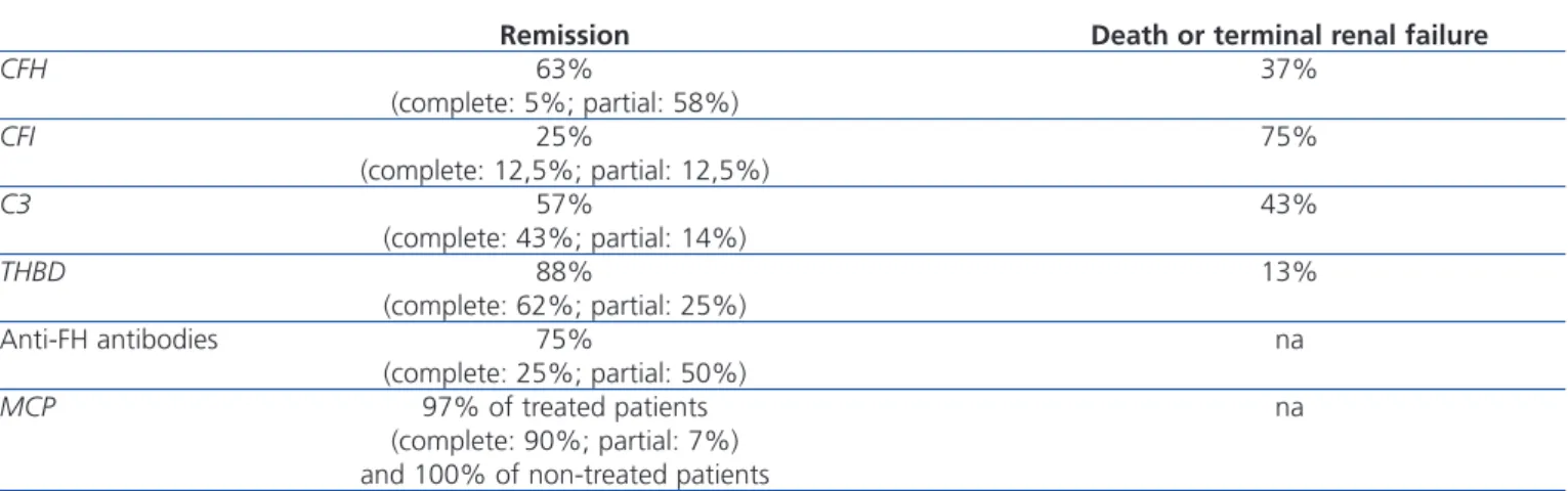
Although information from prospective clinical trials is not available, PT has been empirically the treatment of choice for aHUS for several years, after observing more than three decades ago that this treatment decreases mortality rates in patients with HUS-TTP. Table 5 presents the results of the largest international registry of PT in patients with aHUS (International Registry of Recurrent and Familial HUS/TTP), which included 273 patients diagnosed between 1996 and 2007.15In this registry, the rates of complete haematological
and renal recovery in patients treated with PT were generally lower than 50% (with the exception of patients with THBD and MCP mutations), and these rates were especially low in patients with FH and FI mutations (5% and 12.5%).15
Mortality and progression of TCRF are globally high, occurring in 3 out of 4 patients with FI mutations. Certain observations indicate that early and intensive PE is essential for salvaging patients from aHUS, and maintenance of this type of treatment can prevent disease recurrence and TCRF,14,61but we still have yet to establish the most effective
treatment regimen, nor do we fully understand the long-term impacts of this type of therapy on renal function.
In patients with anti-FH antibodies, it has been shown that concomitant immunosuppressant treatment along with PT can improve results.22,63,64In these cases, an elevated antibody
titre is correlated with a greater risk of recurrence and renal sequelae.22
The appearance of anaphylactic reactions to FFP, hypervolemia, hypertension, heart failure, and hyperproteinaemia are potential complications of PI. The primary complications that arise due to PE are venous access obstruction (6%), hypotension (5%), and allergy (4%),65and
these are most common in paediatric patients.65
Eculizumab
Eculizumab is an IgG2/4κ humanised monoclonal antibody that binds to complement C5 proteins with great affinity, blocking its excision into C5a and C5b and impeding the production of the C5b-9 terminal complement complex (membrane attack complex) (Figure 3).5 In aHUS,
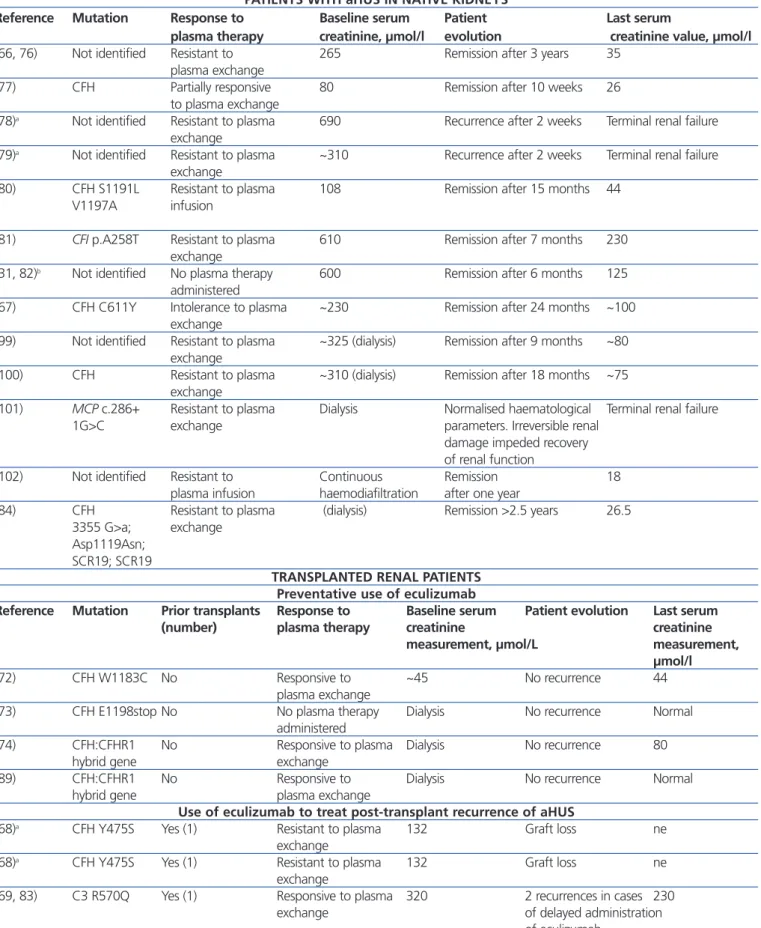
deregulation of the alternative complement pathway leads to uncontrolled activation of this process, provoking damage to endogenous structures through the formation of membrane attack complexes. In this sense, blocking the terminal complement complex with eculizumab quickly and sustainably diminishes this activity, and in many cases of patients with aHUS that receive this treatment, a good clinical response to the drug has been reported (Table 6).31,35,54,66-84
The efficacy and safety of eculizumab as a treatment for aHUS was evaluated recently in two phase II prospective, multi-centre, controlled, open clinical studies of 26 weeks duration, carried out in patients ≥12 years of age and with primary or recurrent (post-KT) disease.85,86One included 17
patients resistant to PT (≥4 sessions/week) (Study C08-002)85
and one included 20 patients on chronic plasma treatment (≥8 weeks) (study C08-003).86The most common mutations
in this sample were related to FH (15 patients) and FI.8Three
patients had MCP mutations, three had C3, and one had FB. We found anti-FH antibodies in 4 patients and no genetic abnormalities were observed in 10 patients. In both studies,
Table 5. Prognosis of patients with atypical haemolytic uraemic syndrome treated with plasma infusion or plasma exchange
Remission Death or terminal renal failure
CFH 63% 37% (complete: 5%; partial: 58%) CFI 25% 75% (complete: 12,5%; partial: 12,5%) C3 57% 43% (complete: 43%; partial: 14%) THBD 88% 13% (complete: 62%; partial: 25%) Anti-FH antibodies 75% na (complete: 25%; partial: 50%) MCP 97% of treated patients na (complete: 90%; partial: 7%) and 100% of non-treated patients
Complete remission: normalisation of haematological parameters and renal function. Partial remission: normalisation of haematological parameters and sequelae of kidney issues.
Anti-FH: anti-complement factor H antibodies; CFH: complement factor H gene; CFI: complement factor I gene; MCP: membrane cofactor protein gene; na: not available; THBD: thrombomodulin gene.
Adapted from Noris.15
the response to eculizumab was similar between patients with mutations and/or ant-FH antibodies and those with no genetic abnormalities. The response was also similar regardless of the type of mutation identified. After 6 months of treatment with eculizumab, rates of haematological normalisation (≥2 consecutive normal measurements of platelets and LDH) reached 76% in resistant cases and 90% of chronic cases. The rates of patients free from TMA events (no reduction in platelets >25%; with no need for PT or new dialysis sessions) were 88% and 80%, accompanied by a significant reduction in the rate of daily interventions for TMA (PE or PI sessions or new dialysis sessions) from baseline values of 6 and 1.5, respectively, to 0 (P<.0001). At the end of the follow-up period, GFR had increased significantly for both patient groups (resistant patients: +31ml/min/1.73m2; P=.0001;
chronic patients: +6.1ml/min.1.73m2; P=.0003), and 80% of
resistant patients who were on dialysis at the start of treatment (4 out of 5) were able to exit dialysis by the end of the treatment period. We also observed that patients who started treatment with eculizumab during the first 10 days following the diagnosis of aHUS exhibited a greater increase in GFR than those who started treatment after 2-4 months (+59ml/min/1.73m2 vs. +7ml/min/1.73m2; P=.03). In
extended studies (62-64 weeks mean follow-up period), the improvement in renal function was maintained.6-9After one
year of eculizumab treatment, proteinuria levels were significantly lower than baseline values (P=.05). Adverse side effects were observed in 19 patients, including 4 severe adverse effects (peritonitis, influenza infection, venous disorder, and severe hypertension). Due to its mechanism of action, eculizumab increases the risk of infection by Neisseria meningitidis, which prompted to vaccination of all patients against Neisseria (tetravalent vaccine) before starting
treatment or antibiotic prophylaxis, with no cases of meningitis produced. Survival was 100% in both studies. Another retrospective study involved 19 paediatric patients treated in clinical practice with eculizumab for a mean 28 weeks.4,11 In this study, 89% of patients obtained normal
platelet levels and 68% were free of TMA events. The rate of interventions for TMA was reduced from ~3/patient/week to 0. Mean GFR increased by ≥15ml/min/1.73m2 in 47% of
patients, and the need for dialysis was eliminated in 50% of patients. The most common adverse effects of treatment were pyrexia (47%), diarrhoea (32%), and upper respiratory tract infections (32%).
Based on these results, the FDA (Food and Drug Administration) and the EMA approved in the United States and Europe, respectively, the indication for eculizumab in the treatment of aHUS.4The recent availability of this drug
in Spain has offered the possibility of substantially improving the management of patients with aHUS, since the approved indication authorises its use as a primary treatment. Here, we present the recommendations for treating aHUS as compiled by the authors of this document based on the available evidence and pertinent clinical experience.
RECOMMENDATIONS FOR THE MANAGEMENT OF ATYPICAL HAEMOLYTIC URAEMIC SYNDROME
Treatment recommendations
Considering the technical difficulties involved in administering PT to paediatric patients (due to body size), as
Table 6. Cases published of patients with atypical haemolytic uraemic syndrome who received eculizumab
PATIENTS WITH aHUS IN NATIVE KIDNEYS
Reference Mutation Response to Baseline serum Patient Last serum
plasma therapy creatinine, µmol/l evolution creatinine value, µmol/l
(66, 76) Not identified Resistant to 265 Remission after 3 years 35
plasma exchange
(77) CFH Partially responsive 80 Remission after 10 weeks 26
to plasma exchange
(78)a Not identified Resistant to plasma 690 Recurrence after 2 weeks Terminal renal failure exchange
(79)a Not identified Resistant to plasma ~310 Recurrence after 2 weeks Terminal renal failure exchange
(80) CFH S1191L Resistant to plasma 108 Remission after 15 months 44 V1197A infusion
(81) CFI p.A258T Resistant to plasma 610 Remission after 7 months 230 exchange
(31, 82)b Not identified No plasma therapy 600 Remission after 6 months 125 administered
(67) CFH C611Y Intolerance to plasma ~230 Remission after 24 months ~100 exchange
(99) Not identified Resistant to plasma ~325 (dialysis) Remission after 9 months ~80 exchange
(100) CFH Resistant to plasma ~310 (dialysis) Remission after 18 months ~75 exchange
(101) MCP c.286+ Resistant to plasma Dialysis Normalised haematological Terminal renal failure
1G>C exchange parameters. Irreversible renal
damage impeded recovery of renal function
(102) Not identified Resistant to Continuous Remission 18
plasma infusion haemodiafiltration after one year
(84) CFH Resistant to plasma (dialysis) Remission >2.5 years 26.5 3355 G>a; exchange
Asp1119Asn; SCR19; SCR19
TRANSPLANTED RENAL PATIENTS Preventative use of eculizumab
Reference Mutation Prior transplants Response to Baseline serum Patient evolution Last serum (number) plasma therapy creatinine creatinine
measurement, µmol/L measurement, µmol/l
(72) CFH W1183C No Responsive to ~45 No recurrence 44
plasma exchange
(73) CFH E1198stop No No plasma therapy Dialysis No recurrence Normal
administered
(74) CFH:CFHR1 No Responsive to plasma Dialysis No recurrence 80
hybrid gene exchange
(89) CFH:CFHR1 No Responsive to Dialysis No recurrence Normal
hybrid gene plasma exchange
Use of eculizumab to treat post-transplant recurrence of aHUS
(68)a CFH Y475S Yes (1) Resistant to plasma 132 Graft loss ne
exchange
(68)a CFH Y475S Yes (1) Resistant to plasma 132 Graft loss ne
exchange
(69, 83) C3 R570Q Yes (1) Responsive to plasma 320 2 recurrences in cases 230
exchange of delayed administration
of eculizumab
well as the potential complications that arise from this strategy, the early use of eculizumab in this population is especially highly indicated, thus avoiding the need for PE. As such, given a suspicion of aHUS in a paediatric patient, it is recommended to provide early treatment with eculizumab as the first choice (Figure 5). In adult patients with the suspicion of aHUS, early eculizumab is also recommended. In the case that the start of this treatment might be delayed, early and intensive PE should be administered until eculizumab is an available option.62
Prior to this point, it is necessary to first vaccinate all patients against Neisseria meningitidis (preferably with
conjugated tetravalent vaccines against serotypes A, C, Y, and W135). In the case that treatment with eculizumab cannot be delayed until obtaining proper vaccination, treatment can still be started along with antibiotic prophylaxis4against Neisseria meningitidis based on the
protocol at each hospital. Considering the more elevated rate of invasive infection by meningococci in paediatric patients, as well as the absence of protection against serotype B infections (which is the most prevalent type due to the systematic vaccination of the other serotypes), these patients should be maintained on antibiotic prophylaxis with penicillin or amoxicillin to accompany
Figure 5. Treatment for atypical haemolytic uraemic syndrome.
PE: plasma exchange; aHUS: atypical haemolytic uraemic syndrome.
Continues table 6. Cases published of patients with atypical haemolytic uraemic syndrome who received eculizumab.
(70) Not specified No Resistant to plasma 323 Remission 238
exchange
(71) CFH S1191L Yes (2) Intolerance to plasma 131 Remission 130
exchange
(103)a Not identified No Resistant to plasma exchange 415 Graft loss na
(35) CFH Yes (1) Resistant to plasma exchange 500 Remission 62
(54) C3 R570W Yes (2) Partially responsive 220 Remission 115
to plasma exchange
(75) CFH E3514Stop No Partially responsive 565 (dialysis) Remission 229
to plasma exchange
(104) Not identified Yes (1) Resistant to plasma 449 (dialysis) Recurrence 5 months na
exchange after suspension of
eculizumab. Graft loss
a Received one single dose of eculizumab. bReduced levels of eculizumab.
CFH: complement factor H gene; CFHR1: complement factor H-related protein 1 gene; CFI: complement factor I gene; na: not
specified; HUS: haemolytic uraemic syndrome; aHUS: atypical haemolytic uraemic syndrome.
Clinical suspicion of aHUS
• Blood sample taken for later analysis prior to the start of treatment • Anti-meningococcus vaccination ± antibiotic prophylaxis Treatment monitored Paediatric patients:
early administration of eculizumab as the treatment of choice ±
Support treatment
Adult patients:
early administration of eculizumab (early and intensive PE until start of eculizumab therapy whenever a delay is warranted)
± Support treatment
eculizumab.2 In adult patients, antibiotic prophylaxis
should be maintained throughout the duration of treatment with eculizumab as based on the attending physician’s judgement and patient characteristics. In paediatric patients, vaccination against Haemophilus influenzae and pneumococci is also necessary, along with strict adherence to local guidelines regarding obligatory vaccinations for each age group.
When patients exhibit a positive response to eculizumab, treatment should be extended (chronic) as advised by the drug technical data sheet.4 It is currently impossible to
provide recommendations regarding the most appropriate duration of treatment, although as our experience with this drug continues to develop, we may be able to evaluate the possibility of treatment removal in certain low-risk patients
on an individual basis. In patients with clinical indications requiring the removal of treatment, subsequent care must include close monitoring for at least 12 weeks in order to detect possible abnormalities suggestive of TMA.4In these
cases, patients should be evaluated for reintroduction to eculizumab and/or treatment with PE.4
When considering the application of PT in patients with aHUS, PE with FFP should be preferentially evaluated (1.5 per volume plasma [60-75ml/kg] per session). Sessions should continue until platelet levels have normalised, haemolysis has stopped, and a sustained improvement in renal function has been observed for several days. Later, 5 sessions per week should be administered during the following 2 weeks, followed by another two weeks of 3 sessions per week and an individualised assessment for considering the appropriateness of continued treatment.2,62
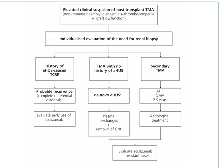
Figure 6. Diagnosis and treatment of thrombotic microangiopathy in kidney transplants.
aNormally, deterioration in renal function with no haematological manifestations.
CMV: cytomegalovirus; CNI: calcineurin inhibitors; TCRF: terminal chronic renal failure; TMA: thrombotic microangiopathy; AHR: acute humoral rejection; HUS: haemolytic uraemic syndrome; aHUS: atypical haemolytic uraemic syndrome.
Elevated clinical suspicion of post-transplant TMA
(non-immune haemolytic anaemia + thrombocytopenia + graft dysfunction)
Individualised evaluation of the need for renal biopsy
History of aHUS-caused TCRF TMA with no history of aHUS Secondary TMA Probable recurrence (complete differential diagnosis) de novo aHUSa AHR CMV BK virus
Evaluate early use of
eculizumab exchanges Plasma + removal of CNI Aetiological treatment Evaluate eculizumab in resistant cases
Immunosuppression therapy should be administered as a concomitant treatment in patients with aHUS and anti-FH antibodies on PT.22,57,63,64The response to treatment in these
cases should be monitored through the evolution of antibody titres.57
Whenever necessary, support measures can be put into place to control hypertension and volemia. For the treatment of anaemia, red blood cell transfusions and/or erythropoietin stimulating factors can be administered. Platelet transfusions should be limited to cases of severe platelet depletion (<30 000mm3) or in rare cases of
severe haemorrhage or in anticipation of invasive procedures with a risk of major blood loss, since this could worsen the TMA phenomena. Another priority is the identification and treatment of possible triggering agents of aHUS. Paediatric patients with aHUS should be transferred to specialised paediatric nephrology centres for treatment from experienced personnel with the availability of a paediatric intensive care unit in order to guarantee appropriate care.
Recommendations for transplantation in patients with atypical haemolytic uraemic syndrome
Prior evaluation
In patients with TCRF and aHUS who are candidates for KT, an exhaustive analysis is first required to determine plasma levels of C3, C4, FH, FI, and FB, as well as MCP expression in peripheral leukocytes.35 A complete genetic analysis
should also be performed in order to detect known complement mutations (and polymorphisms associated with risk factors), along with a screening process for anti-FH antibodies.35Based on these analyses, the patient should be
evaluated on an individual basis for the indications for KT and its concomitant treatment, based on the risk of recurrence of aHUS.
Clinically, aHUS is considered as a contraindication for live kidney donation (especially in patients with high-risk mutations) due to the elevated rates of recurrence and graft loss, as well as the risk that the donor could have undetected complement mutations that would facilitate the development of aHUS.1,53,87 Currently, the advancements in genetic
diagnostics and the use of eculizumab could allow for considering these patients for live kidney donation through an individualised evaluation of select cases. In the case of related donors, a complete genetic-molecular analysis is also required. As a general rule, the transplant should only be considered as a viable option if the recipient mutations known to be associated with aHUS have already been identified, and these mutations have been ruled out in the potential donor.
Treatment and prophylaxis for the recurrence of atypical haemolytic uraemic syndrome following transplantation
Treatment for recurrence of disease in patients who receive kidney transplants for aHUS should be provided under the same framework as for aHUS in native kidneys, based on early administration of eculizumab,6-9,35,54,70,71,75,88while taking
into account the usefulness and convenience of applying PE. Figure 6 displays a management algorithm for the primary causes of TMA in KT.
Before the availability of eculizumab, it was impossible to recommend single kidney transplants in patients with TCRF secondary to aHUS on dialysis with a high risk of recurrence of disease following transplantation (patients with high-risk mutations and/or episodes of recurrence of aHUS). In recent years, several initial pilot studies have been reported with positive experience with the use of eculizumab as prophylaxis in paediatric patients with FH mutations prior to KT from cadaveric donors,72-74,89 suggesting that KT along
with preventative treatment with eculizumab could be the indicated treatment option for these patients. Recently, a study administered prophylactic eculizumab to 9 patients in order to prevent the recurrence of aHUS following KT.88This
study involved 6 paediatric patients and 3 adults with different mutations to the alternative complement pathway (5 FH mutations, 1 C3, and 3 patients with non-homologous genomic reorganisations between CFH and CFHR1 genes). Two patients received post-KT PE and were then transferred to treatment with eculizumab, 2 received eculizumab starting at the week prior to transplantation (live unrelated donor, urgent transplant from cadaveric donor), and the remaining 5 received eculizumab immediately following transplantation. Except for one case involving an early thrombosis that led to graft loss, the other 8 cases had favourable evolution with no recurrences after a mean follow-up period of 14.5 months (mean creatinine: 71.6±44.8µmol). In this context, the recent guidelines from an aHUS research group in France recommended prophylaxis with eculizumab in patients with TCRF secondary to aHUS who are candidates for KT.90
In children with aHUS that have mutations to genes that code for complement factors synthesised in the liver (FH, FB, or FI), liver/kidney and liver transplants have been performed in order to correct the consequences of the genetic defect and prevent disease recurrence. This strategy, combined with perioperative administration of plasma (in order to provide sufficient functional complement factors until the liver graft recovers its ability for synthesis) or eculizumab, has produced successful results in several cases, yielding good liver function and no aHUS recurrence to date.91-95 However, it is essential to
evaluate the risk and the morbidity/mortality associated with these procedures, as well as the possible secondary side effects from the long-term immunosuppression
required by transplant recipients. It is also unclear whether the minimal extra-hepatic synthesis of FH (in adipose tissue, renal tissue, and monocytes) could induce recurrence of aHUS in the post-transplant period.2
Taking into account that calcineurin inhibitors can be associated with post-transplant TMA due to their nephrotoxic effect, these drugs must be used with caution in patients with aHUS who have received transplants. To this end, a treatment regimen free of calcineurin inhibitors based on belatacept and/or mTOR inhibitors could be a possibility, taking into account the immunological risk of the patient, although no conclusive data are available regarding the most appropriate immunosuppression regimen for the at-risk population.
CONCLUSIONS
• aHUS is caused by a deregulation of genetic origin of the activation of the alternative complement system pathway on cell surfaces, leading to the development of systemic TMA. In recent years, several mutations and polymorphisms have been identified in the genes of certain complement factors that have been related to this deregulation.
• This syndrome is characterised clinically by the triad of non-immune microangiopathic haemolytic anaemia, thrombocytopenia, and acute renal dysfunction, com-monly associated with extra-renal manifestations. The prognosis varies based on the type of mutation present, although in general, the syndrome is associated with high mortality rates and occurrence of TCRF, despite standard treatment with PT. There is also an elevated rate of recurrence of aHUS following KT.
• Given clinical signs and symptoms suggestive of TMA, the diagnosis must be orientated towards aHUS if the shiga toxin/STEC test is negative, plasma acti-vity of ADAMTS13 is >5%, and secondary forms of HUS have been ruled out.
• Eculizumab is a monoclonal antibody that inhibits the activation of C5 and the formation of the cell membra-ne attack complex, which is responsible for the dama-ge produced to native cell structures in aHUS. In pros-pective studies involving patients with aHUS, eculizumab effectively interrupted the process of TMA, associated in the long-term with significant hae-matological improvements and recovery of renal func-tion.
• The FDA, the EMA, and the Spanish Agency of Me-dicines have approved the use of eculizumab for the treatment of aHUS, authorising its use as a first line of treatment. The authors of this document recommend its early use in patients with a clinical suspicion of aHUS in native kidneys, as well as in patients with re-current aHUS following KT.
Table 7. Protocol for sample collection for complement
analyses in patients with atypical haemolytic uraemic syndrome.
In the case of samples to be sent to a reference laboratory, it is strongly recommended to contact first with this laboratory and strictly follow their instructions regarding what is required to perform the complement studies.
If no reference laboratory is available and the patient's samples are going to be stored for future analyses, the samples obtained should include:
- 10ml of blood with EDTA
- 10ml of coagulated blood for serum.
- If the patient is a small child and 20ml of blood cannot be extracted, 2-3ml can be obtained for each sample (6-9ml total). - Blood with EDTA must be centrifuged immediately at 3000rpm for 10 minutes at 4ºC, followed by plasma collection in a sterile tube, taking care not to absorb any red blood cells. After labelling five tubes with patient name, date, and a mark indicating EDTA plasma, the plasma is distributed into these tubes for immediate freezing, keeping samples stored at -80ºC. - After removing the EDTA-plasma, store the cellular pellet frozen
at -80ºC. This will be used to obtain DNA.
- Coagulated blood for one hour at room temperature is centrifuged at 3000rpm for 10 minutes at 4ºC, immediately separating the serum from the blood, taking care not to collect any red blood cells in the sterile tube. After labelling five tubes with patient name, date, and a mark indicating serum, the serum is distributed into these tubes for immediate freezing, keeping samples stored at -80ºC.
APPENDIX 1. RECOMMENDATIONS FOR THE TREATMENT OF SECONDARY FORMS OF HAEMOLYTIC URAEMIC SYNDROME
The management of secondary forms of HUS should be based on the primary aetiology of the disease and the administration of PE. In the case of resistance to treatment, eculizumab should be administered (probably on a temporary basis) as a salvage therapy in selected severe cases,96-98although little data is
avail-able to establish a concrete recommendation in this context.
APPENDIX 2. RECOMMENDATIONS OF INTEREST FOR THE DIAGNOSIS OF GENETIC ABNORMALITIES
Although not necessary for the clinical diagnosis of aHUS, a complete evaluation of the complement system is recommend-ed, including plasma levels of all complement factors and a complete genetic analysis of affected patients. Samples should be taken prior to the start of treatment and sent to a reference laboratory (Table 7). Taking into account that complement mu-tations have been identified for STEC-HUS and secondary HUS, it is also recommended that these patients undergo an in-dividualised genetic analysis in order to examine the possible correlation with complement abnormalities/aHUS.2
However, a genetic diagnosis of abnormal complement genes would allow for assessing prognosis on an individual bases, as well as the risk of recurrence of disease, making this a rec-ommended step for all patients. In patients who are potential candidates for KT, the genetic analysis is indispensable. An online database of complement mutations in patients with aHUS is currently available in Spain to facilitate data collec-tion and future studies: https://www.tmaddd.es.
We also recommend taking graft samples from patients who receive kidney transplants due to aHUS for future studies.
Sources of funding and acknowledgements
The research carried out by Dr Rodriguez de Cordoba is financed by the Ministry of Economy and Competition (SAF2010-26583), the Autonomous Community of Madrid (S2010/BMD-2316), and the Fundacion Renal Inigo Alvarez de Toledo.
The authors would like to thank Alexion Pharmaceuticals for their logistic support in carrying out the consensus meetings, as well as editorial support through Ogilvy Healthworld.
Approval
This document has been approved by the Spanish Transplant So-ciety (S.E.T.) and the Spanish SoSo-ciety of Nephrology (S.E.N.) and Spanish Association of Pediatric Nephrology (A.E.N.P.)..
Conflicts of interest
Dr Ariceta, Dr Blasco, Dr Campistol, Dr Praga, Dr Ro-dríguez de Córdoba, and Dr Vilalta have provided consulta-tion and teaching services to Alexion Pharmaceuticals. Dr Torra has formed part of expert consensus committees for aHUS sponsored by Alexion Pharmaceuticals. Dr Espinosa also has participated in clinical studies sponsored by Alex-ion Pharmaceuticals.
None of the aforementioned activities have influenced the elaboration or interpretation of this manuscript, and the au-thors are responsible for all content.
REFERENCES
1. Loirat C, Fremeaux-Bacchi V. Atypical hemolytic uremic syndrome. Orphanet J Rare Dis 2011;6:60.
2. Loirat C, Saland J, Bitzan M. Management of hemolytic uremic syndrome. Presse Med 2012;41(3 Pt 2):e115-35.
3. Caprioli J, Noris M, Brioschi S, Pianetti G, Castelletti F, Bettinaglio P, et al. Genetics of HUS: the impact of MCP, CFH, and IF mutations on
clinical presentation, response to treatment, and outcome. Blood 2006;108(4):1267-79.
4. Alexion Pharmaceuticals I. Soliris (eculizumab). Ficha técnica 2012. 5. Rother RP, Rollins SA, Mojcik CF, Brodsky RA, Bell L. Discovery and
development of the complement inhibitor eculizumab for the treatment of paroxysmal nocturnal hemoglobinuria. Nat Biotechnol 2007;25(11):1256-64.
6. Licht C, Muus P, Legendre C, Douglas KW, Hourmant M, Delmas Y, et al. Eculizumab Is An Effective Long-Term Treatment In Patients with Atypical Hemolytic Uremic Syndrome (aHUS) Previously Receiving Chronic Plasma Exchange/Infusion (PE/PI): Extension Study Results. Blood 2011;118(21):abstr 3303.
7. Greenbaum L, Babu S, Furman RR, Sheerin N, Cohen D, Gaber O, et al. Eculizumab Is An Effective Long-Term Treatment In Patients with Atypical Hemolytic Uremic Syndrome (aHUS) Resistant to Plasma Exchange/Infusion (PE/PI): Results of An Extension Study. Blood 2011;118(21):abstr 193.
8. Greenbaum LA, Babu S, Furman R, Sheerin N, Cohen D, Gaber O, et al. Continued improvements in renal function with sustained eculizumab (ECU) in patients (PTS) with atypical hemolytic uremic syndrome (aHUS) resistant to plasma exchange/infusion (PE/PI). J Am Soc Nephrol 2011;22(suppl):197A abstr TH PO367.
9. Licht C, Muus P, Legendre CM, Douglas K, Hourmant M, Delmas Y, et al. Ph II study of eculizumab (ECU) in patients (PTS) with atypical hemolytic uremic syndrome (aHUS) receiving chronic plasma exchange/infusion (PE/PI). J Am Soc Nephrol 2011;22(suppl):197A abstr TH PO366.
10. Furlan M, Robles N, Lammle B. Partial purificatiion and characterization of a protease from human plasma cleaving von Willebrand factor to fragments produced by in vivo proteolysis. Blood 1996;87:4223-34. 11. Pisoni R, Ruggenenti P, Remuzzi G. Drug-induced thrombotic
microangiopathy: incidence, prevention and management. Drug Saf 2001;24(7):491-501.
12. Siegler R, Oakes R. Hemolytic uremic syndrome; pathogenesis, treatment, and outcome. Curr Opin Pediatr 2005;17(2):200-4. 13. Oakes RS, Siegler RL, McReynolds MA, Pysher T, Pavia AT. Predictors of
fatality in postdiarrheal hemolytic uremic syndrome. Pediatrics 2006;117(5):1656-62.
14. Sellier-Leclerc AL, Fremeaux-Bacchi V, Dragon-Durey MA, Macher MA, Niaudet P, Guest G, et al. Differential impact of complement mutations on clinical characteristics in atypical hemolytic uremic syndrome. J Am Soc Nephrol 2007;18(8):2392-400.
15. Noris M, Caprioli J, Bresin E, Mossali C, Pianetti G, Gamba S, et al. Relative role of genetic complement abnormalities in sporadic and familial aHUS and their impact on clinical phenotype. Clin J Am Soc Nephrol 2010;5(10):1844-59.
16. Sullivan M, Erlic Z, Hoffmann MM, Arbeiter K, Patzer L, Budde K, et al. Epidemiological approach to identifying genetic predispositions for atypical hemolytic uremic syndrome. Ann Hum Genet 2010;74(1):17-26. 17. Westra D, Volokhina E, van der Heijden E, Vos A, Huigen M, Jansen
J, et al. Genetic disorders in complement (regulating) genes in patients with atypical haemolytic uraemic syndrome (aHUS). Nephrol Dial Transplant 2010;25(7):2195-202.
18. Esparza-Gordillo J, Goicoechea de Jorge E, Buil A, Carreras Berges L, López-Trascasa M, Sánchez-Corral P, et al. Predisposition to atypical hemolytic uremic syndrome involves the concurrence of