University of Sassari
Department of Biomedical Sciences
International PhD School in Biomolecular and Biotechnological Sciences Section: Biochemistry and Molecular Biology
Cycle XXVI
Med1/MBD4 DNA repair enzyme and
SUMOylation
Coordinator: Prof. Claudia Crosio
Supervisor: PhD candidate: Prof. Luigi Marco Bagella Valentina Doneddu
Valentina Doneddu
Med1/MBD4 DNA repair enzyme and SUMOylation
Tesi di dottorato in scienze Biomolecolari e Biotecnologiche Ciclo XXVI Università degli studi di Sassari
Valentina Doneddu
Med1/MBD4 DNA repair enzyme and SUMOylation
Tesi di dottorato in scienze Biomolecolari e Biotecnologiche Ciclo XXVI Università degli studi di Sassari
Table of Contents
TABLE OF CONTENTS 6
INTRODUCTION 8
-DNA repair system 9
Direct repair 10
Nucleotide excision repair (NER) 11
Mismatch repair 12
Double strand repair 14
Base excision repair 15
Med1/MBD4 16
-Post-translational modification: SUMOylation 18
The SUMOylation cycle 19
Consensus sites 20
Molecular conseguences of SUMOylation 21
BER system and SUMOylation 22
Valentina Doneddu
Med1/MBD4 DNA repair enzyme and SUMOylation
Tesi di dottorato in scienze Biomolecolari e Biotecnologiche Ciclo XXVI Università degli studi di Sassari
-Cell line and Cell culture 26 -Transfection 26
Valentina Doneddu
Med1/MBD4 DNA repair enzyme and SUMOylation
Tesi di dottorato in scienze Biomolecolari e Biotecnologiche Ciclo XXVI Università degli studi di Sassari
-Cell Lysis 26 -Protein Quantification 27 -Co-immunoprecipitation 27 -Immunoblotting 28 -Drugs treatment 29 -Localization assay 29
-In silico analysis 30
RESULTS 32
-Interaction between Med1/MBD4 and hSUMO1 33
-Interaction between Med1/MBD4 and hSUMO1 after Cisplatin treatment 35
-Interaction between Med1/MBD4 and hSUMO1 after MNU treatment 37
-Interaction between Med1/MBD4 and hSUMO1: activity 38
-Interaction between Med1/MBD4 and hSUMO1: localization 40
CONCLUSIONS 44
REFERENCES 49
Valentina Doneddu
Med1/MBD4 DNA repair enzyme and SUMOylation
Tesi di dottorato in scienze Biomolecolari e Biotecnologiche Ciclo XXVI Università degli studi di Sassari
Introduction
Valentina Doneddu
Med1/MBD4 DNA repair enzyme and SUMOylation
Tesi di dottorato in scienze Biomolecolari e Biotecnologiche Ciclo XXVI Università degli studi di Sassari
1. DNA repair systems
Cells are continuously exposed to stressors that can mine the DNA integrity, forming cleavages or double strand breaks. This causes missed protein interactions regulating gene expression, DNA replication and other cellular processes. In addition, the single base modifications or mismatches that do not follow the Watson and Crick’s rules are dangerous for the DNA integrity.
During evolution, cells have developed DNA repair systems. They start with DNA detection and the Cell cycle arrest until the repair. If the DNA lesion is too serious and the cell is unable to repair the damage, the only way to maintain the DNA integrity is driving the cell to apoptosis.
Most DNA repair systems, share in general transcription factors as DNA helicase, replication factors, DNA polymerase and ligases. Also, if each DNA repair system is specific and able to recognize the single lesion, most of them can cooperate.
Valentina Doneddu
Med1/MBD4 DNA repair enzyme and SUMOylation
Tesi di dottorato in scienze Biomolecolari e Biotecnologiche Ciclo XXVI Università degli studi di Sassari
1. Direct repair (DR)
2. Nucleotide excision repair (NER) 3. Base excision repair (BER) 4. Mismatch repair (MMS) 5. Double strand repair (DBS).
1.1 Direct repair
Direct repair is the simplest system and does not need the template to perform the repair.
Bacteria and Yeast, for example, in a light-dependent reaction, restore directly the damage resulting from UV or cisplatin treatment with several photolyases (1). Surprisingly, even if there is no visible light in most tissue, photolyase binds to cisplatin-damaged DNA. This enzyme has also the ability to respond at oxidative stresses (2; 3).
Only a few types of DNA damage are repaired with this system, such as pyrimidine dimers resulting from exposure to ultraviolet (UV) light and alkylated guanines that have been modified by the addition of methyl or ethyl groups at the O6 position of the purine ring.
In this case, just one enzyme, O6-alkylguanine-DNA–alkyltransferase, transfers alkyl or methyl adducts from oxygen in position 6th of Guanine to an active site of Cysteine. This enzyme is able to
Valentina Doneddu
Med1/MBD4 DNA repair enzyme and SUMOylation
Tesi di dottorato in scienze Biomolecolari e Biotecnologiche Ciclo XXVI Università degli studi di Sassari
work independently to other proteins and without breaks of DNA molecule. The DR is active also for changes induced to UV (4).
1.2 Nucleotide excision repair (NER)
This system is certainly one of the most versatile mechanisms of DNA repair, it is able to identify and process bulky lesions and those that deform double helix, as cyclobutane dimers and 6–4 photoproducts.
Bacteria, yeast and human XP (Xeroderma Pigmentosum) syndrome cells are excellent models to study the NER system.
In human, the NER system shows all its complexity and can be summarized into four stages:
1. Damage recognition/preincision 2. Incision
3. Gap filling 4. Ligation
Valentina Doneddu
Med1/MBD4 DNA repair enzyme and SUMOylation
Tesi di dottorato in scienze Biomolecolari e Biotecnologiche Ciclo XXVI Università degli studi di Sassari
The damage recognition is assigned to the complex: replication protein A (RPA) and XPA and also transcription factors as IIH (TFIIH/XPB/ ERCC3, XPF-ERCC1 and XPG (5).
The incision step is ATP-dependent, it reacts where the excinuclease complex at the 3’ and 5’ of the lesion. XPG, for example, cuts at 3’ while another nuclease; FEN (flap endonuclease) cuts at 5’ (5). The polymerases δ and ε start the synthesis of new nucleotide from free 3’OH extremity; while proliferating cell nuclear antigen (PCNA) is required to fill the gap.
The NER is a multistep process, which involves more than 20 proteins. There are two principal pathways when NER is particularly active:
-TCR-NER (transcription coupled repair)
-GGR-NER (global genome repair)
The first pathway is a highly specific and efficient system, able to recognize and cut out the DNA damage arresting the RNA polymerase II progression. It is particularly sensitive to Cisplatin damages (6).
GGR-NER deals with all repairable lesions throughout the genome; it shares the proteins involved in the initial recognition of lesions with TCR-NER system. In normal situations, the damage recognition enzymes are low expressed, but after damage, a strong upregulation starts through
Valentina Doneddu
Med1/MBD4 DNA repair enzyme and SUMOylation
Tesi di dottorato in scienze Biomolecolari e Biotecnologiche Ciclo XXVI Università degli studi di Sassari
activation of p53 tumor suppressor gene. This transactivation role controls gene expression, XPC and XPE, which are involved only in the GGR-NER (7).
Two disorders are known to be deficient in NER system: XP and Cockayne syndrome.
1.3 Mismatch repair (MMS)
This strategy erases the mistakes that appear during DNA replication and in this particular case the mismatches, insertions/deletions of loops, deaminations of 5-methylcytosine to uracil mining the fidelity of DNA replication. The essential strategy of MMR is similar to the excision repair. In human, two pathways of mismatch repair have been identified:
1. long-patch 2. short-patch
The first cuts large sequence but has no sequence specificity, while the Short-patch repair cuts just one or few nucleotides, keeping in mind the sequence context.
The MMR system was well studied in E.coli (Escherichia coli).
Three steps are involved in this system:
Valentina Doneddu
Med1/MBD4 DNA repair enzyme and SUMOylation
Tesi di dottorato in scienze Biomolecolari e Biotecnologiche Ciclo XXVI Università degli studi di Sassari
2. Excision 3. Resynthesis
During the initiation, MutS recognizes the mismatch and then in a reaction ATP-dependent Mut H interacts with the homodimer MutL. Mut H, now activated, cuts the nucleotide, erroneously inserted during synthesis. Mut H is able to identify the new strand because the adenine present in the GATC site is still not methylated. The DNA polymerase III holoenzyme and DNA ligase make respectively the resynthesis and the ligation (8).
In human, six genes has been identified: hMSH2, hMSH3, hMSH6, hMLH1, hPMS1, and hPMS2.
HMSH2, hMSH3 and hMSH6 are human homologs of E. coli MutS. In a reaction ATP-dependent, these enzymes bind to DNA heteroduplex and promote long-patch MMR. hMSH6, also called G/T mismatch binding protein, is very efficient in recognizing G:T mismatches.
Defects in hMLH1, hMSH2, hMSH6, hPMS1, and hPMS2 are associated with hereditary nonpolyposis colorectal cancer (HNPCC) (9; 10).
1.4 Double strand break repair (DSBR)
The double strand break is usually contemplated as the most lethal DNA lesion. Ionizing radiation (IR), chemotherapeutic drugs, ROS (reactive oxygen species), and chromosomal stresses are the
Valentina Doneddu
Med1/MBD4 DNA repair enzyme and SUMOylation
Tesi di dottorato in scienze Biomolecolari e Biotecnologiche Ciclo XXVI Università degli studi di Sassari
principal causes of this kind of disease. The double strand breaks are usually present when the replicative fork meet single breaks, when the same problem is present during the metabolism of telomeres (11; 12).
The efficient repair of double strand breaks has a fundamental importance in maintaining genomic integrity, cellular viability and, obviously, in preventing tumorigenesis.
Currently, three primary pathways are known:
1. Homologous recombination repair, (HR) is prevalent during G2 phase and in dividing cells and a homologous sister chromatid, as template, must be present. MRE11, RAD50, and NBS1 form a complex able to recognize DSBs and to recruit the protein kinase, called ataxia telangectasia mutated (ATM), which is essential for the subsequent steps of the recombination process.
2. Single strand annealing, more similar to HR.
3. Non-homologous end joining repair, NHEJ is the leader during the G0/G1 cell cycle phase
when a homologous sister chromatid is absent.NHEJ shows three main steps: (a) detection
of the two-ended DSB, (b) process to remove the damage at the break and to expose short stretches of microhomology, and (c) join of two suitable ends (13).
Valentina Doneddu
Med1/MBD4 DNA repair enzyme and SUMOylation
Tesi di dottorato in scienze Biomolecolari e Biotecnologiche Ciclo XXVI Università degli studi di Sassari
Base excision repair is active in removing non-bulky base adducts such as those originated by oxidation, reduction, methylation or fragmentation of bases following oxidative damage or ionizing radiations. The BER enzymes are the first cellular mechanisms recruited to repair damaged DNA. This system is active during all stages of cell cycle both for dividing and non-dividing cells (13).
This is a multistep organization that implicates several proteins. When the damage is identified, the glycosylases remove purines and pyrimidines from DNA, creating an apurinic or apyrimidinic, potentially cytotoxic, site known as AP site (14).
At this point, endonucleases (APE1, APEX, REF-1) incise 3’ (AP lyase) and 5’(AP hydrolase) to the apurinic site. In the last step, DNA polymerases fill the gap and DNA ligase complete the repair action.
The glycosylase enzymes can be classified in mono- or bifunctional enzymes in the matter of their mechanism. TDG (Thymidine DNA glycosylase), UDG (Uracil DNA glycosylase), SMUG and Med1/MBD4 are monofunctional glycosylase which cleave the glycosidic bond using water as a nucleophile to generate apurinic or apyrimidinic (AP) sites. They show specificity to repair deamination of C (cytosine) and or 5-meC (5-methycytosine) residues. The bifunctional DNA glycosylase uses an amino group of enzymes as a nucleophile to form a Schiff base intermediate, that undergoes enzymes-catalyzed β-elimination cleaving the phosphodiester bond 3’ from the
Valentina Doneddu
Med1/MBD4 DNA repair enzyme and SUMOylation
Tesi di dottorato in scienze Biomolecolari e Biotecnologiche Ciclo XXVI Università degli studi di Sassari
abasic site. (15). BER is active during all cell cycle, and has a critical function in dividing and non-dividing cells.
BER system can be summarized in 4 main stages: 1. recognition and excision of an inappropriate base; 2. incision and AP site formation;
3. replacement of the excised nucleotide; 4. sealing of the final nick.
Nowadays, it is known that defects in one or more components of BER system induce cancer predisposition as colorectal cancer (16).
1.5.1 Med1/MBD4
In 1999, Bellacosa and collaborators identified in yeast two-hybrid system Med1 as interactor of MLH1, a protein involved in the mismatch repair (17). This paper shows the structure of Med1 which contains terminus, the methylcytosine binding domain, important for binding an N-terminus to methylated DNA, while a C-terminal contains the catalytic domain presenting a strong homology with the systems of glycosidases and ligase that repair the damage to bacterial DNA. The central structure of the protein is still under investigation.
Valentina Doneddu
Med1/MBD4 DNA repair enzyme and SUMOylation
Tesi di dottorato in scienze Biomolecolari e Biotecnologiche Ciclo XXVI Università degli studi di Sassari
While DNA repair systems, such as NER and MMS, are more complicated, BER removes nitrogenous bases that carry specific injuries. For this purpose, a crucial role is played by the DNA N-glycosylase, which removes damaged bases or those involved in mismatch phenomena through the N-glycosidic bond cleavage. In its modus operandi, Med1/MBD4 inspects the CpG sites in the genome with its MBD domain. The deamination of 5-methylcytosine to thymine in CpG islands creates G: T mismatches that are processed by the catalytic domain of Med1. At this point, the glycosylase activity produces AP sites (apurinic or apyrimidinic sites) to which Med1 remains bound until the systems of polymerase and ligase will not restore the missing base. For this reason MED1/MBD4 is essential to safeguard genomic integrity in CpG sites. (18).
The specificity of DNA-glycosylase enzymes is particular evident during the repair activity. In
effect, the methyl binding domain protein 4 (MBD4/MED1) is very sensitive to act on G:IU
(iododeoxyuridine) but not A:IU, while, the other DNA–glycosylase enzymes are not required to repair this mismatches. (19).
It is also known that MED1/MBD4 shows a strong activity in repairing halogenated pyrimidines comparing top urine bases and is essential for the cytotoxicity of 5-iododeoxyuridine. (20.).
The involvement of Med1/MBD4 in colorectal cancer and ovarian cancer is showed in the hypermethylation of its promoter involving a gene silencing (21; 22).
Valentina Doneddu
Med1/MBD4 DNA repair enzyme and SUMOylation
Tesi di dottorato in scienze Biomolecolari e Biotecnologiche Ciclo XXVI Università degli studi di Sassari
In addition, MED1 is very important in regulating the apoptosis of those cells highly damaged by diverse drugs, especially methylatin agents (23; 24).
2.Post-translantional modification: SUMOylation
Discovered in 1997, the SUMOylation has been the topic of numerous studies focused on understanding its role in different cellular processes such as transcription, replication, chromosome segregation and DNA repair (25; 26).
This post-translation modification has been very often associated to the ubiquitination, because they share the structural conformation and the activation cycle. Recent studies show SUMOylation as a link to the proteosome system.
SUMO is ubiquitously expressed in all eukaryotes and is highly conserved.
The invertebrates code just one gene for the protein SUMO, on the contrary, the vertebrates have showed three genes: SUMO1, SUMO2-3, and SUMO4.
SUMO1,also known as UBL1, PIC1, sentrin, GMP1 and Smt3c, founding member of the family, is
Valentina Doneddu
Med1/MBD4 DNA repair enzyme and SUMOylation
Tesi di dottorato in scienze Biomolecolari e Biotecnologiche Ciclo XXVI Università degli studi di Sassari
ubiquitin. SUMO1 has been discovered as an interactor of Ran-GAP. Ran-GAP is a nuclear protein, but after its sumoylation becomes a cytoplasmatic protein (27).
SUMO2, also called sentrin 2, Smt3b and GMP-related protein and SUMO3, also known as sentrin 3 and Smt3a, considering their high homology of sequence, are usually associated in SUMO2-3. In effect, SUMO-2 and -3 differ from each other only by three residues at the N-terminal sequence.
Both SUMO1 and SUMO2-3 are ubiquitously expressed. On the other hand, the expression of SUMO 4 is circumscribed to human brain, kidney, lymph node and spleen but it is very difficult to detect the endogenous SUMO4 protein. Its function is unclear as well as its activation cycle.
2.1 The sumoylation cycle
The SUMO proteins are synthesized in an inactive isoform. The isopeptidase, called SENPs, cleaves 4 amino acids (AA) for the SUMO1, 11 AA for SUMO2 and 2 AA for SUMO3. This cut shows the functional domain established with 2 glycines in the C-terminus of the proteins.
At this point, in a reaction ATP-dependent, the SUMO adenilate SUMO is activated by E1, activating enzyme. This heterodimer presents two subunits: AOS1 and UBA2.
The tioester bond, between SUMO and E1, is made with a glycine and Cytosine 173 of the UBA2 subunit.
Valentina Doneddu
Med1/MBD4 DNA repair enzyme and SUMOylation
Tesi di dottorato in scienze Biomolecolari e Biotecnologiche Ciclo XXVI Università degli studi di Sassari
Consequently SUMO is transferred on a catalytic cysteine residue which is located on the second cofactor of the SUMOylation cycle, called E2, also know as UBC9, which has the purpose of transporting SUMO to its target protein and for this reason it is called conjugating. The final step is given by the formation of a peptide bond between the glycine residue of SUMO and a lysine residue of the target protein that is within an acceptor site, called consensus site, in which the lysine that makes contact with SUMO is flanked by AA aliphatic and by an Arginine.
Sometime, the SUMOylation cycle requires the presence of another cofactor: E3 (ligase proteins).
The role of this further component is to enhance the identification of the SUMO targets. Usually,
the ligases help the overall SUMO-E2 to approach better the target protein. Recent studies also show a link between the presence of the cofactor E3 and the ubiquitin system and therefore of
protein degradation. In the in vitro experiments, E3 can also be absent.
The SUMOylation cycle relies on a single E1 and E2 enzymes and less of the E3 ligases; on the contrary, the ubiquitination pathway shows 10 E2 and 100 of different E3 (28).
2.2 Consensus sites
Most of target proteins of identified SUMO show an acceptor Lysine within a particular consensus site: ψKX(D/E), ψ is a large hydrophobic residue. (29). This amino acid sequence directly
Valentina Doneddu
Med1/MBD4 DNA repair enzyme and SUMOylation
Tesi di dottorato in scienze Biomolecolari e Biotecnologiche Ciclo XXVI Università degli studi di Sassari
cooperates with the thioester complex E2-SUMO. A crucial role is shown in regulating the right interaction between the complex and the target protein.
When the protein is SUMOylated, its consensus site undergoes a conformational change so that the acceptor Lysine finds a place in a hydrophobic pocket of UBC9. The electrostatic interactions and hydrogen bonds formed help the direct recognition of the consensus site (30).
In the last years, an increasing number of studies showed the presence of particular amino acids near the consensus site. These amino acids can be phosphorylated creating the PDSMs (phosphorylation-dependent SUMO site). In this case, the amino acid is localized at the end of the consensus site: ψKX(D/E)XXSP.
The negatively charged amino acids are present in the NDSMs sites (Negative charged amino acid dependent SUMO site).
2.3 Molecular consequences of SUMOylation
Although SUMO is considered a direct relative of ubiquitin, actually the molecular consequences of the SUMOylation are profoundly different compared to those of the ubiquitination where direct protein degradation is present.
Valentina Doneddu
Med1/MBD4 DNA repair enzyme and SUMOylation
Tesi di dottorato in scienze Biomolecolari e Biotecnologiche Ciclo XXVI Università degli studi di Sassari
The Sumoylated proteins can change their activity and function. For example, the nuclear protein Ran-GAP, when SUMOylated changes its localization, becomes a cytoplasmic protein.
In general, SUMOylation can have three essential molecular effects:
1) it can interfere in the interaction between a target and its partner, and in this case, the interaction is allowed in the absence of SUMO;
2) it may provide a binding site for an interaction partner showing interaction sites and
non-covalent binding sites of SUMO, indicated with the acronym SIM / SBM;
3) it can give life to a conformational change of its target that can modify the protein activity.
The third effect is particularly important in the BER system since in 2002, Primo Schar showed for the first time, the link exiting between the human Thymidine DNA Glycosylase (hTDG) and the SUMOylation reaction. In his work, P. Schar proved that SUMOylation induces a conformational change in hTDG, which increases the turnover of enzyme. This effect seems common to the other BER enzyme (31).
2.4 BER system and SUMOylation
The DNA harbored in the eukariotic cells is continuously stressed by oxidative damage for endogenous or exogenous mechanisms. The first DNA repair system involved in the repair of this kind of abuse is the base excision repair (BER). The relocalization of proteins within the cells is the
Valentina Doneddu
Med1/MBD4 DNA repair enzyme and SUMOylation
Tesi di dottorato in scienze Biomolecolari e Biotecnologiche Ciclo XXVI Università degli studi di Sassari
primary response to execute an appropriate repair response. For this reason, the SUMOylation that can change the protein localization is very important to regulate this process. For example, Ntg1 and Ntg 2 are N-glycosylase andAP/ ligases present in Saccharomyces cerevisiae. Ntg1 is identified in nuclei but also in mitochondria, on the contrary, the localization of Ntg 2 is just nuclear. After an increase of nuclear and mitochondrial, Ntg1 is found to be more present in the nucleus compared to the mitochondria.
Contrariwise, Ntg 2 remains a nuclear localization even following oxidative stress. (32). The SUMOylation plays a central role in determining the transit between nuclei and mitochondria. In effect, high SUMoylated Ntg1 accumulates in the nucleus following the oxidative stress.
SUMOylation, as previously described, is also important to increase the turnover of BER enzymes. For example, after SUMOylation, hTDG changes its conformational structure that involves its
flexible N-terminus and as a consequence loses the ability to bind the DNA (33; 34).Interestingly,
TDG has also been shown to interact with transcription factors, as Jun, suggesting a link between transcription and BER. TDG is also an interactor of transcriptional coactivators CREB-binding protein (CBP) and p300. They enhance different transcription factors, signaling pathways. When TDG is SUMOylated, it loses the ability to interact with this complex (35).
Moreover, XPC complex, which is a DNA damage detector for NER, is able to stimulate the activities of sumoylated TDG and in this case, the enzyme in its SUMOylated form acquires the ability to interact with other partners. (36). It is also know that after the SUMOylation, TDG
Valentina Doneddu
Med1/MBD4 DNA repair enzyme and SUMOylation
Tesi di dottorato in scienze Biomolecolari e Biotecnologiche Ciclo XXVI Università degli studi di Sassari
changes its conformational structure involving its flexible N-terminus and as a consequence it loses the ability to bind the DNA (37; 38).
Considering the consistent information about SUMOylated TDG, its consequences and the poor knowledge about the role of SUMOylation for MED1/MBD4, this thesis has been developed to clarify the effect of this important post-translational modification in this particular enzyme.
Valentina Doneddu
Med1/MBD4 DNA repair enzyme and SUMOylation
Tesi di dottorato in scienze Biomolecolari e Biotecnologiche Ciclo XXVI Università degli studi di Sassari
Valentina Doneddu
Med1/MBD4 DNA repair enzyme and SUMOylation
Tesi di dottorato in scienze Biomolecolari e Biotecnologiche Ciclo XXVI Università degli studi di Sassari
2.1Cell line and Cell culture
The cells used in this work are MCF7, human breast carcinoma. These cells were cultured in DMEM high glucose supplemented with 10% FBS, 2mM L-Glutamine, 0.1 mM non-essential amino acids, 1mM Sodium Pyruvate, 0.01mg/ml Bovine Insulin, 100um/ml Penicillin and 100 Units/ml Streptomycin. Cells were grown at 37 C in 5% of humidity.
2.2Transfection
Cells were plated in 10 mm Petri dishes, 2.4 x 10⁵ cells to obtain 80% of confluence. The transfection was carried out using 10 ug of plasmid DNA, 20 uL of Lipofectamine 2000 (Invitrogene) and Optimem (GIBCO).
Valentina Doneddu
Med1/MBD4 DNA repair enzyme and SUMOylation
Tesi di dottorato in scienze Biomolecolari e Biotecnologiche Ciclo XXVI Università degli studi di Sassari
Cells and all buffers were maintained on ice to minimize the protein degradation and enzyme denaturation. MCF7 were washed three times with cold PBS (Phosphate buffered Saline) and then the lysis was performed with cold RIPA buffer supplemented with 10 mM NEM (N-ethylmaleimide, Sigma), 1:100 PMSF (Phenyl methane sulfonyl fluoride), 1:100 NaVO4 (Sodium ortho-vanadate), 1:100NaPP (Sodium pyrophosphate), 1:1000 PI (Protease Inhibitor), 1:1000 DTT (Dithiothreitol) for 30 minutes on ice. Cells were scraped off the dish and sonicated for 30 seconds. At the end, the pellet was centrifuged for 30 minutes at max speed (14000 rpm, Eppendorf microcentrifuge).
2.4Protein quantification
The protein concentration was identified using Bradford assay (Biorad) and BCA assay (Thermo Scientific Pierce), using BSA as a standard.
Valentina Doneddu
Med1/MBD4 DNA repair enzyme and SUMOylation
Tesi di dottorato in scienze Biomolecolari e Biotecnologiche Ciclo XXVI Università degli studi di Sassari
The immune-blotting analysis was performed using 500 ug of protein lysate in 400 uL of RIPA incomplete buffer. For each protein lysate, 3 uL of T7-tag antibody (By Novagene) were added and incubated for 3 hours at 4°C. After that, 20 uL of A/G beads (by Thermo Scientific) were added for 2 hours at 4°C.
At the end, the mix was gently spun, and the supernatant was discarded. The beads were washed with incomplete buffer for three times.
To the Co-immunoprecipitated, 40 uL of Laemly Buffer (By Novex) were added without reducing agent and it was boiled for 10’.
2.6 Immunoblotting
Immunoblotting was performed using polyacrylamide gels (4-12%), MOPS buffer and protein marker (byNovex). The run was carried out at 150 V for 2 hours and half. The amount of loaded protein was 50 ug each time. Before the transfer step, the PVDF membrane, was hydrated with Methanol first and ddH2O after. The transfer procedure was realized using Transfer Buffer 1X
Valentina Doneddu
Med1/MBD4 DNA repair enzyme and SUMOylation
Tesi di dottorato in scienze Biomolecolari e Biotecnologiche Ciclo XXVI Università degli studi di Sassari
(Stock: 12.5 X - TRIS-BASE 37.5g, glycine 18g, 1 L of dH2O). After one hour of transfer, the membrane was incubated in 5 % milk and TBS-T 0.1%.
The primary antibodies were incubated in 2% milk and TBS-T 0.1 %(Anti-T7 tag, mouse, 1:2500 by-Novagene, Anti-HA tag, rabbit, 1:5000 by Cell Signaling) over night at 4°C. After three washes with TBS-T 0.1%, the membrane was incubated with secondary antibody (goat anti-mouse and goat anti-rabbit by Santa Cruz, 1:4000) in 2% milk and TBS-T 0.1% and developed with ECL system by Pierce.
2.7 Drugs treatment
Methyl-N-nitrosurea (MNU) or Cisplatin were dissolved in DMSO at the final concentration of 10/mg/mL. 24 hours after the transfection, cells were treated with MNU or Cisplatin (2 uM).
Valentina Doneddu
Med1/MBD4 DNA repair enzyme and SUMOylation
Tesi di dottorato in scienze Biomolecolari e Biotecnologiche Ciclo XXVI Università degli studi di Sassari
In order to evaluate the localization, two assays have been performed using commercial kits.
The first assay was performed following the manufacturer’s instructions of Nucbuster Protein Extraction Kit by Millipore. The second assay was carried out using Subcellular Protein Fractionation Kit for Cultured Cells by Thermo Scientific.
2.9 In Silico analysis
Using the free software SUMO plot, it was possible to identify three main sites for covalent SUMOylation of Med1/MBD4. (http://www.abgent.com/sumoplot).
Valentina Doneddu
Med1/MBD4 DNA repair enzyme and SUMOylation
Tesi di dottorato in scienze Biomolecolari e Biotecnologiche Ciclo XXVI Università degli studi di Sassari
Valentina Doneddu
Med1/MBD4 DNA repair enzyme and SUMOylation
Tesi di dottorato in scienze Biomolecolari e Biotecnologiche Ciclo XXVI Università degli studi di Sassari
Valentina Doneddu
Med1/MBD4 DNA repair enzyme and SUMOylation
Tesi di dottorato in scienze Biomolecolari e Biotecnologiche Ciclo XXVI Università degli studi di Sassari
3.1 Interaction between Med1/MBD4 and hSUMO1
In order to verify the possible interaction between the enzyme of the DNA repair, Med1/MBD4 and
hSUMO1, a Co-immunoprecipitation was performed.
MCF7 cells were transfected with the following plasmids:
-the wild-type Med1/MBD4 with the HA tag
-the isoform of Med1/MBD4, mutated in three main sites of covalent sumoylation, combined with the HA tag:
- hSUMO1 associated with the wild-type T7 tag.
As shown in Figure 3.1, Med1/MBD4 interacts with hSUMO1. The band of SUMOylated enzyme is visible around 70 kDa. The mutated isoform of the same enzyme exhibits bands of poliSUMoylation, which are still under study. The first band is always around 70 KDa but the oneof puliSUMOylation is situated around 100 KDa.
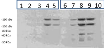
The western-blots (Figure 3.2) confirmed the correct transfection. The HA tag is identifiable in the lines 2,3,4,5 corrisponding to cells transfected respectively with:
Valentina Doneddu
Med1/MBD4 DNA repair enzyme and SUMOylation
Tesi di dottorato in scienze Biomolecolari e Biotecnologiche Ciclo XXVI Università degli studi di Sassari
2: HA-Med1/MBD4 wt;
3: HA-Med1/MBD4 mut 1-2-3;
4: HA-Med1/MBD4 wt + T7-hSUMO1;
5: HA-Med1/MBD4 mut 1-2-3 + T7-hSUMO1.
In lines 4 and 5 numerous bands of poliSUMOylation are encountered, up to 110 KDa until 160 Kda.
The Beta-actin (Fig. 3.3) shows the same amount of loaded proteins.
1 2 3 4 5 -40 kDa -50 kDa -60 kDa -80 kDa -110 kDa
Fig. 3.1: Co-IP Med1/MBD4 and hSUMO1
The Co-IP shows the interaction between Med1/MBD4 and hSUMO1 (line 4 : wild-type isoform of Med1/MBD4 + hSUMO1; line 5: mutated isoform of Med1/MBD4 + hSUMO1; controls: line 1 : untransfected; line 2: Med1/MBD4 wild-type; line 3: mutated Med1/MBD4)
1 2 3 4 5 1 2 3 4 5 -60 kDa -80 kDa -110 kDa -160 kDa a) b) Fig. 3.2: Western-Blot
a) WB with HA ab (Cell signaling); b) WB with T7 ab (Novagene).
Valentina Doneddu
Med1/MBD4 DNA repair enzyme and SUMOylation
Tesi di dottorato in scienze Biomolecolari e Biotecnologiche Ciclo XXVI Università degli studi di Sassari
3.2 Interaction between Med1/MBD4 and hSUMO1 after Cisplatin treatment
Having determined the possibility of Med1/MBD4 of being SUMOylated, the second step was to verify whether the treatment with cisplatin, drug that intercalates DNA and moreover commonly used in clinic as chemotherapeutic, could influence the SUMOylation of Med1/MBD4. Treatment with cisplatin was performed at 24 hours after transfection; the dose administered was 2uM.
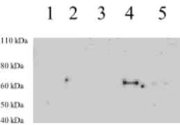
The Co - IP, (fig. 3.4), showed a really interesting figure, in fact while in line 4 (corresponding to HA-Med1/MBD4 wt + T7- SUMO1) the sumoylation band remains around 70 kDa, in line 5 (corresponding to HA-Med1/MBD4 mut 1-2-3 + T7- hSUMO1), sumoylation bands are no longer visible, almost indicating a complete abrogation of interaction of mutant isoform Med1/MBD4 and T7- hSUMO1.
Valentina Doneddu
Med1/MBD4 DNA repair enzyme and SUMOylation
Tesi di dottorato in scienze Biomolecolari e Biotecnologiche Ciclo XXVI Università degli studi di Sassari
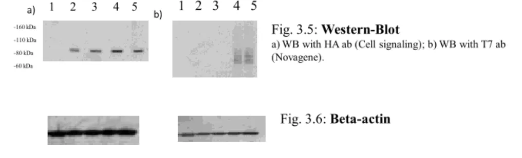
The WB (Fig. 3.5) showed the expression of both isoforms; wild-type and mutated in the three main sumoylation sites, but the bands of poliSUMOylation are completely missing.
In WB with HA ab, the SUMOylation bands are absent while the WB with T7-tag continues to be visible.
The Beta - actin, Fig. 3.6, showed an equal amount of loaded protein.
1 - 2 - 3 - 4 - 5
2uM of Cisplatinum 24h after the transfection
Fig. 3.4 : Co-IP Med1/MBD4 and hSUMO1 after Cisplatin treatment The interaction between Med1/MBD4 and hSUMO1 remain with wild-type isoform of Med1/MBD4 + hSUMO1 (line 4) but is lost with mutated isoform of Med1/MBD4 + hSUMO1 (line 5); controls: line 1 : untransfected; line 2: Med1/MBD4 wild-type; line 3: mutated Med1/MBD4) 1 2 3 4 5 1 2 3 4 5 -60 kDa -80 kDa -110 kDa -160 kDa a) b) Fig. 3.5: Western-Blot
a) WB with HA ab (Cell signaling); b) WB with T7 ab (Novagene).
Fig. 3.6: Beta-actin
Valentina Doneddu
Med1/MBD4 DNA repair enzyme and SUMOylation
Tesi di dottorato in scienze Biomolecolari e Biotecnologiche Ciclo XXVI Università degli studi di Sassari
3.3 Interaction between Med1/MBD4 and hSUMO1 after MNU treatment
With the intent to stress the ability to repair, Med1/MBD4, MCF7 cells were treated 24 hours after transfection with the alkylating agent, MNU, N-methyl-N-nitrosourea at concentration of 2uM. As already observed with cisplatin, the treatment with MNU displayed a total loss of interaction betweenMed1/MBD4 mutated in the three main sites of SUMOylation and hSUMO1. However, the link between the wild-type isoform of Med1/MBD4 and hSUMO1 remained unchanged (Fig 3.7).
Valentina Doneddu
Med1/MBD4 DNA repair enzyme and SUMOylation
Tesi di dottorato in scienze Biomolecolari e Biotecnologiche Ciclo XXVI Università degli studi di Sassari
WB (Fig. 3.8) showed the expression ofMed1/MBD4 and emphasized poliSUMOylation bands for both Med1/MBD4wild-type and its mutated isoform.
The Beta-actin (3.9) was used as control of the amount of loaded protein.
1 2 3 4 5 -40 kDa -50 kDa -60 kDa -80 kDa -110 kDa
Fig. 3.7: Co-IP Med1/MBD4 and hSUMO1 after MNU treatment The Co-IP shows again the interaction between Med1/MBD4 and hSUMO1 (line 4 : wild-type isoform of Med1/MBD4 + hSUMO1; the mutated isoform of Med1/MBD4 + hSUMO1(line 5) is absent; controls: line 1 : untransfected; line 2: Med1/MBD4 wild-type; line 3: mutated Med1/MBD4) 1 2 3 4 5 1 2 3 4 5 -60 kDa -80 kDa -110 kDa -160 kDa a) b) Fig. 3.8: Western-Blot
a) WB with HA ab (Cell signaling); b) WB with T7 ab (Novagene).
Fig. 3.9: Beta-actin
Valentina Doneddu
Med1/MBD4 DNA repair enzyme and SUMOylation
Tesi di dottorato in scienze Biomolecolari e Biotecnologiche Ciclo XXVI Università degli studi di Sassari
The sumoylation is a post-translational modification that alters the activity of its target protein. The purpose of the glycosylase assay was to check whether changes are detected in Med1/MBD4 after SUMOylation activity.
The experiments were precisely made with the help of Genomic Facility of the Fox Chase Cancer Center.
Each assay had planned time using nuclear extracts, obtained with the Nucbuster kit by Millipore, respectively oligonucleotides containing mismatches G:Uand G:Tand the APE enzyme (endonuclease).
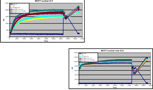
The results of glycosylase activity showed a strong propensity to repair the sumoylated isoform Med1/MBD4, which changed in the three main sites of covalent SUMOylation. This isoform has demonstrated a higher affinity for the repair of G:T mismatch than G:U (Fig 3.10).
Valentina Doneddu
Med1/MBD4 DNA repair enzyme and SUMOylation
Tesi di dottorato in scienze Biomolecolari e Biotecnologiche Ciclo XXVI Università degli studi di Sassari
MCF7 nuclear G:T 0 500 1000 1500 2000 2500 0 2000 4000 6000 8000 10000 12000 14000 16000 18000 20000 Time AU Neg cntr WT HA-MED1 WT HA-MED1 mut 1-2-3 HA-MED1 WT+T7-SUMO HA-MED1 mut 1-2-3+T7 SUMO
Fig. 3.10 : Glycosylase activity
a) oligo G:T; b) oligo G:U. The assay shows a strong activity for SUMOylated Med1/MBD4 mutated and higher affinity for the repair of G:T mismatch than G:U
MCF7 nuclear new G:U
0 2000 4000 6000 8000 10000 12000 14000 0 2000 4000 6000 8000 10000 12000 14000 16000 18000 20000 Time AU Neg cntr WT HA-MED1 WT HA-MED1 mut 1-2-3 HA-MED1 WT+T7-SUMO HA-MED1 mut 1-2-3+T7 SUMO
Valentina Doneddu
Med1/MBD4 DNA repair enzyme and SUMOylation
Tesi di dottorato in scienze Biomolecolari e Biotecnologiche Ciclo XXVI Università degli studi di Sassari
3.5 Interaction between Med1/MBD4 and hSUMO1: localization
When discovered for the first time, the sumoylation was considered the post translational modification capable of migrating protein Ran-GAP to the cytoplasm from the nucleus (Melchior 1999). In order to study the possible changes in location of Med1/MBD4 after sumoylation we used two commercial kits: Nucbuster by Millipore and Subfractionation for cultured cells by Thermo Scientific.
The first kit showed that protein Med1/MBD4, despite being a protein in nuclear localization, when SUMOylated has also the ability to translocate into the cytoplasm. The cytoplasmic localization was observed in both isoforms, wild-type and in the mutant enzyme (Fig. 3.11; 3.12). The kit of Thermo Scientific allows the separation of the proteins present in different nuclear compartments such as:
- membrane proteins
- fraction of cytoplasmic proteins - nuclear extracts
- cytoskeletal proteins - chromatin
Valentina Doneddu
Med1/MBD4 DNA repair enzyme and SUMOylation
Tesi di dottorato in scienze Biomolecolari e Biotecnologiche Ciclo XXVI Università degli studi di Sassari
Fig. 3.11: WB (with HA ab by Cell Signaling) of total lysates extracted with Subcellular
Protein Fractionation Kit for Cultured cells
MCF7 1 not transfected
2 HA-MED1 wt
3 HA-MED1 mut 1-2-3
4 HA-MED1 wt + T7-SUMO1
5 HA-MED1 mut 1-2-3 +T7-SUMO1
-50 kDa -60 kDa -80 kDa -110 kDa -160 kDa -50 kDa -60 kDa -80 kDa -110 kDa -160 kDa 2)Membrane extracts
1)Soluble Cytoplasmic extracts 3) Soluble Nuclear extracts
-50 kDa -60 kDa -80 kDa -110 kDa -160 kDa 1 2 3 4 5 1 2 3 4 5 1 2 3 4 5 MCF7 1 not transfected 2 HA-MED1 wt 3 HA-MED1 mut 1-2-3 4 HA-MED1 wt + T7-SUMO1
5 HA-MED1 mut 1-2-3 +T7-SUMO1
3) Soluble Nuclear extracts
1)Soluble Cytoplasmic extracts
4)Chromatin bound nuclear extracts
Membrane extracts 2) 1 2 3 4 5 1 2 3 4 5 1 2 3 4 5 1 2 3 4 5 -50 kDa -60 kDa -80 kDa -110 kDa -160 kDa -50 kDa -60 kDa -80 kDa -110 kDa -160 kDa -50 kDa -60 kDa -80 kDa -110 kDa -160 kDa -50 kDa -60 kDa -80 kDa -110 kDa -160 kDa
Fig. 3.12: WB (with T7 ab by Novagene) of total lysates extracted with Subcellular Protein
Valentina Doneddu
Med1/MBD4 DNA repair enzyme and SUMOylation
Tesi di dottorato in scienze Biomolecolari e Biotecnologiche Ciclo XXVI Università degli studi di Sassari
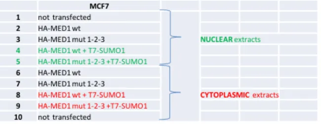
The results confirmed those obtained with the Nucbuster kit. In fact, the sumoylated Med1/MBD4 was found both in the cytoplasmic fraction and in the membrane proteins and of course in the nucleus (Fig. 3.13; 3.14).
MCF7 1 not transfected
2 HA-MED1 wt
3 HA-MED1 mut 1-2-3 NUCLEAR extracts 4 HA-MED1 wt + T7-SUMO1
5 HA-MED1 mut 1-2-3 +T7-SUMO1
6 HA-MED1 wt
7 HA-MED1 mut 1-2-3
8 HA-MED1 wt + T7-SUMO1 CYTOPLASMIC extracts
9 HA-MED1 mut 1-2-3 +T7-SUMO1
10 not transfected 1 2 3 4 5 6 7 8 9 10 α-T7 (Novagene) -50 kDa -60 kDa -80 kDa -110 kDa -160 kDa
Fig. 3.13: WB of total lysates extracted with NucBuster™ Protein Extraction Kit by
Valentina Doneddu
Med1/MBD4 DNA repair enzyme and SUMOylation
Tesi di dottorato in scienze Biomolecolari e Biotecnologiche Ciclo XXVI Università degli studi di Sassari
MCF7 1 not transfected 2 HA-MED1 wt
3 HA-MED1 mut 1-2-3 CYTOPLASMIC extracts 4 HA-MED1 wt + T7-SUMO1
5 HA-MED1 mut 1-2-3 +T7-SUMO1
6 HA-MED1 wt 7 HA-MED1 mut 1-2-3
8 HA-MED1 wt + T7-SUMO1 NUCLEAR extracts 9 HA-MED1 mut 1-2-3 +T7-SUMO1
10 not transfected 1 2 3 4 5 6 7 8 9 10 -50 kDa -60 kDa -80 kDa -110 kDa -160 kDa
α-HA (cell signaling)
Fig. 3.14: WB of total lysates extracted with NucBuster™ Protein Extraction Kit by
Valentina Doneddu
Med1/MBD4 DNA repair enzyme and SUMOylation
Tesi di dottorato in scienze Biomolecolari e Biotecnologiche Ciclo XXVI Università degli studi di Sassari
Valentina Doneddu
Med1/MBD4 DNA repair enzyme and SUMOylation
Tesi di dottorato in scienze Biomolecolari e Biotecnologiche Ciclo XXVI Università degli studi di Sassari
The SUMOylation has aroused great interest in science, precisely because this post-translational modification is able to adjust the complex cellular metabolism, intervening on activity, localization and stability of target proteins.
The great excitement around the SUMOylation concerns the proteins involved in DNA repair. The increased knowledge on the consequences of the modification of enzymes involved in DNA repair may provide useful tools not only for basic research, but also for translational research, especially in oncology.
This work was born from the desire of investigating the effects of SUMOylation of the enzyme involved in DNA repair system of the BER: Med1/MBD4.
Using the SUMOplot software, it was possible to identify 3 main sites of covalent sumoylation, K137 – 215-377 and replace them with R.
The rate of Co -immunoprecipitation showed the ability to interact with Med1/MBD4 hSUMO1. Surprisingly, the interaction remains the same even when you change the three main sites of sumoylation. In this case, in fact, also poliSUMOylation bands appear and this is still the subject of speculation.
Valentina Doneddu
Med1/MBD4 DNA repair enzyme and SUMOylation
Tesi di dottorato in scienze Biomolecolari e Biotecnologiche Ciclo XXVI Università degli studi di Sassari
The interaction was also verified, following treatment ofMCF7 cells, with agents responsible of causing DNA damage as MNU or Cisplatin. Co-immunoprecipitation assays, also in this case, have shown that the interaction between Med1/MBD4 hSUMO1remains unchanged for the wild-type isoform of the protein, whereas it is completely absent in regards to the mutated isoform. These findings suggest that for the sumoylation of Med1/MBD4, non-covalent sumoylation sites are mainly involved.
Further experiments were aimed to clarify the possible changes in activity and localization of Med1/MBD4 after sumoylation.
The glycosylase assays showed in this regard a strongly mutated isoform activity of Med1/MBD4 sumoylated in the repair, with a particular propensity to shelter the oligo G:T than G:U. These data suggest that sumoylation of Med1/MBD4 imparts a strong enzyme repairing activity, especially highlighted in the specificity in the restoration of the oligo G:T. The experiments have shed light on the location of a change in the protein which sumoylated from native nuclear, it also becomes cytoplasmic.
Future studies will be focused on a careful analysis of the sites of sumoylation of non-covalent Med1/MBD4, it will be also studied the biological significance of the change of location and activity of the wild type and mutant isoform of Med1/MBD4.
Valentina Doneddu
Med1/MBD4 DNA repair enzyme and SUMOylation
Tesi di dottorato in scienze Biomolecolari e Biotecnologiche Ciclo XXVI Università degli studi di Sassari
Valentina Doneddu
Med1/MBD4 DNA repair enzyme and SUMOylation
Tesi di dottorato in scienze Biomolecolari e Biotecnologiche Ciclo XXVI Università degli studi di Sassari
Valentina Doneddu
Med1/MBD4 DNA repair enzyme and SUMOylation
Tesi di dottorato in scienze Biomolecolari e Biotecnologiche Ciclo XXVI Università degli studi di Sassari
References
Valentina Doneddu
Med1/MBD4 DNA repair enzyme and SUMOylation
Tesi di dottorato in scienze Biomolecolari e Biotecnologiche Ciclo XXVI Università degli studi di Sassari
1. Sancar A. Mechanisms of DNA excision repair. Science. 1994
2. Fox ME, Feldman BJ, Chu G. A novel role for DNA photolyase: binding to DNA
damaged by drugs is associated with enhanced cytotoxicity in Saccharomyces cerevisiae. Mol Cell Biol. 1994
3. Mitani H, Shima A. Induction of cyclobutane pyrimidine dimer photolyase in
cultured fish cells by fluorescent light and oxygen stress. Photochem Photobiol. 1995
4. Yu Z, Chen J, Ford BN, Brackley ME, Glickman BW. Human DNA repair systems: an
overview. Environ Mol Mutagen. 1999
5. O'Donovan A, Davies AA, Moggs JG, West SC, Wood RD. XPG endonuclease makes the
3' incision in human DNA nucleotide excision repair. Nature. 1994
6. Furuta T, Ueda T, Aune G, Sarasin A, Kraemer KH, Pommier Y.
Transcription-coupled nucleotide excision repair as a determinant of cisplatin sensitivity of human cells. Cancer Res. 2002
7. Hanawalt PC, Ford JM, Lloyd DR. Functional characterization of global genomic
DNA repair and its implications for cancer. Mutat Res. 2003
8. Burdett V, Baitinger C, Viswanathan M, Lovett ST, Modrich P. In vivo
requirement for RecJ, ExoVII, ExoI, and ExoX in methyl-directed mismatch repair. Proc Natl Acad Sci U S A. 2001
9. Genuardi M, Anti M, Capozzi E, Leonardi F, Fornasarig M, Novella E, Bellacosa
A, Valenti A, Gasbarrini GB, Roncucci L, Benatti P, Percesepe A, Ponz de Leòn M, Coco C, de Paoli A, Valentini M, Boiocchi M, Neri G, Viel A. MLH1 and MSH2 constitutional mutations in colorectal cancer families not meeting the standard criteria for hereditary nonpolyposis colorectal cancer. Int J Cancer. 1998
10. Genuardi M, Viel A, Bonora D, Capozzi E, Bellacosa A, Leonardi F, Valle R, Ventura A, Pedroni M, Boiocchi M, Neri G. Characterization of MLH1 and MSH2
Valentina Doneddu
Med1/MBD4 DNA repair enzyme and SUMOylation
Tesi di dottorato in scienze Biomolecolari e Biotecnologiche Ciclo XXVI Università degli studi di Sassari
alternative splicing and its relevance to molecular testing of colorectal cancer susceptibility. Hum Genet. 1998
11. Pandita TK. ATM function and telomere stability. Oncogene. 2002
12. Scott SP, Pandita TK. The cellular control of DNA double-strand breaks. J Cell Biochem. 2006 Dec 15;99(6):1463-75. Review. Erratum in: J Cell Biochem. 2010
13. Iyama T, Wilson DM 3rd. DNA repair mechanisms in dividing and non-dividing cells. DNA Repair (Amst). 2013
14. Damia G, D'Incalci M. Targeting DNA repair as a promising approach in cancer therapy. Eur J Cancer. 2007
15. Schärer OD, Jiricny J. Recent progress in the biology, chemistry and structural biology of DNA glycosylases. Bioessays. 2001
16. Cheadle JP, Sampson JR. MUTYH-associated polyposis--from defect in base excision repair to clinical genetic testing. DNA Repair (Amst). 2007
17. Bellacosa A, Cicchillitti L, Schepis F, Riccio A, Yeung AT, Matsumoto Y, Golemis EA, Genuardi M, Neri G. MED1, a novel human methyl-CpG-binding
endonuclease, interacts with DNA mismatch repair protein MLH1. Proc Natl Acad Sci U S A. 1999
18. Petronzelli F, Riccio A, Markham GD, Seeholzer SH, Stoerker J, Genuardi M, Yeung AT, Matsumoto Y, Bellacosa A. Biphasic kinetics of the human DNA repair protein MED1 (MBD4), a mismatch-specific DNA N-glycosylase. J Biol Chem. 2000
19. Aziz MA, Schupp JE, Kinsella TJ. Modulation of the activity of methyl binding domain protein 4 (MBD4/MED1) while processing iododeoxyuridine generated DNA mispairs. Cancer Biol Ther. 2009
20.Turner DP, Cortellino S, Schupp JE, Caretti E, Loh T, Kinsella TJ, Bellacosa
A. The DNA N-glycosylase MED1 exhibits preference for halogenated pyrimidines and is involved in the cytotoxicity of 5-iododeoxyuridine. Cancer Res. 2006
21. Lucci-Cordisco E, Neri G. Silent beginning: early silencing of the MED1/MBD4 gene in colorectal tumorigenesis. Cancer Biol Ther. 2009
22. Howard JH, Frolov A, Tzeng CW, Stewart A, Midzak A, Majmundar A, Godwin A, Heslin M, Bellacosa A, Arnoletti JP. Epigenetic downregulation of the DNA repair gene MED1/MBD4 in colorectal and ovarian cancer. Cancer Biol Ther. 2009
Valentina Doneddu
Med1/MBD4 DNA repair enzyme and SUMOylation
Tesi di dottorato in scienze Biomolecolari e Biotecnologiche Ciclo XXVI Università degli studi di Sassari
Meropol NJ, Alberti C, Larue L, Bellacosa A. The base excision repair enzyme MED1 mediates DNA damage response to antitumor drugs and is associated with mismatch repair system integrity. Proc Natl Acad Sci U S A. 2003
24. Sansom OJ, Zabkiewicz J, Bishop SM, Guy J, Bird A, Clarke AR. MBD4 deficiency reduces the apoptotic response to DNA-damaging agents in the murine small
intestine. Oncogene. 2003
25. Johnson ES. Protein modification by SUMO. Annu Rev Biochem. 2004
26. Geiss-Friedlander R, Melchior F. Concepts in sumoylation: a decade on. Nat Rev Mol Cell Biol. 2007
27. Delphin C, Guan T, Melchior F, Gerace L. RanGTP targets p97 to RanBP2, a filamentous protein localized at the cytoplasmic periphery of the nuclear pore complex. Mol Biol Cell. 1997
28. Gareau JR, Lima CD. The SUMO pathway: emerging mechanisms that shape specificity, conjugation and recognition. Nat Rev Mol Cell Biol. 2010
29. Rodriguez MS, Dargemont C, Hay RT. SUMO-1 conjugation in vivo requires both a consensus modification motif and nuclear targeting. J Biol Chem. 2001
30. Lin D, Tatham MH, Yu B, Kim S, Hay RT, Chen Y. Identification of a substrate recognition site on Ubc9. J Biol Chem. 2002
31. Lin D, Tatham MH, Yu B, Kim S, Hay RT, Chen Y. Identification of a substrate recognition site on Ubc9. J Biol Chem. 2002
32. Griffiths LM, Swartzlander D, Meadows KL, Wilkinson KD, Corbett AH, Doetsch PW. Dynamic compartmentalization of base excision repair proteins in response to nuclear and mitochondrial oxidative stress. Mol Cell Biol. 2009
33. Steinacher R, Schär P. Functionality of human thymine DNA glycosylase requires SUMO-regulated changes in protein conformation. Curr Biol. 2005
34. Ulrich HD. SUMO modification: wrestling with protein conformation. Curr Biol. 2005
35. Mohan RD, Rao A, Gagliardi J, Tini M. SUMO-1-dependent allosteric regulation of thymine DNA glycosylase alters subnuclear localization and CBP/p300
recruitment. Mol Cell Biol. 2007
36. Shimizu Y, Uchimura Y, Dohmae N, Saitoh H, Hanaoka F, Sugasawa K. Stimulation of DNA Glycosylase Activities by XPC Protein Complex: Roles of Protein-Protein
Valentina Doneddu
Med1/MBD4 DNA repair enzyme and SUMOylation
Tesi di dottorato in scienze Biomolecolari e Biotecnologiche Ciclo XXVI Università degli studi di Sassari
Interactions. J Nucleic Acids. 2010
37. Steinacher R, Schär P. Functionality of human thymine DNA glycosylase requires SUMO-regulated changes in protein conformation. Curr Biol. 2005
38. Ulrich HD. SUMO modification: wrestling with protein conformation. Curr Biol. 2005