UNIVERSITA` DEGLI STUDI DI SASSARI UNIVERSITY OF SASSARI
Department of Biomedical Sciences
PhD Course in Life Sciences and Biotechnologies
XXIX Doctoral cycle
PE_PGRS3: a new player
in Mycobacterium tuberculosis pathogenesis
PhD Student: Basem Battah
2016/2017
Tutor: Prof. Salvatore Rubino Co-tutor: Prof. Giovanni Delogu
Reality cannot compete with imagination
Index
Chapter I : Introduction1. Tuberculosis………...….p2 1.1 The genus mycobacterium………p2 1.2 The M. tuberculosis genome……….…..…..…p3 2. M. tuberculosis virulence and disease process ………..p5 3. BCG vaccine………...p9 4. M. tuberculosis treatment and emergence of MDR and XDR………...p10 5.Cell structure………...p13 5.1 The mycobacterial cell wall………..…..p13 6. The typeVII protein secretion pathways……….…….…….…p15 6.1 Genetic organization of ESX systems and their secreted proteins………...p17 7. PE-PPE protein family………...p19 7.1 PE_PGRS subfamily ………....…...p22 8. Stringent response and phosphate depletion in M.tuberculosis…………...….p27 9. M. tuberculosis between dormancy and reactivation………..……..p32 The aim of the study ………..………...p34 Chapter II: Results
1. Main features of PE_PGRS3 and PE_PGRS4………..p36 2. PE_PGRS3 and PE_PGRS4 are differentially expressed………...…p39 3. PE_PGRS3 has a specific expression under low inorganic phosphate
condition………..….p43 3.1 Quantification of protein expression by measuring the fluorescence of single mycobacteria by FACS-Canto flow cytometer……….p45
3.2 PE_PGRS3 expression increased in low phosphate condition and correlated with RelA in M. smegmatis and M. tuberculosis……….….p47 4. M. smegmatis over expressing PE_PGRS3 were shorter in size than the strains expressing the functional domains ……….……..p50 5. The M. smegmatis expressing PE_PGRS3 and its functional domains have a similar growth rates ……….p53 6. Purified native C-terminal domain of PE_PGRS3 induces specific anti-serum in mice ………...……….…..p53 6.1 Sera from immunized mice specifically recognize C-terminal of
PE_PGRS3………..…..p56 7. PE_PGRS3 could be cleaved at the C-terminal domain secreted or surface
exposed ………...……….…p57 8. The recombinant purified C-terminal of PE_PGRS3 has no cytotoxic effect on the murine macrophages and human alveolar epithelial cell...p58 9. M. smegmatis expressing PE_PGRS3 enhanced entry in macrophages and
alveolar epithelial cells………...….….p58 Chapter III: Material and methods
1. Construction of gene reporter vectors ………...….…..p65 1.1 Construction of plasmids expressing PE_PGRS3 and PE_PGR4 fused with green fluorescent protein (GFP)……….…...p65 1.2 Construction of plasmid expressing PE_PGRS3 under control of the hbhA promoter and of its functional deletion mutants in a plasmid expressing green fluorescence protein (GFP)………...…p66 2. Construction of 6xHis-SUMO fusion expression vector………..…………....p67 3. Bacterial strains media and growth conditions ………...…….…p67
5. Expression of C-terminal domain of the PE_PGRS3………...……..…..p69 5.1 Protein purification………...….….p69 5.2 Purified recombinant C-terminal domain of the PE_PGRS3 LPS free
preparation………...p70 6. Mice immunization………...……...p71 7. SDS_PAGE, Western blotting and immunoblotting………....p71 8. FACS analysis……….………...p72 9. Quantitative reverse transcription - Real time PCR (qRT-PCR)…...p73 10. Confocal microscope and image analysis………..……….p74 11. Cell culture and mycobacteria infection………...…...p75 12. M. smegmatis recombinant strains growth rate measurements ………...p76 13. Cytotoxicity assay ………...………...………....p77 14. Multiple sequence alignments………..…...p77 15. Statistical analysis………..………..…...p77 Chapter VI: Discussion
Abstract
The M. tuberculosis (Mtb) genome contains around 60 pe_pgrs genes, whose role and function remain elusive. In this study, two PE_PGRS proteins with high sequence homology were selected and investigated (PE_PGRS3 and PE_PGRS4), with PE_PGRS3 characterized by the presence of a C-terminal domain rich in arginine. Interestingly, full-length PE_PGRS3 protein is expressed by Mtb strains but not by other MTBC subspecies causing disease in animals. A gene reporter system was developed to investigate in M. smegmatis (Msm) the expression pattern of these genes. Fluorescence microscopy, FACS and transcriptional analysis indicated that the two genes are differentially regulated, with pe_pgrs3 but not pe_pgrs4 being expressed only when mycobacteria are cultivated in low inorganic phosphate (iPhos). Expression of pe_pgrs3 in low iPhos correlated with the upregulation of relA in Msm recombinant strains and Mtb, suggesting that pe_pgrs3 is involved in the stringent response. Overexpression of the PE_PGRS3, and of its functional deletion mutant (PE_PGRS3Ct), in Msm were obtained by expressing these genes under control of hbhA promoter. Interestingly, Msm strains overexpressing PE_PGRS3 showed enhanced ability to entry in macrophages and epithelial cells compared to Msm expressing PE_PGRS3Ct or Msm parental strain. No differences in the ability of these strains to survive intracellularly were measured. These results provide new insights on the role of PE_PGRS3 in TB pathogenesis.
Chapter I
1. Tuberculosis
Tuberculosis (TB), one of the oldest recorded human catastrophes, is still one of the biggest killers among the infectious diseases (WHO, 2016). Despite the worldwide use of a live attenuated vaccine and effective antibiotics, new vaccines and drugs are needed to control the worldwide epidemic of TB that kills two million people each year. In order to develop new anti-tubercular agents, there is a need to study the genetics and physiology of tubercle bacillus and understand the host-pathogen interaction to learn how M. tuberculosis bacteria overcome host defences and cause disease.
In 2015, there were an estimated 10.4 million new (incident) TB cases worldwide, 480. 000 new cases of multidrug-resistant TB (MDR-TB), 1.4 million TB deaths and an additional 0.4 million deaths resulting from TB disease among people living with HIV (WHO, 2016). Therefore, new and effective tools against TB are urgently needed to control the disease at multiple levels.
1.1 The genus mycobacterium
Beside M. tuberculosis, more than 100 other mycobacterial species have been identified (Tortoli, 2006). These can be divided in two groups based on their growth rate: the rapid-growers, which produce visible colonies on solid medium within seven days and the slow – growers, which typically require 10-28 days for visible growth. Genetic analysis of the 16S rRNA genes indicates that the slow - growers have evolved from the fast growers (Fig 1.1) (Reva et al., 2015). This division also reflects the virulence of mycobacterial species, as most fast grower are non-pathogenic, free – living saprophytes whereas the slowly growing group contain a number of important human and animal pathogens.
The latter include, apart from the species belonging to M. tuberculosis complex (M. bovis, M. microti, M. canetti, M. tuberculosis, M. africanum), Mycobacterium leprae, the causative agent of leprosy, M. ulcerans, the etiological agent of buruli ulcer, M. avium, which causes TB in birds and opportunistic infections in immunocompromised humans, and M. marinum, that causes chronic progressive disease in fish and amphibia, skin infections in humans.
Figure 1.1: Species phylogenetic tree of the genus mycobacterium (Reva et al., 2015).
1.2 The M. tuberculosis Genome
The M. tuberculosis H37Rv genome consists of 4.4 x10⁶ bp and encodes approximately 4,000 genes (Fig 1.2). Analysis of the M. tuberculosis genome showed that this bacterium has some unique features. Over 200 genes, accounting to 6% of the total, have been annotated as encoding enzyme involved in the metabolism. Among these approximately 100 are predicted to be implicated in the oxidation of fatty acids, while in comparison E. coli only has 50 enzymes involved in fatty acid metabolism. The large number of enzymes that putatively use fatty acids may be related to the ability of the M. tuberculosis to grow in
Rapid growers Slow growers
infected host tissues, where fatty acids may provide the major carbon source (Smith, 2003). Interestingly, five gene locus encoding Type 7 secretion apparatus
(ESX1-5) were also identified, highlighted an unexpected ability of M. tuberculosis to secrete proteins. Moreover, almost 8% of the genome coding
capacity was devoted to genes belonging to protein with highly similar protein sequence, named PE and PPE.
2. M. tuberculosis virulence and the disease process
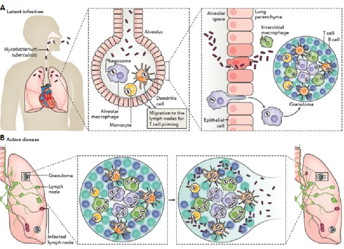
M. tuberculosis usually enters the alveolar pathways of exposed humans in an aerosol droplet, and its first contact is thought to be with macrophages, but it is also possible that bacteria can be initially ingested by alveolar epithelial type II pneumocytes which are found in greater numbers than macrophages in alveoli. In addition, dendritic cells play a very important role in the early stages of infection since they are much better antigen presenters than are macrophages and may play a key role in activating T cells with specific M. tuberculosis antigens. Dendritic cells are migratory and may play an important role in M. tuberculosis dissemination (Smith, 2003) (Fig 1.3).The recognition of M. tuberculosis is mediated by a set of surface receptor, which drive the uptake of bacteria and trigger the innate immune signalling pathways leading to the production of various chemokines and cytokines. Epithelial cells and neutrophils can also produce chemokines in response to the bacterial products, this promotes recruitment of other immune cells, more macrophages, dendritic cells and lymphocyte, to the infection site. They organize in spherical structure with infected macrophages in the middle surrounded by various categories of lymphocytes (mainly CD4+, CD8+). Macrophages can fuse to form multi nucleated giant cells or differentiate into lipid–rich foamy cells. B lymphocytes tend to aggregate in follicular-type structure adjacent to the granuloma. The bacteria can survive for decades inside granuloma with no symptoms of the disease in 90-95% of the cases (Mayra Silva Miranda, 2012). It is known that infected macrophages in the lung, through their production of chemokines, attract inactivated monocytes, lymphocytes, and neutrophils, which cannot kill the bacteria very efficiently. Mycobacteria can escape intracellular killing, multiply and further promote inflammation (van et al., 2002). Then, granulomatous focal lesions composed of macrophage derived giant
cells and lymphocytes begin to form. These processes generally serve as effective means for controlling bacterial spread. As cellular immunity develops, macrophages loaded with bacilli are killed, and this results in the formation of the caseous center of the granuloma, surrounded by a cellular barrier of fibroblasts, lymphocytes, and blood-derived monocytes (Smith, 2003) (Fig 1.3).
Figure 1.3: Mycobacterium tuberculosis infection. (A) The Infection begins when M. tuberculosis enters the lungs
via inhalation, if this first line of defence fails to eliminate the bacteria, M. tuberculosis invades the lung interstitial tissue, either by the bacteria directly infecting the alveolar epithelium or the infected alveolar macrophages migrating to the lung parenchyma. Subsequently, either dendritic cells or inflammatory monocytes transport
M. tuberculosis, migrates to the lymph nodes to recruit the immune cells T and B cells to form a granuloma. (B) If
the bacteria replicated within the growing granuloma with a great load, the granuloma will fail to contain the infection and the bacteria will disseminate to other organs including the brain. At this phase, the bacteria can enter the bloodstream or re-enter the respiratory tract to be released, the infected host is now infectious, symptomatic and
By the time the host immune response properly control bacterial replication, the tubercle bacilli is thought to be able to disseminate by the lymphatics and bloodstream to potentially any organ and tissue.
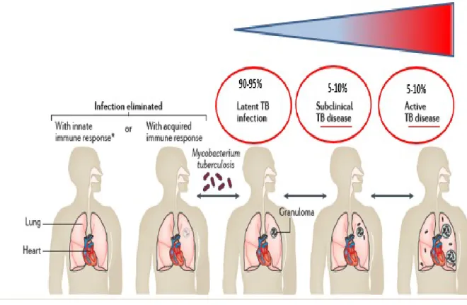
The strength of the host cellular immune response determines whether an infection is arrested here or progresses to the next stages, the resulted enclosed infection is referred to as latent or persistent TB and can persist throughout a person’s life in an asymptomatic and non-transmissible state. In persons with efficient cell-mediated immunity, the infection may be arrested permanently at this point. The granulomas subsequently heal, leaving small fibrous and calcified lesions which is the hallmark of latent tuberculosis (Sandor et al., 2003).
It is estimated that 5-10% of the M. tuberculosis infected subjects may develop disease during the lifetime, either because the infected person cannot control the initial infection or because a person with latent infection may lose the ability to control bacterial replication due to a weakened immune system (use of immunosuppressive drugs, HIV infection, malnutrition, aging, or other factors). Bacterial replication leads the granuloma centre to liquefy by unknown processes and then serves as a rich medium in which the now revived bacteria can replicate in an uncontrolled manner (Smith, 2003), (Russell et al., 2010) (Fig 1.3, 1.4).
3. BCG vaccine
Today the only vaccine available against M. tuberculosis is Bacillus Calmette Guerin (BCG) obtained one hundred years ago by Calmette and Guerin at the Pasteur institute in France, after thirteen years of serial in vitro passage of a M. bovis strain isolated from cattles (Luca and Mihaescu, 2013). Recent genomic
studies showed that the genetic determinants underlying the attenuation of M. bovis BCG is the deletion of long region of genome named Region of deletions
(RD). A total of 14 RD regions have been identified, the most important of which is RD1 that is absent in all BCG strains and correspond to a 9.5 kbp region that in M. tuberculosis encodes 9 genes (Behr et al., 1999), encoding among them two secreted low molecular weight proteins, CFP-10 and ESAT-6, which are transcribed together, that have been recognized as virulence factors and potential vaccine candidates (Brandt et al., 2000). It has also been shown that deletion of RD-1 in M. tuberculosis strains leads to a strong attenuation of pathogenicity in mouse model of M. tuberculosis (Pym et al., 2002). Protection against tuberculosis afforded by BCG is high in children, specifically against the most severe forms of the disease. However, protection is known to decrease over time resulting in variable outcomes of protection ranging from 20% to 80%. BCG protective effects tends indeed to wane in early adolescence (Colditz et al., 1994), due to genetic and environmental factors. There are several TB vaccines in phase I or phase II trials. For example, Ad5-Ag85A vaccine in Phase I clinical trial, AERAS-402/crucel Ad35 vaccine, the recombinant BCG expressing lysteriolysin (Groschel et al., 2014), (Cardona, 2006), and the M72/AS01E vaccine (Marisol Ocampo C, 2015). However, until now a vaccine effective in preventing TB in adults remains elusive (WHO, 2016).
4. M. tuberculosis treatment and emergence of MDR and XDR
Only very few antibiotics, rifampicin, isoniazid, pyrazinamide and ethambutol as first line and ethionamide, fluoroquinolones, streptomycin, aminoglicosides (amikacin and kanamycin) as second line are known to be active against active TB (Laurenzo and Mousa, 2011). Treatment of latent (asymptomatic) infection consists of INH taken for 6-9 months. Rifampin can be used in those exposed to INH resistant strains. The combination of rifampin-pyrazinamide should not be
used because it caused a high rate of liver injury. Multiple drug resistant M. tuberculosis strains (MDR) have emerged primarily in AIDS patients and have
resistance to both INH and rifampin, but some isolates are resistant to three or more drugs. The treatment of MDR organisms includes using of four or five drugs, ciprofloxacin, amikacin, ethionamide and cycloserine. M. tuberculosis strains resistant to INH, rifampin, a fluoroquinolone, and at least one additional drug are called XDR (extensively drug resistant) (levinsin W, 2008).
Within the last 10 years, the mechanism of action of most of the anti-tuberculosis agents have been described, and many studies are beginning to elucidate some of the molecular mechanisms whereby M. tuberculosis becomes resistant.
The genetic basis of resistance for some anti-tuberculosis agents is not fully known. For example, streptomycin resistance emerges through mutations in rrs and rpsL that produce an alteration in the streptomycin binding site. Isoniazid-resistance is caused by modification of KatG, which is the enzyme that activate isoniazid to the active hydrazine derivative. Mutation in KatG lead to high-level resistance to isoniazid (Zhang et al., 1993). A deficiency in enzyme activity produces high-level resistance and is found in more than 80% of isoniazid-resistant strains. Most pyrazinamide isoniazid-resistant organisms have mutations in the pyrazinamidase gene (pncA). Pyrazinamidase is essential in producing the active pyrazinoic acid derivative, and mutants are unable to produce an active drug (Gillespie, 2002), (Ramaswamy and Musser, 1998). Ethambutol resistance in approximately 60% of organisms is due to amino acids replacements at position 306 of an arabinosyltransferase encoded by embB gene (Ramaswamy and Musser, 1998) (Fig 1.5).
Bedaquiline has recently received conditional approval for the treatment of MDR-TB under the trade name Sirturo after the results of two phases, phase II clinical trials and phase III trials was scheduled to begin in 2013, The mechanism of action of bedaquiline is by inhibiting the ATP synthase of M. tuberculosis, which was a completely new target of action for an anti-mycobacterial drug. The only mutation found was in the atpE gene, which encodes the c part of the F0 subunit of the ATP synthase. Nevertheless, in a study to further assess the mechanisms of resistance to bedaquiline in M. tuberculosis, it was found that only 15 out of 53 resistant mutants had mutations in atpE. The other strains do not have
mutations in atpE, which suggests that other mechanisms of resistance are still possible (Huitric et al., 2010).
Delamanid acts by inhibiting the synthesis of mycolic acid and is undergoing clinical evaluation in a phase III trial. Delamanid has more recently shown its safety and efficacy in a clinical evaluation for MDR-TB. It only inhibits methoxy- and keto-mycolic acid while isoniazid also inhibits α-mycolic acid requires reductive activation by M. tuberculosis to exert its activity. In experimentally generated delamanid-resistant mycobacteria, a mutation was found in the Rv3547 gene, suggesting its role in the drug activation (Palomino and Martin, 2014). The increasing prevalence of drug-resistant strains of M. tuberculosis makes the development of novel drugs for tuberculosis and identify potential drug targets an urgent priority. For new drug targets there are several criteria that should be considered. First, the drug target must be essential to bacterial viability, virulence or the persistence of M. tuberculosis in granulomas. Second, targeting a novel pathway not inhibited by existing drugs may reduce the chance of cross-resistance with current drug-resistant strains. Third, targets not conserved in humans may reduce the likelihood of off-target effects. Fourth, the target must be accessible to inhibitors, which is particularly important for penetrating the unique and highly impermeable cell envelope of M. tuberculosis. in general, target that are positioned outside the cytoplasmic membrane will be more accessible (Feltcher et al., 2010).
5. Cell structure
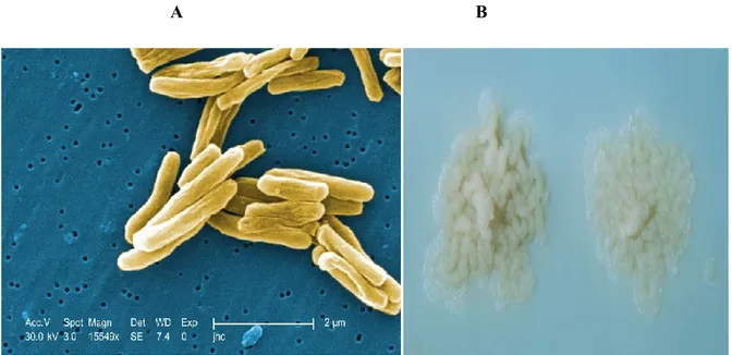
Mycobacterium tuberculosis is a long large non-motile aerobic acid- fast rod-shaped bacterium belonging to the order of actinomycetales (William A.Strohl, 2001). It grows slowly and has a doubling time of 18 hours (levinsin W, 2008). The rods are 2-4 µm in length and 0.2-0.5 µm in width (Fig 1.6A), M. tuberculosis colonies are small and buff colored when grown on solid medium Figure (Fig 1.6B) (Kenneth Todar, 2017).
A B
Figure 1.6 : M. tuberculosis scanning electron micrograph. Mag 15549X (a), Colonies of
M. tuberculosis on Lowenstein-Jensen medium (B) (Kenneth Todar, 2017).
5.1 The mycobacterial cell wall
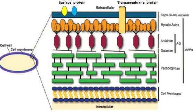
Mycobacterium are surrounded by a unique cell wall with unusual low permeability. A peptidoglycan layer surrounds the plasma membrane and long chain polysaccharides such as lipoarabinomannan (LAM), lipomannan and arabinogalactan covalently link the peptidoglycan layer with the outer membrane.
The mycobacterial outer membrane, also named mycomembrane, is functionally similar the membrane of gram-negative bacteria, though its composition is peculiar. The inner leaflet of the mycomembrane is composed of mycolic acids and the outer leaflet of small glycolipids, sulfolipids and other lipids. The mycomembrane provides strength and impermeable barrier to mycobacteria, due to its high hydrophobicity (Zuber et al., 2008) (Brennan and Nikaido, 1995), (Brennan and Besra, 1997). Many of the drugs used to combat mycobacteria are effective because they specifically target the biosynthesis of the mycobacterial cell wall components.
Figure 1.7: Basic components of the mycobacterial cell wall. MAPc, MA-AG-PG complex (Hett and
6. The type VII protein secretion pathways
The virulence of bacterial pathogens highly depends on the ability to secret proteins and molecules to the bacterial surface, external milieu or directly into host cells (Abdallah et al., 2007). Because the biological membranes in the bacterial cell wall hinder export of proteins, translocation across these barriers is mediated by dedicated proteins secretion systems. Similar to other bacteria, mycobacteria secret proteins across inner membrane via ubiquitous general secretory (sec) pathway or twin-arginine translocation (tat) system (Champion and Cox, 2007). In gram positive bacteria, which have only one lipid bilayer, the Sec/Tat pathways are generally sufficient for protein export. Gram negative bacteria, have evolved a number of specialized secretion systems for transport of protein across the outer membrane. Six pathways, generally known as the type I to type VI secretion systems, either secrete proteins that are delivered into periplasm by sec or Tat system (type II and V) in two step process, or via a signal peptide-independent one-step mechanism across the entire cell envelope (type I, III, IV and VI). Although mycobacterium also contains a diderm cell envelope, they lack type I to type VI pathways and evolved a unique specialized secretion system which known as the ESX or type VII secretion pathway. In M. tuberculosis there are five type VII secretion systems encoded by gene clusters and called ESX1 to ESX5 (Fig 1.8). ESX1 and ESX5 secrete different proteins involved in the virulence of M. tuberculosis, while ESX1 is missing in the attenuated M. bovis vaccine strain Bacille Calmette and Guerin (Delogu et al., 2013). ESX1 is required for the full virulence of M. tuberculosis, which uses this secretion system to escape from the phagosome into the host cell cytosol of infected macrophages where it may persist in a protected environment (Romagnoli et al., 2012). ESX1 mediates secretion many antigens, ESAT-6 and CFP-10, both small highly immunogenic proteins that
form the basis of the immunological diagnosis of M. tuberculosis infection in the interferon-gamma release assays (IGRAs), and appears that the ESX-1 secreted proteins have the ability to disrupt the biological membrane (Brennan and Nikaido, 1995), (Gao et al., 2004). It has also been demonstrated that ESX3 secretion system is responsible for the secretion of some soluble factors required for growth that are probably involved in optimal iron and zinc uptake (Serafini et al., 2009). ESX5 is restricted to the slow growing species, while it is absent from the genome of fast growing bacteria such as M. smegmatis. ESX-5 is found in M. tuberculosis complex (MTBC), M. marinum, M. ulcerans, M. leprae, and M. tuberculosis (Gey van Pittius et al., 2001), and it may represents a secretion
systems specifically evolved to interact with a complex immune system such as that of mammals. Indeed the ESX-5 was shown to play a role in immunomodulation (Abdallah et al., 2008), and induce cell death which facilitate cell to cell spread and it was hypothesized that ESX-5 effectors will interact and manipulate the host cell after ESX-1 mediated escape from phagosome into cytosol during the infection (Abdallah et al., 2011). ESX5 appears to be a major export pathway for PE/PPEs M. marinum proteins, especially for the most recently evolved members, the PE_PGRS and PPE_MPRT proteins (Abdallah et al., 2009), while the role and function of ESX2 and ESX4 are still unknown, the study of the role of ESX systems in TB pathogenesis is one of the major advancements of the last decade in the TB field. The ESX export system represent other potential targets for new anti TB drugs, ESX-1 and ESX-3 are known to be essential for virulence and growth of M. tuberculosis, respectively. An inhibitor that targets a conserved core component of the ESX pathways (EccB, EccD, EccE and MycP), has the potential to disrupt all ESX systems simultaneously, which could reduce evolution of drug resistence. There are also secreted proteins of the ESX system
that may function in the ESX secretion process (EspA and ESAT-6/CFP-10) that could be accessible to inhibition (Feltcher et al., 2010).
Figure 1.8: Protein secretion systems. ESX1 secretes antigens that interfere with the integrity of the
phagosomal membrane, leading to phagosomal rupture and bacterial emission into the cytosol. ESX5 is present only in slow growing mycobacteria (such as M. tuberculosis and M. marinum) and it is thought to be involved in the secretion of proteins (PPE and PE-PGRS) with immunomodulatory properties. ESX3 is involved in Zinc and Iron uptake and homeostasis and as such is essential for growth. The role of ESX2 and ESX4 remain still unknown (Delogu et al., 2013) .
6.1 Genetic organization of ESX systems and their secreted proteins
ESAT-6 (early secreted antigentic target of 6 KDa) and CFP-10 (culture filtrate protein of 10KDa) which are encoded by an operon are both secreted in the culture medium of M. tuberculosis (Berthet et al., 1998), (Sorensen et al., 1995) and belong to WXG 100 family of proteins, characterized by Trp-x-Gly motif. Comparative genomic methods revealed that esat-6 and cfp-10 genes are found in a region known as region of difference 1 (RD1) which is present in virulent strains of M. tuberculosis and M. bovis but absent from the genome of M. bovis BCG and M. microti because of independent deletion events (Daniel, 2006).
Figure 1.9: Genetic organization and gene names of the five ESX loci and espA operon in M.
tuberculosis. The RD1 deletions of M. Bovis BCG and M. Microti are marked in ESX-1 cluster. Ecc stands for ESX conserved component and esp for ESX-1 secretion system (Majlessi et al., 2015).
Phylogenetic analysis and genomic comparison suggest that the five ESX systems ESX-1 to ESX-5 have evolved by duplication events, where the ESX-4 cluster contains the lowest number of genes, is thought to be the most ancestral cluster which duplicated to give arise ESX-1, ESX-2, ESX-3 and finally ESX-5 (Fig 1.9). Interestingly, ESX-5 is restricted to slow growing pathogenic mycobacteria such as M. tuberculosis, M. leprae and M. marinum and absent from the genome of fast-growing M. smegmatis, and seems to be a major secretion pathway of PE and PPE proteins like PPE41 (Abdallah et al., 2009) (Abdallah et al., 2006). Conversely, ESX-1 to ESX-4 clusters are distributed in the genome of mycobacteria and ESX-1 cluster required for secretion of ESAT-6 and CFP-10, encodes a functionally secretion system in mycobacteria such as M. marinum and M. smegmatis (Converse and Cox, 2005).
7. PE-PPE protein family
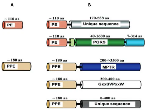
During the analysis of complete genome sequence of Mycobacterium tuberculosis H37Rv, two gene families were identified, encoding proteins with conserved N-terminal domains characterized by motifs Pro-Glu (PE) or Pro-Pro-Glu (PPE) near their respective N-termini, which occupy around 8% of the coding capacity of the genome. These two families consist of 100 and 67 members, respectively and each family has been divided into subfamilies. PE/PPE proteins can consist of only these so called PE and PPE domains (Fig 1.10A), or they may have extended C-termini (Fig 1.10B). In the latter case, the C-terminal domains are composed of unique sequences or of sequences with characteristic glycine-rich repeat, such as those encoded by polymorphic CG-rich-repetitive sequence (PGRS) subfamily of PE proteins and the major polymorphic tandem repeat (MPTR) PPE subfamily mainly encode glycine and aspargine.
Figure 1.10: Domain organization of the PE and PPE proteins. (A) the most ancestral members of these
family consist of only PE/PPE domain. (B) During evolution, the PE/PPE proteins appear to have acquired extended C-terminal domains (Bottai and Brosch, 2009).
B A
Comparative genomic studies of mycobaterial species has shown that pe/ppe genes are largely unique to mycobacteria. Moreover, fast – growing mycobacteria carry only few of these genes and lack genes of the PE_PGRS and PPE-MPTR subfamilies, which are enriched in the genome of slow-growing, pathogenic species (Gey van Pittius et al., 2006). Many members of PE and PPE proteins were shown to localize on the mycobacterial surface, where they are available to interact with host components (Banu et al., 2002), (Malen et al., 2010). The highly polymorphic nature of the C-terminal of these proteins (PE_PGRS and PPE_MPTR) which exhibit the most sequence variation suggests their involvement in the anti-genetic variation (Sampson, 2011). This hypothesis is supported by a study showing that a DNA vaccine expressing the N-terminal PE region of the PE_PGRS33 protein is able to elicit a cellular immune response, while mice immunized with 1818PE_PGRS develop primarily a humoral response (Delogu and Brennan, 2001). It also proved that the PE domain of the PE_PGRS33 is necessary for the subcellular localization while the PGRS domain but not PE domain affect the bacterial shape and colony morphology (Delogu et al., 2004). However, the accumulation of pe/ppe genes into the genomes of mycobacterial pathogens highlights on the important role of these proteins in mycobacterial pathogenesis. Many of pe/ppe genes are located within ESX clusters and both loci are evolutionary correlated. The expansion of PE/PPE family is associated with the duplication of ESX gene cluster and the emergence of repeat proteins PGRS and MPTR is a recent evolutionary event (Gey van Pittius et al., 2006) (Fig. 9). The PE/PPE families and ESX systems also appear to be functionally linked, as several PE and PPE proteins have shown to be substrate to the ESX system. For example, PE35 which is encoded directly upstream of ppe68, esxB (CFP-10) and esxA (ESAT-6), influence the expression of these three genes.
Transposon insertion in the PE35 gene in two independently derived M. tuberculosis strains failed to express PPE68, ESAT-6 or CFP10 and were both
attenuated, while in contrast, transposon mutagenesis or partial deletion of ppe86 did not have attenuated effect in mouse infection model (Delogu G and Cole S.T and borsch R, 2008). Another example is the secretion of PPE42, a hydrophilic protein expressed within the ESX-5 system, which has been shown to induce a strong B cell response in human. PPE41 forms a heterodimeric protein with the neighbouring PE25 and expressed as an operon (Strong et al., 2006). However, while the export of a single PE/PPE couplets has been attributed to the ESX-1 and to all the other ESX systems, (Sani et al., 2010), the ESX-5 seems to be the major export pathway for the most recently evolved proteins such as PE_PGRS and PPE_MPRT, which were shown to play an essential role for the full virulence of M. tuberculosis (Abdallah et al., 2009), (Abdallah et al., 2006), (Ahmed et al., 2015). Moreover, several transcriptional factors have been reported to be involved in the modulation of pe/ppe genes expression (Ahmed et al., 2015), (Mohareer et al., 2011). For instance, the iron dependent regulator ideR, responsible for the induction of genes involved in iron uptake, regulates around 11 pe/ppe genes (Rodriguez et al., 2002). Furthermore, PhoPR, which is a two component system involved in the regulation of genes encoding the type VII secretion system ESX1 and genes involved in synthesis of several cell wall components, regulates pe/ppe genes (Solans et al., 2014).
7.1 PE_PGRS subfamily
All PE_PGRS proteins have a common molecular architecture. The PGRS domain is linked to a highly conserved N-terminal domain that is around 90-100 amino acids along and contains the PE motif (Fig 1.10). The PGRS domain is linked to the PE domain with a linker region 35-40 amino acids long and contains a conserved GRPLI motif (AC domain). This domain may form a putative helix and has been suggested to play a role in the localization of PE_PGRS proteins (Delogu G and Cole S.T and borsch R, 2008) or may serve to anchor the PE_PGRS to a specific, yet unidentified portion of the cell wall. The PE_PGRS is characterized by the presence of multiple tandem repetitions (Gly-Gly-Ala and Gly-Gly-Asn motifs) that vary in number from several tens to hundreds amino acids. These repeats are intercalated by short regions of diverse sequence composition and size, and these differences may be used to define subgroups within PE_PGRS proteins (Delogu G and Cole S.T and borsch R, 2008).
Group A1: which has intercalating sequences of 4-10 amino acids long and present unique C-terminal domain that may extend up to 15-27 amino acids.
Group A2: contains intercalating sequences that may extend up to 20 amino acids. Group A3: contain high number of these intercalating sequence.
Group B: consists of four PE_PGRS which have an intercalating domain that varies in size from 25 to 59 amino acids and containing inside PGRS domain a second GRPLI motif.
Group C: contains a large C-terminal domain that can be as large as 300 amino acids (Fig. 1.11).
However, the PGRS domain cannot simply be considered a repetitive or redundant domain but rather contains a specific sequence interspaced among GGX-GGX regions that make this domains unique for each PE_PGRS protein. The lack of experimental data about PE_PGRS domains make it necessary to study.
Figure 1.11: Division of PE_PGRS proteins into subfamilies (Delogu G and Cole S.T and borsch R,
In the genome of M. tuberculosis 63 open reading frames have annotated as pe_pgrs genes, but a number of them have a frameshift mutations that prevent the synthesis of some functional proteins and two proteins (PE_PGRS62 and 63) lack the typical PGRS domain and the linker region, 51 potentially functional pe_pgrs genes scattered throughout the M. tuberculosis H37Rv genome. pe_pgrs genes were found only in the MTBC or other pathogenic mycobacteria, like M. ulcerans and M. marinum, highlighting their potential involvement in the pathogenesis of M. tuberculosis. Many studies raised the attention on these proteins which are directly involved in the pathogenesis of M. tuberculosis infection and in the evasion from the host immune response (Brennan and Delogu, 2002), (Forrellad et al., 2013), (Lalita Ramakrishnan, 2000). The differences between the genetic organization of pe_pgrs genes and the pe/ppe genes families suggests an autonomous and independent regulation of gene expression for most of PE_PGRS proteins. M. tuberculosis can differently regulate the expression of PE_PGRS. For example pe_pgrs30 gene expression increased following intracellular growth in bone marrow-derived macrophages but not in type-II human pneumocytes, while pe_pgrs9 was induced in both in vitro systems (Iantomasi et al., 2012). Another study showed that PE_PGRS16 and PE_PGRS26 are inversely regulated in macrophages and in mice infected with M. tuberculosis (Dheenadhayalan et al., 2006). Interestingly, M. smegmatis strains expressing PE_PGRS33 and PE_PGRS26 were able to persist at higher level in spleen and liver tissues compared with M. smegmatis expressing PE_PGRS16, suggesting a differential role of these proteins in mycobacterial pathogenesis (Singh et al., 2008). Upregulation of pe_pgrs9, -16 and -30 genes were observed in M. tuberculosis infected macrophages and in host tissues during the chronic step of infection and the upregulation was higher in the spleen compared to the lung infected mice
PE_PGRS47 as an inhibitor of autophagy and a factor contributing to evasion of both innate and adaptive immunity by M. tuberculosis (Saini et al., 2016). Another data have been demonstrated that some sigma factors are involved in the expression of some PE_PGRS like SigA which mediates in-vitro transcription of PE_PGRS33 and their expression is repressed in stress condition (Vallecillo and Espitia, 2009). Therefore, we can summarize that pe_pgrs genes are differently expressed and regulated by M. tuberculosis in host tissue depending on the different environmental conditions that mycobacteria faced during infection.
Both innate and adaptive immune responses play an important role in M. tuberculosis infection, and some studies demonstrated that the PE and PGRS
domains have a role in cellular and humoral immune responses (Cohen et al., 2014). It has been suggested that the PGRS domain could be the target of the host immune response because of its extensive variability, although the link between the genetic variability and antigenic variation is still hypothetical (Delogu G and Cole S.T and borsch R, 2008). Interestingly, the Epstein–Barr Virus nuclear antigen 1 (EBNA1) shows significant similarity with the PGRS domain of PE_PGRS proteins, containing numerous Gly-Ala repeats that are known to inhibit antigen processing and presentation through the major histocompatibility complex I (MHCI pathway) (Cole et al., 1998), (Brennan and Delogu, 2002). PE_PGRS proteins have domains that confer resistance to ubiquitin/proteosome dependent protein degradation and may use this mechanism to evade immune detection and killing of mycobacterium infected cells (Koh et al., 2009). Another study has demonstrated that two PE_PGRS proteins, PE_PGRS 17 and PE_PGRS11, recognize TLR2 and induce the maturation and activation of human dendritic cells, enhancing the ability of dendritic cells to stimulate CD4+ T cells. In this way PE_PGRS proteins could contribute in the initiation of innate immune response during M. tuberculosis infection (Bansal et al., 2010).
Moreover, studies carried out on another PE_PGRS proteins showed that the unique C-terminal domain of the PE_PGRS30, which is around 300 AA, is not required for the full virulence, further implicating the PGRS domain in TB pathogenesis (Iantomasi et al., 2012). In line with these findings, a M. marinum mutant for a pe_pgrs gene (MMAR_0242), encoding for a protein containing an extended and unique C-terminal domain, was shown to be impaired in its ability to survive intracellulary. Attenuation of the mutant was the result of lack of inhibition of phagosomal/lysosomal fusion (Singh et al., 2016). Recently, a study has demonstrated that even a small PGRS region of PE_PGRS33, containing few repeats, can activate the TLR2 depending entry in macrophages (Palucci et al., 2016). These experimental evidences provide support to the role of PE_PGRS proteins in TB pathogenesis.
8. Stringent responses and phosphate depletion in M. tuberculosis
The mycobacterial pathogen M. tuberculosis, has a remarkable adaptation against various physiological and environmental stresses including that induced by drugs. The granuloma formation during the tubercular infection, with the encircling and enclosure of bacilli and infected cells, is a classical example of the physical, chemical and biological changes encountered by M. tuberculosis during infection (Ghosh et al., 2011). Interestingly, M. tuberculosis can survive over years in a latent state and under the pressure of the host immune responses. To resist this harsh environment, M. tuberculosis is able to modulate a number of metabolic processes which are regulated by the so called stringent response. The stringent response has been characterized by a number of studies, and expression of relA has been shown to initiate the expression of a number of genes that may lead to the dormant state. The importance of RelA arises from the fact that it synthesizes the stringent response regulator ppGpp (Guanosine Tetraphosphate) which is essential for the long term survival of M. tuberculosis and persistent infection in mice by altering the expression of antigenic and enzymatic factors that may contribute to
successful latent infection (Dahl et al., 2003), (Sureka et al., 2008). M. tuberculosis and M. smegmatis both have the ability to survive for a long time
under stress condition and partly share the elements of stringent response pathway (Ojha et al., 2000). Recent studies have provided information about the stress signalling pathways in mycobacteria starting from ppK1 and poly phosphate (poly p) which serve as a phosphate donor in the conversion of MprB to MprB-P, facilitate transcription of sigE which regulate the transcription of relA and play a key role in activation of the stringent response in mycobacteria (Sureka et al., 2007) (Fig 1.12.A).
Figure 1.12: The important components of stringent response pathway (A) and phosphate transporter
pathway (B) in M. tuberculosis and M. smegmatis (Ghosh et al., 2011), (Rifat et al., 2009), (Sureka et al., 2007).
A
The two genes mprAB encode the histidine kinase sensor MprB and its cytoplasmic partner response regulator MprA, which responds to the environmental changes sensed by MprB by regulating adaptive transcriptional programs. Poly phosphate kinase 1 (PPK1) catalyses the poly-p providing phosphate to MprAB, possibly to face the phosphate limited environment inside the macrophages (Rifat et al., 2009). MprA-P activates the transcription of sigE, which activates transcription of relAMsm (Sureka et al., 2007). Thus, sigE and
relAMsm are indirectly responsive to poly phosphate levels which increase under stress condition. The relA expression in mycobacteria is controlled by a complex signalling cascade that depends on the amount of polyphosphate present in the cell. High levels of polyphosphate lead to elevated expression of sigE and relAMsm which is correlated with slowed growth and increased isoniazide tolerance (Thayil et al., 2011). This transcriptional network has a positive feedback where MprA-P activates its own transcription. This leads to a high and low level expression of relAMsm which maintain two cell populations. One with high level expression of
relAMsm more likely to exhibit a persister cell phenotype, slow growth and greater resistance to antibiotic and other stresses generated by the host (Dahl et al., 2005). The other cell population with low level expression which has the bias to grow (Boutte and Crosson, 2013). Also in M. tuberculosis the ppk1 is significantly up regulated due to phosphate starvation resulting in the synthesis of inorganic poly-p, the two component system SenX3-RegX3 is known to be activated in phosphate starvation in both M. tuberculosis and M. smegmatis and required for the virulence of M. tuberculosis (Glover et al., 2007), (Parish et al., 2003). RegX3 has been shown to regulate the expression of ppk1 when the phosphorylated RegX3 binds to ppK1 promoter of M. tuberculosis and both SigE and RegX3 were found to regulate the transcription of ppK1 promoter (Sanyal et al., 2013).
In both mycobacteria, it seems that poly-p regulates the stringent response by the MprA-SigE-Rel pathway. The extraordinary ability of M. tuberculosis to survive for a long period in oxygen and nutrient-limited granuloma is facilitated by the stringent response (Boutte and Crosson, 2013). In this phase the so called persister cells are characterized by growth stasis and antibiotic tolerance. The stringent response in M. tuberculosis is controlled by RelAMtb. Phosphate starvation,
hypoxia and activation of the alternative sigma factor sigE, increase relAMtb transcription, which leads to up regulation of (p)ppGpp (Boutte and Crosson,
2013). The RelAMtb enzyme transfers pyrophosphate from ATP to GDP or GTP to
synthesize ppGpp and pppGp, respectively. (p)ppGp then influences numerous metabolic processes. relAMtb also encodes a second catalytic domain that
hydrolyzes (p)ppGp into pyrophosphate and GDP or GTP. It is known that RelMtb
is required for chronic M. tuberculosis infection in mice and demonstrated that the RelAMtb (p)ppGp synthetase activity is required for maintaining the bacteria during
chronic infection where the mutants didn’t persist in mice, while hydrolase mutant RelAMtb during acute or chronic infection in mice was lethal to the infecting
bacteria and this also confirms the distinct role of RelAMtb mediated (p)ppGp
hydrolysis in M. tuberculosis pathogenesis (Weiss and Stallings, 2013). On the other hand over expression of rseA (anti-SigE) attenuated ppK1 expression under phosphate starvation supporting the role of sigE in ppK1 transcription which regulates the sigE expression via MprAB two component systems, so there are multiple feedback loops in this signalling circuit which could be linked to the bistability in the system which could play a key role in M. tuburculosis persistence (Sanyal et al., 2013), (Sureka et al., 2008) (Fig 1.12A).
On the other hand, M. tuberculosis once encounters the phosphate limited environment inside macrophages is able to regulate other genes encoding proteins involved in phosphate transportation. In fact, phosphate limitation is known to restrict M. tuberculosis growth in a dose-dependent manner. M. tuberculosis genes ppK1 and relA were shown to be significantly upregulated after phosphate starvation, followed by inorganic polyphosphate accumulation and M. tuberculosis stringent response stimulation. The phosphate specific transporter operon pstS3-pstC2-pstA1 was induced in phosphate starvation and its expression was
dependent on the two-component regulatory system SenX3-RegX3 (Fig 1.12B). RegX3 appears to regulate the M. tuberculosis phosphate starvation response and
it is essential for bacillus survival during phosphate depletion in the mammalian lung tissue, where the regX3 mutated strains showed a reduced persistence in mouse and guinea pig lungs 56 days after infection. On the other hand, phoY1, pstS1, pstS2, pstC1 and pKnD were not required for M. tuberculosis survival in animal lungs (Rifat et al., 2009). M. tuberculosis pstA1 is essential for virulence in mice and persistence in front of IFN- dependent host immunity (Tischler et al., 2013). Another study showed that the Pst/SenX3-RegX3 system directly regulates ESX-5 secretion at the transcriptional level in response to phosphate availability and defines phosphate limitation as an environmental signal that activates ESX-5 secretion (Elliott and Tischler, 2016). The expression of phosphate starvation response is important for M. tuberculosis persistence to encounter the phosphate limited condition in mammalian lung infection (Rifat et al., 2009). Poly phosphate
deficiency is associated with increased susceptibility to certain drugs by M. tuberculosis and certain polyphosphate levels are required for M. tuberculosis
9. M. tuberculosis between dormancy and reactivation
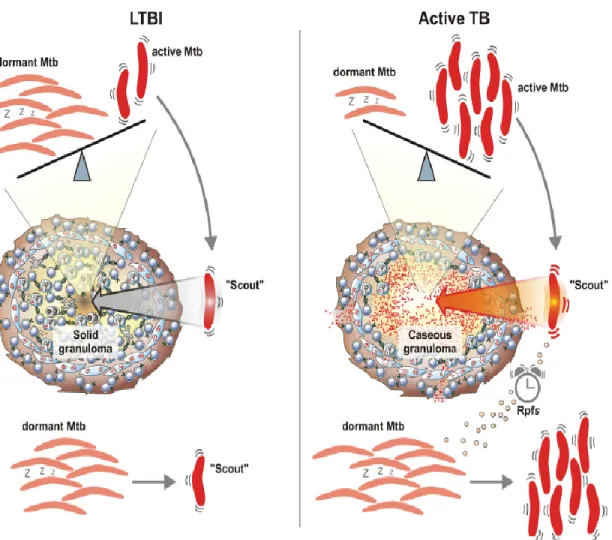
In the human host, M. tuberculosis is equipped for persistence in a dormant stage that cause latent tuberculosis without clinical disease and the term persister is used for M. tuberculosis organisms that are phenotypically resistant to drugs although they are genetically susceptible to these drugs. During latent infection, in addition to the dormant non replicating bacteria, some actively replicating bacteria are present, and the equilibrium balance between dormant/replicating M. tuberculosis determines the development an active or latent TB. In the latent infection, it has been suggested that most bacilli are in a dormant state and few have the ability, depending on the environmental conditions, to “awake”, start replicating. Most of these scouts, under normal condition, are readily killed by the host immune response (Fig 1.13) (Gengenbacher and Kaufmann, 2012). When, for a number of reasons, the host cannot effectively and rapidly kill these scouts, bacilli start replicating and active TB disease may ensue (Chao and Rubin, 2010).
Figure 1.13: Dynamic models for latent tuberculosis infection (Gengenbacher and Kaufmann, 2012).
Identification of target genes and characterization of their respective antigens involved in primary infection, dormancy and reactivation and more in general bacterial factors known to play a key role in this complex interplay with the host, may help us to better understand M. tuberculosis pathogenesis and identify new and more effective tools to control disease at multiple levels.
The aim of the study
The few PE_PGRS proteins that have been so far characterized were found to be involved in key events during TB pathogenesis. These proteins are differentially expressed and were found to be involved in different steps of M. tuberculosis infection and host-pathogen interaction (Brennan and Delogu, 2002), (Sampson, 2011). The aim of the present study is to investigate the role of other PE_PGRSs in M. tuberculosis pathogenesis and gain new insights on the role in the biology of the tubercle bacillus. In this work, PE_PGRS3 and PE_PGRS4 were studied for the first time and have been chosen because they are in the same genome region and close to the ESX3 secretion system gene locus, present a 71.8% similarity and 67% identity, and present unique features. In fact, the presence of a second GRPLI motif, which has been suggested to have a role in anchoring the PE_PGRS domain to the M. tuberculosis outer membrane, has been detected in both proteins (Delogu
G and Cole S.T and borsch R, 2008). Moreover, PE_PGRS3 have a unique C-terminal domain rich in arginine contains ( 80 aa in length contains 30aa
arginine), that may be involved in the interaction with the host extracellular matrix components. In light of these features, we investigated the gene expression profile of these protein and, using recombinant strains, shed light on the role of the different domains in the interaction with a mammal host.
Chapter II
1. Main features of PE_PGRS3 and PE_PGRS4
After a careful analysis of the whole protein family by specific bioinformatics tools, two PE_PGRS proteins were selected: PE_PGRS3 and PE_PGRS4. These proteins have 67% identity and 71.8% similarity, are found in the same gene locus though they seem to be transcriptionally regulated by two different promoters and are located immediately downstream of the ESX3 secretion system gene locus, which is important in M. tuberculosis pathogenesis (Serafini et al., 2009) (Tufariello et al., 2016) (Figure 2.1A and 2.1B). Each protein shows the presence of two GRPLI motifs, which have been suggested to represent the transmembrane domain that anchor the PE_PGRSs to the M. tuberculosis outer membrane (Delogu G and Cole S.T and borsch R, 2008). Moreover, the C-terminal domain of PE_PGRS3 is highly hydrophilic and contains numerous arginine amino acids that are typically found in proteins involved in the interaction with the host extracellular matrix components (Karsdal et al., 2013) (Figure 2.1C).
A
B
C
Figure 2.1: Shows in (A) Schematic representation of pe_pgrs3 and pe_pgrs4 localization on M.
tuberculosis genome. (B) PE_PGRS3 and PE_PGRS4 proteins and their structural characteristic domains. (c) PE_PGRS3 and PE_PGRS4 hydrophobicity analysis using ExPASy tools (ProtScale – Kyte & Doolittle).
Rv0278c
To gain insights on the genetic pressure exerted during MTBC evolution on the pe_pgrs3 gene, multiple sequence alignment was carried out on the pe_pgrs3 gene found in different MTBC strains belonging to different phylogeographic lineages. Sequences were obtained from publicly available databases for the following strains: M. tuberculosis, M. bovis, M. bovis BCG, M. africanum, M. canetti, M. tuberculosis of the EAI lineage. Nucleotides sequence alignments (Fig 2.2A) and amino acids sequence alignments (Fig 2.2B) indicate that not all M. tuberculosis strains have full length PE_PGRS3. Instead, a full length pe_pgrs3 single gene is observed only in M. tuberculosis (new and ancient strains) that cause disease in human but not in those that cause disease in animal (M. bovis), where a duplication events appears to have led to the presence of an extra copy of the pe_pgrs3 gene (putatively expressing a protein of 957 aa). A frameshift due to single base deletion splits this copy into two parts PE_PGRS3a similar to 5` end of Rv0278c and PE_PGRS3b equivalent to the 3` end of the Rv0278c. Hence, it appears that of the two copies of pe_pgrs3 found in M. bovis, none is able to express the full length protein containing the arginine-rich domain. Hence, fully functional pe_pgrs3, including expression of the arginine-rich C-terminal domain, exists only in MTBC strains that cause disease in human but not that cause disease in animal (Fig 2.2).
A B
Figure 2.2: Phylogenetic tree obtained from multiple sequence alignments of PE_PGRS3 between
different MTBC strains (M. tuberculosis, M. bovis, M. bovis BCG, M. africanum, M. canetti, M. tuberculosis of the EAI lineage) using multi-alignments tools (Clustel Omega and Jail view software).
Figure A represents nucleotides sequence alignments, whereas figure B shows amino acids sequence alignments.
2. PE_PGRS3 and PE_PGRS4 are differentially expressed
To start investigating the role of these proteins, we decided to generate a gene reporter system, where the putative promoter and coding sequences of the two selected genes were cloned in mycobacterial shuttle plasmids to be expressed in M. smegmatis. Cloning was started by amplifying pe_pgrs3 and pe_pgrs4 genes with their own promoters from M. tuberculosis H37Rv genome. The primers used are showed in table. 1. The PCR 2.1-T/A cloning vector was used for sub cloning of the two genes and then these genes were inserted in several expression vectors (pMV-based plasmids).
In pMV206, the pe_pgrs genes were fused at 3` with the gene encoding GFP, while in pMV306 the genes were fused to the sequence coding the HA epitope. M. smegmatis mc2155 were electroporated with the pMV306-based vectors and pMV-206 based vectors, alone or in combination, as indicated in figure 2.3, also to assess whether these neighboring genes are co-expressed.
A
B
Figure 2.3: (A) Schematic representation of PE_PGRS3 and PE_PGRS4 protein chimeras, in pMV206
vector (PE_PGRS3GFP, PE_PGRS4GFP) and pMV306 vector ( PE_PGRS3HA, PE_PGRS4HA). (B) M. smegmatis recombinant strains expressing PE_PGRS3, PE_PGRS4 protein chimeras ( M. smegmatis PE_PGRS3GFP, M. smegmatis PE_PGRS4HA, M. smegmatis PE_PGRS3GFP/4HA,
M. smegmatis PE_PGRS4GFP, M. smegmatis PE_PGRS3HA, M. smegmatis PE_PGRS4GFP/3HA).
The recombinant M. smegmatis strains were grown in 7H9/ADC/Tween liquid medium at 37cº and then analyzed at the fluorescence microscope. The M.
M. smegmatis expressing PE_PGRS3-GFP did not show any fluorescence (data not shown). Whole cell lysates from the four recombinant M. smegmatis strains expressing PE_PGRS3-GFP, PE_PGRS4-GFP, PE_PGRS3-GFP/PE_PGRS4-HA, PE_PGRS4-GFP/PE_PGRS3-HA chimeras were prepared and then analyzed by SDS-PAGE and immunoblot probed with anti-GFP and anti-HA specific antibodies. As shown in figure 2.4, we observed a signal corresponding to PE_PGRS4 with anti-GFP and anti-HA (~ 95 KDa for PE_PGRS4-GFP and ~ 79KDa for PE_PGRS4-HA), no signal was observed for PE_PGRS3 with both antibodies (anti-GFP and anti-HA), confirming proper expression of PE_PGRS4 and absence of expression of PE_PGRS3.
Figure 2.4: Shows SDS-PAGE and immunoblotting of the M. smegmatis expressing (PE_PGRS3GFP,
PE_PGRS4GFP, PE_PGRS3GFP/4HA, PE_PGRS4GFP/3HA) whole cell lysates by using anti GFP and anti HA as primary antibody. The blott represents PE_PGRS4-GFP in lane 2,4 and PE_PGRS4-HA, GFP in lane 3,5 respectively.
The lack of expression of the two PE_PGRS3 chimeras, prompted us to verify for a second time by Sanger sequencing the pMV-PE_PGRS3-GFP and-HA plasmids, which however confirmed the presence of a correct sequence. To investigate any
specific condition that could be required for the PE_PGRS3 protein expression, the above mentioned recombinant strains were grown under different stress conditions that are known to be relevant during M. tuberculosis pathogenesis such low pH (pH = 5), low iPhos (~51 µm), low oxygen, low Mg2+, low Fe3+, PBS (Gengenbacher and Kaufmann, 2012). Interestingly, while PE_PGRS4 was always found expressed in the standard 7H9 medium, in Sauton minimal and low iPhos Sauton media, PE_PGRS3 seems not to be expressed in all the conditions tested but when the M. smegmatis PE_PGRS3-GFP strain was grown under low iPhos condition (Fig 2.5). These results indicate that PE_PGRS3 appears to be expressed only under low iPhos and repressed under common growth conditions or other conditions that have been associated with survival of M. tuberculosis in host tissues.
Figure 2.5: Shows the different stress conditions such low pH (pH = 5), low iPhos (~51 µm), low
oxygen, low Mg2+, low Fe3+, PBS that were applied on the recombinant M. smegmatis strains expressing
PE_PGRS3-GFP, PE_PGRS-4GFP, PE_PGRS3-GFP/4HA, PE_PGRS4-GFP/3HA and GFP and were grown in 48 well plate at 37Cº in order to investigate a possible stress source can trigger the protein expression then the fluorescence of the mycobacteria which correspond to the protein expression was
3. PE_PGRS3 has a specific expression under low inorganic phosphate condition
To further investigate the observed PE_PGRS3-specific expression profile, as emerged under growth in low iPhos condition, confocal microscopy analysis was used as a more sensitive system. The recombinant M. smegmatis strains expressing PE_PGRS3-GFP, PE_PGRS4-GFP and, as controls, the recombinant M. smegmatis strain expressing another well-characterized protein of the family (PE_PGRS33-GFP) (Delogu et al., 2004) and M. smegmatis expressing cytosolic GFP, were grown until mid-log phase and then sub inoculations were made for all strains in low iPhos concentration and normal iPhos concentration (which for the sake of simplicity we here arbitrary define as high iPhos) in Sauton medium. After 15 days of incubation at 37C°, plated in chamber slides and then observed at confocal microscopy, the fluorescence microscopy images were analyzed by Image J software. The results obtained confirmed a strong fluorescence for M. smegmatis expressing PE_PGRS3 in low iPhos condition, while no fluorescence was observed when the same strain was grown in high iPhos condition. Conversely, no significant differences in fluorescence intensity were observed when the other recombinant strains were grown in low or high iPhos medium (Fig 2.6). Quantification of the fluorescence intensity as shown in figure 2.7 clearly indicates that PE_PGRS3-GFP is specifically expressed under low iPhos concentration.
A B A B
Figure 2.6: (A) Confocal microscopy images of M. smegmatis expressing PE_ PGRS3, PE_PGRS4
PE_PGRS33 (another well-characterized protein of the PE_PGRSs family) GFP tagged and M. smegmatis GFP grown in high and low iPhos Sauton medium, obtained with x60 objective. (B) Overlapping green channel and transmission microscopy images.
M. smegmatisGFP M. smegmatis PE_PGRS33GFP
M. smegmatis PE_PGRS3GFP M. smegmatis PE_PGRS4GFP
High Phosphate Low Phosphate High Phosphate Low Phosphate
Figure 2.7: Shows confocal microscopy images fluorescence intensity analysis for M. smegmatis
expressing PE_ PGRS3, PE_PGRS4, PE_PGRS33 (another well-characterized protein of the PE_PGRSs family) GFP tagged and M. smegmatis GFP grown in High and low iPhos Sauton medium. A strong fluorescence for M. smegmatis (Msm) expressing PE_ PGRS3-GFP was observed after 15 days of incubation in low iPhos Sauton medium and no fluorescence was observed in high iPhos Sauton medium *P<0.01. No significant difference in fluorescence intensity was observed for M. smegmatis expressing, PE_PGRS33-GFP, PE_PGRS4-GFP in both high and low iPhos Sauton medium. A significant difference was observed for M. smegmatis expressing GFP in low and high iPhos condition***P<0.001. The fluorescence microscopy images were analyzed using image J program.
3.1 Quantification of protein expression by measuring the fluorescence of single mycobacteria by FACS-Canto flow cytometer
To investigate the level of GFP expression at single cell level, we employed the flow cytometry (FACS-canto) for measuring the fluorescence of M. smegmatis expressing PE_PGRS3-GFP, PE_PGRS4-GFP, PE_PGRS33-GFP, GFP and M. smegmatis mc2155wt as a negative control. All strains were grown in a low iPhos Sauton medium until we a observed a maximum and plateau fluorescence at the day 15 for the M. smegmatis PE_PGRS3-GFP. The % of fluorescent cells increased from 0% to arrive 2.5% at the 15th day of incubation. To see if the phosphate is a specific regulator for the expression of M. smegmatis PE_PGRS3-GFP, bacterial cells were washed and suspended in a standard Sauton medium
(high iPhos) and the bacterial fluorescence measured until the 20th day of
incubation. Interestingly, we observed a significant decrease in the % of M. smegmatis PE_PGRS3-GFP showing fluorescence, while we did not observe
significant changes in the fluorescence expressed by the other recombinant strains (Fig 2.8).
Figure 2.8: Shows the FACS results, the fluorescence was measured for M. smegmatis (Msm) expressing
PE_PGRS3-GFP, PE_PGRS4-GFP, PE_PGRS33-GFP, GFP and M. smegmatis mc2155wt grown in low
iPhos Sauton medium. The fluorescence for Msm expressing PE_PGRS4-GFP grown in complete Sauton standard medium - high iPhos (PE_PGRS4+P) was also analyzed to confirm confocal microscopy results. Bacterial cells were washed at the 15th day and resuspended in Sauton standard medium and the
fluorescence was measured until the 20th day of incubation at 37 C°.
The results obtained confirmed that PE_PGRS3, but not the other PE_PGRS proteins analyzed, has a specific expression in low iPhos condition and the phosphate is a specific regulator for the expression of the PE_PGRS3, since
3.2 PE_PGRS3 expression increased in low inorganic phosphate condition and correlated with RelA in M. smegmatis and M. tuberculosis
The inorganic phosphate regulation is a critical element in M. tuberculosis pathogenesis and many studies focused on the importance of the role of iPhos depletion in M. tuberculosis survival and persistence in host tissues, suggesting that iPhos concentration may serve as a trigger for the expression of many genes involved in TB pathogenesis (Rifat et al., 2009). Because of RelA has previously been implicated in the M. tuberculosis transcriptional response to iPhos starvation and considered as a well characterized stringent response mediator which is required for mycobacterial persistence (Sureka et al., 2008), we decided to examine the transcription level of relA and pe_pgrs3 in low and high iPhos medium after 15 days of incubation at 37 C ͦ in the recombinant M. smegmatis strain expressing PE_PGRS3. By using Real time PCR we found that pe_pgrs3 expression was upregulated by 3-4 times in low iPhos conditions respect to the high iPhos concentration and correlated with the expression of relA, which however showed a much more remarkable upregulation (up to 90 times) under low iPhos conditions (Fig 2. 9).