An algorithm based on OmniView technology to reconstruct
sagittal and coronal planes of the fetal brain from volume
datasets acquired by three-dimensional ultrasound
G. RIZZO*†, A. CAPPONI*‡, M. E. PIETROLUCCI†, A. CAPECE†, E. AIELLO†,
S. MAMMARELLA† and D. ARDUINI†
*Fetal Medicine Center Genoma, Rome, Italy; †Department of Obstetrics and Gynecology, Universit `a di Roma ‘Tor Vergata’, Rome, Italy; ‡Department of Obstetrics and Gynecology, Ospedale G.B. Grassi, Rome, Italy
K E Y W O R D S: 3D ultrasound; central nervous system defects; fetal brain; prenatal diagnosis
A B S T R A C T
Objective To describe a novel algorithm, based on
the new display technology ‘OmniView’, developed to visualize diagnostic sagittal and coronal planes of the fetal brain from volumes obtained by three-dimensional (3D) ultrasonography.
Methods We developed an algorithm to image standard
neurosonographic planes by drawing dissecting lines through the axial transventricular view of 3D volume datasets acquired transabdominally. The algorithm was tested on 106 normal fetuses at 18–24 weeks of gestation and the visualization rates of brain diagnostic planes were evaluated by two independent reviewers. The algorithm was also applied to nine cases with proven brain defects.
Results The two reviewers, using the algorithm on normal
fetuses, found satisfactory images with visualization rates ranging between 71.7% and 96.2% for sagittal planes and between 76.4% and 90.6% for coronal planes. The agreement rate between the two reviewers, as expressed by Cohen’s kappa coefficient, was > 0.93 for sagittal planes and > 0.89 for coronal planes. All nine abnormal volumes were identified by a single observer from among a series including normal brains, and eight of these nine cases were diagnosed correctly.
Conclusions This novel algorithm can be used to visualize
standard sagittal and coronal planes in the fetal brain. This approach may simplify the examination of the fetal brain and reduce dependency of success on operator skill. Copyright 2011 ISUOG. Published by John Wiley & Sons, Ltd.
I N T R O D U C T I O N
Central nervous system (CNS) malformations are com-mon in the human fetus, affecting approximately 0.3–1% of live births1,2. Prenatal detection of CNS malformations is important since these anomalies frequently have a severe prognosis and are often associated with chromosomal anomalies and genetic syndromes2.
Despite the high incidence of CNS anomalies and the clinical importance of their prenatal diagnosis, the efficacy of screening is still far from producing satisfactory results, especially when study of the fetal head is limited to axial planes of the brain3. An extended study of fetal CNS anatomy, including sagittal and coronal planes of the brain, may improve the diagnostic efficacy4,5. However, operator experience has a significant impact on the quality and interpretation of fetal CNS imaging as well as on prenatal detection of CNS defects6, thus limiting the routine application of these diagnostic planes.
Three-dimensional (3D) ultrasound has the potential to reduce the dependency of success on operator skill7,8. This technique allows acquisition of volumes containing most anatomic information, which can be reviewed offline sub-sequently by recreating novel or standard planes, allowing a comprehensive study of the fetal brain9 – 14. However, although several algorithms for the 3D reconstruction of the fetal brain have been described9 – 13, these approaches require the operator’s manual ‘navigation’ of the volume acquired, thus necessitating specific experience and skill in 3D orientation and subsequent retrieval of the diagnostic planes.
OmniView (GE Medical Systems, Kretztechnik GmbH, Zipf, Austria) is a new display technology for 3D ultrasound that allows interrogation of volume datasets
Correspondence to: Dr. G. Rizzo, Department of Obstetrics and Gynecology, Universit `a di Roma Tor Vergata, Ospedale ‘‘Fatebenefratelli S. Giovanni Calabita’’, Isola Tiberina 89, 00186 Roma, Italy (e-mail: [email protected])
OmniView and the fetal brain 159
and simultaneous display of up to three independent (non-orthogonal) planes by manually drawing lines from any direction or angle. This has the potential to simplify 3D volume examination, thus further reducing depen-dency on the operator’s skills. Indeed, this technique recently proved successful in the analysis of fetal cardiac volumes15.
The objective of our study was to test whether this approach could be extended to the visualization of diagnostic cerebral planes in normal second-trimester fetuses and in those with CNS defects.
M E T H O D S
We included in this study 110 consecutive singleton low-risk pregnant women undergoing routine second-trimester examination. Furthermore, we selected from our database of fetal brain volumes nine cases with CNS defects (three ventriculomegaly, three agenesis of corpus callosum and three abnormalities of the posterior fossa). All the CNS defects had been confirmed either at autopsy or by prenatal and/or postnatal magnetic resonance imaging. This research project was approved by our institutional review board and all women provided written informed consent to participate.
Brain volumes were acquired with a 4–8-MHz transabdominal probe (Voluson 8, GE Healthcare, Milwaukee, WI, USA) using a previously reported technique16, starting with a transverse view of the fetal head at the level of the transcerebellar axial plane. The angle of insonation between the incident ultrasound beam and the cerebral midline was kept at approximately 45◦ in order to minimize the acoustic shadow of the skull base on the brain structures of reconstructed planes. The sweep acquisition angle was set between 45◦ and 60◦according to gestational age in order to include the entire fetal brain within the volume. Volumes were acquired during fetal rest and maternal apnea, in ‘maximum’ quality mode. We used a standardized setting of two-dimensional (2D) imaging with: harmonics, on (low level); cross beam, 1; speckle reduction, 3; and dynamic contrast, 7.
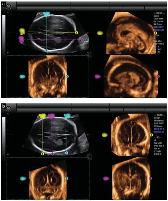
We developed a standardized algorithm to retrieve sagittal and coronal diagnostic planes from normal brain volume datasets. This was achieved by retrospective offline review of datasets using 4D View (Version 10.0, GE Medical Systems) and applying the OmniView tech-nology. The algorithm consisted of the following steps: 1) Volume datasets were adjusted in order to obtain
in Panel A an axial view of the fetal brain at the level of the lateral ventricles (transventricular plane). The reference point was placed in the middle of the cavum septi pellucidi. Volume contrast imaging (VCI) was activated, with a thickness of 2 mm, in order to improve contrast resolution (Figure 1a, Panel A). 2) A first (yellow) line was drawn horizontally on the
midline across the cavum septum pellucidi, allowing visualization of the midsagittal plane (Figure 1a, Panel 1).
3) A second (fuchsia) line was drawn parallel to the first line at the level of the lateral ventricle, to obtain a parasagittal plane (Figure 1a, Panel 2). This line could be moved from the distal to the proximal ventricle to obtain both parasagittal planes.
4) A third (turquoise) line was drawn perpendicular to the first line and passing through the cavum septi pellucidi, obtaining the coronal transcaudate plane (Figure 1a, Panel 3).
5) To obtain the remaining diagnostic coronal planes the generated OmniView lines were removed and three new vertical lines were drawn, one anterior to the cavum septi pellucidi, one immediately posterior to it and one at the level of the lateral ventricles, obtaining transfrontal (Figure 1b, Panel 1), transthalamic (Figure 1b, Panel 2) and transcerebellar (Figure 1b, Panel 3) planes. Videoclips S1 and S2 show examples of the application of this algorithm.
Independent analysis of the volumes was later performed by two reviewers with experience in CNS post-processing (G.R. and A.C.). They then classified independently the diagnostic planes that they had obtained as being either satisfactory or unsatisfactory for obtaining diagnostic-quality images according to the criteria reported in Table 1.
The nine cases with CNS anomalies were anonymized and included randomly alongside a series of 11 brain volume datasets from normal fetuses. One of the reviewers (A.C.), blind to whether each case was normal or abnormal and to the types of CNS defect that had been included in the abnormal group, analyzed with the help of our algorithm this series of 20 cases. He was asked to determine the normal from the abnormal volumes and to provide a possible diagnosis for the anomalies.
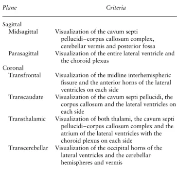
Table 1 Criteria followed by the reviewers to classify fetal brain sagittal and coronal planes as satisfactory or not satisfactory
Plane Criteria
Sagittal
Midsagittal Visualization of the cavum septi pellucidi–corpus callosum complex, cerebellar vermis and posterior fossa Parasagittal Visualization of the entire lateral ventricle and
the choroid plexus Coronal
Transfrontal Visualization of the midline interhemispheric fissure and the anterior horns of the lateral ventricles on each side
Transcaudate Visualization of the cavum septi pellucidi, the corpus callosum and the lateral ventricles on each side
Transthalamic Visualization of both thalami, the cavum septi pellucidi–corpus callosum complex and the atrium of the lateral ventricles with the choroid plexus on each side
Transcerebellar Visualization of the occipital horns of the lateral ventricles and the cerebellar hemispheres and vermis
Figure 1 Example, using a normal fetal brain, of application of our new algorithm to reconstruct sagittal and coronal planes of the fetal brain from volume datasets acquired by three-dimensional ultrasound. First, the transventricular axial view of the fetal brain was obtained (Panel A). (a) The OmniView function was activated and three lines were drawn, allowing display of midsagittal (yellow line), parasagittal (fuchsia line) and coronal transcaudate (turquoise line) views in Panels 1, 2 and 3, respectively. (b) These three lines were then removed and redrawn, allowing display of coronal transfrontal (yellow line), coronal transthalamic (fuchsia line) and coronal transcerebellar (turquoise line) views in Panels 1–3, respectively.
The agreement rate between the two reviewers was evaluated using Cohen’s kappa coefficient. The time required for the analysis was recorded and differences between reviewers were analyzed by Mann–Whitney U-test. P < 0.05 was considered statistically significant.
R E S U L T S
The characteristics of the population studied are reported in Table 2. In four cases brain volumes were not recorded, due either to extensive fetal movements or to unfavorable fetal position. Table 3 reports the frequencies with which each type of CNS view was judged satisfactory according to the two reviewers. Overall, the median time to analyze each volume dataset was 72.5 (range, 40–125) s and there
Table 2 General characteristics of the 106 normal pregnancies evaluated using the new algorithm
Characteristic Median (range)
Maternal age (years) 31 (18–45)
Parity 1 (0–4)
Gestational age at ultrasound (weeks) 21.3 (18.1–23.8) Body mass index (kg/m2) 23.6 (18.2–41.8)
was no statistically significant difference between the two reviewers (median, 67 s vs. 74 s, P= 0.318).
Regarding the series of 20 containing normal and abnormal datasets, the reviewer identified correctly all nine of the pathological CNS volumes and made a
OmniView and the fetal brain 161
Table 3 Frequency of satisfactory views obtained of the fetal brain, stratified by reviewer and type of plane, in 106 normal pregnancies evaluated using a new algorithm based on OmniView technology to reconstruct sagittal and coronal planes of the fetal brain
Frequency of satisfactory views (%) Plane Reviewer 1 (G.R.) Reviewer 2 (A.C.) Kappa (95% CI) Sagittal Midsagittal 93.4 96.2 0.95 (0.91–0.99) Proximal parasagittal 71.7 74.5 0.93 (0.88–0.98) Distal parasagittal 92.5 90.6 0.94 (0.87–0.99) Coronal Transfrontal 88.7 90.6 0.93 (0.89–0.96) Transcaudate 84.0 82.1 0.89 (0.82–0.98) Transthalamic 76.4 78.3 0.92 (0.87–0.96) Transcerebellar 87.7 85.8 0.93 (0.88–0.99) Agreement between reviewers evaluated by Cohen’s K coefficients (kappa).
correct diagnosis in eight (88.8%) of these cases. None of the normal volumes was identified incorrectly as being abnormal.
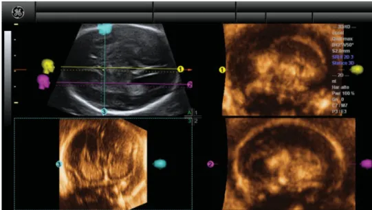
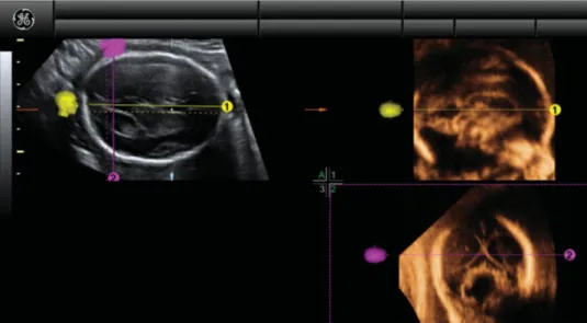
Examples of fetuses with complete agenesis of the cor-pus callosum, borderline ventriculomegaly and posterior fossa anomaly (classical Dandy–Walker malformation) are given in Figures 2–4.
D I S C U S S I O N
In this study we developed a simple algorithm to visualize sagittal and coronal planes of the fetal brain by applying OmniView technology to reslice fetal brain volume datasets obtained with 3D ultrasound during the second trimester. Using the algorithm, we showed that these sonographic planes can be obtained easily in most cases.
Current guidelines for the screening of CNS anomalies suggest limiting the investigation to the axial brain planes on which the falx, thalami, cavum septi pellucidi, lateral ventricles with choroid plexus, cerebellum and cisterna magna should be evident4,17,18. These axial planes, however, do not allow evaluation of the corpus callosum, cerebellar vermis or other midline brain structures. As a consequence, anomalies concerning these structures are frequently missed in screening programs. Their identification is, however, of great clinical interest since these defects are severe and are frequently associated with other structural malformations and chromosomal or genetic diseases2,3.
Therefore, the addition of sagittal and coronal planes to the fetal brain evaluation has been suggested as an integral part of the study of the fetal CNS4,6. However, visualization of these additional planes requires either a transvaginal approach, when this is possible according to fetal position, or a transabdominal approach, using a transfrontal view through the metopic suture. However, not only can both approaches be time consuming, but the success rate of obtaining the correct views is greatly dependent on both the operator’s expertise and the fetal position. As a consequence, they are generally only used for referral cases in specialized centers, where a dedicated neurosonography examination is performed.
The advantages of the 3D over the 2D approach in visualizing coronal and sagittal planes must be emphasized. 3D ultrasonography offers the potential of acquiring a volume of the fetal head and then, by subsequently reconstructing the image through the multiplanar technique, allows visualization of the appropriate sagittal and coronal views, thus reducing the difficulties which are encountered in the 2D approach. The main limitation of this approach is the complex anatomy of the fetal brain, which requires the operator to be skilled in 3D orientation and subsequent retrieval of
Figure 2 Example of application of our new algorithm in a fetus with complete agenesis of the corpus callosum. In Panel A, the
transventricular axial view of the fetal brain is shown. The OmniView function was activated and three lines were drawn, allowing display of midsagittal (yellow line), parasagittal (fuchsia line) and coronal transcaudate (turquoise line) views in Panels 1–3, respectively.
Figure 3 Example of application of our new algorithm in a fetus with classical Dandy–Walker malformation. In Panel A, the
transventricular axial view of the fetal brain is shown. The OmniView function was activated and two lines were drawn, allowing display of sagittal (yellow line) and coronal transcerebellar (fuchsia line) views in Panels 1 and 2, respectively.
Figure 4 Example of application of our new algorithm in a fetus with bilateral borderline ventriculomegaly. In Panel A, the transventricular axial view of the fetal brain is shown. The OmniView function was activated and three lines were drawn, allowing display of distal parasagittal (yellow line), coronal transcerebellar (fuchsia line) and proximal parasagittal (turquoise line) views in Panels 1–3, respectively. the diagnostic planes from acquired volumes5,10,11,14,19.
The use of sequential slices of fetal brain obtained using tomographic ultrasonographic imaging (TUI) has been suggested to facilitate this approach20,21. Although TUI allows automatic slicing of the volume datasets, displaying multiple parallel images, the lines producing these slices are rigid and equidistant from each other and cannot be drawn manually. Despite the possibility of TUI being combined with VCI to improve image quality and the images obtained being manually rotated, there is no possibility of obtaining a simultaneous display of orthogonal sectional planes, thus limiting its clinical application.
OmniView technology instead allows reslicing of the structure that has been acquired, along lines that can be
generated manually by the operator from any direction or angle, thus providing a more flexible and effective approach in the study of volume datasets. Indeed, our study illustrates how it is possible to achieve all of the diagnostic sagittal and coronal planes simply by drawing lines on the axial transventricular view of the fetal head, a view obtained routinely in screening ultrasound examinations.
We observed the lowest rate of visualization for the proximal parasagittal plane, a limitation that occurs with 2D ultrasound also and is caused by difficulties in visualizing clearly the proximal atria due to the ossification of cranial bones and their shadowing effect, hampering complete visualization of the proximal brain anatomy. These findings suggest that 3D ultrasound
OmniView and the fetal brain 163
is susceptible to the same intrinsic limitations in scanning windows as those that hamper conventional 2D ultrasound imaging. Nonetheless, our algorithm allowed proper visualization of the proximal parasagittal plane in at least 70% of cases. Acquisition of multiple volumes with different angles of insonation may further reduce the frequency of suboptimal visualization.
Of utmost importance is the rate of agreement achieved between the reviewers in processing the volume datasets. This finding suggests a clinically acceptable repeatability of fetal brain examination using the 3D technique with this algorithm. Furthermore, the short time (median, 70 s) necessary to retrieve the diagnostic planes suggests the ease with which this technique could be applied clinically.
In the abnormal cases, the algorithm allowed the CNS defects to be distinguished and the correct diagnosis to be reached in most of the cases studied. However, although our results are encouraging, some caution is necessary in their interpretation before applying these findings to clinical practice. We tested the algorithm on a limited number of CNS anomalies and further studies on larger numbers of brain defects are necessary to evaluate its diagnostic efficiency. However, its ability to display views deviating from those expected in normal brains is promising and suggests a potential role for the identification of fetal brains suspicious for congenital anomalies.
Recent data suggest that abnormal CNS volumes can be acquired remotely and interpreted accurately by referral centers22. Nonetheless, caution is still necessary in the analysis of reconstructed 3D images of the fetal brain, since imaging artifacts cannot always be excluded23.
The algorithm proposed here may provide a simple means with which to display planes that are difficult to visualize transabdominally, but the transvaginal or transabdominal transfontanellar route will still be necessary for confirmation when anomalies are suspected and/or poor image quality is obtained.
At this time, we recommend that the diagnosis of CNS anomalies should not be made solely on the basis of 3D volume reconstructions. When a CNS malformation is suspected based on a 3D ultrasound volume, it is still prudent and highly recommended for an expert in ultrasonography to perform an accurate evaluation of the fetus. In such situations it is necessary to obtain high-resolution images sufficient to allow detection of subtle but relevant anomalies of the fetal brain; at the moment this can only be achieved using a 2D transvaginal or transabdominal transfontanellar approach.
A further limitation of this study is that the CNS volume datasets were acquired by operators with expertise in fetal CNS imaging, whose skills might be greater than those of the average sonographer. As a consequence, the success rate of post-processing examination of CNS volumes may be lower in datasets acquired in peripheral centers during routine second-trimester ultrasonographic examinations. However, recent studies of volumes obtained from
fetal brains by operators performing routine second-trimester ultrasound screening found that more than 90% of datasets were of sufficiently high quality as to allow satisfactory diagnostic neurosonographic views24.
In conclusion, we have developed a novel algorithm, based on OmniView technology, that can be used to visualize standard sagittal and coronal planes in the fetal brain. This approach may simplify the examination of the fetal brain and, by reducing operator dependency, may help in the incorporation of these diagnostic planes into routine second-trimester examinations.
R E F E R E N C E S
1. von Wendt L, Rantakallio P. Congenital malformations of the central nervous system in a 1-year birth cohort followed to the age of 14 years. Childs Nerv Syst 1986; 2: 80–82.
2. Chitty LS, Pilu G. The challenge of imaging the fetal central nervous system: an aid to prenatal diagnosis, management and prognosis. Prenat Diagn 2009; 29: 301–302.
3. Garne E, Dolk H, Loane M, EUROCAT. EUROCAT website data on prenatal detection rates of congenital anomalies. J Med Screen 2010; 17: 97–98.
4. International Society of Ultrasound in Obstetrics & Gynecology Education Committee. Sonographic examination of the fetal central nervous system: guidelines for performing the basic examination and the fetal neurosonogram. Ultrasound Obstet Gynecol 2007; 29: 109–116.
5. Timor-Tritsch IE, Monteagudo A. Transvaginal fetal neu-rosonography: standardization of the planes and sections by anatomic landmarks. Ultrasound Obstet Gynecol 1996; 8: 42–47.
6. Monteagudo A. Fetal neurosonography: should it be routine? Should it be detailed? Ultrasound Obstet Gynecol 1998; 12: 1–5.
7. Benacerraf BR, Shipp TD, Bromley B. Three-dimensional US of the fetus: volume imaging. Radiology 2006; 238: 988–996. 8. Abuhamad AZ. Standardization of 3-dimensional volumes
in obstetric sonography: a required step for training and automation. J Ultrasound Med 2005; 24: 397–401.
9. Monteagudo A, Timor-Tritsch IE, Mayberry P. Three-dimensional transvaginal neurosonography of the fetal brain: navigating in the volume scan. Ultrasound Obstet Gynecol 2000; 16: 307–313.
10. Correa FF, Lara C, Bellver J, Remohli J, Pellicer A, Serra V. Examination of the fetal brain by transabdominal three-dimensional ultrasound: potential for routine neurosonographic studies. Ultrasound Obstet Gynecol 2006; 27: 503–508. 11. Kalache KD, Eder K, Esser T, Proquitt´e H,
Stoltenburg-Didinger G, Hartung JP, Bamberg C. Three-dimensional ultra-sonographic reslicing of the fetal brain to assist prenatal diagnosis of central nervous system anomalies. J Ultrasound Med 2006; 25: 509–514.
12. Pilu G, Segata M, Ghi T, Carletti A, Perolo A, Santini D, Bonasoni P, Tani G, Rizzo N. Diagnosis of midline anomalies of the fetal brain with the three-dimensional median view. Ultrasound Obstet Gynecol 2006; 27: 522–529.
13. Pooh RK, Pooh KH. Fetal neuroimaging. Fetal Matern Med Rev 2008; 19: 1–31.
14. Vi ˜nals F, Mu ˜noz M, Naveas R, Giuliano A. Transfrontal three-dimensional visualization of midline cerebral structures. Ultrasound Obstet Gynecol 2007; 30: 162–168.
15. Yeo L, Romero R, Jodicke C, Ogg`e G, Lee W, Kusanovic JP, Vaisbuch E, Hassan S. Four-chamber view and ‘swing tech-nique’ (FAST) echo: a novel and simple algorithm to visualize standard fetal echocardiographic planes. Ultrasound Obstet Gynecol 2011; 37: 423–431.
16. Rizzo G, Pietrolucci ME, Capponi A, Arduini D. Assessment of corpus callosum biometry at 18–32 weeks of gestation by three dimensional ultrasound. J Ultrasound Med 2011; 30: 47–53. 17. American Institute of Ultrasound in Medicine. AIUM
prac-tice guidelines for the performance of an antepartum obstetric ultrasound examination. J Ultrasound Med 2010; 29: 157–166.
18. Salomon LJ, Alfirevic Z, Berghella V, Bilardo C, Hernandez-Handrade E, Johnsen SL, Kalache K, Leung KY, Malinger G, Munoz H, Prefumo F, Toi A, Lee W. Practice guidelines for performance of the routine mid-trimester fetal ultrasound scan. Ultrasound Obstet Gynecol 2011; 37: 116–126.
19. Bornstein E, Monteagudo A, Santos R, Strock I, Tsymbal T, Lenchner E, Timor-Tritsch IE. Basic as well as detailed neurosonograms can be performed by offline analysis of three-dimensional fetal brain volumes. Ultrasound Obstet Gynecol 2010; 36: 20–25.
20. Pilu G, Ghi A, Segata M, Perolo A, Rizzo N. Three-dimensional ultrasound examination of the fetal central nervous system. Ultrasound Obstet Gynecol 2007; 30: 233–345.
21. Zalel Y, Yagel S, Achiron R, Kivilevich Z, Gindes L. Three-dimensional ultrasonography of the fetal vermis at 18 to 26 weeks’ gestation: time of appearance of the primary fissure. J Ultrasound Med 2009; 28: 1–8.
22. Rizzo G, Abuhamad A, Benaceraff B, Chaoui R, Corral E, D’Addario V, Espinoza J, Lee W, Merce L, Pooh R, Sepul-veda W, Sinkovskaya E, Vi ˜nals F, Volpe P, Pietrolucci ME, Arduini D. Collaborative study on three-dimensional ultra-sonography for the prenatal diagnosis of central nervous system defects. J Ultrasound Med 2011; (in press).
23. Malinger G, Lerman-Sagie T, Vi ˜nals F. Three-dimensional sagittal reconstruction of the corpus callosum: fact or artifact. Ultrasound Obstet Gynecol 2006; 28: 742–743.
24. Rizzo G, Pietrolucci ME, Capece G, Cimmino E, Colosi E, Ferrentino S, Sica C, Di Meglio A, Arduini D. Satisfactory rate of postprocessing visualization of fetal cerebral axial, sagittal, and coronal planes from three-dimensional volumes acquired in routine second trimester ultrasound practice by sonographers of peripheral centers J Matern Fetal Neonatal Med 2011; Jan 13. [Epub ahead of print].
S U P P O R T I N G I N F O R M A T I O N O N T H E IN T E R N E T
The following supporting information may be found in the online version of this article:
Videoclip S1 Example of application of the OmniView algorithm to a normal fetus (same as that in
Figure 1). Having obtained the transventricular axial view of the fetal brain, the OmniView function was activated and three lines were drawn, allowing display of midsagittal (yellow line), parasagittal (fuchsia line) and coronal transcaudate (turquoise line) views in Panels 1, 2 and 3, respectively.
Videoclip S2 The three lines shown in Videoclip S1 were then removed and redrawn, allowing display
of coronal transfrontal (yellow line), coronal transthalamic (fuchsia line) and coronal transcerebellar (turquoise line) views in Panels 1–3, respectively.