DOTTORATO DI RICERCA IN
Scienze Ambientali: tutela e gestione delle risorse naturali
Ciclo XXIII
Settore scientifico-disciplinare di afferenza: BIO 09/ FISIOLOGIA
“Pharmaceutical residues in aquatic systems: mode
of action and effects on mussel physiology”
Presentata da:
Sara Buratti
Coordinatore Dottorato
Relatore
Prof. Enrico Dinelli
Prof.ssa Elena Fabbri
Dedicated to my parents,
Emilia and Sauro
Summary
Abstract... 5
1. Introduction... 7
1.1. Pharmaceuticals as “emerging contaminants”: an issue of concern from different sources. ... 9
1.2. Pharmaceuticals and legislation: what does regulation say? ... 19
1.3. Pharmaceuticals tested in this study ... 25
1.3.1. Carbamazepine ...25
1.3.2. Propranolol ...30
1.3.3. Oxytetracycline ...32
1.3.4. Copper ...34
1.4. Experimental setup: laboratory mussels exposure ... 36
1.5. Why the choice of mussels as sentinel organisms?... 37
1.6. Selected battery of biomarkers ... 39
1.6.1. The neutral red retention assay...39
1.6.2. Cytochemical assays: accumulation of lipofuscins (LF) and neutral lipids (NL)...41
1.6.3. Antioxidant enzymes activities...43
1.7. Cyclic AMP-related cascade reactions... 45
1.8. Expression of HSP70 proteins... 47
1.9. The MEECE Programme... 48
2. Aim of the work... 51
3. Materials and methods ... 55
3.1. Tested substances ... 57
3.1.1. Carbamazepine ...57
3.1.2. Propranolol ...57
3.1.3. Oxytetracycline ...58
3.1.4. Copper ...58
3.2. Experimental animals and holding conditions ... 58
3.3. Experimental setup and mussel exposure ... 59
3.3.1. Exposure to carbamazepine...59 3.3.2. Exposure to propranolol ...59 3.3.2.1. L-PROP ...59 3.3.2.2. DL-PROP...59 3.3.3. Exposure to oxytetracycline ...60 3.3.3.1. Preliminary test...60 3.3.3.2. Final test ...60 3.3.4. Exposure to copper...61 3.4. Tissues preparation... 61 3.5. Battery of biomarkers... 62 3.5.1. Stress on stress...62
3.5.2. Neutral red retention assay ...62
3.5.3. Cytochemical assays: accumulation of lipofuscins (LF) and neutral lipids (NL)...63
3.5.4. Antioxidant enzymes activity ...64
3.5.5. Malondialdehyde (MDA) assay...65
3.7. Cyclic AMP-related cascade reactions ... 66
3.7.1. Cyclic AMP content... 66
3.7.2. PKA activity... 67
3.8. Statistical analysis ... 67
4. Results ... 69
4.1. CARBAMAZEPINE ... 71
4.1.1. Lysosomal membrane stability (NRR assay) ... 71
4.1.2. Cytochemical assays ... 71
4.1.2.1. Accumulation of lipofuscins ... 71
4.1.2.2. Accumulation of neutral lipids ... 72
4.1.3. Antioxidant enzymes activity... 72
4.1.3.1. Catalase ... 72
4.1.3.2. Glutatione S-transferase ... 73
4.1.4. MDA assay... 74
4.1.5. Cyclic AMP-related cascade reactions... 74
4.1.5.1. cAMP content... 74
4.1.5.2. PKA activity... 75
4.1.6. Sensitivity of biomarkers and cAMP-related parameters to CBZ exposure... 75
4.2. L-PROPRANOLOL and DL-PROPRANOLOL ... 77
4.2.1. Lysosomal membrane stability ... 77
4.2.2. Cytochemical assays ... 78
4.2.2.1. Accumulation of lipofuscins ... 78
4.2.2.2. Accumulation of neutral lipids ... 78
4.2.3. Antioxidant enzymes activity... 79
4.2.3.1. Catalase ... 79
4.2.3.2. Glutatione S-transferase ... 80
4.2.4. Cyclic AMP-related cascade reactions... 81
4.2.4.1. cAMP content... 81
4.2.4.2. PKA activity... 82
4.2.4. Sensitivity of biomarkers and cAMP-related parameters to PROP exposure ... 83
4.3. OXYTETRACYCLINE... 84
4.3.1. Preliminary experiment... 84
4.3.1.1 Lysosomal membrane stability (NRR assay) ... 84
4.3.1.2. Stress on stress ... 85
4.3.1.3. Cytochemical assay: accumulation of lipofuscins... 86
4.3.2. Final experiment ... 87 4.3.2.1. HSP70 determination ... 87 4.3.2.1.1. Exposure to OTC at 16°C... 88 4.3.2.1.2. Exposure to OTC at 20°C... 89 4.3.2.1.3. Exposure to OTC at 24°C... 90 4.3.3. Statistical analysis ... 91 4.4. COPPER ... 92 4.4.1. HSP70 determination ... 92 4.4.1.1. Exposure to Cu at 16°C... 92 4.4.1.2. Exposure to Cu at 20°C... 93 4.4.1.3. Exposure to Cu at 24°C... 94 4.4.2. Statistical analysis ... 95
5. Discussion... 97
5.1. CARBAMAZEPINE ... 99 5.2. PROPRANOLOL... 103 5.3. OXYTETRACYCLINE... 109 5.4. COPPER ... 1136. Conclusions... 117
7. Activity of research at Cádiz University... 121
7.1. Introduction... 123
7.2. Materials and methods ... 127
7.2.1. Study site ...127
7.2.2. Sediment characterization ...128
7.2.3. In situ study: selection of organisms and animals handling ...129
7.2.4. Lysosomal membrane stability (LMS): the neutral red retention (NRR) assay...130
7.2.5. Statistical analysis ...132
7.3. Results ... 132
7.3.1. Chemical characterization of sediments ...132
7.3.2. Ecotoxicological responses ...135
7.4. Discussion and conclusion ... 139
7.5. References... 143
8. References... 149
Abstract
Pharmaceutical residues contaminate aquatic ecosystems as a result of their widespread human and veterinary usage. Since continuously released and not efficiently removed, certain pharmaceuticals exhibit pseudo-persistence thus generating concerns for the health of aquatic wildlife.
This work aimed at assessing on mussels Mytilus galloprovincialis, under laboratory conditions, the effects of three pharmaceuticals, carbamazepine (antiepileptic), propranolol (!-blocker) and oxytetracycline (antibiotic), to evaluate if the human-based mode of action of these molecules is conserved in invertebrates. Furthermore, in the framework of the European MEECE Programme, mussels were exposed to oxytetracycline and copper at increasing temperatures, simulating variations due to climate changes.
The effects of these compounds were assessed evaluating a battery of biomarkers, the expression of HSP70 proteins and changes in cAMP-related parameters.
A decrease in lysosomal membrane stability, induction of oxidative stress, alterations of cAMP-dependent pathway and the induction of defense mechanisms were observed indicating the development of a stress syndrome, and a worsening in mussels health status. Data obtained in MEECE Programme confirmed that the toxicity of substances can be enhanced following changes in temperature. The alterations observed were obtained after exposure to pharmaceuticals at concentrations sometimes lower than those detected in the aquatic environment. Hence, further research is advisable regarding subtle effects of pharmaceuticals on non-target organisms.
Furthermore, results obtained during a research stay in the laboratories of Cádiz
University (Spain) are presented. The project aimed at measuring possible effects
of polluted sediments in Algeciras Bay (Spain) and in Cádiz Bay, by assessing
different physiological parameters in caged crabs Carcinus maenas and clams
Ruditapes decussatus exposed in situ for 28 days. The neutral red retention assay
was adapted to these species and proved to be a sensitive screening tool for the assessment of sediment quality.
1.1. Pharmaceuticals as “emerging contaminants”: an issue of
concern from different sources.
The presence of pharmaceuticals and personal care products (PPCPs) in the environment has become a recent research topic. The presence of these compounds in the environment is known since decades, but it was only in the mid 90s with advances in analytical techniques that important knowledge on environmental contamination by those compounds grew. Chromatographic-detection techniques improved powerfully, enabling detection limits within the ng/L to µg/L range and allowed researchers to quantify a large number of medicines components (i.e. drugs and excipients) in the environment, thus making the scientific community to reflect on this contamination type as a potential issue meriting concern and further investigation (Santos et al., 2010). The attention to PPCPs originally focused on their occurrence and monitoring, primarily in surface/ground waters and untreated/treated sewage. This research was driven principally by environmental analytical chemists, as new instrument technologies expanded the types of molecules that could be easily identified, as instrument sensitivity increased, and as detection limits of analytical methods were lowered. Approximately during the late 1990s, the interest began to expand to waste treatment and fate/transport. In the last decade, more attention was turned to ecotoxicology, pollution prevention, and environmental stewardship. Likewise, the scope of environmental compartments under investigation has expanded from primarily waters to now include sediments (and suspended particulates), sewage sludge (and biosolids), air (e.g., PPCPs sorbed to suspended particulates), and biota.
Confirming this growing interest, the annual rate of published articles directly relevant to PPCPs has grown exponentially since the middle of 1990s (Cleuvers, 2003; Daughton, 2007) (Fig. 1.1).
Fig. 1.1. Percentage of published studies on different therapeutic classes, expressed in relative percentage, described on 183 articles published between 1996 and 2009. From Santos et al., 2010.
Pharmaceuticals are a class of emerging environmental contaminants that are extensively and increasingly being used in human and veterinary medicine. These chemicals are designed to have a specific mode of action, and many of them for some persistence in the body. These features among others make pharmaceuticals to be evaluated for potential effects on aquatic flora and fauna (Fent et al., 2006). Some of the chemicals released into the environment may have endocrine-disrupting effects in living organisms, including humans (Nikolaou et al., 2007). By itself, the word “pharmaceutical” refers to a chemical prepared or dispensed in pharmacies and which treats or prevents or alleviates the symptoms of disease or physiologic function. But in the environment not only these compounds are found. PPCPs include all chemicals used for humans, domestic animals, or agricultural crops that treat disease, alter or improve physiological, cosmetic, or emotional function, appearance, or status, prevent illness (prophylaxis) or maintain health, help in the diagnosis or monitoring of health or disease, or serve to formulate the active ingredient into a commercial product (e.g., excipients and delivery vehicles). These chemicals include human and veterinary prescription and medications and also diagnostic agents (e.g. X-ray contrast media, radiopharmaceuticals), vaccines, “nutraceuticals” (bioactive dietary supplements) and vitamins. Drug consumption originates also from illegal usage; illicit drugs, in particular, comprise an unknown but possibly significant fraction of total drug usage, and consequently contribute to
individual environmental residues and to the overall environmental loading of PPCPs (Fig. 1.2).
Fig. 1.2. Therapeutic classes detected in the environment, expressed in relative percentage. Data collected from 134 articles published between 1997 and 2009. From Santos et al., 2010.
The universe of PPCPs includes the parent form of the chemicals (the active ingredients or prodrugs, inactive precursors that are converted to the active form by normal metabolic processes), and furthermore their bioactive metabolites and transformation products (including conjugates) (Daughton, 2007).
Pharmaceuticals are conceived primarily to have particular physiological modes of action and frequently to resist to inactivation before exerting their intended therapeutic effect. However, these same properties are paradoxically responsible either for bioaccumulation and toxic effects in aquatic and terrestrial ecosystems (Fent et al., 2006). In a different way from some conventional pollutants (such as pesticides, detergents, fuels, metals and other compounds), medicines are continuously delivered at low levels which might give rise to toxicity even without high persistence rates (Santos et al., 2010).
Pharmaceuticals can be classified according to their purpose and biological activity, like antibiotics, analgesics and anti-neoplastics. Classification according to chemical structure is used mainly for the active pharmaceutical ingredients within sub-groups of medicines, for example within the group of antibiotics. Other
classifications refer to the mode of action; in this case, chemical structures of molecules within the same group can be very different and therefore their environmental fate can be different too (Kümmerer, 2009).
Some drugs are excreted essentially unaltered in their free form, others are metabolized to various extents, still others are converted to more soluble forms by formation of conjugates (Daughton and Ternes, 1999). Many pharmaceuticals indeed undergo a structural change in the body of humans and animals, respectively. The result of such a process is metabolites. After their excretion and introduction into the environment, both parent compounds and metabolites can undergo structural changes by a variety of biotic and non-biotic processes. Pharmaceuticals can be incompletely transformed by organisms such as bacteria and fungi in the environment as well as by light and other abiotic chemical processes. In general, it is supposed that metabolism and transformation of active pharmaceutical ingredients lead to decreased toxicity. In some cases however, metabolism could give origin to more active compounds, for example in the case of prodrugs. This could happen also after photolysis and other oxidizing processes (Kümmerer, 2009) (Fig. 1.3).
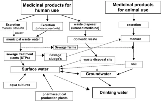
The active pharmaceutical ingredients used in medicine as well as the additional compounds in medical formulations may enter the environment by different routes (Fig. 1.4). These include several different non point sources such as manufacturing plants, effluents from sewage treatment plants, waste and landfill runoff.
Fig. 1.4. Scheme showing possible sources and pathways for the occurrence of pharmaceutical residues in the aquatic environment. From Heberer, 2002.
The consumption of pharmaceuticals is substantial. In the European Union about 3000 different substances are used in human medicine such as analgesics and antiinflammatory drugs, contraceptives, antibiotics, beta-blockers, lipid regulators, neuroactive compounds and many others. Also a large number of pharmaceuticals are used in veterinary medicine, among them antibiotics and antiinflammatory drugs. Pharmaceuticals are excreted after application in their native form or as metabolites and enter aquatic systems via different ways. The main pathway from humans is ingestion following excretion and disposal via wastewater. Residues of the parent PPCPs (as well as sometimes a complex array of metabolites) are either excreted or are dislodged from skin by sweating, bathing, or swimming (Daughton, 2007). Municipal wastewater is therefore the main route that brings human pharmaceuticals after normal use and disposal of unused medicines into the environment. Hospital wastewater, wastewater from manufacturers and landfill leachates may contain significant concentrations of these drugs. Pharmaceuticals
not readily degraded in the sewage treatment plant are being discharged in treated effluents resulting in the contamination of rivers, lakes, estuaries and rarely, groundwater and drinking water. Where sewage sludge is applied to agricultural fields, contamination of soil, runoff into surface water but also drainage may occur. In addition, veterinary pharmaceuticals may enter aquatic systems via manure application to fields and subsequent runoff, but also via direct application in aquaculture (fish farming) (Fig. 1.5). Investigations conducted in surface waters pointed out that common illicit drugs also contaminate the aquatic environment of populated areas (Zuccato et al., 2008); Zuccato et al. (2005) for example reported that kilograms of cocaine residues travel daily down the River Po (Italy).
Fig. 1.5. Origins and fate of PPCPs in the environment. 1, usage by individuals and pets; 2, release of hospital wastes; 3, release to private septic/leach fields; 4, release from agriculture; 5, direct release to open waters via washing/bathing/swimming; 6, industrial manufacturing waste or release from clandestine drug labs and illicit drug usage; 7, leaching from landfill or cemeteries; 8, release from aquaculture; 9, release from agriculture; 10, environmental transport/fate of pharmaceuticals. Adapted from Daughton, 2006.
Pharmaceuticals have been found in the effluents from medical care units, municipal sewages and the effluents of sewage treatment plants, in surface water, seawater, ground waters and in drinking water (Heberer, 2002; Kümmerer, 2009; Murray et al., 2010; Pal et al., 2010). These molecules have also been detected in runoff from landfill sites and even in the arctic environment (Kallenborn et al., 2008). The concentrations of pharmaceuticals in surface water and the effluents
from sewage treatment plants have been shown to lie in the ng/l to µg/l range (Kümmerer, 2009; Murray et al., 2010).
In contrast to the extensive literature describing the occurrence and persistence of pharmaceuticals in freshwater systems, little attention has been paid to their prevalence and quantification in marine ecosystems. In this perspective, Wille et
al. (2010) developed and validated a quantitative analytical method for
pharmaceuticals in seawater. On the basis of data on the current use in Belgium, 13 environmentally relevant pharmaceuticals were selected from five different therapeutic classes. These included four antibiotics (sulfamethoxazole, ofloxacin, trimethoprim and chloramphenicol), four nonsteroidal anti-inflammatory drugs (NSAIDs) (mefenamic acid, diclofenac, salicylic acid, and ketoprofen), two !-blockers (propranolol and atenolol), two lipid regulators (bezafibrate and clofibric acid) and one psychiatric drug (carbamazepine).
After administration, some pharmaceuticals are not completely metabolized. The unmetabolized parent drugs and some metabolites are subsequently excreted from the body via urine and faeces. In places with sewage systems, these pharmaceutical residues enter the WWTPs via wastewater (Zhang et al., 2008).
A tangentially related issue regarding sources is the fate of the packaging materials used for PPCPs, such as the materials used for plastic vials, IVs, and syringes, including the drug residues contained therein. Incineration and weathering of these materials are processes perhaps leading to a number of additional unknown products (Daughton, 2009).
The most obvious pathway for environmental contamination of medicines is via the unaltered excretion in urine and faeces although other anthropogenic mechanisms should be assumed, namely:
a) Metabolism post-consumption; since many drugs are metabolised as the organism attempts to convert hydrophobic compounds into more easily excreted polar residues. Their bioconversion into one or more metabolites can occur throughout Phase I and Phase II reactions (Fig. 1.6). Phase I reactions include oxidation, reduction and hydrolysis to modify the original molecule structure by introducing functional groups more receptive to phase II reactions. Phase II reactions (or conjugation reactions) consist of the addition of endogenous groups (like glucuronic acid, sulphate, glutathione, etc.) to receptive functional groups present in the original molecule or in its metabolite derived from phase I.
b) Diagnostic compounds; such as X-ray contrast media are directly discharged in their native forms.
c) Household disposal; either topic formulations or unused medicines (out-of-date or unwanted) are discarded through the sink/toilet or via waste collection, before being taken to landfill sites where they appear as terrestrial ecosystem contaminants. Alternatively, they may possibly leak into surrounding water compartments.
d) Impacts due to anthropogenic activities; as, for instance, sewage treatment plant (STP) sludge, which can carry non-suspected drugs and is frequently used as a fertilizer on agricultural land; veterinary medicines, which are also excreted in urine and faeces by animals before being spread onto land via manure application as fertilisers. Apart from the potential for direct soil contamination, there is also the risk of run-off with heavy rain, thus potentially contaminating both the surrounding surface and groundwater. Other example of an anthropogenic activity is aquaculture, whose pharmaceuticals employed, as well as their metabolites and degradation products, are directly discharged into surface waters. Another important source of environmental contamination by pharmaceuticals is the effluents of pharmaceutical production facilities (Santos et al., 2010).
Fig. 1.6. Schematic representation of pharmaceutical biotransformation to increase their polarity. Adapted from Daughton and Ternes, 1999.
Wastewater treatment systems are designed for collection and transportation of wastewater and to reduce organic matter that may cause oxygen depletion in the recipient surface water, with the aim of reducing nutrients (nitrogen and phosphorus) that can cause over-fertilisation of recipient lakes, streams and the sea. When pharmaceuticals, or the remains of pharmaceuticals after use, enter the wastewater treatment plants where their fate is governed by the physical, chemical and biological properties of the substance and of the treatment processes in use at the treatment plant. The treatment plants of today are not designed for the reduction or degradation of pharmaceuticals. The fate of the pharmaceuticals in the
plants is therefore determined by the character of the substance itself and of the processes used at particular plants. In some cases the pharmaceuticals can be biologically degraded, if the process conditions are favourable at the plant.
Three main mechanisms have to be taken into account in order to understand the fate of the substances in wastewater treatment systems: evaporation of volatile compounds, compounds ability to adhere to particles and solubility of the pharmaceuticals in water (la Cour Jansen and Ledin, 2009).
What is known today is that very few pharmaceuticals are volatile. This means that evaporation from the plants is not significant. The majority of substances are water soluble and will pass through the plants, unless they are degraded. The major method for handling sludge today is anaerobic digestion, but it was observed that destruction of pharmaceuticals by anaerobic digestion is effective only in a few cases. As a consequence, most of the pharmaceuticals have to be handled in the water treatment part of the STP. Also in this case, biodegradation of these molecules depends on their chemical properties and on the capability of bacteria of degrading them. If pharmaceuticals adhere to particles, physical methods can be used; however, in this case only the result obtained is the separation from the wastewater with no destruction of pharmaceuticals. Besides the removal of the pharmaceuticals all other particles and substances adsorbed to particles are removed. That means in general an improved removal of many other substances from the wastewater, such as particulate organic matter, nitrogen and phosphorus, heavy metals and also pathogens, but it must be kept into consideration that these particles will need further handling. Another way of processing these molecules is the chemical approach. Chemical methods can result in complete or partial destruction of the pharmaceuticals. Partial destruction can lead to the formation of new substances, which are normally less harmful than the original chemical. Examples exist however where newly formed degradation products are more harmful than the parent compound. For example, degradation experiments with carbamazepine have shown the formation of several transformation products including acridine and acrodone; the latter compound is one example of a molecule that have non-wanted biological effects of greater importance then the mother compound since it is known to cause DNA damage and exhibits greater ecotoxicity (la Cour Jansen and Ledin, 2009).
The evaluation of removal efficiency in STPs (by comparing influent and effluent contents) has been studied in detail, showing removal rates that can differ by up to 99% (Santos et al., 2010). Some studies showed that removal of the parent compound ranged from 7% (e.g. for the antiepileptic drug carbamazepine) to 96% (for the !-blocker propranolol) and that most of the removal efficiencies averaged about 60% (Daughton and Ternes, 1999).
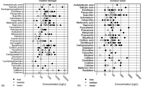
Hence, pharmaceuticals outgo STP barriers and reach water streams where they are detected at concentrations in the range of ng/L to µg/L (Fig. 1.7).
Fig. 1.7. Concentration of pharmaceuticals in treated wastewater (a) and surface water (b). From Fent et al., 2006.
Although most pharmaceuticals are designed to target specific metabolic pathways in humans and domestic animals, they can have numerous often unknown effects on metabolic systems of non-target organisms, especially invertebrates. Although many non-target organisms share certain receptors with humans, effects on their physiology are usually unknown. It is important to recognize that for many drugs, their specific modes of action even in the target species are also unknown. For these drugs, it is impossible to predict what effects they might have on non-target organisms. Without knowing the mode of action, coupled with not knowing the
possible receptors, it is impossible to design rational toxicity testing procedures at the organism level.
Moreover, most pharmaceuticals are racemic mixtures. For a specific optically active drug, it is theorized that only one of its optical isomers is responsible for the desired physiologic therapeutic effects; the other isomers are at best inactive, or even worse, responsible for many of the untoward side effects observed (Daughton and Ternes, 1999; Kasprzyk-Hordern, 2010).
The current knowledge indicates that residues of pharmaceuticals at trace quantities are widespread in aquatic systems. Pharmaceuticals in the environment are suggested to pose only a low risk for acute toxicity. For chronic effects, the situation may be different, but there is a considerable lack of information. Investigation of multigenerational life-cycle effects or at various life stages is lacking, although many aquatic organisms are exposed for their entire life. There is a need to focus on long-term exposure assessment regarding specific modes of action of pharmaceuticals to better judge the implications of pharmaceutical residues in aquatic systems. Only after filling these gaps, more reliable environmental risk assessments with much lower uncertainty can be performed (Fent et al., 2006).
1.2. Pharmaceuticals and legislation: what does regulation say?
In spite of the sizeable amounts of human drugs released to the environment, concise regulations for ecological risk assessment are largely missing (Fent et al., 2006). This probably arises because available data is insufficient to quantify a precise contamination profile. Abundant conclusive studies concerning chronic toxicity are also lacking so that it becomes impossible to infer the risks of long-term exposure of pharmaceuticals and their metabolites on fauna and flora (Santoset al., 2010).
For more than 20 years, European legislation has required an environmental risk assessment within the pharmaceutical authorization procedure. Only in the last few years, regulatory agencies have issued detailed guidelines on how pharmaceuticals should be assessed for possible unwanted effects on the environment (Fent et al., 2006). The European Union Directive 92/18/EEC introduced for the first time the
requirement for an environmental risk assessment, as a prerequisite to obtain marketing authorization for veterinary pharmaceuticals. For this purpose, the European Agency for the Evaluation of Medicinal Products (EMEA) published a “Note for Guidance” where guidelines to assess the environmental risk of veterinary medicinal products are established. The European Commission extended its concerns to pharmaceuticals for human use by publishing the Directive 2001/83/EC, which was subsequently amended by the Directive 2004/27/EC. These directives established that marketing authorization for new medical products for human use should be accompanied by an environmental risk assessment, whose guidelines were set out by EMEA. Nevertheless, the environmental impact does not provide sufficient grounds for a refusal (Santos et al., 2010). Environmental risk assessment of both veterinary and human pharmaceuticals is assessed in a step-wise approach, divided into two phases. In phase I drug environmental concentrations are estimated; phase II is an assessment of physical, chemical and ecotoxicological properties. In addition, an exposure threshold value or “action limit” separates the exposure estimation in the first phase from the test requirements in the subsequent second phase. The goals of the risk assessment include protection of the aquatic and terrestrial ecosystems, groundwater, microorganisms in sewage treatment plants and top predators.
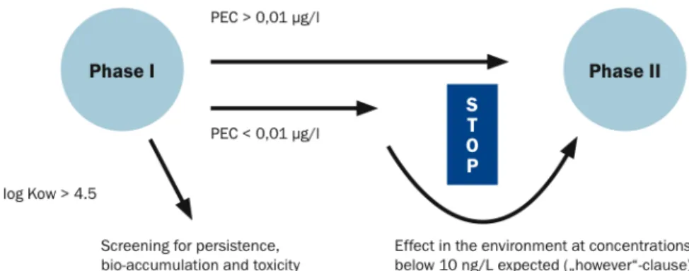
The EMEA guidance document for human pharmaceuticals came into force in 2006. In phase I, the drug concentration expected to occur in the aquatic environment is calculated based on the maximum daily dose. If this value is below a defined action limit of 0.01 µg/L it is assumed that this specific medicinal product is unlikely to represent a risk to the environment. In this case the assessment procedure does not continue. If on the other hand the calculated concentration in surface water exceeds the action limit of 0.01 µg/L a phase II environmental fate and effect analysis is required.
When a high risk of toxic effects is supposed (e.g. in the case of potential endocrine disruptors), a phase II assessment should however be carried out irrespective of the predicted environmental concentration. Furthermore, a screening for persistence, bioaccumulation and toxicity is necessary if the octanol-water partition coefficient (log Kow) exceeds 4.5. This identifies a potential risk for bioaccumulation (Fig. 1.8).
Fig. 1.8. Environmental Risk Assessment of human medical products. From Rechenberg, 2009. Natural substances like vitamins, electrolytes, amino acids, peptides, proteins, carbohydrates, lipids and herbal medicinal products are exempted from environmental assessments.
In the second phase, information on the physical, chemical, and ecotoxicological properties are obtained and assessed in relation to the extent of the environmental exposure. This phase is further split into two tiers, Tier A and Tier B.
In Tier A, possible fate and effects of the pharmaceutical and/or its major metabolites are evaluated; if no risk is identified at the Tier A level, the assessment is considered completed. Conversely, if a risk is recognised at the Tier A level, then an extended ecotoxicity data set is required to be submitted at the subsequent Tier B level. This step focuses on the effects on fauna and flora within environmental compartments that are likely to be affected. However, medicinal products for human use only require Phase II studies if the predicted environmental concentration in surface water is equal to or above 0.01 µg/L (Rechenberg, 2009; Santos et al., 2010).
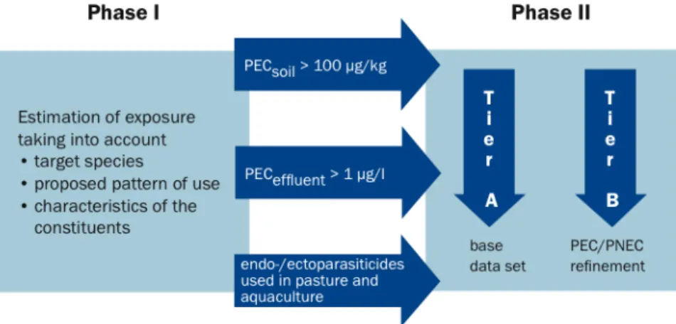
Authorisation of a human medicinal product, however, can explicitly not be denied if an environmental risk is identified. For veterinary pharmaceuticals the legal situation is different: authorisation may be denied, or may be limited to certain application areas, if evidence for an environmental risk exists (Fent et al., 2006). In 1996 the EMEA adapted a comprehensive Note for Guidance to assist the applicant in the evaluation of the environmental risk assessment for veterinary medicinal products. A final detailed Technical Guidance Document was drawn up for Europe and came into force in 2007. Similar to the first phase of the environment risk assessment of human pharmaceuticals, phase I identifies veterinary medicinal products that require a more extensive investigation of their
potential environmental effects on non-target organisms. In this step, the potential extent of exposure to the environment from the product, its active substances and other ingredients should be assessed. The evaluation has to take into account the target species, the proposed pattern of use, characteristics of the constituents of the veterinary pharmaceutical and the method of administration. Phase I is further divided into assessments of drugs used in the aquatic and terrestrial systems respectively. With respect to the aquatic branch, any veterinary medicinal product intended for use in open systems are subjected to phase II. The “action limit” settled for pharmaceuticals used in aquatic environment is 1 µg/L, while for terrestrial sector this limit is set up at 100 µg/kg. If these values are exceeded, a phase II assessment is required. As for human pharmaceuticals, pahse II is further divided into two tiers (Fig. 1.9) (Rechenberg, 2009; Santos et al., 2010).
Fig. 1.9. Environmental Risk Assessment of veterinary medical products. From Rechenberg, 2009. Aiming at estimating potential hazardous compounds, Huggett et al. (2003) proposed the “fish plasma model”. It is based on the comparison of the estimated fish plasma concentration of a given drug to the human therapeutic plasma concentration. The assumption is that the closer the estimated fish plasma concentration and the human therapeutic concentration are, the higher the risk is that the pharmaceutical elicits an effect in fish. Another concept focuses on the homology between human drug targets and potential targets in lower vertebrates; in this case it is assumed that the risk for adverse effects in the environment increases with the degree of homology between human drug target and potential drug targets in lower vertebrates. Moreover, several quantitative structure activity
response (QSAR)-based concepts are developed to aid in the development process of pharmaceuticals (Christen et al., 2010).
In previous studies (Fent et al. 2006; Owen et al., 2007), it was hypothesized that the effects of pharmaceuticals should be better identified by focusing on their mode of actions, or to guide species and toxicity endpoint selection for testing or for development of biomarkers.
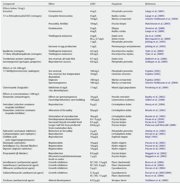
Christen et al. (2010) develop a concept for the identification of highly active compounds that could be used within the risk assessment process for human pharmaceuticals in the framework of the EMEA guideline (2006). The concept was concentrated on human pharmaceuticals, however, it could presumably be applied for veterinary pharmaceuticals in a similar way. As effect concentrations were divided by assessment factors to extrapolate the potential adverse effects of a compound to the ecosystem from laboratory tests on single species to derive predicted no effect concentrations for regulatory purposes, substances with effect concentrations higher than 10 ng/L were also taken into consideration. Even if legislation requires deeper investigations at environmental concentrations higher than 0.01 µg/L, it was demonstrated that there are also compounds which show adverse effects under this value (Tab 1.1).
Tab. 1.1. Summary of pharmaceuticals exerting effects on aquatic organisms at low concentrations. The lowest levels found for lowest effect concentrations (LOEC) are shown. From Christen et al., 2010.
In phase II, a tailored risk assessment taking into account the mode of action is required. Therefore, it is important to identify compounds that could be expected to act at low concentrations according to their modes of action. Such highly active pharmaceuticals with predicted no effect concentrations at 10 ng/L or below are termed in Christen’s work highly active compounds (HC). The risk assessment proposed is a stepwise approach consisting of three major steps, based on three principles: 1) the identification of important human drug targets and of the mode of action of the pharmaceutical; 2) the homology between the human target and the possible targets in lower vertebrates and invertebrates (which are often highly conserved through the evolutionary scale); 3) the importance of the affected
pathway for the target (or non-target) species. A flow chart of the present concept is outlined in Fig. 1.10.
Fig. 1.10. Flow scheme and criteria for the identification of HC among human pharmaceuticals in a stepwise mode of action concept. From Christen et al., 2010.
1.3. Pharmaceuticals tested in this study
1.3.1. Carbamazepine
5H-dibenzo[b,f]azepine-5-carboxamide Formula C15H12N2O
Mol. mass 236.269 g/mol
Fig. 1.11. Structural form of carbamazepine.
In the present study, one of the pharmaceuticals which were taken into consideration was carbamazepine (CBZ) (Fig. 1.11), an anticonvulsant and mood stabilizing drug used primarily in the treatment of epilepsy, bipolar disorder, and
trigeminal neuralgia (Garcia-Morales et al., 2007). CBZ is also used in bipolar depression. This drug is relatively lipophilic, with an octanol/water partition coefficient of 2.2. Reports indicate that CBZ is persistent when released to the environment, and its average removal efficiencies by wastewater treatment plants (WTPs) are below 10% (Zhang et al., 2008). No biodegradation of CBZ at low concentrations in either salt or freshwater was found (Stamatelatou et al., 2003). As a result of its persistence in both municipal wastewater sand surface waters, CBZ is proposed as a suitable anthropogenic marker of urban effluents (Clara et
al., 2004). The drug was indeed detected with few exceptions in influents and
effluents from WTPs, surface waters, and even seawater. Concentrations as high as 6.3 µg/L have been found in wastewaters; average concentrations up to 2.3 µg/L have been measured in effluent samples; values in the range of 0.1–1 µg/L have been reported for surface waters, with levels up to 1.1 and 0.03 µg/L in ground water and drinking waters, respectively (Fent et al., 2006) (Tab. 1.2).
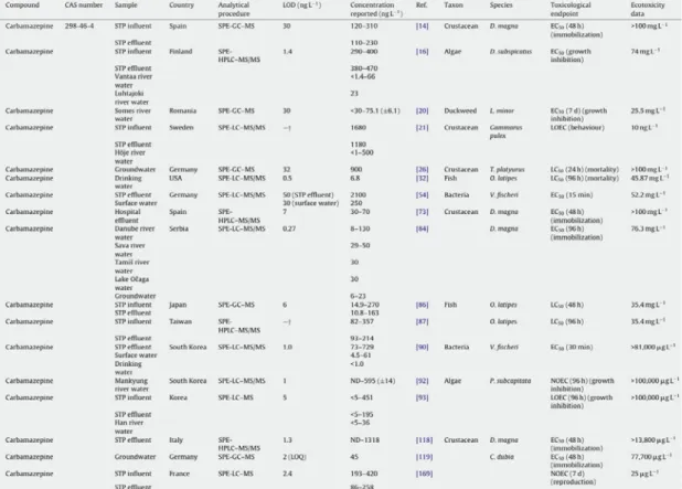
Tab. 1.2. Examples of concentrations (ng/L) of CBZ measured in different aquatic environments. From Santos et al., 2010.
Approximately 72% of orally administered carbamazepine is absorbed, while 28% is unchanged and subsequently discharged through the faeces. After it is absorbed, carbamazepine is heavily metabolized by the liver: only about 1% of dosage leaves the body in an unaltered form. The metabolites of this drug undergo enterohepatic cycling and finally are excreted with urine. The elimination half-life time of carbamazepine is dose-dependent, but is usually in the range of 25–65 hours post-administration. Its important urine metabolites are 10,11-dihydro-10,11-expoxycarbamazepine (CBZ-epoxide) and trans-10,11-dihydro-10,11-dihydroxycarbamazepine (CBZ-diol) (Fig. 1.12). Importantly, the former is pharmaceutically just as active as its parent drug. Furthermore, CBZ-diol is also a metabolite of oxcarbazepine which is a derivative of carbamazepine, with an extra oxygen atom on the dibenzazepine ring (Theisohn and Heimann, 1982). Therefore, one may expect to observe a higher concentration of CBZ-diol in water bodies. Unfortunately, limited studies have been conducted on those metabolites in the aquatic environment to date. Miao and Metcalfe (2003) and Miao et al. (2005) investigated the occurrences of carbamazepine metabolites in a WTP effluent and a surface water. They observed that the concentration of CBZ-diol was approximately three times that of carbamazepine. Therefore, more studies should be conducted on the environmental fate of carbamazepine metabolites (Zhang et
al., 2008).
Fig. 1.12. Identied metabolites of carbamazepine and their percentages of oral dosage. Adapted from Zhang et al., 2008.
Clara et al. (2004) studied the degradation and the removal rates of CBZ in several Austrian sewage treatment plants (STPs), with different membrane systems, measuring its concentration in influent and effluent stream flows. They found only in one case a removal rate over 20%, while in all other plants the effluent concentrations were within the same range as those in the influent or even higher. At two STPs effluent concentrations were twice as high as the influent concentrations.
These findings completely agreed with the results that were obtained by the same research group from laboratory experiments.
Moreover, neither in the post-treatment nor during infiltration or in the groundwater a significant removal was detectable. The decrease of CBZ concentrations in the groundwater was only due to dilution processes. The results of the groundwater sampling confirmed that CBZ is a very persistent pharmaceutical active compound, which is not subjected to any degradation or adsorption, neither in waste-water treatment nor during the underground/groundwater passage.
Preuß et al. (2001) obtained comparable results. They studied the behaviour of CBZ during artificial groundwater recharge and reported poor removal during soil passage, too. This behaviour makes CBZ a suitable wastewater marker. In addition, CBZ has the advantage that it is detectable also in higher dilutions.
For all these reasons, carbamazepine showed to be a very persistent substance and seems to be a qualified parameter for detecting wastewater in the aquatic environment. It is neither subjected to degradation nor to adsorption processes during wastewater treatment and as a consequence, these characteristics qualify CBZ as a suitable marker for anthropogenic influences on the aquatic environment. The persistence of this molecule represents also a potential risk factor for drinking water reserves and has already been tracked from wastewater to drinking water. To avoid such contaminations technologies have to be developed that allow their removal.
Antiepileptic drugs act on the central nerve system by decreasing the overall neuronal activity. CBZ achieves it by blocking voltage-dependent sodium channels of excitatory neurons (Fent et al., 2006).
The mechanism of action of carbamazepine and its derivatives is relatively well understood. Voltage-gated sodium channels are the molecular pores that allow
neurons to generate action potentials. After the sodium channels open to start the action potential, they inactivate, essentially closing the channel. CBZ stabilizes the inactivated state of sodium channels, meaning that fewer of these channels are available to subsequently open, making brain cells less excitable.
Fig. 1.13. Toxicity data of carbamazepine in the literatures. EC50: concentrations that cause 50% of effect ; NOEC: no observed effect concentration; PNEC: predicted no-effect concentrations. Adapted from Zhang et al., 2008.
As previously described, CBZ is widely present in water bodies. Therefore, it is necessary to evaluate its impact on the ecosystems where it is present. Many studies have assessed its ecotoxicology (Fig. 1.13). Ferrari et al. (2003) studied the toxic effects of carbamazepine on bacteria, algae, microcrustaceans, and fish. They observed that it had a relatively limited acute ecotoxicity on the tested organisms. In the worst case of acute toxicity tests, concentration that causes 50% of effect (EC50) was 13800 µg/L on D.magna over 48 hours. However, chronic tests
displayed higher toxicity than acute tests. In this case, the worst no observed effect concentration (NOEC) for CBZ was 25 µg/L on B. calyciflorus over 48 hours. In contrast, Andreozzi et al. (2002) found no toxicity of carbamazepine on algae
Ankistrodesmus braunii and also found that the concentration of carbamazepine
progressively decreased in the culture of the algae. After 60 days, over 50% of the substance had disappeared from the medium. Furthermore, no significant amounts of CBZ could be detected in A. braunii cells during the course of the experiment. The authors assumed that carbamazepine was taken up by algal cells and entered into biochemical processes. The chronic effects are still not clear. Long term studies are needed for a better characterization of chronic ecological effects. The synergistic toxicity of these drugs with other compounds also needs more studies.
Furthermore, information on the ecological effects of their metabolites and intermediates is also needed (Zhang et al., 2008).
1.3.2. Propranolol
(RS)-1-(isopropylamino)-3-(1-naphthyloxy)propan-2-ol
Formula C16H21NO2
Mol. mass 259.34 g/mol
Fig. 1.14. Structural form of propranolol.
The second phamaceutical to which mussels were exposed in this work was propranolol (PROP) (Fig. 1.14). Propranolol is available in generic form as propranolol hydrochloride, and it is soluble in water. PROP is a non-selective !-blocker (adrenergic receptor) antagonist, blocking !1 and !2-receptors, mainly used
in the treatment of hypertension, to treat high blood pressure and other related heart condition illnesses. Due to similarities between human and other vertebrate beta-adrenergic receptors, consequences of !-blocker exposure in vertebrate wildlife, such as fish, are easier to predict. Fish, like other vertebrates, possess !-receptors in the heart, liver and reproductive system so that prolonged exposure to drugs belonging to this therapeutic class may cause deleterious effects (Santos et
al., 2010). However, little is known regarding the presence of this receptor in
invertebrates although the presence of a !-adrenergic-like receptor in some molluscs has been demonstrated and predicted to have a role in bivalve larval metamorphosis, feeding behaviour in the gastropod Aplysia and bioluminescence regulation in brittlestars. In ascidian larvae (Ciona savignyi), the receptor has been localised and has a role in initiating metamorphosis. Nevertheless, studies conducted in crustacean point to a lack of the !-adrenergic receptor in this group (Solé et al., 2010).
Propranolol and other human and animal pharmaceuticals reach the aquatic environment through a variety of routes. However, the majority of pharmaceuticals are present in the aquatic environment due to incomplete removal
at sewage treatment works (STWs). In Germany it was found that 96% of propranolol was removed from the influent compared to the effluent, yet the 4% remaining is largely why propranolol has been found ubiquitously in rivers and streams in America and Europe at concentrations in the ng/L range, with maximum and median concentrations reaching 590 ng/L and 12 ng/L, respectively (Giltrow et
al., 2009).
Although !-blockers act on beta-adrenergic receptors (!-ARs), they can differ greatly in their specificity and lipophilic properties; for example, propranolol has a relatively high log Kow of 3.48, whereas atenolol has a considerably lower log Kow of 0.23 (Owen et al., 2007). Published data show propranolol, out of all the !-blockers investigated, to be the most toxic to aquatic organisms. For example, invertebrate LC50 values for metoprolol and propranolol range from 64 to > 100
mg/L and 0.8 to 29.8 mg/L, respectively, showing that propranolol is harmful to invertebrates at much lower concentrations than metoprolol (Cleuvers, 2003; Huggett et al., 2002). In fish studies, the published data on propranolol are limited. However, a report by Huggett et al. (2002) showed propranolol to be relatively toxic with LOEChatchability and egg production values of 0.5 µg/L.
Photodegradation of propranolol has been evaluated with half-life of 16.8 days (Cleuvers, 2005).
While propranolol is extensively metabolised in humans, oral ingestion leads to excretion of some of the unabsorbed parent compound, which has consequently been detected in USA and European Sewage Treatment Work (STW) effluents (Owen et al., 2009).
Despite the high rate of removal by sewage treatment plants (about 96% (Fent et
al., 2006)), PROP has been detected in STP effluents at concentrations from 30 to
373 ng/L and in surface waters at levels of ng/L. This pharmaceutical has also been found in hospital effluent (Spain) at concentrations that can reach 6.5 µg/L (Tab. 1.3).
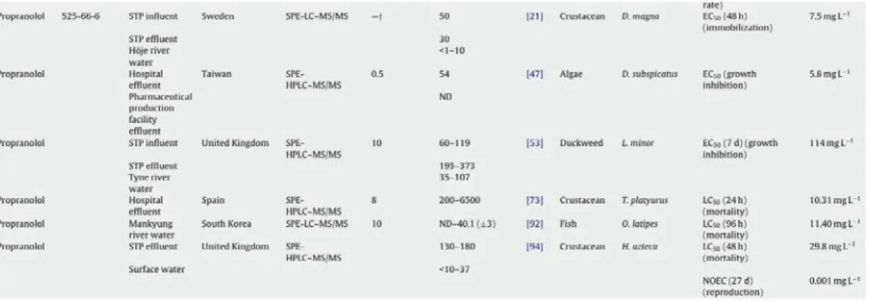
Tab. 1.3. Examples of concentrations (ng/L) of PROP measured in different aquatic environments. From Santos et al., 2010.
1.3.3. Oxytetracycline
(4S,4aR,5S,5aR,6S,12aS)-4-(dimethylamino)-3,5,6,10,11,12a-hexahydroxy 6methyl1,12dioxo1,4,4a,5,5a,6,12,12aoctahydrotetracene -2-carboxamide Formula C22H24N2O9Mol. mass 460.434g/mol Fig. 1.15. Structural form of oxytetracycline.
Oxytetracycline (OTC) (Fig.1.15) was the second of the broad-spectrum tetracycline group of antibiotics to be discovered.
OTC belongs to the tetracycline class of antibiotics, which includes tetracycline, chlortetracycline, doxycycline, minocycline, and glycylcyclines. This class of antibiotics represents broad-spectrum agents that act against a range of Gram-positive and Gram-negative bacteria by inhibiting protein synthesis. The tetracyclines have played an important role in human and veterinary medicine, and some have been used as growth promoters for livestock and aquaculture (Li et al., 2010). OTC works by interfering with the ability of bacteria to produce proteins that are essential to them. Without these proteins the bacteria cannot grow, multiply and increase in number. Oxytetracycline therefore stops the spread of the infection and the remaining bacteria are killed by the immune system or eventually die.
Oxytetracycline is a broad spectrum antibiotic that is active against a wide variety of bacteria. However, some strains of bacteria have developed resistance to this antibiotic, which has reduced its effectiveness for treating some types of infection. OTC is still used to treat infections caused by chlamydia (e.g. the chest infection psittacosis, the eye infection trachoma, and the genital infection urethritis) and infections caused by mycoplasma organisms (e.g. pneumonia). It is also used to treat acne, due to its activity against the bacteria on the skin that cause this disease (Propionebacterium acnes). It is used to treat flare-ups of chronic bronchitis, due to its activity against the bacteria usually responsible, Haemophilus influenzae. Antibiotics released into the aquatic environment are of concern since: a) they could contaminate raw, treated and recycled water used for drinking, irrigation and recreation; b) they could potentially accelerate widespread bacterial resistance to antibiotics; c) they can have negative effect on important ecosystem bacteria (through death or inhibition). Between 30% and 90% of an administered dose of most antibiotics to humans and animals are excreted in the urine as the active substance (Rang et al., 1999). Antibiotics are predominantly water soluble and enter the aquatic environment through sewage systems following consumption and excretion by humans and via effluent from farms, abattoirs and landfills (Daughton and Ternes, 1999; Costanzo et al., 2005).
Antibiotics are widely used to treat infections in humans and are applied intensively for veterinary purposes. Because they are poorly metabolized in the body and incompletely degraded in wastewater treatment plants, antibiotics are continuously introduced into the environment. Consequently, in spite of their relatively short environmental half-lives, they are ubiquitous in aquatic environments (van der Grinten et al., 2010).
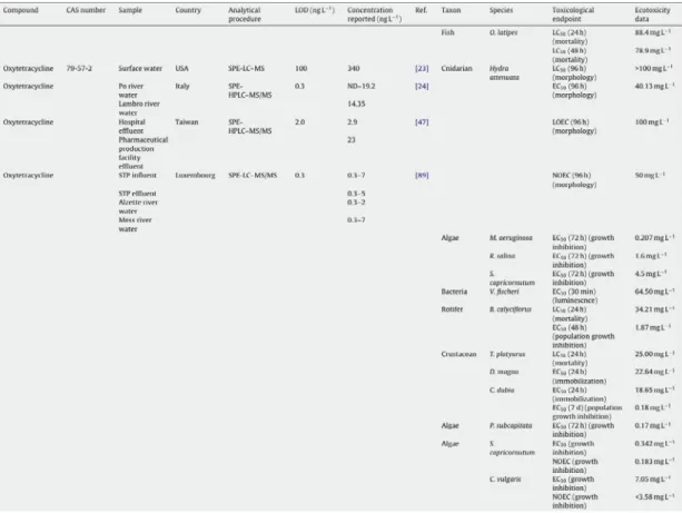
Antibiotics belonging to different classes have been found in different aquatic environments (Tab. 1.4). Oxytetracycline was detected in the Po and Lambro rivers (Italy) at concentrations up to 248.90 and 24.40 ng/L respectively, in combination with tetracycline in American STP influents (47 µg/L) and effluents (4.2 µg/L) and in surface waters (340 ng/L) (Santos et al., 2010).
Tab. 1.4. Examples of concentrations (ng/L) of OTC measured in different aquatic environments. From Santos et al., 2010.
1.3.4. Copper
In the contest of MEECE Programme, mussels were exposed to increasing concentrations of copper.
Copper is an essential micronutrient, it is a cofactor for many enzymes and other proteins implicated in an array of biological processes required for growth, development and maintenance (Trevisan et al., 2011), but it can also have adverse effects on aquatic organisms if bioavailable forms of Cu reach toxic concentrations. Knowing only the total concentration of Cu (i.e., bioavailable and nonbioavailable forms) in the aquatic environment is inadequate to accurately determine the probability of adverse effects. The concentrations of the bioavailable forms of Cu must be determined to better predict potential adverse effects. Numerous studies in fresh water have demonstrated that complexation of Cu with organic and inorganic ligands as well as competition with dissolved cations (Ca2+, Mg2+, Na+, K+, H+) for binding and uptake pathways modify Cu bioavailability and
thus Cu toxicity. Several contemporary studies have similarly demonstrated the need to quantify the bioavailable fraction to assess potential Cu toxicity and risk in salt water. Factors that modify Cu bioavailability have been incorporated into a biotic ligand model (BLM). This model has been modified and adopted by the U.S. EPA for determining the freshwater Cu quality criteria for the protection of aquatic organisms and their uses. No such accommodation for factors that modify copper bioavailability has been adopted for water quality criteria applied to marine waters. However, it has clearly been shown that dissolved organic matter (DOM) greatly affects Cu bioavailability in salt water to two sensitive species of mussels (Arnold
et al., 2010).
Increased concerns regarding the use of oceans as a site for the disposal of anthropogenic wastes and the large scale use of copper and other metals in antifouling boat paint have prompted evaluation of metal toxicity to marine species. Today, there is a considerable body of research outlining the toxicity of various metals to marine species. However, available data often exhibit wide variation in sensitivity. For instance mussel larvae of the genus Mytilus are widely accepted to represent one of the most sensitive marine organisms to Cu and therefore play a critical role in environmental regulations. Yet, the US EPA's (2003) ambient water quality criteria document reports mean acute values (EC50s in 48 hours developmental tests) of 21.4 g/L dissolved Cu (at 20 ppt salinity) for
Mytilus edulis larvae but 6.1 g/L dissolved Cu (at 28–30 ppt) salinity for Mytilus
spp. respectively. This variation could reflect either the importance of water chemistry in influencing the bioavailability of the metal, differences in physiological sensitivity of the organisms at different salinities, or true inter-species differences. Furthermore, many studies have recently presented evidence that the toxicity of dissolved copper to Mytilus spp. is a function of the dissolved organic carbon (DOC) concentration of the test sample (Nadella et al., 2009). It is known also that high concentrations of Cu may cause oxidative stress by impairing antioxidant defenses and increasing the generation of ROS leading to lipid peroxidation and DNA damage (Trevisan et al., 2011).
1.4. Experimental setup: laboratory mussels exposure
In the aquatic environment, organisms are exposed to mixtures of contaminants at different concentrations, that can interact among them and threat animals health. As previously described, among these sources of contamination, also pharmaceuticals are assuming increasing importance and attention.
As to pharmaceuticals exposure, many acute tests and studies were performed, showing effects only at concentrations many times higher than those detected in the environment. Instead, chronic tests are necessary, to understand if these substances constitute a danger for the organisms which are continuously exposed to them even if at very low concentrations.
This kind of survey would be very difficult in natural environment, because of the manifold variables that are involved. To better understand the effects induced by each pharmaceutical, mussels were exposed under laboratory conditions, in order to exclude the interference of other factors.
After acclimation, mussels were exposed to the different contaminants in aquaria containing artificial seawater with continuous aeration at 16°C (Fig. 1.16) renewed every day; they were fed once a day and the drug was dispensed at the same hour every day.
1.5. Why the choice of mussels as sentinel organisms?
Aquatic organisms are particularly important targets for pharmaceuticals, as they are exposed via wastewater residues over their whole life. Standard acute ecotoxicity data have been reported for a number of pharmaceuticals, however, such data alone may not specifically address the question of environmental effects, and subsequently in the hazard and risk assessment (Fent, 2003). The current lack of knowledge holds in particular for chronic effects that have only very rarely been investigated (Fent et al., 2006).
Bivalves are important members of aquatic ecosystems and markedly interact with water and sediment. These sessile and long-lived organisms filter large quantities of surface water for feeding and respiration. They are therefore particularly susceptible to environmental stressors, including point source and diffuse contamination, water-level variations and climatic changes (e.g., temperature fluctuations in shallow water) (Gagné et al., 2006).
Bivalve molluscs, in particular mussels, are useful sentinel organisms to assess chemical pollution in aquatic environment. They are sessile, filter feeding, widely distributed and abundant in coastal and estuarine areas, able to accumulate several classes of pollutants, thus providing a time-integrated picture of their bioavailability. For such characteristics, these organisms are widely used in Mussel Watch monitoring programmes, where chemical analyses are integrated with the use of biomarkers, to evaluate molecular, biochemical and cellular effects induced by pollutants (Bocchetti and Regoli, 2006).
Bivalves have an open vascular system; the haemolymph pervades most organs, favouring direct exposure to the external environment and hence to contaminants. Long-term exposure to contaminants emanating from various sources (urban and industrial wastewaters) could compromise immune function and progressively lead to infectious diseases and cancerous disorders (Gagné et al., 2006). Mussel haemolymph does not coagulate; haemocytes play several functions, in particular they represent the immune defense system, and are important for the accumulation of nutrients, metabolic products and respiratory pigments.
Haemocytes enclose lysosomes, cellular organelles that contain about 40 acid hydrolase enzymes to break up waste materials and cellular debris. Their size varies from 0.1–1.2 m. The membrane around the lysosome allows the digestive
enzymes to work at the 4.5 pH as they require; this pH differential with respect to cytosol is mantained by pumping protons (H+ ions) from the cytosol across the membrane via proton pumps and chloride ion channels. The lysosomal membrane protects the cytosol, and therefore the rest of the cell, from the degradative enzymes within the lysosome. The cell is additionally protected from any lysosomal acid hydrolases that leak into the cytosol, as these enzymes are pH-sensitive and do not function as well in the alkaline environment of the cytosol. In addition to degrading molecules taken up by endocytosis, lysosomes digest material derived from two other routes: phagocytosis and autophagy. In phagocytosis, specialized cells, such as macrophages, take up and degrade large particles, including bacteria, cell debris, and aged cells that need to be eliminated from the body. Such large particles are taken up in phagocytic vacuoles (phagosomes), which then fuse with lysosomes, resulting in digestion of their contents. The lysosomes formed in this way (phagolysosomes) can be quite large and heterogeneous, since their size and shape is determined by the content of material that is being digested.
Lysosomes are also responsible for autophagy, the gradual turnover of the cell’s own components. The first step of autophagy appears to be the enclosure of an organelle (e.g., a mitochondrion) in membrane derived from the endoplasmic reticulum. The resulting vesicle (an autophagosome) then fuses with a lysosome, and its contents are digested.
The lysosomal-autophagic system appears to be a common target for many environmental pollutants, as lysosomes accumulate many toxic metals and organic xenobiotics, which perturb normal function and damage the lysosomal membrane. In fact, autophagic reactions frequently involving reduced lysosomal membrane integrity or stability appear to be effective generic indicators of cellular well-being in eukaryotes (Moore et al., 2008). Autophagy is often considered to be primarily a survival strategy in multi-cellular organisms, which may be initiated by stressors (e.g., restricted nutrients, hyperthermia, hypoxia and salinity increase), but recent evidence indicates that autophagy is much more than just a survival process and is, in fact, intimately involved in cell physiology. Physiological responses and pathological reactions to environmental stressors in mollusc digestive cells and haemocytes frequently involve destabilizing changes in the lysosomal membrane and the induction of autophagy. Other related lysosomal perturbations can also
occur, such as lysosomal swelling, accumulation of lipid and age pigments or lipofuscins: all of these changes have been described in molluscan hepatopancreatic digestive cells. In eukaryotic cells, the first tier of defense against oxidative damage is provided by xenobiotic transporters, biotransformation enzymes and antioxidant enzymes, such as superoxide dismutase and catalase (Livingstone, 2001; Moore, 2004). Lysosomal autophagy provides a second line of defense, by removing oxidatively damaged proteins, inappropriately folded glycosylated proteins and impaired organelles, and even portions of the nucleus and DNA.
In the field of aquatic toxicology lysosomes have attracted considerable attention in recent years because they a) were shown to be the target for a wide range of contaminants, b) are easy to visualize in blood cells and in reacted tissue cryosections, and c) are present in all nucleated cells and therefore are not species-specific (Viarengo et al., 2007).
Cytochemistry and histochemistry have been used as the main tools in the study of environmentally induced alterations in lysosomes of lower animals for several reasons (Moore, 1990; Moore and Simpson, 1992). Exposure to contaminants gives rise to fairly rapid and characteristic pathological alterations such as enlargement of the digestive cell and hepatocyte lysosomes, which are correlated with increased fragility of the lysosomal membrane, excessive build up of unsaturated neutral lipids (lipidosis) in the lysosomal compartment and accumulation of lipofuscins (lipofuscinosis) (Moore et al., 2008).
1.6. Selected battery of biomarkers
1.6.1. The neutral red retention assay
Neutral red is a lipophilic dye and as such will freely permeate the cell membrane. Within cells the compound becomes trapped by protonisation in the lysosomes and accumulated in these organelles, where it can be visualised microscopically. The degree of trapping of this lysosomotropic marker depends on the pH of the lysosome as well as the efficiency of its membrane associated proton pump. The
acid environment of lysosomes is maintained by a membrane Mg2+-ATPase dependent H+ ion proton pump, the neutral red retention assay reflects on the efflux of the lysosomal contents into the cytosol following damage to the membrane and, possibly, impairment of the H+ ion pump. So any impairment of this latter system will result in a reduction of the dye retention assay. Studies indicate that, similarly to the cytochemical method described above, the neutral red retention assay is sensitive to the main classes of chemical pollutants (UNEP/RAMOGE).
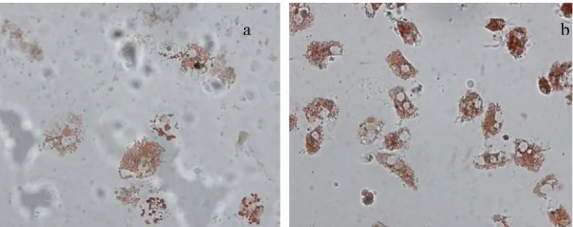
According to this in vitro methodology, the dye is sequestered into the lysosomal compartment when living cells are preloaded with neutral red (NR); if the lysosome membranes are damaged NR leaks out into the cytosol where it can be visualized under the microscope. The time taken for the dye to leak out into the cytosol is related to the degree of membrane damage. In case lysosome membranes are severely damaged (Fig. 1.17a) the dye will leak out within 15 min of incubation, whereas healthy lysosomes (Fig. 1.17b) retain it for up to 180 min (Lowe and Pipe, 1994). The NRR assay has to be considered “a stress on stress test”, in that neutral red is itself toxic for the cells and therefore further damages lysosomal membranes that are potentially already damaged. The additional damage caused by the dye results in a total membrane failure in the case of severely impacted cells, and also in membrane failure and leakage in healthy cells but in this case only after 150–180 min (Viarengo et al., 2007).
Reduced lysosomal membrane stability has to be considered as an indicator of a general physiological stress. Viarengo et al. (2007) proposed the lysosomal membrane stability, assessed either by NRR or by the histochemical technique, as a robust Tier 1 screening biomarker for Environmental Impact Assessments in a 2-Tier approach.