© 2007 Acta Dermato-Venereologica. ISSN 0001-5555 DOI: 10.2340/00015555-0149 Acta Derm Venereol 87
82 Letters to the Editor
Sir,
Acquired ichthyoses can either reflect an underlying severe disorder or may be an atypical expression of skin diseases such as cutaneous T-cell lymphomas (1). Otherwise, lymphomatous infiltrates in the skin can cause heterogeneous cutaneous symptoms and lesions, which may range from invisible features to massive tu-mour nodules. Mycosis fungoides is an epidermotropic cutaneous T-cell lymphoma, which accounts for almost 50% of all primary cutaneous lymphomas. Subtypes and variants may complete its wide clinicopathological spectrum (2). Ichthyosiform mycosis fungoides is a rare variant of mycosis fungoides observed in 1.8% of cases (3–5). We describe here a case of ichthyosiform mycosis fungoides in a 30-year-old man who presented a wide spread ichthyosiform appearance coexisting with limited conventional papular lesions.
CASE REPORT
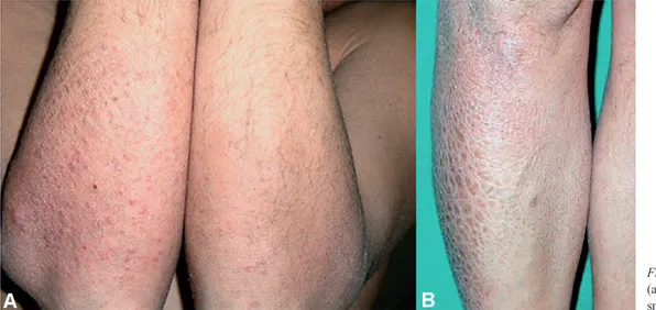
The patient was referred for ill-defined ichthyosis-like, hypo-hyperpigmented patches of 4 years duration. Pru-ritus was common and severe and crusted excoriations were present on the extremities. The ichthyosiform lesions were spread on the arms and legs (Fig. 1), trunk, back and lower abdomen. Clinical examination was otherwise unremarkable without any enlarged lymph
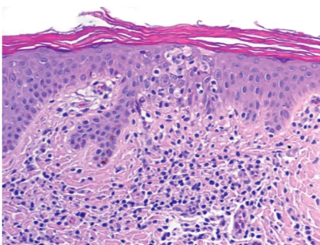
nodes. Histological features of skin biopsies obtained from different ichthyosiform sites disclosed a picture of acquired ichthyosis (orthokeratotic hyperkeratosis, focal parakeratosis, acanthosis and thinned granular layer) combined with epidermotropic lymphomatous infiltration (lymphocytes exocytosis in the basal layer, small epidermal lymphocytic micro-abscesses and lichenoid infiltrate of atypical cerebriform lymphocytes in the upper dermis) (Fig. 2). Immunostaining showed a T-helper phenotype of the infiltrating cells. They were CD2-, CD3-, CD4-, CD45RO-positive but CD8-, CD20- and CD30-negative. Polymerase chain reaction analysis of the T-cell receptor γ-gene disclosed mono-clonal rearrangement in the lesion. Biopsy samples of the papular lesions did not show any difference in the histopathological features. Staging procedures, includ-ing routine blood cell counts, biochemical analyses and cervicothoracoabdominal computed tomography, ruled out a systemic involvement of the disease. The diagnosis of mycosis fungoides, stage Ib, was establish-ed. A combined treatment with acitretin, 25 mg daily, and interferon α-2a, 3 million units 3 times a week, is now at 6 months’ duration. To date, absence of disease progression and 50% improvement in the ichthyosiform appearance has been noted. On the other hand, the last control biopsy still showed signs of lymphomatous epidermotropism.
Ichthyosiform Mycosis Fungoides: A Neoplastic Acquired Ichthyosis
Luca Bianchi1, Marina Papoutsaki1, Augusto Orlandi2, Luigi Citarella1 and Sergio Chimenti1
Departments of 1Dermatology and 2Anatomic Pathology, Tor Vergata University of Rome, Policlinico Tor Vergata (PTV), Viale Oxford 81, IT-00133
Rome, Italy. E-mail: [email protected] Accepted June 13, 2006.
Fig. 1. Ichthyosiform appearance on (a) the arms, with limited irregular small papules, (b) and legs.
83 Letters to the Editor
DISCUSSION
Acquired ichthyoses or ichthyosiform eruptions appear as adherent hyperkeratotic hyper- or hypo-pigmented patches widespread or mostly affecting the extremities in the adult life. The age of onset depends on the as-sociated underlying condition. The aetiologies include autoimmune and endocrinological disorders, infectious diseases, nutritional disorders, renal failure, bone marrow transplantation, side-effects of drugs and ma-lignancies (1). When an acquired ichthyosis is related to neoplasms, mostly Hodgkin’s and non-Hodgkin’s lymphomas, it can behave as a paraneoplastic syndrome or as a specific clinical variant of the lymphoma, the latter showing combined ichthyosiform and lymphoma-tous features in the same lesion. Ichthyosiform mycosis fungoides is a rare variant of the cutaneous lymphoma: less then 20 cases are reported in the literature (2–5). Conventional cutaneous manifestations of mycosis fungoides over years or decades of their slow clinical progression include patches, papules, plaques and tumours, which define the classical form of Alibert-Bazin. Lymph nodes and visceral organs may become involved in the later stages of the disease, which are associated with an aggressive clinical course. Subtypes and vari-ants complete the wide clinicopathological spectrum of mycosis fungoides (2). Subtypes have distinct
clinico-pathological features, such as folliculotropic mycosis fungoides, pagetoid reticulosis and granulomatous slack skin. Variants are considered those forms which have a clinical behaviour similar to that of classical mycosis fungoides. These forms include a long list of dermatological lesions (2–5). Among them acanthosis nigricans-like, angiocentric/angiodestructive, bullous, hyper/hypopigmented, unilesional, syringotropic, erythrodermic, granulomatous, keratosis lichenoides chronica-like, interstitial, mucinous, poikilodermatous, palmaris et plantaris, verrucous, dyshidrotic, with eruptive cysts, pigmented purpura-like, pustular, papular, perioral dermatitis-like, zosteriform, invisible and ichthyosiform mycosis fungoides (2–5). Ichthyosiform appearance can be isolated or combined with otherwise classical or atypical clinical lesions.
In our patient, on the nape of the neck, over the shoulders and on the arms, limited irregular small scaly patches and papules (Fig. 1a), similar to conventional expressions of mycosis fungoides, could be observed. Whereas, manifestations of folliculotropic mycosis fungoides, such as follicular papules, patchy alopecia, comedo-like and milia-like lesions, rarely described in the ichthyosiform variant, were not detectable. Ichthy-osiform mycosis fungoides is characterized by indolent course, good prognosis and response to non-aggressive therapies such as topical treatment, psoralen and ultra-violet light A (PUVA) therapy or combined treatment with alpha interferon and retinoids. The short period of therapy in our patient does not allow us to validate this observation. After 6 months, both the neoplastic acquired ichthyosis and the conventional papular lesions are controlled by the treatment proposed.
REFERENCES
1. Humbert P, Dupond JL, Agache P. Acquired ichthyosis. Ann Dermatol Venerol 1988; 115: 937–942.
2. Kazakov DV, Burg G, Kempf W. Clinicopathological spec-trum of mycosis fungoides. J Eur Acad Dermatol Venereol 2004; 18: 397–415.
3. Badawy E, D’Incan M, Majjaoui SE, Franck F, Fabricio L, Dereure O, et al. Ichthyosiform mycosis fungoides. Eur J Dermatol 2002; 12: 594–596.
4. Hodak E, Amitay I, Feinmesser M, Aviram A, David M. Ichthyosiform mycosis fungoides: an atypical variant of cutaneous T-cell lymphoma. J Am Acad Dermatol 2004; 50: 368–374.
5. Marzano AV, Borghi A, Facchetti M, Alessi E. Ichthyosiform mycosis fungoides. Dermatology 2002; 204: 124–129.
Fig. 2. Combined histological features of acquired ichthyosis (orthokeratotic hyperkeratosis, mild acanthosis and thinned granular layer) and epidermo-tropic lymphomatous infiltration (lymphocytes exocytosis in the basal layer, small epidermal lymphocytic micro-abscesses and lichenoid infiltrate of atypical cerebriform lymphocytes in the upper dermis) (haematoxylin and eosin (HE) ×200).