UNIVERSITÀ DEGLI STUDI DI ROMA
"TOR VERGATA"
FACOLTA' DI MEDICINA E CHIRURGIA
DOTTORATO DI RICERCA IN MICROBIOLOGIA MEDICA E
IMMUNOLOGIA
XXII
CICLO DEL CORSO DI DOTTORATO
GENETIC CHARACTERISTICS OF THE
INFLUENZA A PANDEMIC (H1N1) 2009 VIRUS
CIRCULATING IN ITALY
Dottoranda: Dott.ssa Angela Di Martino
A.A. 2009/2010
Tutor : Dott.ssa I. Donatelli
ABSTRACT
Influenza viruses cause annual epidemics and occasional pandemics that have claimed the lives of millions. The emergence of new strains will continue to pose challenges to public health and the scientific communities. The recent flu pandemic, caused by a swine-origin A/H1N1 influenza virus, presents an opportunity to examine virulence factors, the spread of the infection and to prepare for major influenza outbreaks in the future.
As part of the intensified surveillance carried out during the influenza pandemic, the sequences of 133 pandemic A/H1N1 strains, from patients showing different clinical outcame (12 from fatal, 24 from severe and 97 from mild cases) have been examined at WHO National Influenza Centre (NIC) located at the National Institute of Health (Istituto Superiore di Sanità, ISS). Phylogenetic analysis of the new strain, showed that the virus circulating in Italy, combines genetic information related to different swine influenza viruses. Segments HA and NP are related to swine influenza viruses isolated in North America. Segments NA and M are related to swine influenza viruses isolated in Eurasia. Specific markers for virulence and pathogenicity have been evaluated in the viral genome. Sequence analysis of the isolates of the 2009 A/H1N1 viruses, to date, has not identified molecular features known to confer increased transmissibility or virulence in studies of other influenza A viruses, suggesting that previously unrecognized molecular determinants could be responsible for the transmission among humans.
Particular attention has been paid to the specific mutation in the viral haemagglutinin D222G, initially reported in association with fatal cases in several countries. On the basis of our observations, the majority of severe and fatal cases investigated in Italy did not carry the D222G substitution, and it was also observed in few mild cases, suggesting that this mutation is not required for a severe outcome.
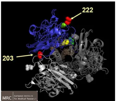
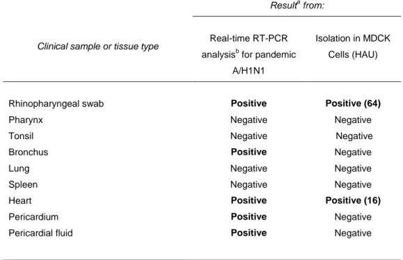
We also describe a fatal case of myopericarditis presenting with cardiac tamponade in a previously healthy 11-year-old child, which has not shown the D222G mutation. Pandemic H1N1 2009 influenza A virus sequences were identified in throat and myocardial tissues and pericardial fluid, suggesting damage of myocardial cells directly caused by the virus.
The first case of oseltamivir-resistance among the influenza pandemic A/H1N1 strains circulating in Italy since the beginning of the pandemic is also reported.
ABSTRACT
I virus influenzali sono responsabili di epidemie annuali e di più rare pandemie occasionali, che in passato hanno causato la morte di milioni di persone. La comparsa di nuovi ceppi virali rappresenta una continua minaccia per la salute pubblica e una sfida per la comunità scientifica. La recente pandemia influenzale, causata da un virus di sottotipo A/H1N1 di origine suina, costituisce un ulteriore strumento per lo studio dei fattori di virulenza e della diffusione dell'infezione in preparazione a futuri eventi pandemici.
Nell'ambito dell‟attività di sorveglianza virologica dell'influenza, intensificata durante la pandemia influenzale, le sequenze di 133 ceppi pandemici A/H1N1, isolati da pazienti che mostravano diversi esiti clinici (12 casi fatali, 24 gravi e 97 lievi) sono state esaminate presso il WHO-National Influenza Centre (NIC) dell'Istituto Superiore di Sanità (ISS).
L‟analisi filogenetica dei ceppi in studio, ha mostrato che i virus pandemici circolanti in Italia, derivano dal riassortimento genetico di virus influenzali diffusi tra i suini. I segmenti genici HA e NP mostrano una stretta correlazione con i ceppi isolati nei suini del Nord America, mentre i segmenti M ed NA correlano maggiormente con i ceppi suini circolanti in Eurasia. La presenza di specifici marcatori molecolari per virulenza e patogenicità è stata valutata all‟interno dell‟intero genoma virale. L'analisi delle sequenze degli isolati analizzati, non ha evidenziato alcuna caratteristica molecolare coinvolta in una maggiore trasmissibilità o virulenza, suggerendo che preesistenti determinanti molecolari potrebbero essere responsabili della elevata trasmissibilità. Particolare attenzione è stata dedicata alla mutazione nel gene della emagglutinina virale D222G, inizialmente riportata da molti paesi in associazione a casi clinici ad esito fatale. Sulla base delle nostre osservazioni, solo pochi tra i casi fatali e gravi, analizzati in Italia, presentano la sostituzione D222G, osservata anche in casi lievi, suggerendo che questa mutazione non è strettamente associata ad infezioni ad esito fatale.
In questo studio è inoltre descritto un caso di miocardite ad esito fatale in un bambina di 11 anni. Il virus pandemico, non presentante la mutazione D222G, è stato identificato nei tessuti della gola, del miocardio e nel fluido pericardico, il che suggerisce un danno alle cellule del miocardio causato direttamente dal virus.
Viene anche riportato il primo isolamento di un ceppo pandemico A/H1N1 resistente all‟Oseltamivir in Italia a partire dall'inizio della pandemia.
KEYWORDS: influenza A virus, genetic reassortment, swine, pandemic,
ACKNOWLEDGEMENTS
First of all, I would like to express my sincere gratitude to my supervisor Dr. Isabella Donatelli, Director of the italian National Influenza Centre. She provided me with many helpful suggestions, important advice and constant encouragement during the past years.
I would also like to thank my colleagues Laura Calzoletti, Concetta Fabiani, Marzia Facchini, Tiziana Grisetti, Annapina Palmieri, Simona Puzelli, Domenico Spagnolo for the lovely working environment and their kind support whenever necessary.
I wish to extend my thanks to my friends for their help and friendship over the years.
Special thanks go to my parents and my sister for all their love, their constant support, encouragement and endless patient.
i
INDEX
1.
INTRODUCTION
pag. 11.1. ORTHOMYXOVIRIDAE
” 21.1.1 Structure of influenza A virus ” 3
1.1.2. Viral life cycle ” 13
A. Entry into the host cell ” 13
B. Transcription and replication of the viral genome ” 14 C. Assembly and budding at the host cell’s plasma membrane ” 16
1.2. ANTIGENIC VARIATION OF INFLUENZA A VIRUSES
” 201.2.1. Antigenic drift ” 20
1.2.2. Antigenic shift ” 21
1.2.3. RNA recombination ” 22
1.3. ECOLOGY OF INFLUENZA A VIRUSES
” 241.3.1. Reservoirs of influenza A viruses in nature
“ 24
1.3.2. Emergence of pandemic influenza viruses ” 29
1.3.3. A novel swine-origin influenza A virus H1N1 in humans ” 36 1.3.4. Virological influenza surveillance in Italy and the 2009
pandemic A/H1N1 virus
”
392. AIM OF THE STUDY
” 423. MATERIALS AND METHODS
“ 43
ii
3.2.METHODS
pag. 443.2.1. Isolation methods “ 44
A. Embryonated egg culture ” 44
B. Cell culture ” 45
3.2.2. Identification of viral isolate ” 46
Haemagglutination test (HA) ” 46
3.2.3. Antigenic characterization ” 47
A. Haemagglutination inhibition test (HI) ” 47
B. Neuraminidase inhibition test (NI) ” 48
3.2.4. Molecular characterization ” 50
A. Detection of viral RNA ” 50
B. Reverse transcription (RT) ” 51
C. PCR amplification ” 51
3.2.5. Sequence and phylogenetic analysis ” 52
3.2.6. Real time (RT-PCR) for pandemic H1N1 2009 influenza virus ” 53
4. RESULTS
” 584.1. Evolutionary characterization ” 58
4.2. Analysis of viral pathogenicity determinants ” 61
4.3. Detection of molecular markers of antiviral resistance ” 73 4.4. CASE REPORT: fatal myopericarditis associated with pandemic
A/H1N1 virus infection ” 75
5. DISCUSSION
” 781
1. INTRODUCTION
Influenza viruses are among the most common causes of human respiratory infections and they cause high morbidity and mortality.
Human epidemic influenza is caused by influenza type A and B viruses, which continually undergo antigenic change in their surface antigens. Seasonal influenza is an acute and recurring respiratory disease occuring, in particular, during winter months. The disease has high morbidity rates for people of all ages and particularly high mortality rates for children, adults over 60 years old, patients with chronic illnesses.
Influenza A viruses are also responsible for sporadic pandemics that usually cause higher mortality rates than seasonal influenza epidemics. Pandemic influenza occurs, at irregular and unpredictable intervals, when a new influenza A virus emerges for which there is little or no immunity in the human population; the virus causes serious illness and spreads easily from person-to-person worldwide, such as in 1918, when over twenty million deaths occurred. It is the result of a major antigenic change which occurs only in Influenza A viruses.
Based on history, influenza is and will continue to be a serious threat to public health. The rapid evolution of influenza viruses highlights the importance of surveillance in identifying novel circulating strains; to this aim, a global surveillance of influenza is actually maintained by a network of laboratories sponsored by the World Health Organization.
2
1.1. ORTOMYXOVIRIDAE
Influenza viruses are enveloped RNA viruses belonging to the Ortomyxoviridae family. According to the identity of two internal viral proteins, the nucleoprotein (NP) and matrix (M1) protein, influenza viruses are classified in three antigenically distinct groups, named A, B and C.
Influenza A and B viruses can cause serious diseases in humans, whereas only minor illness are due to type C viruses.
Type B and C are mainly found in humans, whereas influenza A viruses can infect a large spectrum of vertebrate hosts represented by both avian and mammalian species, human being included (Lamb et al., 1996).
Both the wide host range and the animal reservoir place influenza A viruses in a central position among the zoonotic emerging infectious desease (Morens et al., 2004).
3
1.1.1.
STRUCTURE OF INFLUENZA A VIRUS
Influenza A virus has a lipid bilayer envelope, within which are eight RNA genomic segments, each of which is associated with the trimeric viral RNA polymerase (PB1, PB2, PA) and coated with multiple nucleoproteins (NPs) to form the viral nucleoproteins (vRNPs).
The outer layer of the lipid envelope is spiked with multiple copies of HA, NA and a small number of M2, whereas the M1 molecules keep vRNPs attached to the inner layer. According to the major differences of the main virus surface proteins, at the present time a total of 16 HAs (from H1 to H16) and 9 NAs (from N1 to N9) are known, and both HA and NA groups comprise different subtypes that, in general, are not cross-reactive serologically (Fouchier et al., 2005).
The influenza virus virions (Fig.1) are known to display a number of shapes, with the most abundant one being roughly spherical. The viral envelope is made up of a lipid bilayer that contains three of the viral transmembrane proteins: HA, NA, and M2. This lipid bilayer is derived from the host‟s plasma membrane and is known to contain both cholesterol-enriched lipid rafts and non-raft lipids (Scheiffele et al., 1992; Nayak et al., 2009). HA is the most abundant envelope protein at approximately 80 percent, followed by NA, which makes up around 17 percent of the viral envelope proteins. M2 is a very minor component of the envelope, with only 16 to 20 molecules per virion. Sitting just underneath the viral lipid membrane is M1, which forms a matrix holding the viral ribonucleoproteins (vRNPs). These vRNPs are the core of the virus and are made up of the viral negative stranded RNAs, which are wrapped up around NP and very small amounts of NS2. At one end of the vRNPs are the three polymerase proteins (PB1, PB2 and PA) that make up the viral RNA polymerase complex (Nayak et al., 2004).
4
5
Viral genome:
Influenza A is an enveloped virus with a genome of negative sense, single-stranded, segmented RNA. Influenza A contains eight segments that encode for 11 viral genes (Tab. 1). The three largest RNA segments encode the three viral RNA-dependent RNA polymerase (RdRP) proteins: polymerase acidic protein (PA), polymerase basic protein 1 (PB1) and PB2. The RNA seg-ment for PB1 also encodes a small 87-residue nonstructural protein, PB1-F2, which has apoptotic functions (Chen et al., 2001).
The three intermediate-size RNA segments encode the hemagglutinin (HA), the neuraminidase (NA) and the nucleoprotein (NP). The larger of the remaining two segments encodes the M1 matrix protein and the M2 ion-channel protein, and the smaller one encodes two nonstructural proteins, NS1 and NS2 (also known as nuclear export protein, NEP) (Fields et al., 2007).
6
Tab. 1 Genes and gene products of Influenza A virus
Gene segment nucleotides Protein product molecular weight (dalton) function 1 2341 PB2 86.000
recognizes and binds the 5‟cap structures from host mRNAs for use as viral mRNA transcription primers
2 2341
PB1 83.000
responsible for elongation of the primed nascent viral mRNA and also as elongation protein for template RNA and vRNA synthesis
PB1-F2 10.500 appears to enhance virus-induced cell death
3 2233 PA 85.000
induces proteolytic degradation of coexpressed proteins, suggesting a role in virion RNA synthesis
4 1778
HA1 50.000
responsible for attachment of virus to cells and fusion of virus and cell membranes; cleavage into HA1 and HA2 is required for infectivity
HA2 30.000
5 1565 NP 56.000 internal protein associated with RNA and polymerase proteins
6 1413 NA 50.000 responsible for release of virus from infected cells
7 1027
M1 27.000 involved in both virus assembly and budding
M2 11.000 proton channel protein contributes to virus disassembly
8 890
NS1 27.000 non-structural protein, viral interferon antagonist
NS2 13.000 contributes to the extra-nuclear exportation of newly synthesized RNPs
7
Hemagglutinin:
The HA protein is an integral membrane protein and the major surface antigen of the influenza virus virion. It is responsible for binding of virions to host cell receptors and for fusion between the virion envelope and the host cell.
A newly synthesized ~70-kDa HA is cleaved into HA1 (~50-kDa) and HA2 (~27-kDa) subunits, which are disulfide linked. This cleavage is accomplished by host-produced trypsinlike proteases and is required for infectivity because virus-cell fusion is mediated by the free amino terminus of HA2.
HA molecules form homotrimers during maturation. The three-dimensional structure of a complete HA trimer has been determined. In essence, each HA molecule consists of a globular head on a stalk. The head is made up exclusively of HA1 and contains the receptor-binding cavity as well as most of the antigenic sites of the molecule. The stalk consist of all of HA2 and part of HA1. The carboxy-terminal region of HA2 contains the hydrophobic transmembrane sequence (Fig.2).
Owing to error prone viral RNA polymerase activity, influenza virus HA is subject to a very high rate of mutation. Selection for amino acid substitutions is driven at least in part by immune pressure, as the HA is the major target of the host immune response. Although the amino acids making up the receptor-binding site are highly conserved, the remainder of the HA molecule is highly mutable. In nature there are presently 16 recognized subtypes of HA (Murphy & Webster, 1996; Rohm et al., 1996, Fouchier et al., 2005).
HA is, also, the major determinant of host range restriction. The HAs of human influenza viruses preferentially bind sialyloligosaccharides terminated by N-acetylsialic acid linked to galactose by the α 2,6 linkages (NeuAcα 2,6Gal), whereas avian and equine influenza viruses preferentially recognize receptors terminating in an α 2,3 linkage (NeuAcα 2,3Gal). In the host
8 parenchyma of different species, presence, quantity and distribution of these cell receptors can vary influencing the viral tissue tropism; this is mailnly respiratory in mammals and both respiratory and intestinal in avian species (Horimoto et al., 2001; Zambon, 2001). In fact, NeuAcα 2,6Gal linkages are predominant in the human respiratory tract, whereas NeuAcα 2,3Gal are characteristic of horse trachea and duck intestine. The presence of both types of linkages in the epithelial cells of the pig trachea allows this species to be infected with human and avian viruses (Ito et al., 1998).
Neuraminidase:
The NA protein is also an integral membrane glycoprotein and a second major surface antigen of the virion. NA is a mushroom-shaped tetrameric protein, anchored to the viral membrane by a single hydrophobic sequence of some 29 amino acids near the N-terminus. Treatment of virions with pronase liberates a 200-kDa protein containing 4 identical and glycosylated polypeptides that have all of the antigenic and enzymatic properties of membrane-bound neuraminidase (Colman, 1994) (Fig. 2).
Like HA, NA is highly mutable with variant selection partly in response to host immune pressure. Nine subtypes of NA have been identified in nature (Murphy & Webster, 1996; Rohm et al., 1996). The host cell receptors for influenza A and B viruses are cell surface sialic acids. The predominant type of sialic acids is N-acetylneuraminic acid (Neu5Ac), which is the biosynthetic precursor for most of the other types. In nature, Neu5Ac is mostly linked to the penultimate galactose residues of carbohydrate side chains via α(2,3)- or α(2,6)-linkages.
Both Neu5Ac α(2,3)-Gal and Neu5Ac α(2,6)-Gal molecules can be recognized as a receptor by influenza viruses but human viruses prefer α(2,6)-linked sialic
9 acid, whereas avian and equine viruses predominantly recognize α(2,3)-linked sialic acid.
NA (belonging to the sialidase family) cleaves the α-ketosidic linkage between terminal Neu5Ac and the galactose residues on the host cell surface. Experimental evidence is available to support a role for the enzyme in facilitating release of progeny virions from the surface of infected cells, where they would otherwise aggregate as a result of interactions between the hemagglutinin and sialic acid on the surface of the infected cell and on the progeny virion envelope (Palese et al., 1974; Palese & Compans, 1976; Griffin & Compans, 1979; Griffin et al., 1983).
It also serves as an important antigenic site. The NA carries several important amino acid residues which, if they mutate, can lead to resistance against neuraminidase inhibitors.
10
Fig. 2 Surface glycoprotein of the virion (HA and NA).
H TRIMER
N TETRAMER
Antigenic sites (covering head) catalytic site head stalkCell-binding site in surface pocket
Antigenic sites
Fusion activation
site Hydrophobic regions
(membrane anchoring sites)
Lipid bilayer
Matrix protein
11
Other viral proteins:
The NP interacts with RNA as a structural component of the virion and also associates with the three polymerase proteins to form the transcriptase complex (Lamb, 1989). The NP has also been implicated in the switch from mRNA to vRNA synthesis in the infected cells (Schapiro & Krug, 1988; Portela & Digard, 2002 ).
The three polymerase proteins function as a complex in the nucleus of the infected cells, where transcription and replication of influenza virus RNA occur. The PA subunit of the influenza virus polymerase complex is a phosphoprotein that induces proteolytic degradation of coexpressed proteins, suggesting a role in virion RNA synthesis(Sanz-Ezquerro et al, 1998).
PB1 is responsible for elongation of the primed nascent viral mRNA and also as elongation protein for template RNA and vRNA synthesis, PB2 is known to function during initiation of viral mRNA transcription as the protein which recognizes and binds the 5‟cap structures from host mRNAs for use as viral mRNA transcription primers (Nakagawa et al, 1996).
M1 protein is the most abundant protein in the influenza virus virion. Matrix protein forms a shell surrounding the virion nucleocapsids, underneath the virion envelope. It plays an important role in initiating progeny virus assembly. The mRNA for M2 is also transcribed from RNA segment 7. It is derived from the colinear (M1) transcript by splicing. M2 is an integral membrane protein, which acts as a proton channel to control the pH of the Golgi during HA synthesis and to allow acidification of the interior of the virion during virus uncoating (Yasuda et al., 1993).
The NS1 is a phosphoprotein and has two nuclear localization signals. In the nucleus, the NS1 interferes with the splicing as well as the nuclear export of cellular mRNAs. In the later time of the infection, the NS1 is present in the cytoplasm and associates with the polysomes. The NS1 binds to the 5'UTR of
12 some viral mRNAs and stimulates translation. In addition , NS1 is probably able to suppress the interferon response in the virus- infected cell leading to unimpaired virus production. The NS2 protein plays an important role in the nuclear export of the vRNPs interacting with the RNP-M1 complex and the nucleoprotein.
13
1.1.2. VIRAL LIFE CYCLE
A. Entry into the host cell
HA is a homotrimer that forms spikes on the viral lipid membrane. These spikes of HA bind to sialic acid found on the surface of the host cell‟s membrane (Webster et al., 1992; Skehel & Wiley, 2000) (Fig. 3).
The HA precursor, HA0, is made up of two subunits: HA1, which contains the receptor binding domain, and HA2, which contains the fusion peptide. These subunits are linked by disulphide bonds (Huang et al., 2003). Two major linkages are found between sialic acids and the carbohydrates they are bound to in glycoproteins: α(2,3) and α(2,6). These are extremely important for the specificity of the HA molecules in binding to cell surface sialic acid receptors found in different species. Viruses from humans recognize the α(2,6) linkage, whereas those from avians and equines recognize the α(2,3) linkages. Those from swine recognize both (Rogers & Paulson, 1983; Connor et al. 1994).. This explains the importance of swine being a good mixing vessel for avian and human influenza viruses, hence producing dangerous pathogenic viruses. Upon binding to the host cell‟s sialic acid residues, receptor-mediated endocytosis occurs and the virus enters the host cell in an endosome. The endosome has a low pH of around 5 to 6, which triggers the fusion of the viral and endosomal membranes. The low pH induces a conformational change in HA0, leading to maintenance of the HA1 receptor-binding domain but exposing the HA2 fusion peptide. This fusion peptide inserts itself into the endosomal membrane, bringing both the viral and endosomal membranes into contact with each other. The acidic environment of the endosome is not only important for inducing the conformation in HA0 and, thus, fusion of the viral and endosomal membranes but also opens up the M2 ion channel. M2 is a type III transmembrane protein that forms tetramers, whose transmembrane
14 domains form a channel that acts as a proton-selective ion channel (Holsinger et al., 1991; Pinto et al., 1992)
Opening theM2 ion channels acidifies the viral core. This acidic environment in the virion releases the vRNP from M1 such that vRNP is free to enter the host cell‟s cytoplasm (Bullough et al., 1994; Pinto & Lamb, 2006).
B. Transcription and replication of the viral genome
Influenza viral transcription and replication occurs in the nucleus; therefore, after being released into the cytoplasm, the vRNP enter the nucleus. The viral proteins that make up the vRNP are NP, PA, PB1, and PB2. All of these proteins have known nuclear localization signals (NLSs) that can bind to the cellular nuclear import machinery and, thus, enter the nucleus. To date, it is unclear which NLS is the most important for vRNP nuclear entry.
The influenza viral genome is made up of negative sense strands of RNA. In order for the genome to be transcribed, it first must be converted into a positive sense RNA to serve as a template for the production of viral RNAs. Replication of the genome does not require a primer; instead, the viral RNA dependent RNA polymerase (RdRp) initiates RNA synthesis internally on viral RNA, this is possible, as the extreme 5‟ and 3‟ ends of the genome exhibit partial inverse complementarity. Mature cellular messenger RNAs (mRNAs) have a 5‟ methylated cap and a poly (A) tail. It is known that the vRNPs have poly(A) tails but no 5‟ caps. It was determined that the 5‟methylated caps of the viral mRNAs actually belonged to the cellular mRNAs. That discovery lead to the formulation of the “cap-snatching” mechanism (Plotch et al., 1981).
The viral RdRp is made up of three viral proteins: PB1, PB2, and PA. PB2 has endonuclease activity. It binds to the 5‟ methylated caps of cellular mRNAs
15 and cleaves the cellular mRNAs‟ 10 to 15 nucleotides 3‟ to the cap structure. This cellular capped RNA fragment is used by the viral RdRp to prime viral transcription (Li et al., 2001).
Cellular RNA Polymerase II (Pol II) binds to DNA and starts transcription. Six but two of the viral segments encode for one protein. Segments 7 and 8 encode for two proteins each due to splicing. Segment 7 encodes for M1 and M2; whereas, segment 8 encodes for NS1 and NS2. M2 and NS2 are the spliced products and generally are found in much lower abundance than NS1 and M1 (Amorim & Digard, 2006). The virus uses the host cell‟s splicing machinery to express both of these proteins (Engelhardt & Fodor, 2006). Despite influenza‟s need for the cellular splicing machinery, it prevents the host cell from using its own splicing machinery for processing the host cell mRNAs. NS1 binds to U6 small nuclear RNAs (snRNAs) (Lu et al., 1994) and other splicing components, causing them to re-localize to the nucleus of infected cells. In this way, influenza is able to inhibit splicing of cellular mRNAs. It also has been shown to bind to a novel protein called NS1 binding protein (NS1-BP), causing it to re-localize to the nucleus in infected cells (Wolff et al., 1998).
The mechanism of polyadenylation of viral mRNAs is very unusual. Cellular mRNAs are polyadenylated through cleavage at the polyadenylation signal (AAUAAA) by cleavage and polyadenylation specificity factor (CPSF) and subsequent addition of a poly(A) tail at the 3‟ end of the mRNA. Viral mRNAs do not contain this sequence; instead, the viral RdRp remains bound to the 5‟ end of the template viral RNA, leading to steric blockage at the end of viral RNA synthesis (Hagen et al., 1994).
Each viral segment has a stretch of five to seven U residues approximately 17 nucleotides from the 5‟ end, and this forms the basis of the viral polyadenylation signal. Interestingly, NS1 inhibits the nuclear export of
16 cellular mRNAs by preventing cellular mRNAs from being cleaved at the polyadenylation cleavage site.
It is known that only negative sense vRNPs are exported from the nucleus (Shapiro et al., 1987). vRNPs appear to be exported out of the nucleus via the CRM1 dependent pathway through the nuclear pores. NP has been shown to interact with CRM1 directly, although no GTP hydrolysis activity could be detected. This indicates an unusual method of export if the binding of NP to CRM1 is critical for export of the vRNPs. M1 is known to interact directly with the vRNPs through the C-terminal end of the protein. It has been shown that the N-terminal portion of M1 can bind to NS2, thus masking the NLS. NS2 also has been shown to bind to CRM1 with the accompanying GTP hydrolysis that normally occurs in a CRM1- dependent export pathway. It is hypothesized that M1 binds to the negative sense vRNPs, as well as binding to NS2. In turn, NS2 binds to CRM1, and through this “daisychain” complex, the vRNPs are exported out of the nucleus (Akarsu et al., 2003).
A. Assembly and budding at the host cell’s plasma membrane
After the vRNPs have left the nucleus, all that is left for the virus to do is form viral particles and leave the cell. Since influenza is an enveloped virus, it uses the host cell‟s plasma membrane to form the viral particles that leave the cell and go on to infect neighbouring cells. Virus particles bud from the apical side of polarized cells. Because of this, HA, NA, and M2 are transported to the apical plasma membrane. It has been shown through deletion and mutational analysis that the tail of M2 is extremely important in the formation of viral particles. Viruses that had the M2 tail deleted or partially mutated produced elongated particles.
17 M1, which is present underneath the lipid bilayer, is important in the final step of closing and budding off of the viral particle (Martin & Helenius, 1991). There are two models that have been hypothesized to explain the packaging of viral genomic segments into virions: the random packaging model (Bancroft & Parslow, 2002) and the specific packaging model (Smith & Hay, 1982).
The former predicts that viral genomic segments are randomly packaged into virions; whereas, the latter predicts that there are signals present in the viral segments dictating which segments are to be packaged into the virions.
Packaging signals have been identified in the 5‟ and 3‟ non-coding and coding regions of some of the viral segments (Fujii et al.,2003), thus leaning toward the specific packaging model. One of the most important steps that must occur before the newly made viral particle can leave the plasma membrane is the cleavage of sialic acid residue from glyco-proteins and glycolipids. NA removes these sialic acids. Without this process, the viral particle would not be released from the plasma membrane .
HA, the major surface antigen, allows virus infection when it is cleaved at a specific site by host trypsin-like enzymes in two subunits HA1and HA2.
Cleavage of the HA by cellular trypsin like proteases is necessary for viral infectivity and liberates a hydrophobic, highly conserved amino terminus of HA-2. Under conditions of low pH such as exist in endosomal vesicles, a conformational change occurs in the HA which results in the amino terminus of HA-2 being exposed on the outside of the molecule.
The ability of the HA to be cleaved by cellular proteases has been correlated with virulence of avian influenza viruses in bird (Webster and Rott, 1987). Host trypsine-like enzymes cleave HA in two subunits , HA1 and HA2. This proteolytic activation is essential for infectivity and spread of virions in the host organism. HP avian strains, to date represented by H5 and H7 subtypes, are cleaved in a broad range of tissues by ubiquitous proteases and cause systemic infections. Avian influenza viruses of the H5 or H7 subtypes with
18 genome sequences coding for multiple basic amino acids at the cleavage site of the haemagglutinin molecule similar to that observed for other HPAI viruses, indicating that the haemagglutinin molecule can be cleaved by a host ubiquitous protease.
On the contrary, LP avian influenza viruses (AIVs) are cleaved in only a few cell type, usually restricted to epithelial cells of the respiratory or gastrointestinal tract, and cause localized infections.In the HPAIVs the presence of multiple basic amino acid motifs at the HA cleavage site is a relevant virulence factor, lacking in most LPAIVs that usually have a single basic amino acid in this position.
When LP H5 or H7 subtypes infect reared land-based birds these virus can mutate to high pathogenicity, increasing virulence by the insertions of polybasic residues at the HA cleavage site.
19
20
1.2. ANTIGENIC VARIATION OF INFLUNZA A VIRUSES
The world of the RNA viruses includes biological organisms characterized by the highest mutation rate known in nature. RNA viruses have an intrinsically high RNA polymerase error rate, and during a replication cycle on average 1 error in 104 bases occurs. For this reason RNA virus populations can also be named quasispecies populations that include different genetic and phenotypic variants, arising during replication phases when competitive and selective mechanisms occur. Influenza viruses could represent quasispecies population in particular when they jump from their natural hosts (aquatic birds) to other non-reservoir hosts, moving from a well adapted host/parasite balance, where viruses show an apparent genetic stability, to an unstable evolutive relationship. During this interspecies transmission a rapid modification of the HA surface antigen usually occurs (Garcia et al., 1996) even if, during the outbreaks that occurred in Hong Kong in 1997, the initial rapid evolution of H5N1 viruses in chicken involved all gene products but the HA glycoprotein (Zhou et al., 1999). threet, and RNA recombination.
1.2.1. ANTIGENIC DRIFT
As previously described, genetic drift of influenza A viruses is due to point mutations in the RNA segments coding for all the virus gene products; however it is most relevant for the surface proteins HA and NA, causing the accumulation of small antigenic changes that represent the antigenic drift. This antigenic change mainly occurs in mammalian influenza viruses, and it is usually higher for human influenza A viruses compared to the swine and equine ones (Webster et al., 1992). In particular, in humans this process is responsible for annual epidemics due to the random emergence of variants
21 selected by the host immune response; it usually occurs during inter-pandemic periods, when viral variants belonging to the same HN subtype, such as H1N1 or H3N2, circulate (De Jong et al., 2000).
In avian hosts antigenic drift is very limited, in particular in the aquatic bird reservoir where a well adapted host/parasite balance exists at the population level, whereas it can occur in non-adapted avian hosts. This latter situation has been observed in Asia during the severe H5N1 chicken epidemics and in Mexico when H5N2 poultry outbreaks occurred between 1993 and 2002. Moreover, the widespread associated vaccination program likely facilitated the infection persistence in the field, allowing the emergence of virus lineages able to escape the population immune pressure (Hinshaw et al., 1980).
Antigenic drift results in minor antigenic changes (single or few aminoacids only) favoured by the high mutation rate of RNA viruses.
1.2.2. ANTIGENIC SHIFT
Antigenic shift is a major mutation due to mechanisms of genetic exchange and it is typical of segmented RNA viruses; it results from the simultaneous infection of a single cell with two or more antigenically distinct influenza A viruses which can exchange RNA segments and finally can originate novel strains and/or HN subtypes; reassortment of two influenza A viruses, each having eight RNA segments, can theoretically produce 256 different recombinations .
Human influenza pandemics can originate from genetic reassortment of viral RNA segments, causing antigenic shift of previously circulating HN serotypes.
22 For example, before 1968 an H2N2 influenza virus caused annual epidemics in the human population and when the new virus A/Hong Kong/68 (H3N2) appeared, it replaced the previously circulating H2N2 subtype and rapidly spread worldwide. Compared to the earlier H2N2 virus, the new pandemic strain had, as surface antigens, a new H3 haemagglutinin and an old N2 neuraminidase. Further genetic and biochemical characterization showed that six out of eight RNA segments of the Hong Kong pandemic virus derived from the previously circulating H2N2 virus and both the gene segments coding for the H3 haemagglutinin and PB1 protein were discovered to be recently introduced in the human population, likely from an avian donor (Horimoto & Kawaoka, 2001).
In this context, the presence in swine trachea cells of both α 2-3 and α 2-6 terminal sialic acid linkages make this species susceptible to both human and avian influenza A viruses and various evidence strongly indicates that pigs can represent, during viral co-infections, an ideal “mixing vessel”, able to originate novel pandemic viruses (Lipatov et al., 2004).
The rarity of antigenic drift in waterfowl is a consequence of well adapted host/parasite interactions. However, ducks can increase the antigenic variability of avian influenza viruses (AIVs) by genetic reassortment. This event determines the genesis of recombinant viruses, as demonstrated in experimental and natural co-infections.
1.2.3. RNA RECOMBINATION
Finally, the third mechanism causing antigenic variability of influenza A viruses is RNA recombination; this is typical f other RNA viruses such as retroviruses and it was demonstrated for the first time for avian influenza during the outbreaks that occurred in Chile in 2002. On that occasion, an
23 H7N3 lowly pathogenic (LP) AIV mutated to high pathogenicity acquiring virulence by a 30 nucleotide insertion at the cleavage site. This event was likely due to recombination between the HA and nucleoprotein genes of the LPAIV (Suarez et al., 2004).
24
1.3.
THE ECOLOGY OF INFLUENZA A VIRUSES
1.3.1 RESERVOIRS OF INFLUENZA A VIRUSES IN NATURE
The ecology of influenza A viruses is dynamic and complex involving multiple host species and viral genes.
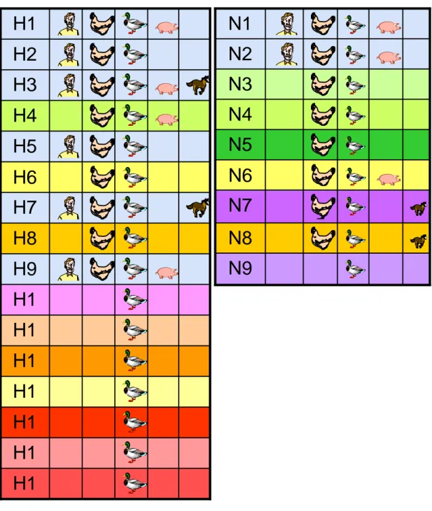
Influenza A virus can infect a wide range of species, including both avian and mammalian hosts. At the present time, aquatic birds are generally indicated as the natural reservoir of the gene pool of influenza A viruses, which they perpetuate in nature, from one year to the next, by well-adapted host/parasite relationships. Influenza A viruses are preferentially endemic in water birds, such as ducks, geese, and shore birds (gulls), which usually do not fall ill from this infection. All 16 known hemagglutinin (HA) and 9 neuraminidase (NA) subtypes of influenza A viruses have been isolated from wild waterfowl and seabirds (Webster et al, 2006 ) (Fig. 4).
The presence of all subtypes influenza A viruses in wild aquatic birds poses serious health risks to a wide range of animal species. Few distinct subtypes have been isolated from pigs, horses, seals, whales and human beings (Fig. 5). Equines can be only infected with influenza viruses H3N8 and H7N7, although the latter has not been detected in horses in recent years and may have disappeared completely (Paillot et al., 2006). Dogs can also be infected with the H3N8 equine variant (Dubovi et al., 2008; Crawford et al., 2005). A variety of influenza viruses have been found in aquatic mammals (H1, H3, H4, H7, and H13 containing variants). The avian flu H5N1 outbreak in cats, leopards and tigers demonstrated that it was also possible to infect felids with this strain (Keawcharoen et al., 2004; Leschnik et al., 2007).
Asymptomatic infection in ducks are due to the circulation of lowly pathogenic avian influenza viruses (LPAIVs) mainly transmitted by the faecal-oral route (Webster et al., 1992). Through their movement and migrations,
25 wild birds carry the virus to various countries and environments where new hosts and/or parasite populations can be present. According to the main bird migratory routes, spatial and/or temporal segregation exists among wild bird populations of the world. This leads to the existence of numerous avian influenza strains that are basically grouped by several sublineages streaming in a few different geographical lineages, mainly represented by the Eurasian, Australian, North and South America clades (Suarez et al., 2004; Gorman et al., 1990). However, gene segment reassortment between different geographical clades have been reported, showing the existence of some contact areas among avian populations of different continents. The mixing, at a single location, of various populations of migratory birds could also explain the genetic variation recently described in AIVs isolated from natural reservoir species (Spackman et al., 2005; Chen et al., 2006): the selection pressure, naturally occurring at low level in these well adapted hosts, may increase when several bird groups having specific influenza immunity share different viruses.
Although some of these subtypes are non-pathogenic/nonvirulent within their natural hosts and have been present in these animal reservoirs for many centuries, various subtypes are highly virulent within their natural host species and to other species (Webby et al., 2007). For example, the changing role of the highly pathogenic avian influenza virus (HPAIV) H5N1 subtype in both wild and domestic ducks has recently been documented as a potential public health hazard because they are zoonotic agents with the theoretical ability – after genetic adaptation – of a human-to-human transmission (Hulse-Post et al., 2005).
Among domestic species utilized for meat and egg production, land-based birds (such as chickens and turkeys) are highly susceptible to avian influenza viruses. Moreover in these birds the well-adapted host/parasite interaction, typical of the aquatic avian reservoir, tends to change into an unstable balance
26 characterized by clinical disease ranging from mild respiratory symptoms to lethal illness forms able to kill 100% of the infected groups (Easterday et al., 1997). Fatal cases are usually due to the H5 and the H7 virus subtypes that, introduced in domestic avian species, can evolve into highly pathogenic (HP) strains responsible for systemic disease.
Commercial poultry farms, “wet markets”, (where live birds and other animals are sold), backyard poultry farms, commercial and family poultry slaughtering facilities, swine farms, human dietary habits and the global trade in exotic animals have all been implicated in the spread of influenza A viruses (Greger, 2006). The “wet markets” of Southeast Asia, where people, pigs, ducks, geese and chickens (and occasionally other animals) are in close proximity pose a particular danger to public health (Webster, 2004; Bush, 2005; Greenfeld, 2006;Lau et al, 2007).
27
H1
5
H1
6
H1
4
H1
3
H1
2
H1
1
H1
0
H9
H8
H7
H6
H5
H4
H3
H2
H1
N9
N8
N7
N6
N5
N4
N3
N2
N1
28
Fig. 5 Interspecies transmission of influenza A viruses.
Solid lines; frequent and/or confirmed transmission events. Dotted lines; possible and/or occasional transmission events.
29
1.3.2. EMERGENCE OF PANDEMIC INFLUENZA VIRUSES
Pandemics are epidemics that rapidly spread on a worldwide scale, caused by pathogens against which humans have no immunity that infect a large part of the population and lead to associated serious illnesses.
Influenza pandemics occur when an influenza virus with a hemagglutinin, against which there is little or no existing immunity, emerges in the human population and efficiently transmits from human to human. Three human influenza pandemics occurred in the twentieth century, in 1918, 1957, and 1968. Human influenza pandemics are caused by emerging influenza viruses from non-human reservoirs. From the three influenza pandemics of the twentieth century, the 1918 pandemic was possibly caused by an influenza virus with an avian origin (Taubenberger et al., 2005; Rabadan et al., 2006) and the other two, in 1957 and 1968, were caused by new strains that were combinations of avian and human viruses through the process of reassortment (Lindstrom et al., 2004; Scholtissek et al., 1978)
There are two major reassortant hypotheses:
the avian influenza virus transmits to humans first and then reassorts with human influenza viruses;
both the avian and human influenza viruses infect and reassort in an unknown mammal, for example pigs; then the novel reassortant virus is transmitted to humans (Ito et al, 1998).
30
Introduction of an avian influenza virus in toto into the human
population
Taubenberger and his colleagues (2005), after analyzing the complete genome of the 1918 Spanish flu virus, proposed that the 1918 virus was not a reassortant virus (like those of the 1957 and 1968 pandemics), but more likely an entirely avian-like virus which crossed the species barrier to humans without an intermediate mammal and infected and adapted to humans. However, it should be noted that recent studies have found that the receptors preferentially binding avian influenza viruses are located in the lower portions of the human respiratory system (Shinya et al., 2006; Gambotto et al, 2008), explaining how avian influenza viruses can directly infect humans and also why human-to-human transmission with non-adapted avian influenza viruses is rather limited. If changes were to occur in virus receptor binding affinity of avian influenza viruses that permitted replication of avian influenza viruses also in the upper human respiratory system, this would result in efficient human-to-human transmission and the possibility of a pandemic (van Riel, 2006).
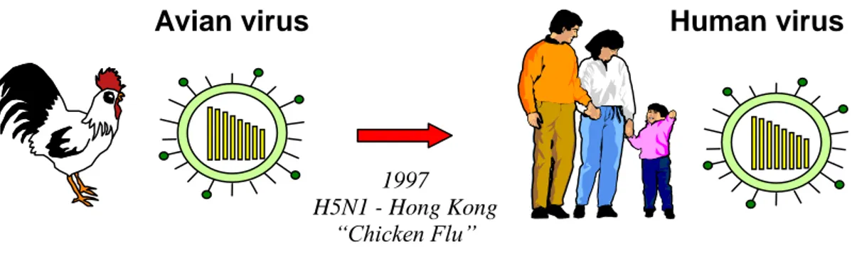
The first documented instance of human infection with avian influenza A virus H5N1 occurred in Hong Kong in 1997. Infection was confirmed in 18 individuals, 6 of whom died. Infections were acquired by humans directly from chickens, without the involvement of an intermediate host. An influenza virus (H5N1) known to infect only birds previously was found to infect human causing disease and death. Prior to the human outbreak, the H5N1 virus was found to cause extensive death in chickens in three farms in Hong Kong (Shortridge et al., 1998) (Fig. 6).
31
Then, in early 2003, a father and his nine-year-old son, who had been visiting relatives in Fujian Province in China, were hospitalized when they returned to Hong Kong with H5N1 infection. A second of the father‟s children – an eight-year-old girl – had died of an undiagnosed respiratory illness, while visiting in China. The father subsequently succumbed, the hospitalized boy recovered. The virus responsible was found to be a mutated strain of the H5N1 virus that had first surfaced in human beings in Hong Kong, in 1997.
The largest outbreak of subtype H7 infections in humans to date occurred in the spring of 2003, when an HPAI (H7N7) virus was detected in commercial poultry farms in the Netherlands and necessitated the culling of >30 million birds (Fouchier et al., 2004). All internal genes of this virus were of avian origin and were found to be related to low pathogenicity viruses detected during surveillance of ducks in the region in 2000. Eighty-six persons involved in the culling operation and 3 of their family members who had not been in contact with infected poultry had virologically confirmed subtype H7 illness, which suggests that limited
human-to-Avian virus Human virus
1997 H5N1 - Hong Kong
“Chicken Flu”
Fig. 6 Introduction of an avian influenza virus in toto into the human
32 human transmission of the avian virus also had occurred. Among these persons, 78 had conjunctivitis, 5 had conjunctivitis and respiratory symptoms, 2 had respiratory symptoms only, and 1 died, a veterinarian who had visited several infected farms and in whom an acute respiratory distress syndrome and pneumonia developed (Koopmans et al., 2003; Meijer et al., 2004).
From late 2003 H5N1 influenza viruses spread in an unprecedented manner across Asia, affecting poultry in Vietnam, Thailand, Indonesia, China, Japan, South Korea, Cambodia, and Laos. Hundreds of millions of chickens and ducks were culled in an effort to stop the spread. The outbreak appeared largely under control in March 2004. However, in July 2004 the virus reemerged in Thailand, Vietnam, and China and was isolated for the first time in Malaysia.
According to the World Health Organization (WHO) (http://www.who.int/ csr/disease/avian_influenza/country/cases_table_2009_05_15/en/ index.html)., as of May 2009, 424 human infections with H5N1 have been confirmed, resulting in 261 deaths. Although several family clusters of H5N1 virus infection have been described, sustained human-to-human infection has not occurred. Hence, these H5N1 viruses are characterized by a high mortality rate but inefficient spread among humans. It should be noted that 90% of H5N1 cases (348) and 91% of H5N1-related deaths (223) have been in only five countries: Indonesia, Vietnam, Egypt, Thailand and China. Nevertheless, since 2005, the WHO Pandemic Alert Level dealing with the H5N1 HPAIV epidemic has remained at Phase 3: “Human infection(s) with a new subtype, but no human-to-human spread, or at more rare instances of spread to a close contact” (WHO, 2005). Human-to-human transmissions of HPAIV H5N1 have been found within individual families in Thailand, Indonesia and most probably China (Ungchusak et al, 2005; Normile, 2007). The epidemic situation in these countries has come close to a Phase 4 Pandemic Alert:
33 “Small cluster(s) with limited human-to human transmission but spread is highly localized, suggesting that the virus is not well adapted to humans” (WHO, 2005). However, because the virus has remained localized within these families, the WHO has not raised the alert level.
34
The pig as a mixing vessel for influenza viruses
The obvious potential of creating novel reassortant influenza viruses in pigs has led to the “mixing vessel” theory (Fig. 7). The theory was first proposed by Scholtissek and his colleagues (1985) based on the understanding that human influenza A viruses do not spread easily to birds and vice versa, whereas the species barrier to pigs is rather low (Scholtissek, 1990; 1996). The antigenic and genetic similarities between certain subtypes of avian, swine and human influenza viruses and the susceptibility of swine to avian and human influenza viruses form the basis of this theory. Most avian and human influenza viruses preferentially bind to specific receptor types having SA α-2,3 Gal (avian receptor) - or SA α-2,6 Gal (mammalian receptor) - terminated saccharides, respectively (Rogers and Paulson, 1983; Rogers and D‟Souza, 1989). Both receptors have been found in the tracheal epithelium of swine (Ito et al, 1998), providing solid molecular evidence for pigs as “mixing vessels” for human and avian influenza viruses, therefore, pigs are considered as an intermediate host for the adaptation of avian influenza viruses to humans or as mixing vessels for the generation of genetically reassortant viruses.
This phenomenon was responsible for the 1957 pandemic when the human H1N1 strain that had been circulating since 1918 reassorted to become a human H2N2 strain with new PB1, HA, and NA segments of avian origin. Also, in 1968, the reassortment of the PB1 and HA segments created a new human H3N2 strain which is currently co-circulating with the human H1N1 strain that reappeared in 1977 (Nakajima et al., 1978; Scholtissek et al., 1978). Analysis of the 1957 H2N2 pandemic strain found that the emergent virus resulted from the acquisition by previously circulating human H1N1 of three new gene segments of avian origin (the H2 gene, the N2 gene, and one other). Similarly, the 1968 pandemic H3N2 virus acquired two new genes from an avian virus closely related to viruses isolated from ducks in Asia in 1963.
35
Fig. 7 Pig as a mixing vessel.
Genetic shift 1957 H2N2 “Asian” 1968 H3N2 “Hong Kong”
Avian virus Human virus
Reassortant virus 2,6 2,3 - 2,6 2,3 Co-infection: human and avian viruses
36
1.3.3. A NOVEL SWINE-ORIGIN INFLUENZA A VIRUS H1N1 IN
HUMANS
During April 2009, a novel H1N1 virus was detected in epidemiologically unrelated cases of influenza-like illness in California and was subsequently recognized to be the cause of a major outbreak of respiratory disease in Mexico that had been ongoing for some weeks previously. The virus was found to be an H1N1 virus that was antigenically and genetically unrelated to human seasonal influenza viruses and genetically related to viruses known to circulate in swine.
As of 25th of April, the virus caused over 17919 deaths in 214 countries in the Americas, Europe, Asia and Australasia (WHO). On June 11, 2009, the World Health Organization raised its pandemic level to the highest level, Phase 6, indicating widespread community transmission on at least two continents. The virus contains a novel constellation of gene segments, the nearest known precursors being viruses found in swine and it likely arose through reassortment of two or more viruses of swine origin. H1N1, H1N2 and H3N2 subtype swine influenza viruses have occasionally infected humans before but such zoonotic transmission events did not lead to sustained human-to-human transmission in the manner this swine-origin influenza virus has done. Clinical disease generally appears mild but complications leading to hospitalization can occur, especially in those with underlying lung or cardiac disease, diabetes or those on immunosuppressive therapies. Children and young adults appear to be the most affected, perhaps reflecting protection in the elderly owing to exposure to H1N1 strains before 1957.
The current swine flu has emerged from reassortment of gene segments from North American and Eurasian swine strains that have been undetectably circulating in humans for a long period of time (Garten et al., 2009). The H1-subtype HA of swine flu differs substantially from recent H1 HAs of seasonal
37 influenza A viruses. Consequently, most of the human population lacks immunological protection against this virus, resulting in a pandemic. It is unusual for a pandemic virus to have the same HA subtype as currently circulating seasonal strains.
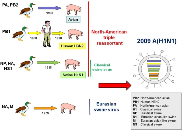
Genetic analysis of 2009 H1N1 viruses isolated in North America, Europe and Asia revealed quadruple reassortant swine influenza A viruses that have not been recognized previously in pigs or humans. The virus resulted from the reassortment of North American H3N2 and H1N2 swine viruses (triple reassortment viruses: avian/swine/human with Eurasian swine viruses) (Morens et al., 2009) . Sequence analysis also suggests that PB2 and PA genes originated from American H3N2 avian virus; a PB1 originated from H3N2: HA, NP, and NS genes originated from classical swine virus: and NA and M genes originated from Eurasian swine virus.
The NA and M gene segments are in the Eurasian swine genetic lineage. Viruses with NA and M gene segments in this lineage were originally derived from a wholly avian influenza virus and thought to have entered the Eurasian swine population in 1979 (Pensaert et al., 1981), continue to circulate throughout Eurasia, and have not been previously reported outside Eurasia. The HA, NP, and NS gene segments are in the classical swine lineage. Viruses that seeded this lineage are thought to have entered swine around 1918 and subsequently circulated in classical swine viruses and triple reassortant swine viruses (Olsen, 2002). The PB2 and PA gene segments are in the swine triple reassortant lineage. Viruses that seeded this lineage, originally of avian origin, entered swine in North America around 1998. Finally, the PB1 gene segment is in the swine triple reassortant lineage. This lineage of PB1 was seeded in swine from humans at the time of the North American swine triple reassortment events (Zhou et al., 1999) and was itself seeded from birds around 1968 (Kawaoka et al.,1989). However, the human-like PB1 gene and the avian-like PB2 and PA genes have been circulating in pigs since
38 1997/1998 (when triple reassortant swine viruses were first isolated), and have likely undergone adaptation to pigs. These viruses do not possess markers associated with high pathogenicity.
Figure 8 summarizes the host and lineage origins for the gene segments of the 2009 A/H1N1 virus.
39
1.3.4. VIROLOGICAL INFLUENZA SURVEILLANCE IN ITALY
AND THE
2009 PANDEMIC A/H1N1 VIRUS
In Italy, influenza surveillance is routinely based on integrated epidemiological and virological national networks. Seasonal virological surveillance is carried out by the WHO National Influenza Centre (NIC) located at the National Institute of Health (Istituto Superiore di Sanità, ISS), which coordinates the activities of 15 collaborating laboratories. In case of emergency, further 12 hospital laboratories are involved in the surveillance activities. The NIC performs quality control assessment and laboratory validation activities specifically aimed to strengthen the diagnostic capabilities of the Italian laboratory network. When a pandemic occurs, the major task of the NIC is to rapidly detect and/or confirm cases of influenza and perform virus characterisation. In response to the spread of the pandemic A/H1N1 virus in the United States and Mexico, virological surveillance activities throughout Italy were maintained effective beyond the usual deadline (week 17) of seasonal influenza surveillance. Since 28 April 2009, the Ministry of Health (MoH) undertook a number of actions, including the recommendations to enhance surveillance activities and laboratory confirmation of suspected and probable cases, which were published as a national guidance document [Ministry of Health of Italy. Influenza A (H1N1). Azioni del Governo
[Actions of the government] [in Italian]. Available from:
http://www.ministerosalute.it/dettaglio/approfondimentoFocusNuovo.jsp?id=1 3&sub=1&lang=it&area=influenzaA]. The main scope of the guidance was the early identification of individuals presenting with influenza-like illness and recent history of travel to the affected areas and the adoption of population distancing measures (early isolation of cases and precautionary school closure) and antiviral prophylaxis of close contacts of cases, in order to contain the spread of pandemic A/H1N1 virus cases in the country. In particular, a
seven-40 day period of isolation at home of travellers coming back from affected areas, although asymptomatic, was initially recommended. According to the above document, pharyngeal and/or nasal swabs should be collected by family and/or hospital doctors from each suspected case (i.e. a case fitting the clinical and epidemiological criteria (Commission Decision of 30 April 2009 amending Decision 2002/253/EC laying down case definitions for reporting communicable diseases to the Community network under Decision n° 21/19/98/EC. 2009/363/EC. Official Journal L 110/58. 01.05.2009. Available from:http://eurlex.europa.eu/LexUriServ/LexUriServ.do?uri=OJ:L:2009:110:0 058:0059:EN:PDF) and two separate aliquots of the samples should be sent – one to the regional reference laboratory and another one to the NIC. Since 20 May 2009, following the updated MoH recommendations, only specimens from probable cases (i.e. cases with positive test results for influenza A virus) should be sent for influenza pandemic A/H1N1 virus confirmation by NIC. The notification of confirmed cases of infection to the MoH is done by the NIC.
As of 10 June 2009, the number of cases of influenza pandemic A/H1N1 virus infection reached 27,737 in 74 different countries, with 141 deaths. On 11 June 2009 the WHO raised the level of pandemic alert to phase 6.
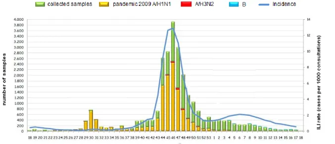
The pandemic influenza A/H1N1 virus, emerged in Italy in April 2009, has clearly been the predominant virus circulating among the population while seasonal influenza A/H1N1, A/H3N2 and B viruses circulated simultaneously to a lesser extent.
From week 18/2009 (April 2009) to week 18/2010 (April 2010), 13.309 influenza virus detections have been reported by the Italian laboratory network: 13.258 were influenza A (99,6%) and 51 (0,4%) were influenza B. Of the influenza A viruses, 12.903 (97,3%) were subtyped, with 12.981 being pandemic A/H1N1 and 12 A/H3N2 (Fig. 9).
41 The pandemic virus has been characterized by mild and self –limiting
disease
in the over-whelming majority of cases. However, severe and fatal cases,
many of them with primary viral pneumonia, have been occurring in age
groups where such clinical outcomes are very rarely seen in seasonal
influenza. It is important to better understand what viral and host-related
factors determine this dichotomy.
Fig. 9 Number of specimens collected and specimens positive for influenza, by
42
2. AIM OF THE STUDY
The sequences of 133 pandemic A/H1N1 strains, from patients showing different clinical outcame (12 from fatal, 24 from severe and 97 from mild cases) have been examined at the Italian WHO National Influenza Centre (NIC) located at the National Institute of Health (Istituto Superiore di Sanità, ISS) in order to provide additional information on the genetic characteristics of pandemic A/H1N1 viruses circulating in Italy.
The aim of this study was to carry out a molecular characterization, by
sequencing the entire genome of 133 pandemic A/H1N1 samples,
collected between May 2009 and April 2010 in Italy
, to determine the
evolutionary relationships of their gene segments, compared to other
recent pandemic A/H1N1 virus sequences obtained from GenBank and
to some recent Italian swine and European human seasonal isolates.
Amino acid sequence analysis was also performed, with the aim to detect particular mutations potencially altering virus antigenicity or pathogenecity and to identify molecular markers of virulence and sensitivity to antivirals.
43
3. MATERIALS AND METHODS
Upper respiratory specimens such as nasopharyngeal aspirates or nasopharyngeal swabs, throat or nose swabs are suitable for the detection of influenza viruses.
Word Heath Organization recommends that suspected clinical cases of pandemic H1N1 influenza A infection are confirmed by:
(1) the isolation and identification of swine-origin influenza viruses (S-OIV), (2) the detection of a fourfold rise of neutralization or haemagglutination inhibition antibodies to S-OIV,
(3) specific RT-PCR assays that differentiate S-OIV from seasonal influenza viruses.
(World Health Organization. WHO information for laboratory diagnosis of new influenza A(H1N1) virus in humans. http://www.who.int/csr/resources/publications/swineflu/WHO Diagnostic Recommendations H1N1 20090521.pdf)
3.1 MATERIALS
Respiratory specimens
Specimens for virus detection or isolation should be collected within 3 days after the onset of symptoms and rapidly transported to the laboratory. A nasopharyngeal aspirate, nasal swab, nasal wash, nasopharyngeal swab, or throat swab are all suitable for diagnosis.
The timing of specimen collection is very important since the yield is the highest for respiratory specimens obtained within four days of onset of symptoms. Different types of respiratory specimens can be used. Nasal washes and nasopharyngeal aspirates tend to be more sensitive than pharyngeal swabs. Swabs should be transported in virus transport medium to prevent desiccation. All specimens should arrive at the laboratory as soon as possible to avoid any degradation. Transportation in virus transport medium on ice or with
44 refrigeration at 2-8° C is recommended if any delay in transportation is expected.
Pharyngeal and/or nasal swabs collected by family and/or hospital doctors from each suspected case were sent to the NIC.
Blood specimens
Blood (whole blood, serum) specimens were also collected for the purpose of antibody serology (determining the presence of antibodies to influenza).
3.2 METHODS
3.2.1 ISOLATION METHODS
The S-OIV H1N1 can be isolated in MDCK cells in the presence of trypsin (as for other seasonal influenza viruses) or in embryonated hens egg. Virus culture is recommended to be carried out in 3 or 2 with BSL-3 practice.
A. Embryonated egg culture
Since this technique requires the supply of fertilized chicken eggs and special incubators it is no longer used for the routine diagnosis of influenza infection. However egg isolation provides high quantities of virus and is a very sensitive culture system. Reference laboratories therefore utilise this culture system to ensure high sensitivity and to enable the production of virus stocks for epidemiological monitoring.