CORSO DI DOTTORATO IN
“FISIOPATOLOGIA dell’APPARATO CARDIOVASCOLARE e RESPIRATORIO”
Università degli Studi di Pisa Direttore Prof. A. Mussi
“A new extraction technique in lead transvenous
removal : The Internal Transjugular Approach.”
Candidate: Dr. F. Lapira Tutor: Prof. A. Balbarini
Cardiac and Thoracic Department, Cisanello Hospital, Pisa.
INTRODUCTION
As know, the recent expansion of implanted devices for cardiac pacing and defibrillation and the rise of mean age is consequently associated to increase in device-related complications number. However , the management of infection or malfunction leads has been modified by the introduction in the clinical pratice of transvenous lead removal, a technique able to treat all the leads-related complications1,
The indications, facilities and training to removal chronically implanted leads have been codified from the NASPE Policy Consensus Conference in 20002.
The transvenous lead extraction is a complex procedure, often associated to rare but severe complications; it needed by availability of new technique and materials in continous evolution and by the possibility of different approaches, beside resulting hardly dependent from the staff experience3.
Actually, two different techniques in transvenous leads removal are used: the extraction with mechanical dilators or with powered sheaths. The first one, was introduced by Byrd in the late ’80s; it is performed with polypropylene sheaths and other dedicate tools (locking stylets, Lasso loop-retriver, Catcher, Transfemoral workstation etc..), that allow to apply longitudinal forces of pression, traction and countertraction, useful to
adherences dissection above the lead all over the venous tree and inside the heart4,5. The literature report a high success rate of this technique, with a low major-complications’ incidence6. On the other hand, the powered techniques, developed in the ‘90s, use a source of energy (Laser or Radiofrequency) delivered at the edge of a special sheath; the advantage is to make dissection of binding sites easier and faster. However, the success rate from literature is similar to the mechanical one, but the powered removal procedures are more expansive and more frequently associated to serious complications as tamponade, hemothorax, pulmonary embolism, led migration and death7,8.
Independently from the choice of one technique, there are two possible conventional approachs: the superior one (through the original venuos entry site, when the proximal end of the lead is exposed) or the inferior one (through the femoral vein, useful for example in case of total intravascular leads)9.
Percutaneous lead removal has been still associated with a small but significant procedural failure, morbidity and mortality. Many factors can influenced effectiveness, complexity, duration and outcome. We can describe clinical factors (related to the patient and lead story) and technical factors (structural or acquired characteristics of the leads)10.
As known from the literature, the success rate of removal is strongly affected by the presence of fibrotic and calcified scar tissue develops
above the lead, by the impossibility of advancing a stylet into the lead usually caused from damage (previous or new damage during the dilation), or by the presence of free-floating intravascular leads11.
According to these observations, in order to resolve these specific troubles, we have been developing a modified percutaneous mechanical dilatation technique: the Internal Transjugular Approach (ITA). Really, this method is not a new technique, just a modification of the standard one, using as different approach the right internal jugular vein. In our personal experience12, it presents some advantages, because it results able to overcome the most important procedural difficulties (strongly adherences, damage or intravascular leads), improving success rate and reducing complications of the transvenous lead extraction13.
AIM OF THE STUDY.
The aim of this study is to evaluate if the Internal Transjugular Approach can enhance the success rate of the standar mechanical technique of transvenous lead extraction, without an increase of complications incidences (evaluation of effectiveness and safety), useful in case of particulary difficult procedures.
MATERIALS and METHODS
POPULATION
Between October 2005 and July 2007, we evaluated consecutive patients admitted at our Electrophysiology Laboratory for submission to transvenous lead extraction procedures. The study-inclusion criterions were the indications to the procedures, according to the currently used NASPE Guidelines.
The indications to transvenous leads extraction are divided in Class I (general agreement that leads should be removed) and Class II (leads are often removed but there is some divergence of opinion with respect to the benefit versus risk of removal) and moreover they are classified as following:
I-a) Sepsis (including endocarditis) secondary to an infection of the pacing system;
I-b) Retained lead, lead fragment or extraction hardware;
I-c) Occlusion of all useable veins, with the need to implant a new lead;
I-d) Lead interfering with the operation of another implanted device; II-a) Localized pocket infection, erosion or chronic draining sinus that does not involve the transvenous portion of the lead;
II-b) Occult infection (no source can be found, pacing system is suspected);
II-c) Chronic pain at the pocket or lead insertion site not manageable without lead removal;
II-d) A lead that, due to its design or its failure, may pose a threat to the patient;
II-e) Interference with the treatment of a malignancy;
II-f) Traumatic injury for which the lead may interfere with reconstruction;
II-g) Non-functional leads in young patient.
The clinical and pacing notes of all patients were reported into a structured database and then evaluated. In particular, we recorded:
a) patient clinical data (age, sex, case history and comorbidity);
b) lead data (leads total number; trade-name, implant period, pacing or defibrillating type, site of pacing, intravascular or exposed location, access vein, eventually previous procedures or damages);
c) indication to removal (according to the Guidelines).
We randomized the patients, through a simple randomization order, into two Groups:
A) Standard Group,
The Group-A patients and theirs relatives leads were submitted to a standard removal procedure, according to the global experience, through the mechanical dilation technique describe in literature.
The Group-B patients were approached with the same standard procedure as first step, only in case of failure we performed our personal technique, using the Internal Transjugular Approach (ITA), as second step.
PROCEDURE
The procedures were performed in the Cardiac Electrophysiology Laboratory, in Cardiac and Thoracic Department, at the Cisanello Hospital (Pisa). We obtained informed consent, also regarding the possibility to use the availables different approaches, in order to obtain the clinical success of the procedure.
Before the procedure, all the patients were submitted to 2P X-Ray and TransthoracicEchography, than during the procedure (with local anaesthesia o general sedation) were used the application of cutaneous pads for defibrillation, transvenous temporary pacing, invasive arterial blood pressure and pulse oximetry monitoring. In our Hospital was also possible worked with the availability of cardiothoracic surgery stand-by.
The procedures can been supported by intracardiac echography (ICE), performed using catheters equipped with an echo-transducer at the tip, introduced through the femoral vein. This is a recent technological
improvement very useful to monitoring the procedure, to determinate the real relationship between leads and anatomical structure, to detect the presence of vegetations and their outcome during dilation, finally to monitor the possible occurrence of early complications14. However, it result expensive and needed a dedicated operator, than we used ICE only in selected cases (such as difficult, old, intravascular or multiple leads and suspicion of vegetations).
The Standard mechanical dilation technique was performed using the extraction system provided by Cook Vascular Inc. (Leechburg, PA, USA), equipped by polypropylene sheaths in different size (from 7 to 14 F) with the possibility of telescopic combination too, locking stylets and other specific tools (such as Transvenous workstation, tip deflecting wire, basket and loop retriever). The technique consist of a combination of different forces: traction by the stylet, rotation alternatively clockwise and counter-clockwise by the inner dilating sheaths and countertraction at the tip of the lead by the outer telescopic one. The result could be the mechanical dissection of the adherences around the body lead, the detachment of the tip lead from the cardiac wall and the total removal from the venous tree.
In case of exposed lead, i.e. when the proximal end is out of the venous entry site and accessible from the pace-maker pocket, we always used a superior approach as first choice. After the lead preparation (cutted and freed out of its venous entry site, than inserted the stylet and secured
by suture material), we performed the mechanical dilation in order to dissect the binding sites, following the course of the body lead until the tip. (Fig. 1)
In case of free-floating leads, according to the global experience, we used as first choice an inferior approach, performed by the Transfemoral workstation (an extra-long sheath, 16 F, and other dedicated intravascular tools).(Fig. 2). When the lead tip result free, inside the venous system, through femoral vein can be used a Lasso loop-retriver to catch the tip and than pulled it back into the workstation, in order to remove the lead, also in case of adherences dilatation necessity (performed by use of the workstation sheath itself). When the lead present proximal end totally intravascular, not accessible from the pace-maker pocket and anchored tip in the heart, as the standard inferior approach described in leterature, we tried with Transfemoral workstation too15. In this case a deflecting wire is introduced trough the femoral vein in order to grasped the body lead and pulled the free proximal end in an inferior position, then trying to catched and dilated the lead from the low.
According on our study design, only in case of standard technique failure and only in the randomised Group-B patients, we performed the Internal Transjugular Approach (ITA). (Fig. 3). In order to performed ITA, the right internal jugular vein was percutaneously cannulated using an 11 F introducer (Avanti, Cordis Corp., Miami, USA). After remove stylet and
suture from the lead (obviously only in case of exposed leads, no in intravascular ones), by a control tip deflecting wire (Cook Vascular Inc., Leechburg, PA, USA) introduced through the right femoral vein, the body lead was catched and pulled below, in order to slipping the proximal end at right atrium or superior vena cava level, making it free-floating. Then, through a Lasso loop-retriver (Osypka GmbH, Grentzig-Whylen, Germany) advanced into the jugular vein, the free-floating proximal end of the lead was captured and exteriorized in laterolcervical region. Now, a percutaneous procedure for exposed leads can been peformed, using dilating sheaths. (Fig. 4)
According on the NASPE Guidelines, all the procedure results were defined based on the Radiological outcome (for one single lead) as:
- complete success (removal of the whole lead),
- partial success (removal of the lead except a fragment of less than 4 centimeters from the tip),
- failure (a significant fragment is left, or stop the procedure because of a major complication).
Complications were classified as major or minor ones, according on the guidelines too. The major complications are tamponade, hemothorax, pulmonary embolism, led migration and death
RESULTS
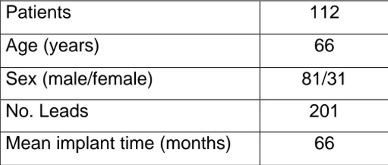
Between October 2005 and July 2007, we evaluated 112 consecutive patients (81 males, mean age 66.2 years) admitted at our E.P Laboratory for submission to transvenous lead extraction procedures (201 leads considered for removal, mean implant time 66 months). (Table 1).
The patients were previously randomized with a simple order, into two Groups: A) Standard Group,
B) Internal Transjugular Approach (ITA) Group.
The Standard group was formed by 56 patiens, of which 42 males (mean age 68 years); the procedure was performed for 98 leads (mean lead number for patient 2.15), with mean implant time 73 months. Pacing leads were 79 and implantable cardiac defibrillating 19; 32 were atrial, 58 ventricular and 8 coronary sinus; only 4 were free-floating leads with proximal end intravascular, of which 1 atrial and 3 ventriculars.
The ITA group was formed by 56 patiens too, of which 39 males (mean age 65 years); the procedure was performed for 103 leads (mean lead number for patient 2.2), with mean implant time 63 months. Pacing leads were 89 and implantable cardiac defibrillating 14; 37 were atrial, 56 ventricular and 10 coronary sinus leads; only 6 were free-floating leads with proximal end intravascular, of which 4 atrial and 2 ventriculars (Table 2).
As demonstrated by the T-student statistical analysis, the two group didn’t have significant differences in theirs characteristics.
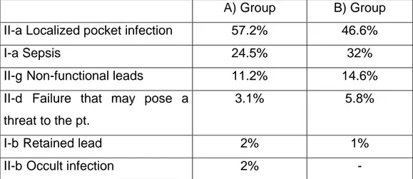
According to the guidelines classification, in our population the indications to removal in A-Standard Group patients were: II-a) Localized pocket infection, erosion or chronic draining sinus that does not involve the transvenous portion of the lead (57.2%); I-a) Sepsis (including endocarditis) secondary to an infection of the pacing system (24.5%); II-g) Non-functional leads in a young patient (11.2%); II-d) A lead that, due to its design or its failure, may pose a threat to the patient (3.1%); I-b) Retained lead, lead fragment or extraction hardware (2%); II-b) Occult infection, i.e. no source can be found but pacing system is suspected (2%). Indications to removal in B-ITA Group patients were as following: II-a) 46.6%; I-a) 32%; II-g) 14.6%; II-d) 5.8%; I-b) 1%; No occult infection (Table 3).
Transvenous extraction was attempted in all the 98 leads belonged to the A-Group, with a total removal in 92 cases (success rate 93.9%), a partial removal in 1 case (1.02%) and 5 failure procedure (unsuccess rate 5.1%). On the other hand, the transvenous extraction attempted in all the 103 leads of B-Group was completely successful in 100 cases (success rate 97.1%), a partial success was obtained in 1 lead (1.9%) and 1 lead was not removed (unsuccess rate 1%). The comparative overall results are explicated in Table 4.
In the B-group the partial removal of two leads was achieved during the first step, before the cross-over to ITA; one lead was not removed because of a major complication (light cardiac tamponade) that stopped the procedure, before the cross-over to ITA, too. The jugular approach was performed as second step in 8 out of 103 leads, in which the standard technique of dilatation alone resulted ineffective (potential not success); all these 8 leads were totally removed (success rate of ITA 100%). The mean implantation time was 88 months; 4 of these were free-floating leads with intravascular proximal end (2 atrial and 2 ventricular), 4 were ventricular exposed leads (3 of which defibrillating leads); the leads location were 2 atrial and 6 ventricular.
The failure of standard technique in these 8 leads, that needed the cross-over to Internal Transjugular Approach, was due to different factors: impossibility to dilation free-floating leads through the inferior approach because of the lead course itself (4 pacing leads,of which 2 atrial and 2 ventricular); lead damage, with impossibility of advancing the stylet (only 1 ventricular pacing lead); calcified scar tissue that imprison the body lead strongly (2 defibrillating lead with adherences at Tricuspidalic or proximal coil’s level); difficulty to performed dilatation because of the rib-clavicular narrow or the Subclavian-Anonima corner (1 defibrillating lead). (Table 5).
In our total population, major complication occurred in 2 cases (incidence rate 1.7%): one light pericardial effusion, treated with medical
therapy, and one cardiac tamponade with ventricular fibrillation and cardiac arrest, successfully treated with pericardiocentesis. Minor complications were: 2 lead implanted dislodgement, 18 cases of hypotension, 2 substained Arrhythmias (not requiring electrical cardioversion), 1 haematoma at the pacemaker pocket and 1 at the venous entry. However, no complications were directly related to the jugular approach. (Table 6)
DISCUSSION
According to the global experience, our results confirm that transvenous lead extraction is actually a procedure effective and safe, able to resolve the device-related complications in patients with indications as codified in the current Guidelines.
The success rate from the literature has a range from 96% and 90%, the failure from 0.6% and 3%, the major complications rate from 2.6% and 1.3%. The most recent results of the US extraction Database were reported for 6420 leads in 4090 patients: 93% of the leads were completely extracted, 5% partially extracted and 2% not removed. Major complications occurred in 1,6% of patients, including a 0,2% mortality rate16. Another recent paper reported the US experience with laser sheaths; 2561 pacing and defibrillator leads were treated in 1684 patients at 89 sites in the United States. Of the leads, 90% were completely removed, 3% were partially removed, and the balance were failures. Major perioperative complications (tamponade, hemothorax, pulmonary embolism, lead migration and death) were observed in 1,9% of patients, with in-hospital death in 0,8%. Minor complications were observed in an additional 1,4% of patients.17
Our results with standar mechanical technique are similar to the data described in literature and the major complication observed are also comparable. (Table 7)
The Aims of our study was to evalueted the effectiveness and safety of a particular approach, the Internal Transjugular one, that is really a modification of the standard mechanical dilatation and if this approach is able to enhance the standard technique itself about the success rate.
This peculiar approach has been recently describe in literature18 19 in personal experiences and it could resolve some situations that affected the difficult procedures, for example the presence of free-floating or difficult exposed leads, representing a real improvement in transvenous lead extraction technique.
As our study design show, we have compared two similar group; the first step was the same for all the patients, i.e. all the leads were approached with the standard technique using the more suitable approach , the superior or the inferior one, as describe in the most of the literature studies. Only in the B-Group, in case of standard approach failure, the procedure did not stopped and we performed a second step, trying to remove the lead through Internal Transjugular Approach. In order to guarantee the same treatment to all the patiens, we reserved ITA only as second possibility; the obvious consequence was that the cross-over to ITA was performed in more difficult cases, in which the normal procedure resulted not successed.
In the B-Group, by using the standard tecnhique, we totally removed 92 leads, partially removed 2 leads and 9 were the leads not removed; only
8 out of these 9 were submitted to the second step through Internal transjugular Approach, because in one case the procedure was stopped at the first step because of pericardial effusion and did not gone over. These leads were not removed with standard approaches because of theirs characteristics, that made difficult the procedure.
According to the current opinions, the factors that strongly affected the succes rate of removal are the presence of free-floating leads, calcified scar tissue and the impossibillity to advance the stylet into the lead. By analysing the 8 not-removed leads in B-Group, treated with ITA as second step, we found that these were free-floating leads, damaged leads, leads imprisoned in strong adherences or not dilatable leads in the narrow rib-clavicular space. All the 8 not removed leads with standard technique were totally removed after cross-over on ITA, without major complications.
From a simple comparison between the A and B Group, we can observe a further significant increase from the yet elevated 93.9% success rate for the standard mechanical dilatation, to the more elevated 97.1% success rate considering the possibility of a cross-over to Internal Transjugular Approach in case of failure, without addictioned complications.
As deduced from the literature experiences, the critical points conditioning the transvenous lead extraction procedures are the difficult sheath advancement (because of tight binding sites due to scar or calcified tissue, narrow corners in the lead course, tight space between the clavicle
and the first rib) or the impossibility to introduce the stylet (because of lead damage or intravascular location of the lead)20 21.
In the critical case of exposed leads with tenacious adhesions sites the mechanical dilatation result often difficult because of the lead cours. In particular, the mechanical force of rotation and countertraction in addition to the traction one are not completely applied longitudinally over the body lead, when the vein entry site is the subclavian one; moreover, the dilation is more difficult in case of defibrillating leads, because of theirs major diameters and especially when the sub-clavicular space result very narrowed. When the lead is exposed in laterocervical site, during the Transjugular approach, it assume a new straight course from the jugular vein to the right atrium or ventricle; then, the mechanical dilatation result more easy, allowing the longitudinal axis of the lead, the traction and countertraction forces are more effective and the narrow subclavian-clavicular space is by-passed. Besides, these conditions appeared to reduce the risk of complications, usually related to mechanical forces not longitudinally applied on the venous wall (risk of veins laceration) or to excessive countertraction on the lead tip (risk of myocardial invagination and rupture).
Moreover, in case of free-floating lead, after holding the central body using Transfemoral workstation, the proximal end can be exposed in laterocervical space via the jugular approach and thus it can be submitted
to a standard procedure as in normal exposed leads, by-passing the specific difficulty of the intravascular leads.
In our experience the possibility to adapt the technique to the specific situation may determine some advantages, improve the succes rate of the difficult procedures and reduce the complications risk.
CONCLUSIONS
The technological improvement in the field of transvenous lead extraction is a needed consequence to resolve the increase of the indications to this procedure, also due to the recent increase of the number of implanted devices and theirs related complications.
In case of malfunction or infection the solution may be the replacement of device, the repositioning or addiction of a new lead, finally the lead extraction before a new implant. The last one rapresent the best treatment, because it can completely resolve the trouble. Transvenous lead extraction is on the other hand a complex procedure, actually effective, but potentially burdened by severe complications and strongly dependent from the staff experience. Removal techniques are still considered to be in evolution in order to reach a gold standard, high success rate and low incidence of complications too.
The Internal Transjugular Approach could represent, according on our experience, an useful modification to the standardized mechanical technique, able to improve the effectiveness of transvenous lead extraction expecially in case of difficult exposed or free-floating leads, increasing success rate and safety.
FIGURES
Fig.1 Superior Approach
Fig. 2. Inferior Approach
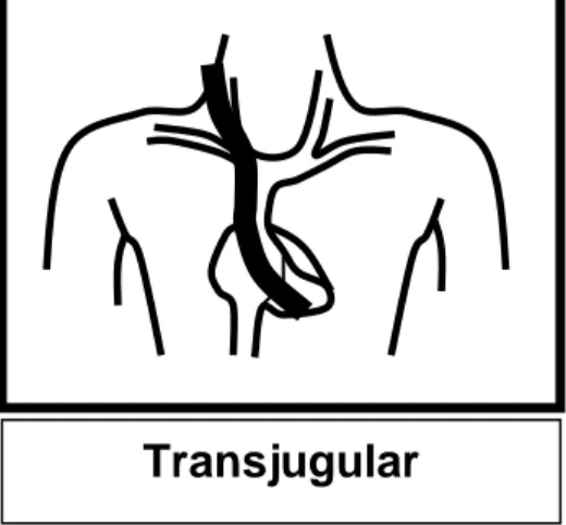
Fig. 3 Transjugular Approach
Superior Venous
Inferior Venous Entry
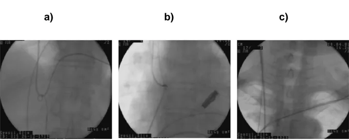
Fig. 4.Internal Transjugular Approach
a) The lead is made free floating in the venous system by using the tip deflecting guide wire introduced via the femoral vein
b) The proximal end of the lead is caught by the lassos and the lead is then exposed through the internal jugular vein
c) Once the lead is exposed, a standard procedure is performed by using mechanical sheaths
TABLES
Table 1.Global Population
Patients 112
Age (years) 66
Sex (male/female) 81/31
No. Leads 201
Mean implant time (months) 66
Table 2. Randomized two Groups
A) Group B) Group
No.Patients 56 56
Mean age (years) 68 65
Sex (M/F) (42/14) (39/17)
No. Leads 98 103
Mean implant time (months) 63 73 Pacing/Defibrillating leads 79/19 89/14 Location: Atrial Ventricular Coronary sinus 32 58 8 37 56 10 Esposed/Intravascular 94/4 97/6
Table 3. Indications to removal
A) Group B) Group II-a Localized pocket infection 57.2% 46.6%
I-a Sepsis 24.5% 32%
II-g Non-functional leads 11.2% 14.6% II-d Failure that may pose a
threat to the pt.
3.1% 5.8%
I-b Retained lead 2% 1%
II-b Occult infection 2% -
Table 4. Results
A) Group B) Group Total removal 92 leads (93.9%) 100 leads (97.1%) Partial removal 1 lead (1.02%)- 2 lead (1.9%) Failure 5 leads (5.1%) 1 lead (1%)
Table 5. Causes of standard technique failure in the sub-group of 8 leads that were submitted on the second step with ITA in the B) Group.
Free-floating leads 4 pacing leads
Damage 1 pacing lead
Strongly adherences 2 defib leads Rib-clavicular narrow 1 defib. lead
Table 6.Global complications.
Major - 1 pericardial effusion
- 1 cardiac tamponade with VF and cardiac arrest Minor - 2 lead implanted dislodgement
- 18 hypotension
- 2 subtained arrhythmias non requiring el. Cardiovertion - 1 haematoma at the PM pocket
- 1 haematoma at the venous entry
Table 7. Comparison between our results and the most recent literature.
US Extraction Database US experience-Laser Wilkoff NASPE 2000 Byrd PACE 25,2002 Our experience Pts/leads 4090/6420 1684/2561 2104/3355 1684/2561 112/201 Rate of removal (%) 93% 90% 94% 90% 93.9%(Standard) 97.1% (ITA) Major complications 1.6% 1.9% 1.3% 1.9% 1.7%
REFERENCES
1 Del Rio A, Anguerra I, Miro JM et al.: Surgical Treatment of pacemaker and defibrillator
lead endocarditis: impact of electrode lead extraction on outcome. Chest 2003 Oct; 124 (4):1451-9.
2 Love CJ, Wilkoff BL, Byrd CL, Belott PH, Brinker JA, Fearnot NE, et alt. NASPE Policy
Steatment. Recommendation for Extraction of chronically implanted transvenous pacing and defibrillation leads. PACE 2000; 23(4):544-551.
3 Bracke F.A., Meijer A, Van Gelder L.M: Pacemaker lead complications: when is
extraction appropriate and what can we learn from published data? Review. Heart 2001; 85:254-259.
4 Byrd CL, Schwartz SJ, Hedin NB, Goode LB, Feranot NE, Smith HJ. Intravascular lead
extraction using locking stylets and sheaths. Pacing Clin Electrophysiol 1990;13:1871-1875.
5 Byrd CL, Schwartz SJ, Hedin NB. Lead extraction: Indications and techniques. Cardiol
Clin 1992;10: 735-748.
6 Byrd CL, Schwartz SJ, Hedin NB (1991): Intravascular techniques for extraction of
permanent pacemakers leads. J thoracic Cardiovasc Surg 101: 989-997.
7B. Wilkoff, C. Byrd, C. Love, et alt: Pacemaker Lead extraction with the Laser Sheath:
result of pacing lead extraction with the excimer sheath (PLEXES) trial. J Am Coll Cardiol. 1999 May; 33(6):1671-6.
8 Gilligan DM, Dan D: Excimer laser for pacemaker and defibrillator lead extraction:
technique and clinical results. Laser Med Sci. 2001; 16(2):113-21.
9 Byrd CL, Schwartz SJ, Hedin NB et al.: Inferior vena cava extraction technique.(abs)
10 Heidi JM, Neal EF, Byrd CL, Wilkoff BL et al (1994): Five-years experience with
intravascular lead extraction. PACE 17:2016-2020.
11 Bongiorni M.G, Soldati E, Arena G et al (1998): The transvenous removal of permanent
pacing and defibrillator leads: indications, methods and results. Cardiologia 43:1105-1109.
12 Bongiorni MG, Soldati E, Arena G, Ratti M, Gherarducci G, Mariani M. Transvenous
removal of permanent electrocatheters for heart stimulation and defibrillation. Cardiologia 1999;44(Suppl 1 Pt 1):395-398
13 Bongiorni MG, Soldati E, Arena G et al. Percutaneous extraction of infected
pacemaker/ICD leads: which technological advances and results? In Raviele A, ed.
Cardiac Arhythmias 2001. Proceedings of the 7th International Workshop on Cardiac
Arrhythmias. Milan: Springer-Verlag Italy, 2001: 614-7.
14Bongiorni MG, Arena G, Soldati E, et al. Pacemaker/ICD leads infection: what are the
main intracardiac echocardiographic features? In Raviele A, ed. Cardiac Arhythmias 2001. Proceedings of the 7th International Workshop on Cardiac Arrhythmias. Milan: Springer-Verlag Italy, 2001: 618-22.
15
Verma A, Wilkoff BL. Intravascular pacemaker and defibrillator lead extraction: a state-of-the-art review. Heart Rhythm 2004;1:739-745.
16Byrd CL, Wilkoff B.Technique.and devices for extraction of pacemaker and implantable
cardioverter-defibrillator leads. In: Ellenbogen KA, Kay GN, Wilkoff BN, eds. Clinical cardiac pacing and defibrillation. Philadelphia, PA: WB Saunders, 2002:695-709.
17Byrd CL, Wilkoff BL, Love CJ, Sellers TF, Reiser C. Clinical study of the laser sheath for
lead extraction: the total experience in the United States. Pacing and Clin Electrophysiol 2002; 25:804-8.
18 M.G. Bongiorni, G. Giannola, G. Arena, E. Soldati, C. Bartoli, F. Lapira, G.Zucchelli, A.
Di Cori. Pacing and implantable cardioverter-defibrillator transvenous lead extraction. Ital Heart J 2005; 6 (3): 261-266.
19
M.G. Bongiorni, G. Giannola, G. Arena, E. Soldati, C. Bartoli, F. Lapira, G.Zucchelli, A. Di Cori. Effectiveness and safety of a personal approach to transvenous lead extraction. Heart Rythm 2005; May (2) Issue 1S, 281
20 Byrd CL, Wilkoff BL, Love CJ, Sellers TD, Turk KT, Reeves R, Young R, Crevey B,
Kutalek SP, Freedman R, Friedman R, Trantham J, Watts M, Schutzman J, Oren J, Wilson J, Gold F, Fearnot NE, Van Zandt HJ. Intravascular extraction of problematic or infected permanent pacemaker leads: 1994-1996. U.S. Extraction Database, MED Institute. Pacing Clin Electrophysiol 1999;22:1348-1357
21 Smith HJ, Fearnot NE, Byrd CL, Wilkoff BL, Love CJ, Sellers TD. Five-years experience
with intravascular lead extraction. U.S. Lead Extraction Database. Pacing Clin Electrophysiol 1994;17(11 Pt 2): 2016-2020.