Alma Mater Studiorum · Universit`
a di Bologna
Scuola di Scienze
Dipartimento di Fisica e Astronomia Corso di Laurea Magistrale in Fisica
Structure-function studies of TRPV1
alcohol modulation
Relatore:
Prof. Gastone Castellani
Correlatore:
Prof. Erik Lindahl
Dr. Rebecca J. Howard
Presentata da:
Giulio Rosano
Contents
Abstract iii Acknowledgments viii Dedication ix 1 Background 1 1.1 Neurons . . . 11.1.1 Principles of electrical models of membranes and ion channels . . . 3
1.1.2 Ion channels are membrane proteins . . . 4
2 Introduction 6 2.1 TRPV1, general architecture and characteristics . . . 6
2.1.1 TRPV1 and ethanol . . . 9
2.2 Allosteric modulation and alcohol cut-off . . . 10
2.3 Aim of the thesis . . . 12
3 Methods and Materials 13 3.1 Two-electrode voltage clamp electrophysiology . . . 13
3.2 The use of Xenopus Laevis oocytes as expression system . . . 15
3.2.1 mRNA synthesis . . . 16
3.3 TEVC recording . . . 18
3.4 Molecular dynamics system set-up . . . 20
3.4.1 Simulate biological processes . . . 21
3.4.2 Simulation parameters . . . 22 3.4.3 RMSD . . . 23 3.4.4 RMSF . . . 24 3.4.5 Visualization software . . . 25 4 Results 26 5 Discussion 31 6 Ethical consideration 34 References 35
Abstract
The TRPV1 channel is a member of the Transient Receptor Potential (TRP) family of cation channels and is responsible for the perception of different noxious stimuli. It is mainly found in the peripheral terminals of primary sensory neurons where pain is produced and in various brain regions such as hypothalamus, cerebellum and cerebral cortex. It can be activated by a large number of physical and chemical stimuli and the most known ones include capsaicin (the active component of hot chili peppers), noxious heat (greater than 42◦ C), voltage and acidity.
There is now evidence of how ethanol can potentiate TRPV1 activity when activated by low pH (inflammation) and capsaicin, and how it can lower the temperature threshold activation when activated by heat. It is also known that different alcohol molecules can affect many biophysical processes, including the kinetics of ion channels acting as positive allosteric modulators, therefore, it is important to determine how the proprieties of such receptors change when alcohol molecules interact with the system. Following this path, the aim of this project is to optimize a protocol which can later be used for a simpler TRPV1 expression on Xenopus laevis oocytes in order to perform experiments of electrophysiology (TEVC). Once this is achieved, using the TEVC method, it will be possible to determine how alcohol modulates TRPV1 by figuring out the alcohol cut-off size (the maximum size alcohol that can bind and modulate to the channel) that could indicate a direct alcohol binding site on the channel. Through this step and others that
will follow in this direction, a new way of understanding pain can be accomplished by formulating more pain-inhibiting drugs that can act on additional specific sites.
Sommario
Il canale TRPV1 `e un membro della famiglia dei canali ionici Transient Receptor Potential (TRP) ed `e responsabile per la percezione di diversi stimoli nocivi.
`
E localizzato principalmente nelle terminazioni nervose periferiche dei neuroni senso-riali primari dove `e generata la sensazione di dolore e in varie regioni del cervello come l’ippotalamo, il cervelletto e la corteccia cerebrale.
Pu`o essere attivato da un grande numero di stimoli fisici e chimici, i pi`u conosciuti includono la capsaicina (il componente attivo del peperonicino piccante), temperatura nociva all’uomo (maggiore di 42◦ C), voltaggio e acidit`a.
C’`e ora evidenza di come l’etanolo possa potenziare l’attivit`a di TRPV1 quando `e attivato da basso pH (infiammazione) e capsaicina, e sia in grado di abbassare la soglia di attivatione della temperatura quando attivato dal calore. `E anche risaputo che diverse molecole alcoliche possano interferire con diversi processi biofisici, inclusa la cinetica dei canali ionici, `e quindi importante determinare come le propriet`a di questi recettori cam-biano quando le molecole d’alcool interagiscono con il sistema. Seguendo questo percorso, questo studio si prepone di ottimizzare il protocollo per una migliore e pi`u semplice espressione del recettore sugli ociti Xenopus laevis per eseguire successivamente degli es-perimenti elettrofisiologici. Una volta ottimizzato il processo, usando la tecnica del Two Electrode Voltage Clamp (TEVC) sar`a possibile determinare come diverse molecole di alcool modulino TRPV1, puntando alla scoperta di un cut-off (la dimensione massima della molecola che pu`o interagire col sistema) che possa indicare una chiara e diretta
in-terazione della molecola di alcool con il recettore. Attraverso questo studio e altri che seguiranno in questa direzione, un nuovo modo di studiare, capire e tracciare il dolore sar`a possibile attraverso lo sviluppo di nuovi agenti antidolororifici che possano agire in siti specifici addizionali.
Acknowledgments
I would like to thank my internal and external supervisors, Gastone Castellani, Re-becca J. Howard and Erik Lindahl for their patience and caring. All the people from the Molecular Biophysics Stockholm group that helped me in this new, and exciting, field of research. Among them, my coworker Helen, who helped me in the everyday life in the lab.
All the international people I met along the way that I can now call friends. All of you made everything easier in the cold Sweden.
Thanks to Robert and Flurin, my flatmates, who were always present and supportive, expecially when I needed the most.
Thanks to Sofie, who thought me that six months in Stockholm are not the end, but the beginning of something.
My parents and my grandma, whitout you I wouldn’t be the person I am now. Last but not least, the man who stole my laptop from the library one week before the thesis submission. You made everything more exciting and pricey.
Alla mia famiglia, al loro amore e supporto incondizionato.
Chapter 1
Background
1.1
Neurons
One of the core components of the central and peripheral nervous system are cells called neurons. Unlike other cells, neurons are excitable cells and can receive sensory inputs from the environment, integrate and transform the information and send adequate motor responses to the body. As a means of transport, excitable cells make use of tiny electrical currents as was famously discovered in the 18th century by the natural scientist Luigi Galvani who observed muscle contractions in detached frog legs upon application of an electrical current [1]. The fundamental aspects of electrical excitability of neurons were uncovered.
The small currents used for signal transmission are generated through small and con-trolled changes in the ion permeability of the cell membrane. As most of other cells, neu-rons at rest keep an electrical potential difference, called membrane potential, of around -70 mV between the inside and the outside of the cell membrane. Molecular pumps are responsible for maintaining this membrane potential that build up the excess of positive charges on the outside of the cell. Changes in the membrane potential of a neuron leads
to a so-called excited cell that can in turn excite neighboring neurons by transmitting the changes in membrane potential.
The transmission of excitability creates the basis for communication between these sensory cells. A neuron consists of a cell body that contains the nucleus, dendrites that receive incoming information from other cells and axons that spread signals toward the next cell. Communication between two neurons occurs at synapses. A single axon can be branched and participate in multiple synapses with various post-synaptic cells. In this sense, one neuron can receive thousands of inputs from pre-synaptic cells (Figure 1.1).
Figure 1.1: Illustration of neurons connected to each other via synapses
If the net effect of small currents arriving from the dendrites is large enough to de-polarize the membrane to around -55 mV, voltage-gated ion channels are opened up and depolarize the membrane even further. In a domino-like way, channels further along the axon get activated, thus leading to the propagation of an action potential towards the axon tips where it can trigger a synaptic potential in the post-synaptic cell.
Ion channels, then, are the core for this excitability of cells as they allow the change of the membrane’s electrical state. The next paragraph will describe the electrical properties of cell membranes entirely through simple yet efficient electrical models.
1.1.1
Principles of electrical models of membranes and ion
chan-nels
As ions strongly prefer an aqueous environment over the hydrophobic one of the lipid bilayer, the membrane of the cells acts as an very thin insulating layer that separates in-ternal and exin-ternal conducting solutions and can be viewed as a parallel-plate capacitors. The active separation of charges across a capacitor creates a potential difference (E) mea-sured in Volts and calculated as E = Q/C, where Q is the amount of charge transferred and C the capacitance, which reflect the ability of the membrane to store charge.
Spanning the insulating lipid bilayer, ion channels acts as conductors and allows spe-cific ions to pass through the other side of the cell. The flow of ions (charges) leads to a current (I) that depends both on the membrane potential as well as on the conductance (g) that serves as a measure for the efficiency of flow of current though the pore and is specific to the ion channel. At equilibrium, Ohm’s law describes the relation of those terms:
I = gE (1.1)
Ohm’s law can be also written as
I = E
R (1.2)
where the resistance R is the reciprocal conductance. Pure lipid bilayers have a con-ductance close to zero and the passage of charges without the presence of membrane proteins is negligible. The ion channels and the membrane can be seen as a classical RC circuit with a resistor and capacitor connected in parallel where an applied potential over the membrane discharges through ion channels and therefore decreases over time. The decrease-rate of the membrane potential under a current IC can be calculated using
dE dt = IC C = − E RC (1.3)
This principles laid the foundations for a more complete understanding of ion channels and how they can be studied through electrophysiological techniques.
1.1.2
Ion channels are membrane proteins
As briefly introduced above, ion channels are ion-permeable gates placed in the lipid membrane of cells. These channels open and close in response to specific electrical, chemi-cal or mechanichemi-cal signals. Gating of ion channels (transition of channels between different states) involves a temporary change in the channel structure, that is a conformational change in the protein that in turn changes the membrane permeability and allow the current to flow, as a result of net driving forces acting on different ion species. If the structure of a protein changes, energy needs to be supplied to cause a change in confor-mation. This conformational change can be driven in different ways, that also distinguish the ion channel type. They can be opened by changes in the electrical potential across the membrane (voltage-gated ion channels) or by the binding of a ligand (usually in the extracellular domain) such as a hormone or a neurotransmitter (the ligand-gated ion channels). Voltage-gated ion channels play a crucial role in generating and facilitating the transmission of information, in the form of action potentials (fast changes in the mem-brane potential that travel in coherent directions, as described above) within the central and peripheral nervous system. Each channel is usually selective for one ion type, such as sodium, calcium, potassium, or chloride.
From 1978 several voltage-dependent ion channels were isolated, extracted and later their sequence was deducted. A basic pattern emerged from the large number of cloned sodium, calcium and potassium channels. These functional channels are made up of four subunits (potassium channels) or one protein with four homologous domains (sodium and
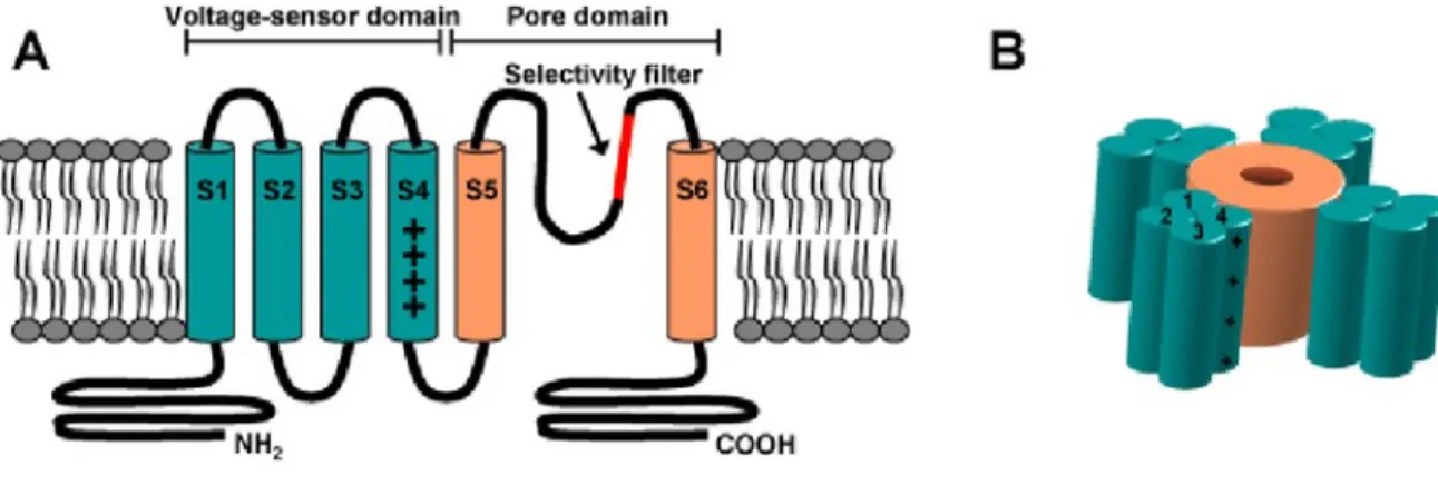
calcium channels). Each one of the domains has six transmembrane (tm) segments and one pore loop. The fifth and sixth transmembrane segments and the pore loop were found to be responsible for ion conduction (Figure 1.2). Many of the channels have additional subunits that modify the basic functions and define their family, but they are not necessary for voltage sensing and ion conduction [2].
Figure 1.2: General architecture of a voltage-gated ion channel. (A) shows the basic sub-unit. Segments S1 through S4 are called the voltage sensor domain (green) of the channel. The region between S5 and S6 forms the pore domain (orange) with the selectivity filter (red). (B) is a schematic view where the four identical subunits assembled form the ion channel (From Elinder et al. 2008 [3]).
These channels contains a large family of targets for drug development; with multiple genetic links to human disease, it is no surprise that they have received considerable attention especially in the last years where experimental techniques allow us to target and study them individually.
Chapter 2
Introduction
2.1
TRPV1, general architecture and characteristics
Transient receptor potential (TRP) channels represent one of the largest family of voltage-gated ion channels. This large receptor family consists of 30 different channels divided into six distinct subfamilies [4]. One of the unique features of TRP channels is the wide variety of exogenous and endogenous stimuli that activate these receptors. Excess heat, changes in pH, changes in pressure, and even molecules in commonly eaten foods (allicin in garlic, capsaicin in chili peppers) are just a few types of stimuli that can activate receptors in the TRP channel family. When TRP channels are activated there is a slow depolarization of a cell as well as a rise in intracellular Na+ and Ca2+. The exact
mechanism of TRP channel activation still remains elusive and as a result less detail is known about this process.
The transient receptor potential channel subfamily V member 1, the vanilloid receptor-1 (TRPVreceptor-1), is a trans-membrane protein and it was originally found in the primary terminals of the sensory neurons of the Dorsal Root Ganglion (DRG), mainly in the nociceptive neurons of the peripheral nervous system [5].
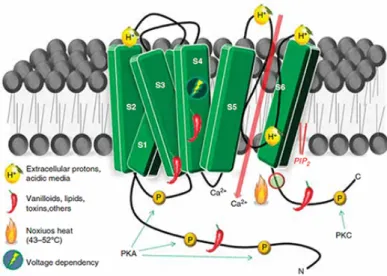
The topology of the channel is similar to that of voltage-gated ion channels. It is a homotetramer formed by four subunits, each of them contains six transmembrane α-helices (S1-S6), and one S5-P-S6 pore loop helix, with N- and C- termini in the intracellular domain (Figure 2.1). The structure was recently resolved to 2.95 ˚A resolution with electron cryo-microscopy (cryo-EM) technique [6].
Figure 2.1: Topographic structure of the TRPV1 receptor and postulated domains for the well known direct activation and indirect modulation.
TRPV1 is a primary sensory receptor involved in the transmission and modulation of pain in the peripheral nerve terminals (nociception), as well as the integration of diverse painful stimuli. It can be activated by many physical and chemical stimuli such as cap-saicin (the active component of chili peppers) and the increase of ambient temperature that can produce pain in humans (noxious heat - typically greater than 42◦ C) [5], volt-age [7], inflammatory volt-agents (extracellular acidity) [8], animal toxins such as the DkTx [9], Oxytocin [10] and others. A very peculiar feature of the receptor is that prolonged application of agonists (such as protons) may cause TRPV1 activity to decrease, leading to alleviation of pain [11].
Recent experiments demonstrated that TRPV1 can exhibit two types of desensitiza-tion. It is still unclear if both of them are triggered by the same mechanism, but they can frequently occur in conjunction: the first type is a kind of acute and fast desensitization,
which is a diminished response during a continuous vanilloid application; tachyphylaxis is the second and slower way of desensitizing the channel, which is a reduction in response to repeated applications of the agonist [12]. Desensitization is a process believed to occur predominantly via a procedure that should involve Ca2+, since it is abolished by the use
of calcium-free conditions. It may be a consequence of the fact that TRPV1 has a sig-nificant Ca2+ permeability, allowing excessive Ca2+ influx through the channel to trigger inhibitory feedback process and probably to even provoke local cell damage [5].
It is important to note that the desensitization of the channel is a very complex mechanism that involves multiple and different pathways. A well-established mechanism of desensitization caused by Ca2+ influx is the reduction of PIP2 (required for the channel
activity in the cell) caused by PLC that splits the phospholipid [13]. There is also evidence that the considerable amount of Ca2+influx lead by TRPV1 activation can also play a role
not only in the desensitization of the channel itself but it can inhibit other voltage-gated Ca2+ channels [14]. Since the channel is responsible for the transmission of painful stimuli, consequences of this desensitization are thought to trigger the paradoxical analgesic effect of agonists such capsaicin on TRPV1. Various studies are now concentrating on developing and optimizing agonists application that may cause the desired desensitization of TRPV1 while containing the side effects of such desensitization. One example is the development of creams and patches with either low or high concentration of capsaicin (0.1 % and 8 % respectively) in order to help and assist patients during surgery recovery [15].
This study tested the activation of TRPV1 led by extracellular acidic condition. Hence, its important to address that high proton concentrations (pH < 6) can be found in humans tissues when they are exposed to some kind of injury like infection and/or inflammation (the latter is probably the best-known endogenous TRPV1 agonist) or during an ischemia. It may be that this local acidosis can contribute to pain and hyperalgesia in disease states. Studies have shown that protons modulate TRPV1 activity by two distinct mechanisms [8][16]. The first way of interaction was found to be potentiation of response when TRPV1
is activated by other stimuli such as capsaicin or heat. Regarding the heat activation, an important conclusion was drawn during a decrease of extracellular pH. An acid attack, in fact, not only produced a larger response at noxious temperatures, but the threshold temperature activation of the channel was intensely reduced. Data shows that with a pH of 6.3 the lower level activation of the channel drops to temperatures as low as 35◦ C while at the same temperature a solution of pH 7.4 showed no activation of the channel. This result suggested that pH similar to the one found in inflamed tissues can actually activate TRPV1 at normal body temperature (around 36◦ C) and that protons can be considered one of the many TRPV1 agonists [17]. The second mechanism that regulates TRPV1 activity induced by proton is the direct activation of the channel that occurs at even lower pH values (pH < 6).
It is still not clear how protons directly activate TRPV1 but various studies were done in this direction in order to better understand the problem. It is clear though that activation via capsaicin and protons occurs in two different ways. Capsaicin binds in the transmembrane domain into the vanilloid-binding side-pocket while electrophysiological recording on both Xenopus laevis oocytes and HEK293 cells shows that application of protons in the extracellular side of the receptor increases the probability of the channel to open when interacting with two extracellular sites: one is believed to involve T633 in the pore helix, and the other one, V538, in the linker between S3 and S4. Another difference between the two agonists seems to be the fact that in order to achieve an open state and a fully conductive pore, capsaicin can bind to a single binding site in one subunit, while protons must bind to all four subunits [18].
2.1.1
TRPV1 and ethanol
Following this path, studies have also focused on how other agents can be part of the play. Ethanol is in fact believed to elicit and potentiate TRPV1 responses causing, among others, the burning-like sensation when drinking high concentration of alcohol.
The fact that the activation of a thermoreceptor is enrolled in ethanols burning sensation is supported by the fact that ethanol is found to activate primary sensory neurons that cause the release of neuropeptides in the esophagus, spinal cord or skin. This potentiation in the response of the vanilloid receptor-1 was found when activated by protons, capsaicin and heat when tested on HEK293 cells.
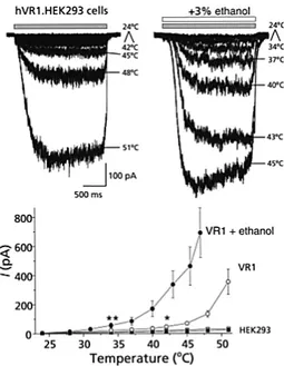
In an important study of 2002, electrophysiology recordings were performed on the wild-type (as control) and on the hVR1-expressing cells, showing a clear ethanol-driven modulation of the channel response. In the case of heat stimulation, a large shift of the threshold activation from ∼ 42◦C to ∼ 34◦C was observed (that is near the temperature of the tongue), which it can explain the human body temperature-related sensory responses when we are exposed to alcohol [19] (Figure 2.2).
In 2009 a behavioural study showed that the deletion of TRPV1 in mice significantly increases their ethanol preference and consumption, reduces the sedative effect of ethanol and shortens the recovery from acute ethanol intoxication [20]. It has been suggested that the increased ethanol consumption could be due to the reduced ethanol-induced burning sensations.
Given the widespread distribution of TRPV1 in both the peripheral and the central nervous system, activation of this channel by ethanol may be important for some periph-eral or central actions of ethanol. These studies laid the foundations for a more complete understanding of the problem of alcohol modulation on TRPV1.
2.2
Allosteric modulation and alcohol cut-off
The transmission of the effect induced by binding to one functional site in a receptor to a change in another topologically distinct site, is referred to as allostery.
These sites are termed as the orthosteric site, where the endogenous agonist binds producing a response as channel activation; the spatially non-overlapping allosteric site,
Figure 2.2: Shifting of the threshold activation of TRPV1 lead by ethanol (From Trevisani et al., Nature Neuroscience (2002)).
where allosteric ligands bind; the biologically active site that refers to the proteins effective site of biological function - in the case of ion channels, the ion conducting pore [21]. To distinguish between allosteric modulators the terms positive and negative allosteric modulators are used, which either increase or decrease the effect of the channel activation, respectively. It is important to note that modulators have no direct effect on the function of the protein target rather than modulate them.
Understanding the characteristics and dynamics of these binding pockets, interacting residues in the distinct conformational states and transitions between these states would not only advance scientific knowledge, but crucially inform our understanding of drug interactions. In this study different-sized primary alcohol molecules were used as positive allosteric modulators in order to figure out how they modulate TRPV1 activation.
It is known, in fact, that primary alcohols act as anesthetics and that as the size of the carbon chain increases, so does the potency of the anesthetics. This happen only up to a certain size, after which alcohols with longer carbon chains decline in potency and efficacy or are equally potent with the (n − 1)-alcohol molecules [22]. This point is
the so-called ’cut-off effect’ and indicates the maximum length of the carbon chain of the alcohol molecule that can bind and modulate the activity of the ion channel. If the cutoff phenomenon exists a critical alcohol binding site of finite lenght that will accommodate only alcohols below a certain limit of molecular volume will be observed.
In this study, I focused on the proton activation of TRPV1, and the Two-Electrode Voltage Clamp (TEVC) electrophysiology method was used to record current from the activation of the channel in Xenopus laevis oocytes. Two-Electrode Voltage Clamp is an electrophysiological technique used to control the membrane potential of cells large enough to be patched by two micro-electrodes. By doing so it is possible to study the properties of membrane proteins, especially ion channels when isolated and treated them singularly [23] (more details on this technique will be given in the Methods and Materials section).
2.3
Aim of the thesis
The main part of the thesis aims to optimize the process of TRPV1 expression on Xenopus laevis oocytes to help future studies in this field to quickly and better express the protein on the cells. Testing different concentrations of different-sized primary alcohol molecules to find out a potentiation-concentration dose-response will later help determine the length of the mentioned cut-off effect and possibly a direct binding site of a specific primary alcohol molecule.
Part of the project focused on building, launching and analysing molecular dynamics simulations of the TRPV1 open state from cryo-electron microscopy structure [6]. This simulation should be stable enough in order to be later used into different ways, such as the introduction of alcohol molecules to combine the results of the electrophysiological studies and molecular dynamics insight.
Chapter 3
Methods and Materials
3.1
Two-electrode voltage clamp electrophysiology
The two-electrode voltage clamp (TEVC) technique used in this study was developed by Alan Hodgkin, Andrew Huxley and Bernard Katz [24]. Their work in elucidating the role of voltage-gated ion channels and ionic mechanisms of action potentials led Hodgkin and Huxley to share The Nobel Prize in Physiology or Medicine in 1963.
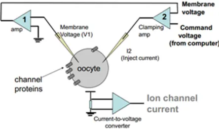
TEVC is an electrophysiological technique used to control the membrane potential of cells large enough to be patched by two micro-electrodes. By doing so it is possible to study the properties of membrane proteins, especially ion channels [23]. It uses two intracellular microelectrodes: one electrode is used to measure the internal potential of the cell and a current electrode for current injection is used to regulate the potential of the membrane. In this manner, and setting the membrane potential at desired values, one can record the membrane current flow in order to analyse the ion channel activity. Micromanipulators are used to lower the tips of the two electrodes until they enter through the oocyte membrane into the cytosolic environment (Figure 3.1).
The membrane potential is set to a certain value (in this case to -70 mV in order to simulate the resting potential of a neuron) and is constantly measured by the voltage
electrode that is connected to a voltage follower. This signal is fed into a clamping amplifier that compares the measured voltage (VM) to the set value (VC). When ion
channels are activated, ions cross the membrane and induce a change in VM.
Figure 3.1: Semplified scheme of the TEVC system.
The clamping amplifier detects the deviance from VC, and a current proportional
to the difference between VM and VC is forced to flow through the current electrode.
This current that needs to keep the voltage constant is equal but opposite to the current flowing through the sum of open channels in the membrane, which means that VM will
be kept very close to VC. This current is later measured, amplified and plotted. Oocytes
expressing ion channels on their surface are placed into a recording chamber connected to a perfusions system. A steady flow of suitable buffer (Ringer’s solution) is provided by pumps at the in- and outlets of the chamber. The perfusion buffer can be changed to a buffer of different composition in order to test channel activation and modulation.
3.2
The use of Xenopus Laevis oocytes as expression
system
A very important study led by Gurdon in 1971 showed that unfertilized oocytes of fe-male South African toads Xenopus Laevis can translate foreign injected RNA into proteins [25]. The advantages of this in vivo method to assay protein synthesis were immediately clear. The cultured living oocyte synthesizes foreign proteins for days, the protein accu-mulates in the oocyte cytoplasm and later are correctly sorted, targeted and assembled into the oocytes cell membrane.
Since their first use, they have proven to be a great expression system for membrane proteins for different reasons. In fact, with a size of about 1.2 mm these cells are large enough that they allow the injection of RNA or cDNA directly into the cytoplasm or nucleus, respectively. In 1977 Mertz and Gurdon demonstrated that SV40 DNA injected into Xenopus Laevis oocyte nuclei produced SV40 mRNA, and this mRNA was translated into recognizable SV40 proteins [26].
Neurophysiologists and biochemists have taken advantage of the X. laevis oocyte to clone and investigate a large section of receptors and ion channel proteins.
The native oocyte only expresses low levels and small numbers of endogenous channels on the surface, and it is therefore possible to study exogenous proteins without consider-able interference from background effects of endogenous membrane proteins. Additionally, oocytes are relatively tough and do not require sophisticated culturing procedures. It is important to notice, however, that the cell membrane of Xenopus oocytes differs signif-icantly from mammalian cells in both structure and composition, which could possibly affect the protein folding and function. Still, given its simplicity and robustness, the oocyte expression system is one of the best tool so far for the functional study of mem-brane proteins, in particular when used in combination with electrophysiology techniques like in this study.
3.2.1
mRNA synthesis
In order to express TRPV1 channels on the oocytes, a few steps are needed before the injection procedure. Plasmid DNA containing the membrane protein gene was pre-pared and then linearized before the (in vitro) mRNA synthesis process. mRNA for oocyte injection was synthesized from TRPV1-pcDNA3 8305 bp plasmid DNA (Figure 3.2) (Addgene, MA, USA; GeneArt, Thermo Scientific, MA, USA) that contains multiple restriction sites, T7 RNA polymerase promoter, TRPV1 gene, ampicillin, kanamycin and neomycin resistance genes.
Figure 3.2: Schematical representation of TRPV1-pcDNA3 8305 plasmid. Regions are marked for the TRPV1 gene, T7 promoter region, NotI cleavage site, ampicillin, kanamycin and neomycin resistance genes.
NotI restriction enzyme was used for linearization and mRNA synthesis was performed using the Thermo Fisher mMessage mMachine kits following the user guide (mMESSAGE
cloned cDNA is subcloned into a plasmid expression vector containing a bacteriophage promoter (recognized specifically by an RNA polymerase encoded by a bacteriophage; in this case che T7 promoter) [27].
It was chosen to inject mRNA instead of DNA because mRNA does not require as deep of an injection (in the nucleus), and thus is not as invasive for the oocyte. Also because with mRNA there is a higher likelihood of sufficient protein production [28].
The quality of RNA is crucial. Expression in the oocyte is the most rigorous test of RNA quality. Even if RNA that appears undegraded on a gel, it may not be good enough for expression purposes. Therefore, the method was meticulously followed. It was then necessary to check the purity and to quantify the actual concentration of the mRNA obtained from the synthesis on the Nanodrop spectrometer. Following the protocol, in order to have good expression of the channels only concentrations with product ≥ 250 ng/µL could be used for injection.
3.2.2
Xenopus Laevis oocytes injection
The mRNA obtained from the synthesis is then injected into the cytoplasm of each of the oocytes with a Nanoject II microinjector through a ∼20 µm thin capillary needle (Figure 3.3) that was previously pulled with Narishige PC-10 puller.
The dimension of the tip of the needle is of fundamental importance. Having a tip too wide might kill the oocyte during the injection process, while having a tip too narrow can result in the impossibility of filling the needle from the RNA drop. The needle, before loading the tip with RNA, must be backfilled with mineral oil to seal the pipette from air. Different concentrations of RNA (5 ng, 10 ng, 20 ng and 30 ng) were injected into the oocytes in a 50.6 nL total solution of TRPV1 RNA in order to find the optimal concentration for the best expression of the ion channel on the cells. The injected oocytes were then kept separately in 1.75 mL wells on a 48-well plate with incubation medium (88 mM NaCl, 10 mM HEPES, 2.4 mM NaHCO3, 1 mM KCl, 0.91 mM CaCl2, 0.82 mM
MgSO4, 0.33 mM Ca(NO3)2, pH 7.5) and incubated in 13◦ C from one to two weeks in
order to promote proper expression of the channels.
Figure 3.3: Picture of an oocyte taken from the built-in camera of the microscope during the injection process. The white arrow shows the point where the injection take place: beetween the animal pole (dark green) and the vegetal pole (light green) of the cell.
3.3
TEVC recording
The method used to record the current due to the activation of the channel is the Two-Electrode voltage Clamp as introduced above. Here borosilicate glass electrodes (Harvard Apparatus, MA, USA) were pulled with a PC-10 Narishige Puller (Narishige Group, Tokyo, Japan) and filled with 3M KCl to obtain a resistance of 5-50 MΩ. OC-725C voltage clamp (Warner Instruments, CT, USA) was used to clamp the oocytes at a membrane potential of -70 mV and kept constant to reproduce the natural neuron environment. Once the oocyte was in position and clamped, different buffers with different properties were perfused with a flow rate ∼1 mL/min into the system. Different responses were measured when different solutions were perfused across the oocytes.
The output of TEVC data are current traces (digitized using Axon CNS 1440A Digi-data and pCLAMP 10 software (Molecular Devices, Sunnyvale, CA)) that form peaks in the negative direction when ions flow from the outside to the inside of the ion channels
and positive when the flow is in opposite direction. If these currents are big enough to be distinguished from noise and possible background effects upon the pH applications (and alcohol modulation), the resulting peaks can be analyzed based on size and its change over time. As the measured current is the sum of all currents flowing though the chan-nels expressed on the surface, the current amplitude is highly dependent on expression level at the specific point of recording and varies between oocytes and days. In order to minimize the influence of changes in expression level, the data presented in this thesis are based on the comparison between relative currents. In this case, to establish the effect of allosteric modulators on TRPV1, every modulation of the channel response was preceded and followed by activation at a set pH. As it has been shown that the effect of allosteric modulators is greater at lower levels of activation [29], modulators are normally tested at a level of activation where the channel conducts 10 % of their maximal current (EC10).
In this case the value was found to be at pH 5 (Figure 3.4).
Figure 3.4: Normalized response of pH dose response showing (EC10) to be pH 5.
After the oocyte was in position and clamped, the protocol started with a 8.5 pH solution with calcium Ringer’s buffer used as running buffer, after that, variables were removed one at the time. In fact, the choice of a followed application of a zero-calcium buffer was made in order to find the most uninhibited response possible from the
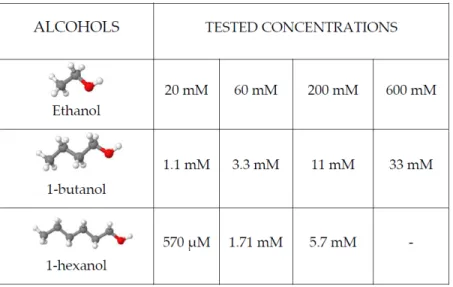
chan-nel activation and to wash out any residual calcium left from the running buffer. For activating the channel, after 5 minutes of running buffer, 1-minute applications of pH 8.5 calcium-free buffer and 1-minute application of pH 5 calcium-free buffer were used as control and to account as a rundown. Five minutes of washing out the oocyte with running buffer and letting the membrane potential reach the steady state and alcohol modulation can be tested. To do so, 1-minute of the same pH 8.5 calcium-free buffer application was followed by 1-minute of pH 8.5 calcium-free buffer with alcohol. After the two pH 8.5 applications, the channel can be activated and modulated with a pH 5 calcium-free solution mixed with alcohol molecules. Different concentrations of different primary alcohol were tested (Figure 3.5) and a modulation-concentration response curve was carried out.
Figure 3.5: Alcohol molecules tested and corresponding different concentrations
3.4
Molecular dynamics system set-up
While experimental procedures measure the outcome of an event at a time resolution from several hundred microseconds to milliseconds, molecular dynamics (MD) simulation can investigate the dynamic changes in the femto-to-microsecond time scale and can
of-fer insights into the underlying atomistic changes related to a specific event, therefore building a bridge between static molecules from structure determination and the result-ing functional effects observable by electrophysiology. For large systems such as this one where the protein is in a membrane environment surrounded by water and ions, these cal-culations require substantial computational power. A local computing cluster maintained by TCBlab was used for setting up the system in GROMACS version 2018.1 [30].
In order to have a more complete understanding of alcohol modulation on TRPV1, part of this project focused on setting up the system for a Molecular dynamics simulation that can later be used for introducing ethanol molecules and find out how and where they interact with the protein.
Going through details of MD is far beyond the scope of this study but it is therefore important to define what are the inputs and outputs for GROMACS in setting-up the system.
3.4.1
Simulate biological processes
A molecular dynamics simulation solve Newton’s equation of motion for a system of N interacting atoms:
mi
∂2r i
∂t2 = Fi, i = 1 . . . N (3.1)
where the forces are the negative derivatives of a potential function V (r1, r2, . . . , rN):
Fi = −
∂V ∂ri
(3.2) Here forces between the particles and their potential energies are often calculated using molecular mechanics force fields. Other than the mass of each particle, the force on each particle is needed in order for the accelerations and velocities to be calculated. In MD this is often achieved though force fields. Force fields are related to potential energy
surfaces, with a specific energy at each point. This energy surface can be generated by quantum or classical mechanics. Quantum mechanical calculations are very heavy and involve certain undesired approximations, such as treating the system as 0 degrees Kelvin. As a result, energy calculations of large biological system such as a protein in a membrane like this are only available today through classical mechanics. The equations are solved simultaneously in small time steps (e.g. femtoseconds). For example a simulation of 100 ns will be composed of 108 snapshots.
3.4.2
Simulation parameters
The model of the protein used was a TRPV1 open state in complex with DkTx and RTX (PDB ID: 5IRX) at resolution 2.95 ˚A [6]. The protein was embedded in POPC homogenous lipid bilayers with extended Berger parameters [31] using the CHARM-GUI Membrane Builder method [32] [33], TIP3P water model [34] and surrounded by 100 mM of NaCl solution in order to neutralize the total charges of the systems. The overall sixe of the system was a 150x150x150 ˚A3 cubic box with periodic boundary conditions. For
the simulations, CHARM36 force field was used.
Once the system is assembled it is necessary to evaluate and check that the three-dimensional structure has no artifact such as steric clashes (unnatural overlap of any two non-bonding atoms in the protein structure) or inappropriate geometry. The structure is relaxed through a process called energy minimization (EM) in which the system is relaxed to the closest local energy minimum; if this minimum is reached then the structure is rea-sonable in terms of geometry and solvent orientation. The system was energy minimized for 5000 steps (1 fs time step).
After EM it is necessary to equilibrate the solvent and the ions around the protein. Here the solvent is heterogenous system, with ions and both lipids and water molecoules. During this process water has to re-orient around the lipid headgroups and any exposed parts of the protein, the lipids have to orient themselves around the protein as well. The
equilibration process is carried out with a position restraining force on heavy atoms of the proteins. The utility of these restraints is to maintain the initial structure while the rest of the environment (solvent, ions, hydrogen atoms) around the main system of interest relaxes. In this case, as the equilibration phase continues, the restraint force constants were gradually reduced and then eliminated at the end of the process from heavy atoms and the backbone of the protein. The goal of the equilibration phase is to relax the system and its environment such that production dynamics can occur without restraints. Equilibration was conducted in two phases. The first phase is referred as a ”canonical” ensemble (constant Number of particles, Volume and Temperature - NVT) where the system needs to reach and stabilize to the desired temperature of 300 K. The system was NVT equilibrated for 5 milion steps (1 fs time step) equivalent to 5 ns. The second equilibration phase aims to equilibrate with respect to pressure and is conducted under an NPT ensemble, where the Number of particles, Pressure, and Temperature are all constants (”isothermal-isobaric” ensemble). Pressure was set to 1 bar and during NPT equilibration 75 ns were simulated (2 fs time step).
After the two equilibration phases the system is ready to run without restraints and to produce MD for data collection. MD simulation without restraints ran for 400 ns in total.
These steps were repeated three times on the same model in order to obtain three independent replicas of the same structure.
The final trajectories were then visualized, analyzed and displayed with VMD [35].
3.4.3
RMSD
To evaluate the stability of the MD simulation the Root-Mean-Square Deviation (RMSD) method was used. It is defined as:
RMSD = s PNatoms i=1 (ri(t1) − ri(t2))2 Natoms (3.3)
where N atoms is the number of atoms whose positions are compared and ri(t) the
position of the atom i at time t, that will correspond to different sets of coordinates. Since calculating the RMSD over the time of the simulation length will give a measure of the difference between two states, it is important to set the frame reference and choose the right alignment method for the protein. In this case the first frame of the simulation was chosen as the reference frame and the protein was aligned on his backbone to exclude every fluctuation due to a general moving of the whole system. Reaching a plateau after a certain amount of time that correspond to a low value of RMSD indicates that the system (or the selected part of it) is stable and that the deviation from its reference frame are acceptable.
3.4.4
RMSF
Another great tool for estimating which part of the protein is fluctuating more than others is the Root-Mean-Square Fluctuations (RMSF). Is a measure of the deviation between the position of a particle i and its reference position. It is defined as:
RMSF = s PT tj=1(ri(tj) − r ref i )2 T (3.4)
where T is the time which over the system is averaged and rrefi is the reference position of the particle i. In this case it is the time average position of the same particle (riref = ri).
The difference from the RMSD evaluation lies from the fact that RMSF is averaged over time, giving a value for each particle (residues of the protein in this case) rather than the change over time of the system. RMSF will show the fluctuation of the protein for each residue and this will give insight about the stability and flexibility of the system for the desired part.
3.4.5
Visualization software
The final trajectories were then visualized, analyzed and displayed using VMD [35]. The software was also used to render different images used in this study and for analyzing RMSD and RMSF of different trajectories as described above.
Chapter 4
Results
Since a large part of this project was dedicated to the optimization process of express-ing TRPV1 channels on Xenopus laevis oocytes, very much time was dedicated in order to find the right parameters (concentration of injected mRNA and the corresponding incubation period) that might reach this goal.
Figure 4.1: Typical spectral pattern for RNA sample quantification and purity checks.
Figure 4.1 shows the output of the Nanodrop spectrometer for a 2.16 µg/µL mRNA sample concentration that was later used for injection. The 260/280 and 260/230 ratios are used to assess the purity of the sample. A ratio of ∼2 is generally accepted as a pure RNA sample. Having a peak at higher absorbance might indicate the presence of
contaminants that can interfere with the protein expression later during the incubation period of the oocytes.
Figure 4.2 shows a recording sample of two trials of different concentrations of 1-butanol and ethanol. As visible from the curves, primary alcohol molecules do not activate the channel alone when introduced into the system, they, in fact, potentiates the activation response when pH 5 buffer is perfused through the oocyte. It can be noted from the concentrations written above the alcohol applications that 1-butanol potentiates more TRPV1 response compared to ethanol since the concentrations used are at least one magnitude lower.
Figure 4.2: Recording sample of both 1-butanol (top) and ethanol (bottom) applications. pH 5 was applied in between each modulation as control.
Figure 4.3 shows the potentiation of TRPV1 activation with different alcohol molecules at different concentrations. From this data, it is clear that 1-hexanol is more potent than 1-butanol that is, in turn, more potent than ethanol. Hence, it is not possible to figure out a clear cut-off effect yet, because an increase of the length of carbon chain actually has an effect on the modulation in the potency of TRPV1 response, but preliminary the cut-off effect is beyond 1-hexanol. It is important to note that the error on 570 µM 1-hexanol is
high, so lower concentrations on 1-hexanol must be tested in order to have a more precise modulation at low concentration.
Figure 4.3: Modulation of TRPV1 by short and long chain alcohol. Data represent mean s.e. of n = 2-5 oocytes
Figure 4.4 shows the ribbon diagram of the TRPV1 model used for this project in its open structure (PDB ID: 51RX) with each of the identical subunits coloured differently in two different views. From side, where the transmembrane domain is shown, and from top, where the pore tunnel is visible.
Figure 4.4: Ribbon diagram of TRPV1. Left side indicating the transmembrane domain (TMD). Right side shows the top view of the channel.
Root-mean-square deviation (RMSD) vs simulation time is plotted in Figure 4.5. Fig-ure 4.5 (a) shows values for the three different replicas that were produced, revealing that the system is not fully equilibrated and RMSD values for each trajectory are relatively high. The similar behaviour of the three replicas give us enough confidence to keep on study one of the replicas. Figure 4.5 (b) shows the RMSD of replica 1 for the different domain of the protein, revealing that the transmembrane domain (TMD) of the channel, that is believed to be the target region for interaction with alcohol, is highly stable in contrast to the overall structure of the protein.
Figure 4.5: RMSD vs time. (a) shows RMSD for the whole protein for three different replicas. (b) shows different domain of the protein in replica 1.
A closer look at the structure can be done by evaluating the RMSF values for each residue of the protein as shown in Figure 4.6. RMSF values are averaged over 400 ns and show that the transmembrane domain of the structure, together with the capsaicin binding pocket that could be a candidate site for alcohol interaction, is very stable as first suggested from RMSD values above. Extracellular loops and pore loops are shown; together with the intracellular region of the protein these are the most flexible part of the structure.
Figure 4.7 is a visual of the system embedded with lipids in a POPC membrane showing the different regions of the protein.
Figure 4.6: RMSF of replica 1. Different colours refer to different subunit of the protein. The transmembrane domain, intracellular domain and loop regions are indicated together with the capsaicin binding pocket. Residues truncated from the structure are not shown.
Chapter 5
Discussion
One of the biggest challenges of this project was to determine the right parameters in order to obtain optimal expression of TRPV1 channels on the oocytes membrane for the purpose of performing TEVC recordings. Several weeks were used to establish the exact amount of mRNA injected in the oocytes and the corresponding incubation period. After a large number of trials, it was established that starting from an RNA stock concentration of ∼ 2 µg/µL and 10 ng injection of mRNA in a 50.6 nL solution for each oocyte should assure good expression of the channel after an incubation period of 8 - 9 days. Data recorded from TEVC shows clear evidence that a change in the length of the carbon chain of primary alcohol influence TRPV1 response lead by protons (pH 5), but data collected and shown here are not enough to indicate the exact cut-off point. The modulation response shows that increasing the length of the alcohol chain results in lower concentration needed to modulate the channel to 100% potentiation. It is therefore clear that the location of the cut-off effect is beyond 1-hexanol and lower concentrations and longer chain alcohols must be tried in order to figure out the exact position of the cut-off effect.
RMSD and RMSF values from the MD simulations of the open state shows that the overall structure of the system is not fully equilibrated up to a 400 ns trajectory and a longer simulation is probably needed to reach that point. If the alcohol binding site is in
the vicinity of the capsaicin binding pocket (in the transmembrane domain) as believed, then the RMSD values of the target portion reaches a stable plateau around 2 ˚A. This gives enough confidence to state that the system is ready to be introduced with alcohol molecules.
The equilibrated system in MD simulation provides a basis for running the unre-strained simulation to confirm the stability of the built model and track the dynamics of both the structure and the alcohol binding pocket through longer timeframes. The model remains a relevant system for further characterization studies of allosteric binding sites and channel gating.
The discovery of an alcohol cut-off effect on TRPV1 response driven by pH can shed light on the understanding of how different alcohol molecules interact with the channel. The alcohol potency found here was 1-hexanol >1-butanol> ethanol, and in order to reach a cut-off effect a more complete relation between primary alcohol molecules longer than 1-hexanol and modulation must be carried out. Gradually increasing the carbon chain of the alcohol will not produce any further potentiation on the channel response after the cut-off point has been reached.
Paradoxically, it now clear that the best TRPV1-based therapeutics so far are agonists (like capsaicin) that activate the channel and initially cause pain. Applied chronically, these agonists presumably work by chronically depolarizing the neurons, inactivating the voltage-gated channels needed to support action potential propagation. Prolonged expo-sure to high concentrations of TRPV1 agonists, or modulating the channel in the less invasive way, can cause the nerve terminals to retreat from the area of application. Phys-iologically, this can be very helpful in pain regulation by creating drugs that can target the site-specific binding site and can lead to a more specific way of pain inhibition.
Studying allostery is a difficult task, incorporating a wide range of techniques to de-termine the role and pharmacological profiles of ligands acting on specific receptors. The mechanisms of modulation, channel gating through different functional states,
identifi-cation of ligand-interacting residues needs interdisciplinary approaches to gain insight. Integrating experimental electrophysiology with constant advances in a) structure de-termination methods, more specifically the potentials of cryogenic electron microscopy (cryo-EM) to capture native-like membrane proteins covering a range of functional states, b) computational power through MD simulations, will allow characterization of these processes at atomic resolution.
The experimental electrophysiology results showed here and the MD system provide a basis towards this integration by determining the expression and biophysical properties of TRPV1 receptor in response to its activation through extracellular acidity and allosteric modulators of n-alcohols.
Chapter 6
Ethical consideration
Only unfertilized eggs were used in this study, limiting ethical issues regarding experi-mental work performed on a developed organism. Xenopus laevis oocytes were purchased from a widely used and professional animal care facility (EcoCyte BioScience). This mini-mizes risks to animals from in-house lab environment and leaves proper animal handling to qualified professionals. This project is a step further to benefit the health and well-being of people, because understanding ion channel function is critical to designing better neu-ropharmaceuticals, including general anesthetics, and hopefully one day for determining the causes of neurological diseases.
References
[1] Marco Bresadola. Medicine and science in the life of Luigi Galvani (1737-1798). Brain Research Bulletin, 1998.
[2] Zheng Jie and Trudeau Matthew C. Handbook of Ion Channels. CRC Press, 2015. [3] Sara I. B¨orjesson and Fredrik Elinder. Structure, function, and modification of the
voltage sensor in voltage-gated ion channels, 2008.
[4] I. S. Ambudkar, X. Liu, and B. Paria. Transient Receptor Potential Channels. In Encyclopedia of Biological Chemistry: Second Edition, pages 412–417. Elsevier Inc., 2 2013.
[5] MJ Caterina, Schumacher MA, Tominaga M, Rosen TA, Levine JD, and Julius D. The capsaicin receptor: a heat-activated ion channel in the pain pathway. Nature, 1997.
[6] Yuan Gao, Erhu Cao, David Julius, and Yifan Cheng. TRPV1 structures in nanodiscs reveal mechanisms of ligand and lipid action. Nature, 2016.
[7] Martin J. Gunthorpe, Mark H. Harries, Rab K. Prinjha, John B. Davis, and Andrew Randall. Voltage- and time-dependent properties of the recombinant rat vanilloid receptor (rVR1). Journal of Physiology, 2000.
[8] S. Ryu, B. Liu, J. Yao, Q. Fu, and F. Qin. Uncoupling Proton Activation of Vanilloid Receptor TRPV1. Journal of Neuroscience, 27(47):12797–12807, 2007.
[9] Chanhyung Bae, Claudio Anselmi, Jeet Kalia, Andres Jara-Oseguera, Charles D Schwieters, Dmitriy Krepkiy, Chul Won Lee, Eun-Hee Kim, Jae Il Kim, Jos D Faraldo-G´omez, and Kenton J Swartz. Structural insights into the mechanism of activation of the TRPV1 channel by a membrane-bound tarantula toxin. eLife, 2016.
[10] Yelena Nersesyan, Lusine Demirkhanyan, Deny Cabezas-Bratesco, Victoria Oakes, Ricardo Kusuda, Tyler Dawson, Xiaohui Sun, Chike Cao, Alejandro Martin Cohen, Bharath Chelluboina, Krishna Kumar Veeravalli, Katharina Zimmermann, Carmen Domene, Sebastian Brauchi, and Eleonora Zakharian. Oxytocin Modulates Nocicep-tion as an Agonist of Pain-Sensing TRPV1. Cell Reports, 2017.
[11] Filip Touska, Lenka Marsakova, Jan Teisinger, and Viktorie Vlachova. A ”cute” desensitization of TRPV1. Current pharmaceutical biotechnology, 2011.
[12] L. Vyklick´y, K. Nov´akov´a-Touˇsov´a, J. Benedikt, A. Samad, F. Touˇska, and Vik-torie Vlachova. Calcium-dependent desensitization of vanilloid receptor TRPV1: A mechanism possibly involved in analgesia induced by topical application of capsaicin, 2008.
[13] Jing Yao and Feng Qin. Interaction with phosphoinositides confers adaptation onto the TRPV1 pain receptor. PLoS Biology, 2009.
[14] Valentina Comunanza, Emilio Carbone, Andrea Marcantoni, Emanuele Sher, and Daniel Ursu. Calcium-dependent inhibition of T-type calcium channels by TRPV1 activation in rat sensory neurons. Pflugers Archiv European Journal of Physiology, 2011.
[15] B. Nilius and A. Szallasi. Transient Receptor Potential Channels as Drug Targets: From the Science of Basic Research to the Art of Medicine. Pharmacological Reviews,
[16] Thomas K. Baumann and Melissa E. Martenson. Extracellular Protons Both Increase the Activity and Reduce the Conductance of Capsaicin-Gated Channels. The Journal of Neuroscience, 2018.
[17] Makoto Tominaga, Michael J. Caterina, Annika B. Malmberg, Tobias A. Rosen, Heather Gilbert, Kate Skinner, Brigitte E. Raumann, Allan I. Basbaum, and David Julius. The cloned capsaicin receptor integrates multiple pain-producing stimuli. Neuron, 1998.
[18] Adina Hazan, Rakesh Kumar, Henry Matzner, and Avi Priel. The pain receptor TRPV1 displays agonist-dependent activation stoichiometry. Scientific Reports, 2015. [19] M. Trevisani, D. Smart, M. J. Gunthorpe, M. Tognetto, M. Barbieri, B. Campi, S. Amadesi, J. Gray, J. C. Jerman, S. J. Brough, D. Owen, G. D. Smith, A. D. Randall, S. Harrison, A. Bianchi, J. B. Davis, and P. Geppetti. Ethanol elicits and potentiates nociceptor responses via the vanilloid receptor-1. Nature Neuroscience, 5(6):546–551, 2002.
[20] Y. A. Blednov and R. A. Harris. Deletion of vanilloid receptor (TRPV1) in mice alters behavioral effects of ethanol. Neuropharmacology, 2009.
[21] Jean Pierre Changeux and Arthur Christopoulos. Allosteric modulation as a unifying mechanism for receptor function and regulation, 2017.
[22] James K. Alifimoff, Leonard L. Firestone, and Keith W. Miller. Anaesthetic potencies of primary alkanols: implications for the molecular dimensions of the anaesthetic site. British Journal of Pharmacology, 1989.
[23] Carsten A. Wagner, Bjrn Friedrich, Iwan Setiawan, Florian Lang, and Stefan Br¨oer. The use of Xenopus laevis oocytes for the functional characterization of heterolo-gously expressed membrane proteins, 2000.
[24] A. L. Hodgkin and A. F. Huxley. A quantitative description of membrane current and its application to conduction and excitation in nerve. Bulletin of Mathematical Biology, 1990.
[25] J. B. Gurdon, C. D. Lane, H. R. Woodland, and G. Marbaix. Use of frog eggs and oocytes for the study of messenger RNA and its translation in living cells. Nature, 1971.
[26] J. E. Mertz and J. B. Gurdon. Purified DNAs are transcribed after microinjection into Xenopus oocytes. Proceedings of the National Academy of Sciences of the United States of America, 1977.
[27] Nathan Dascal. The use of xenopus oocytes for the study of ion channel. Critical Reviews in Biochemistry and Molecular Biology, 1987.
[28] Srinivasan P. Venkatachalan, Jeremy D. Bushman, Jos L. Mercado, Feyza Sancar, Kelly R. Christopherson, and Andrew J. Boileau. Optimized expression vector for ion channel studies in Xenopus oocytes and mammalian cells using alfalfa mosaic virus. Pflugers Archiv European Journal of Physiology, 2007.
[29] Asba Tasneem, Lakshminarayan M. Iyer, Eric Jakobsson, and L. Aravind. Identi-fication of the prokaryotic ligand-gated ion channels and their implications for the mechanisms and origins of animal Cys-loop ion channels. Genome biology, 2005. [30] Mark Abraham, Berk Hess, David van der Spoel, and Erik Lindahl. GROMACS
User Manual-5.0.7. Www.Gromacs.Org, 2015.
[31] Oliver Berger, Olle Edholm, and Fritz J¨ahnig. Molecular dynamics simulations of a fluid bilayer of dipalmitoylphosphatidylcholine at full hydration, constant pressure, and constant temperature. Biophysical Journal, 1997.
[32] Sunhwan Jo, Taehoon Kim, and Wonpil Im. Automated builder and database of protein/membrane complexes for molecular dynamics simulations. PLoS ONE, 2007. [33] Sunhwan Jo, Taehoon Kim, Vidyashankara G. Iyer, and Wonpil Im. CHARMM-GUI: A web-based graphical user interface for CHARMM. Journal of Computational Chemistry, 2008.
[34] William L. Jorgensen, Jayaraman Chandrasekhar, Jeffry D. Madura, Roger W. Im-pey, and Michael L. Klein. Comparison of simple potential functions for simulating liquid water. The Journal of Chemical Physics, 1983.
[35] William Humphrey, Andrew Dalke, and Klaus Schulten. VMD: Visual molecular dynamics. Journal of Molecular Graphics, 1996.