0394-6320 (2011) Copyright © by BIOLIFE, s.a.s. This publication and/or article is for individual use only and may not be further reproduced without written permission from the copyright holder. Unauthorized reproduction may result in financial and other penalties
31 (S)
The main factor in determining the mechanical properties of bone is the collagen configuration in the matrix and corresponding orientation of mineral crystallites (1).According to classical histology, bone tissue may be classified in relation to the spatial orientation of collagen fibers. Two different types of bone have been recognized: oven-fibered bone and parallel-fibered bone (lamellar or non-lamellar) (2).Woven bone has been estimated to contain four to eight times more osteocytes than lamellar bone (3). Osteocytes plays a crucial role in maintaining the mechanical quality of bone, and osteocyte density could be considered as an alternative index in
assessing bone quality (4). The coefficient of variation of osteocyte lacunar density (number osteocyte lacunae per bone area) increases linearly with age, and aging bone tissue is characterized by increased heterogeneity in the spatial organization (numbers) of osteocytes (5). So, the bone mass is determined by the control of osteocyte number (6).
It is known that static loads have no effects on bone mass; however, experimental information makes it plausible that amplitude, rate, frequency and duration of loading are all important for bone metabolism (7). The osteocytes, through their network, provide a stimulus to
HISTOMORPHIC-METRIC EVALUATION OF AN IMMEDIATELY LOADED IMPLANT RETRIEVED FROM HUMAN MANDIBLE AFTER 2 YEARS
T. TRAINI1*, M. DANZA2*, I. ZOLLINO3, R. ALTAVILLA4,
A. LUCCHESE3, V. SOLLAZZO4, G. TRAPELLA5, G. BRUNELLI.3, F. CARINCI3
1EIS-international educational society and private practice, San Benedetto del Tronto, Italy 2Dental School, University of Pescara-Chieti, Italy
3Department of D.M.C.C.C., Section of Maxillofacial and Plastic Surgery, University of Ferrara, Ferrara, Italy 4Orthopedic Clinic, University of Ferrara, Ferrara, Italy
5Department of D.M.C.C.C., Section of Neurosurgery, University of Ferrara, Ferrara, Italy *These two authors equally contributed to this paper
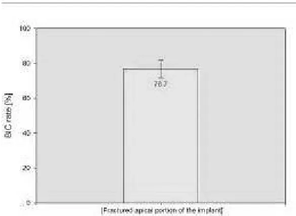
The aim of the present study was to evaluate the interplay between microstructure and function of the bone around an immediately loaded implant retrieved from human maxilla after 23 months due to fracture. A spiral implant of 3.3 mm x 15 mm was placed in a male 53 years old in the anterior region of the mandible bone (4.1) and it was processed for histology. The specimen was analyzed under the confocal scanning laser microscope (CSLM) and brightfield light microscope (LM) equipped with circularly polarized light (CPL). The BIC rate was 76.7± 4.9 (mean ±SD). Many cement lines indicates an high remodeling rate of the bone. The transverse collagen fiber orientation (CFO) (mean ±SD) under the lower flank of the thread near the tread tip was 55.2 ± 4.8 x 10⁴
pixel while the longitudinal CFO was 45.8 ± 2.3 x 104 pixel (P<.05). In the inter-threads region the transverse CFO
(mean ±SD) was 36.4 ± 2.4 x 104 pixel while the longitudinal CFO was 65.6 ± 6.5 x 104 pixel (P<.05). The osteocytes
numbers (mean ±SD) was 205 ± 45 in the peri-implant bone and 144 ± 53 in the native bone (P=.007). After 2-years of loading the SLA spiral implant was well osseointegrated but still surrounded by woven bone. The osteocytes density was significantly higher in the peri-implant bone than in the native bone. The transverse collagen fibers were significantly associated with the lower flank of the implant threads, while the longitudinal collagen fibers were more represented in the straight surface of the implant. The implant fracture was correlated to crestal bone resorbing and subsequent fatigue yielding.
Key Words: Circularly Polarized Light Microscopy, collagen fiber orientation, bone implant contact rate, implant fracture, spiral implant
Corresponding author: Prof. Francesco Carinci, M.D
Department of D.M.C.C.C Section of Maxillofacial and Plastic Surgery University of Ferrara Corso Giovecca 203 44100 Ferrara ITALY E-mail: [email protected]
Web: www.carinci.org
resin. Undecalcified longitudinal cut sections of 50 µm were prepared by using a cutting and grinding TT system (TMA2, Grottammare, AP, Italy). The sections were double stained with toluidine blue and fuchsine acid for some samples and toluidine blue stains for others to be analyzed.
Transmitted Light Microscopy (LM)
The histomorphometry was used to evaluate the amount of bone implant contact rate (BIC%). The investigation was carried out in a transmitted brightfield Light Microscope Axiolab (Zeiss Oberchen, Germany) connected to a high-resolution digital camera (FinePix S2 Pro, Fuji Photo Film Co. LTD. Minato-Ku, Japan). An Histometric software package with image capturing capabilities (Image-Pro Plus 6.0, Media Cybernetics Inc., Bethesda, MD, USA) was used. To ensure accuracy, the software was calibrated for each experimental image using a software feature named “Calibration Wizard” which reports the number of pixel between two selected points (diameter or length of the implant). The linear remapping of the pixel numbers was used to calibrate the distance in millimeters.
Circularly Polarized Light Microscopy (CPLM)
Birefringence was used to evaluate the collagen fiber orientation (CFO) of the bone matrix around the implants. The measurements using polarized light were concentrated mainly under the thread tip along the lower flank of the thread and in the inter-threads region. Unstained sections (before staining procedure) were used. The CFO was evaluated by a means of a light microscope (Axiolab, Carl Zeiss, Jena, Germany) equipped with two linear polarizer and two quarter wave plates arranged to have transmitted circularly polarized light, connected to a high-resolution digital camera (FinePix S2 Pro, Fuji Photo Film Co. LTD. Minato-Ku, Japan). The Collagen fibers aligned perfectly transverse to the direction of the light propagation (parallel to the plane of the section) appeared “white-blue” due to a change in the refraction of exiting light whereas the collagen fibers aligned along the axis of light propagation (perpendicular to the plane of the section) appeared “red-yellow”, because no refraction occurred.
Confocal Scanning Laser microscopy
In order to evaluate the osteocytes/ lacunae density the specimens were stained using basic fuchsin (14) than evaluated under a confocal scanning laser microscope (CSLM - TCS-SP, Leica Microsystems, Wetzlar, Germany) with a 20 x magnification objective lens. A 568 nm wavelength excitation light was used to view the fluorescent die. The digitized images were stored in format JPEG with NxM = 3024 x 2016 grid of pixels for a 24 bit. The osteocytes density (OD) was evaluated as follow OD = Ot.Lc/BAr where Ot.Lc was the number of osteocytes or lacunae counted while BAr was the bone area investigated.
Statistical analysis
One person (TT) performed all the measurements. Intra-examiner variability was controlled by carrying out 2 measurements for each index. When for the same index the difference in the two performed readings exceeded 15% the measure was repeated. Statistical analysis was performed by
the `basic multicellular units’ (BMUs) of osteoblasts and osteoclasts at the trabecular surface either to increase or to reduce net local bone mass (2).
Bone is a dynamic tissue that undergoes remodeling process that occurs not only in response to microdamage, but also as a part of the normal metabolic process of bone maintenance (1, 8) , and to readjust the concentration of minerals in the blood stream (9). In the remodeling phase, new bone is laid down on the resorbed surface of old bone, creating an interface with itself on a continual basis.
The collagen network may lead to significant variation in the mechanical competence of the bone. Longitudinal collagen fiber orientations, for example, correlate with the modulus and strength of bone (10). Moreover, the mechanical loading is an important determinant of collagen fiber orientation, and it could alter osteoblasts in a manner that causes a characteristic orientation in the fibers they synthesize (11). Therefore, determining the orientation of collagen fibers in human bone tissues is indispensable when studying the relationship between the physical properties and the structure (12).
However few studies have, until now, focused on the relationship between collagen fiber orientation and loaded dental implants although the orientation of the collagen fibers enable evaluation of the quality of osseointegration and bone remodeling (13).
The orientation of collagen fibers in bone could help obtain information about the relation among implant design, distribution of stress applied to the bone, and the growth of bone (12). Although many hypotheses exist about why marginal bone change occurs around implant, the mechanism of marginal bone loss is no clear known. The aim of this case report was to analyze the course of osseointegration speculating the bone-to-implant contact (BIC) rate, the osteocytes density and the collagen fiber organization in one immediately loaded implant retrieved due to fracture (overloading) after 23 months of function.
MATERIALS AND METHODS
A SLA (Sand-blasted, Large grit, Acid-etched) surface implant (Arrow Press fixture, Alpha Bio LTD, Petah-Tikva, Israel) of 3.3 mm x 15 mm was placed in a male 53 years old in the anterior region of the mandible bone (4.1) in June 2008. The implant was immediately loaded with an acrylic resin restoration. Final restoration in glass-ceramic fused to zirconia was placed two months later. The implant-restoration undergo to fracture in July 2010 (after 23 months).
Specimen’s processing
The retrieved specimen was fixed in 4% formalin pH 7.0 for 10 days and then transferred to a solution of 70% ethanol until processing. The specimen was dehydrated in increasing concentrations of alcohol up to 100%, infiltrated and embedded in LR White (London Resin Company, Berkshire, England)
means of the computerized statistical package (Sigma Stat 3.5, SPSS inc. Ekrath, Germany). The difference between the osteocytes lacunar densities was made by a means of Mann-Whitney Rank Sum Test since the normality test failed (P <
0.050). One-Way ANOVA test was used in the inference of CFO since both normality and equal variance tests passed (P = 0,325) and (P = 0,748) respectively.
The evaluation of multiple comparison procedures after the ANOVA was made using Holm-Sidak method. A P value of under 0.05 was considered statistically significant.
RESULTS
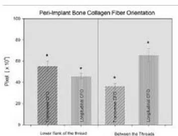
The peri-implant bone was mainly of woven type with only few osteons (Fig. 1.1). Many cement lines indicates an high remodeling rate of the bone (Fig.1.2 ). The BIC rate of the implant retrieved after 23 months was 76.7± 4.9 (mean ±SD) (Fg.2). The osteocytes numbers (mean ±SD) was 205 ± 45 in the peri-implant bone and 144 ± 53 in the native bone away 0.7mm from implant surface (P=.007) (Figg. 3.1 ; 3.2 and 4). The transverse CFO (mean ±SD) under the lower flank of the thread near the tread tip (Fig. 5 A,A1) was 55.2 ± 4.8 x 104 pixel while the longitudinal
CFO was 45.8 ± 2.3 x 104 pixel (P<.05) In the
inter-threads region (Fig. 5 B,B1). the transverse CFO (mean ±SD) was 36.4 ± 2.4 x 104 pixel while the longitudinal
CFO was 65.6 ± 6.5 x 104 pixel (P<.05) (Fig. 6).
DISCUSSION
The implant fracture is the most catastrophic failure of implant components because it usually causes the loss of the implant. Nevertheless, an osseointegrated fractured implant represents a very useful opportunity to study, in humans, the effects of overloading of the peri-implant bone microstructure.
Many studies are in progress to prevent failure of osseointegration and to enhance the longevity of osseointegrated implants. In the present study a spiral implant was investigated.
Fig. 1.1. Light microscopic view of bone implant contact. In (A) immediate loaded implant retrieved after 23 months due to fracture (overloaded) I, implant. Toluidine blue and fuchsine acid staining; original magnification x 18. In (B) higher magnification of the area (square) outlined in (A); several cement lines indicating remodeling bone areas near the implant surface are visible; Hc, Havers canal; I,implant. Toluidine blue and fuchsine acid staining; original magnification x 100.
Fig. 1.2. Light microscopic view of the remodeling area near the implant surface; view of the front of bone deposition, black arrows indicates pre-osteocytes embedded into the osteoid matrix white arrows indicate the osteoblastic rim. I, implant; B, mineralized bone; O, osteoid . Azure II and toluidine blue staining; original magnification x 200.
Bone tissue formation around implants is related to several variables (15) and, among them, the collagen configuration in extracellular matrix and the corresponding orientation of mineral crystallites reflect the mechanical microenvironment at the time of bone formation (13, 16). Consequently, the quantity and orientation of
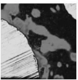
Fig. 3.1. Confocal laser micrographs of the bone around dental implants after 23 months. The implant appears in green, the mineralized bone matrix appears in black while the marrow spaces and the osteocytes appear in red. Basic fuchsine staining; original magnification x 100.
Fig. 3.2. (A) light microscopic view of native bone (away from implant surface at least 0.7mm). Toluidine blue and fuchsine acid staining; original magnification x 12. In (B) higher magnification under light microscope of the area arrowed in (A); inside the marrow space is evident an osteogenesis activity with several osteoblasts around a newly formed bone area. Toluidine blue and fuchsine acid staining; original magnification x 200. In (C) higher magnification under confocal laser microscope of the area arrowed in (A); the mineralized bone matrix appears in dark gray while the marrow spaces and the osteocytes appears in white. Basic fuchsine staining; original magnification x 200. In (D) higher magnification under light microscope of the area arrowed in (A); two bone vascular channel are visible; Azure II and orange G staining; original magnification x 200.
Fig. 4. The osteocyte lacunar density (Ot.Lc.N/ BAr) comparison
between native bone (away from implant surface at least 0.7mm) and peri-implant bone after 23 moths. Mann-Whitney Test.
Fig. 5. Polarized light microscopic images of the bone near the implant retrieved after 23 months. in (A) the bone facing the lower flank of the thread (near the tip), where the load was transferred to bone by compressive vectors, the CFO appeared mainly transverse (white-blue)than longitudinal (white-red). In (A1) the computer separation of the two CFO orientation. In (B) the bone facing the inter-threads, were the load was transferred to bone mainly by shear vectors, the CFO was mostly longitudinal (white-red). In (B1) the computer separation of the two CFO orientation.
collagen fibers around implant give a reliable measure of osseointegration. While the mineral bone phase primarily guarantees the stiffness of bone, the spatial orientation of collagen fibers contributes to bone toughness and strength (mechanical properties) (11). However, the important and complex interactions of extracellular matrix macromolecules like collagen fibers with mineral deposits under mechanical loads are still poorly known.
The different mechanical loading acts on osteoblasts activity which in turn modify collagen fibers orientation they synthesize: bone under compression shows oblique- or transversely oriented collagen, whereas bone under tension shows longitudinally oriented collagen (17).
In the present study the peri-implant bone was mainly of woven type with only few osteons. However, many cement lines indicates an high remodeling rate of the bone despite two years of immediate load. Our current observations fit well with the emerging concept that osteocyte number may influence bone remodeling. Hernandez et al. (18) suggest that woven bone with increased lacunar density may undergo remodeling at an accelerated rate. In his study periosteal woven bone formed via intramembranous osteogenesis, either in response to mechanical loading was 921 ±204 cells/mm2 and in the periosteal buttressing
region of the fracture callus was 1138 ± 168 cells/mm2.
These findings demonstrated that lacunar density in woven bone varies and appears to be elevated in endochondrally derived woven bone adjacent to marrow space.
In our study the BIC rate of the implant retrieved after 23 months was 76.7± 4.9 (mean ±SD). This result is comparable to that obtained in previous study by Traini et al. (13) in which the BIC percentage of the implant retrieved after 5-year loading was of 81.6% ± 1.5%.
Statistical differences were detected between osteocytes numbers in the peri-implant and native bone. Vashishth et al. (5) reported that the coefficient of variation of osteocyte lacunar density increases linearly with age, and that aging bone tissue is characterized by increased heterogeneity in the spatial organization (numbers) of osteocytes. The osteocyte syncytium within bone is responsible for sensing load and regulating functional bone adaptation via the lacuno-canalicular network and intercellular gap junctions (19).
Additional difference was found between the transverse CFO under the lower flank of the thread and the longitudinal CFO. Also transverse and longitudinal CFO in the inter-threads region were statistically different. In a previous study Traini et al. showed that the transverse collagen fibers were significantly associated with the lower flank of the implant threads, while in the straight surface of the implant the longitudinal collagen fibers were more represented. The predominance of transverse collagen fibers under the lower flank of the dental implant threads is an important factor because they can better resist the compressive loads. The different collagen fiber orientation is due to their different capacity in the absorption of energy, and in this way the loading stresses, tensile or compressive, are distributed (1).
ACKNOWLEDGMENT
This work was supported by FAR from the University of Ferrara (FC), Ferrara, Italy, and from Regione Emilia Romagna, Programma di Ricerca Regione Universita, 2007–2009, Area 1B: Patologia osteoarticolare: ricerca pre-clinica e applicazioni cliniche della medicina rigenerativa Unita Operativa n. 14, and PRIN 2008 (F.C.).
REFERENCES
1. Traini T, Degidi M, Caputi S, Strocchi R, Di Iorio D, Piattelli A. Collagen fiber orientation in human peri-implant bone around immediately loaded and unloaded titanium dental implants. J Periodontol 2005; 76:83-9. 2. Traini T, Pecora G, Iezzi G, Piattelli A. Preferred collagen
fiber orientation human peri-implant bone after a short- and long-term loading period: a case report. J Oral Implantol 2006; 32:177-81.
3. Yi JM, Lee JK, Um HS, Chang BS, Lee MK. Marginal
Fig. 6. The CFO comparison for transverse and longitudinal collagen fiber by both lower flank of the thread and between threads. It was noted a statistical significant association for both transverse CFO to the lower flank of the thread and of longitudinal CFO to the inter-threads area. In general, the transversal CFO was related to the Implant threads while the longitudinal CFO to the inter-threads part of the implant.
0394-6320 (2011) Copyright © by BIOLIFE, s.a.s. This publication and/or article is for individual use only and may not be further reproduced without written permission from the copyright holder. Unauthorized reproduction may result in financial and other penalties
37 (S)
11. Kalmey JK, Lovejoy CO. Collagen fiber orientation in the femoral necks of apes and humans: do their histological structures reflect differences in locomotor loading? Bone 2002; 31:327-32.
12. Osaki S, Tohno S, Tohno Y, Ohuchi K, Takakura Y. Determination of the orientation of collagen fibers in human bone. Anat Rec 2002; 266:103-7.
13. Traini T, De Paoli S, Caputi S, Iezzi G, Piattelli A. Collagen fiber orientation near a fractured dental implant after a 5-year loading period: case report. Implant Dent 2006; 15: 70-6.
14. Bentolila V, Boyce TM, Fyhrie DP, Drumb R, Skerry TM, Schaffler MB. Intracortical remodeling in adult rat long bones after fatigue loading. Bone 1998; 23:275-81. 15. Misch CE. Bone density: A key determinant for clinical
success. In Contemporary implant dentistry. Misch CE, ed. Mosby Chicago, 1999; 109-18.
16. Wang X, Bank RA, TeKoppele JM, Agrawal CM. The role of collagen in determining bone mechanical properties. J Orthop Res 2001; 19:1021-6.
17. Riggs CM, Lanyon LE, Boyde A. Functional associations between collagen fibre orientation and locomotor strain direction in cortical bone of the equine radius. Anat Embryol (Berl) 1993; 187:231-8.
18. Hernandez CJ, Majeska RJ, Schaffler MB. Osteocyte density in woven bone. Bone 2004; 35:1095-9.
19. Knothe Tate ML, Adamson JR, Tami AE, Bauer TW. The osteocyte. Int J Biochem Cell Biol 2004; 36:1-8.
bony changes in relation to different vertical positions of dental implants. J Periodontal Implant Sci; 40:244-8. 4. Ma YL, Dai RC, Sheng ZF, Jin Y, Zhang YH, Fang LN, Fan
HJ, Liao EY. Quantitative associations between osteocyte density and biomechanics, microcrack and microstructure in OVX rats vertebral trabeculae. J Biomech 2008; 41: 1324-32.
5. Vashishth D, Verborgt O, Divine G, Schaffler MB, Fyhrie DP. Decline in osteocyte lacunar density in human cortical bone is associated with accumulation of microcracks with age. Bone 2000; 26:375-80.
6. Vashishth D, Gibson G, Kimura J, Schaffler MB, Fyhrie DP. Determination of bone volume by osteocyte population. Anat Rec 2002; 267:292-5.
7. Piattelli A, Ruggeri A, Franchi M, Romasco N, Trisi P. An histologic and histomorphometric study of bone reactions to unloaded and loaded non-submerged single implants in monkeys: a pilot study. J Oral Implantol 1993; 19:314-20. 8. Martin RB. Functional adaptation and fragility of the
skeleton. In Bone loss and osteoporosis: an anthropological perspective. Agarwal SC, Stout SD, eds. Kluwer Academic/ Plenum Publishers. New York, 2004; 121-38.
9. Ortner DJ. Aging effects on osteon remodeling. Calcif Tissue Res 1975; 18:27-36.
10. Martin RB, Lau ST, Mathews PV, Gibson VA, Stover SM. Collagen fiber organization is related to mechanical properties and remodeling in equine bone. A comparison of two methods. J Biomech 1996; 29:1515-21.