I
NTRASPHENOIDAL
E
NCEPHALOCELE
A
SSOCIATED WITH
C
EREBROSPINAL
F
LUID
F
ISTULA AND
S
UBDURAL
H
EMATOMAS
: T
ECHNICAL
C
ASE
R
EPORT
Bernardo Fraioli, M.D.
Department of Neuroscience, Neurosurgery, University of Rome “Tor Vergata,” Rome, Italy
Carlo Conti, M.D.
Department of Neuroscience, Neurosurgery, University of Rome “Tor Vergata,” Rome, Italy
Pierpaolo Lunardi, M.D.
Department of Neuroscience, Neurosurgery, University of Rome “Tor Vergata,” Rome, Italy
Giovanni Liccardo, M.D.
Department of Neuroscience, Neurosurgery, University of Rome “Tor Vergata,” Rome, Italy
Mario Francesco Fraioli, M.D.
Department of Neuroscience, Neurosurgery, University of Rome “Tor Vergata,” Rome, Italy
Francesco Saverio Pastore, M.D.
Department of Neuroscience, Neurosurgery, University of Rome “Tor Vergata,” Rome, Italy Reprint requests: Bernardo Fraioli, M.D., Department of Neuroscience, Neurosurgery, Via Montpellier, 1, 00133 Rome, Italy.
Email: [email protected] Received, June 17, 2002. Accepted, February 12, 2003.
OBJECTIVE AND IMPORTANCE: Intrasphenoidal encephalocele is a rare clinical
entity that is often complicated by rhinorrhea, recurrent meningitis, and headache, but
in no case has the association of rhinorrhea with subdural hematomas been described.
A surgical procedure to stop persistent cerebrospinal fluid leakage is reported.
CLINICAL PRESENTATION: A 59-year-old man sought care for intractable
rhinoli-quorrhea of 6 months’ duration. Cranial computed tomographic and magnetic
reso-nance imaging scans revealed a basal posterior frontal bony defect and an evocative
image suggesting intrasphenoidal encephalocele.
INTERVENTION: A transnasal transsphenoidal surgical procedure was performed; the
encephalocele was removed, and the sphenoid sinus was filled with an inflatable
pouch made of synthetic dura mater containing abdominal fat. Postoperative reduction
of the rhinoliquorrhea, but not its total disappearance, was observed. Total
disappear-ance was achieved only after endonasal, transmucosal inflation of the pouch with
human fibrin glue. One of the subdural hematomas disappeared spontaneously, and
the other was treated by a surgical procedure.
CONCLUSION: The possible role of the presented technique in the treatment of
cerebrospinal fluid leakage is discussed.
KEY WORDS: Cerebrospinal fluid rhinorrhea, Intrasphenoidal encephalocele, Transsphenoidal approach
Neurosurgery 52:1487-1490, 2003 DOI: 10.1227/01.NEU.0000065183.05896.9C www.neurosurgery-online.com
I
ntrasphenoidal encephalocele is a rare clinical entity that is usually caused by trauma, tumor, intracranial infection, or a surgical procedure involving the sellar region, the parasellar region, or the paranasal sinus. When none of these causes can be identified, the encephalocele’s pathogenesis can be at-tributed, as in the case we present here, to the existence of a small defect in the bone, prob-ably congenital in nature, located in the mid-dle cranial fossa, through which cerebral tis-sue may protrude (4, 5, 17, 22). In the available literature, we were able to find descriptions of only 12 cases of this entity (1, 6, 9, 10, 16, 20, 23, 25). Rhinorrhea, recurrent meningitis, and headache are mentioned as the complications of encephalocele, but in no case has the asso-ciation of rhinorrhea with subdural hematoma (SDH) been described. This finding makes the case we present here unique.CASE REPORT
The patient, a 59-year-old male doctor, had experienced rhinorrhea and occasional
retro-orbital pain and frontal headache for approx-imately 6 years. No previous trauma or other significant abnormalities were reported. The neurological examination revealed nothing abnormal. The leaking nasal fluid was con-firmed by a glucose test to be cerebrospinal fluid (CSF) (47 mg/100 ml). The first contrast-enhanced encephalic magnetic resonance im-aging (MRI) scan, obtained 20 days before admission to our department, failed to dem-onstrate the location of a fistula and did not detect a blood collection. However, it revealed a fusiform formation in the sphenoid sinus with signal characteristics similar to those of the cerebral parenchyma (Fig. 1). A high-resolution computed tomographic (CT) scan revealed a communication between the right posterolateral side of the sphenoethmoidal planum and the sphenoid sinus (Fig. 2). Con-sidering the length of the patient’s clinical his-tory and the therapeutic procedures per-formed in the past, which included prolonged lumbar drainage performed at another insti-tution 3 months earlier, surgery was pro-posed. Surprisingly, control MRI scans, which
were taken before surgery to assess increased CSF leakage, revealed two large bihemispheric SDHs (Fig.
3).
OPERATIVE
TECHNIQUE
The sphenoid sinus was exposed through a transna-sal transsphenoidal micro-surgical approach disclos-ing, in the right section, a fusiform mass (1.5⫻ 0.7 cm)with a fine translucent coating; the small mass protruded through a bony defect onto the inferoposterolateral ethmoidal cells. CSF leakage around the mass was also evident (Fig. 4). The lesion was removed, and duraplasty was performed with human fibrin glue and synthetic dura mater. Then a pouch was made by wrapping synthetic dura mater around abdom-inal fat; this was inserted into the sphenoid sinus as a “plug,” which resulted in reduction, but not disappearance, of the CSF leakage (Fig. 5). This was not evident during the operation, but only postoperatively, when the recurrence of rhinoliquorrhea, even though reduced, was observed. A histological examina-tion revealed that the removed lesion consisted of central nervous system tissue.
The patient underwent an endonasal, transmucosal surgical procedure while under CT scan control, as previously de-scribed (12). A 12-gauge lumbar puncture needle was inserted through the sphenoid sinus into the pouch, and human fibrin glue was injected under pressure (Fig. 6). After this surgical procedure was performed, the fistula disappeared. During the next 2 weeks, CT and MRI control scans revealed progressive reduction until the SDH disappeared on the left side; the one on the right side persisted, so it was surgically drained. At a follow-up examination at 20 months, the patient showed no signs of rhinorrhea, and a CT scan demonstrated that SDHs were absent.
DISCUSSION
The association of rhinor-rhea, SDHs, and intrasphe-noidal encephalocele repre-sents an event not described, to our knowledge, in the available literature. It raises a diagnostic problem regard-ing the location of the fistu-lous connection and thera-peutic questions regarding the choice of surgical proce-dure. The bony defect was localized by high-resolution CT scan (4, 8, 23). The MRI scan did not visualize the fis-tulous connection, but itcon-tributed to the identification of the intrasphenoidal encepha-locele (10, 23).
The choice of the surgical approach was not simple, as we envisaged both a transcranial and a transsphenoidal approach. We were persuaded that the choice of the transsphenoidal approach was not contingent upon the particular expertise of one of us (BF) with this technique (11). In our opinion, it is advisable that a surgeon with an equivalent confidence in both the transsphenoidal and the transcranial approach be at first inclined toward a less invasive procedure. The additional risks of the intracranial procedure are not negligible, and they are both generic and related to the possible manipulation of vas-cular and nervous structures. Undoubtedly, the transcranial route should be the first choice when a wide cranial base bone defect requires an osteoplasty completed with a large pericra-nial vascularized flap; the transsphenoidal approach probably cannot ensure an adequate closure in these cases. Neverthe-less, in intracranial procedures for the treatment of CSF fistula, recurrences are not rare (2, 4, 5, 25). In the present case, the bone defect was actually limited and easy to reach transsphe-FIGURE 1. Axial T2-weighted MRI
scan revealing fluid inside the sphe-noid sinus and a mass with signal features similar to those of the cere-bral parenchyma (arrow).
FIGURE 2. Sagittal (A) and coronal (B) CT images (bone window)
revealing communication between the sphenoethmoidal planum and the sphenoid sinus (arrows).
FIGURE 3. Fluid-attenuated inversion
recovery MRI scan revealing bilateral hemispheric subdural hematomas.
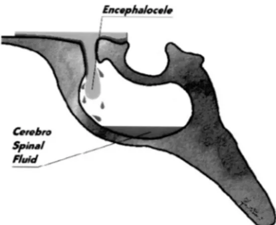
FIGURE 4. Drawing of the sphenoid sinus (sagittal view), showing the
bone defect of the sphenoethmoidal planum and encephalocele associated with a CSF fistula.
F
RAIOLI ET AL.
noidally, so we could accomplish a procedure that was both extracerebral and extrasellar.
The technique we present differs remarkably from the per-cutaneous technique previously published, which consists of filling the sphenoid sinus with fibrin glue under CT guidance (12). In this case, our technique permitted the introduction into the sphenoid sinus of a dural handmade pouch wrapped in autologous fat tissue. It is possible to further inflate the pouch percutaneously, if necessary. During the operation, the dural pouch was introduced only partially filled; otherwise, its vol-ume would not have fit into the sphenoid sinus. However, the leakage of CSF immediately stopped, so further intraoperative inflation did not seem necessary. The postoperative recurrence of leakage, even though reduced, prompted us to further inflate the pouch percutaneously. In the future, we intend to inflate the pouch to its maximum volume intraoperatively.
Regarding the association of CSF fistula and SDH, the liter-ature provides some possible explanations for the mechanisms inducing the formation of SDH in the presence of a CSF fistula and for its regression after leakage interruption (3, 7, 13, 18, 19). The reexpansion of brain parenchyma after surgery could have progressively restored the craniocerebral proportions, previously impaired by the leakage. This phenomenon might
have hindered the proliferation of the neocapillary network of the SDH outer membrane responsible for expansion of the hematoma, possibly also reducing fibrinolytic activity. Finally, these events may have resulted in neomembrane atrophy and progressive hematoma fluid resorption (14, 15, 19, 21, 24). However, these hypotheses do not explain why the contralat-eral hematoma persisted in our patient. Finally, although the follow-up period in our case may be considered to be too short, the convincing rationale of the technique leads us to conclude that the result will be lasting.
REFERENCES
1. Abiko S, Aoki H, Fudaba H: Intrasphenoidal encephalocele: Report of a case. Neurosurgery22:933–936, 1988.
2. Albernaz SM, Horton DW, Adkins YW, Garen DP: Intrasphenoidal enceph-alocele. Otolaryngol Head Neck Surg 104:279–281, 1991.
3. Alemohammad S, Bouzarth WF: Intracranial subdural hematoma following lumbar myelography. J Neurosurg 52:256–258, 1980.
4. Beckhardt R, Setzen M, Carras R: Primary spontaneous cerebrospinal fluid rhinorrhea. Otolaryngol Head Neck Surg 4:425–432, 1991.
5. Bernstein MJ, Roland Thomas Jr, Persky SM: Sphenoid cranial base defects in siblings presenting with cerebrospinal fluid leak. Skull Base Surg 7:193– 197, 1997.
6. Buchfelder M, Fahlbusch R, Huk JW, Thierauf P: Intrasphenoidal encephaloceles, a clinical entity. Acta Neurochir (Wien) 89:10–15, 1987. 7. Chokroverty S, Mayo CM: Spontaneous resolution of subdural hematoma.
Dis Nerv Syst29:704–706, 1968.
8. Chow JM, Goodman D, Mafee MF: Evaluation of CSF rhinorrhea by com-puterized tomography with metrizamide. Otolaryngol Head Neck Surg 100:99–105, 1989.
9. Daniilidis J, Vlachtsis K, Ferekidis E, Dimitriadis A: Intrasphenoidal enceph-alocele and spontaneous CSF rhinorrhea. Rhinology 37:186–189, 1999. 10. Deasy NP, Jarosz JM, Al Sarray S, Cox TC: Intrasphenoid cephalocele: MRI
in two cases. Neuroradiology 41:497–500, 1999.
11. Fraioli B, Esposito V, Santoro A, Iannetti G, Giuffrè R, Cantore G: Transmaxillosphenoidal approach to tumors invading the medial compart-ment of the cavernous sinus. J Neurosurg 82:63–69, 1995.
12. Fraioli B, Pastore FS, Floris R, Vagnozzi R, Simonetti G, Liccardo G, Giuffre R: Computed tomography-guided transsphenoidal closure of postsurgical cerebrospinal fluid fistula: A transmucosal needle technique. Surg Neurol 48:409–412, 1997.
13. Gannon E, Cook AW, Browder EJ: Resolving subdural collections. J Neurosurg19:865–869, 1962.
14. Ito H, Komai T, Yamamoto S: Fibrinolytic enzyme in the living walls of chronic subdural hematoma. J Neurosurg 48:197–200, 1978.
15. Ito H, Yamamoto S, Komai T, Mizukoshi H: Role of local hyperfibrinolysis in the etiology of chronic subdural hematoma. J Neurosurg 45:26–31, 1976. 16. Jabre A, Tabaddor R, Samaraweera R: Transsphenoidal
meningoencephalo-cele in adults. Surg Neurol 54:183–188, 2000.
17. Kaufman B, Yonas H, Witt RJ, Miller CF: Acquired middle cranial fossa fistulas: Normal pressure and nontraumatic origin. Neurosurgery 5:466– 472, 1979.
18. Markwalder TM: Chronic subdural hematomas: A review. Neurosurgery 54:637–645, 1981.
19. McCullough DL, Fox JL: Negative intracranial pressure hydrocephalus in adults with shunts and its relationship to the production of subdural hema-toma. J Neurosurg 40:372–375, 1974.
20. Myssionek D, Cohey NL: Intrasphenoidal meningoencephalocele: A case report. Am J Otolaryngol 8:391–394, 1987.
21. Naganuma H, Fukamachi A, Kawakami M, Misumi S, Nakajima H, Wakao T: Spontaneous resolution of chronic subdural hematoma. Neurosurgery 19:794–798, 1986.
22. Schick B, Draf W, Kahle G, Weber R, Wallenfang T: Occult malformations of the skull base. Arch Otolaryngol Head Neck Surg 123:77–80, 1997. FIGURE 5. Drawing of the insufficient intrasphenoidal filling of autologous fat
wrapped by synthetic dura mater. The CSF leakage is reduced but still exists.
FIGURE 6. Drawing of the CT-guided pressure injection of human fibrin
glue into the customized dural pouch.
I
NTRASPHENOIDALE
NCEPHALOCELE23. Shetty P, Shroff M, Fatterpekar G, Sahani D, Kirtane M: A retrospective analysis of spontaneous sphenoid sinus fistula: MR and CT findings. AJNR Am J Neuroradiol21:337–342, 2000.
24. Suzuki J, Takaku A: Nonsurgical treatment of chronic subdural hematoma. J Neurosurg33:548–553, 1970.
25. Willner A, Kantrowitz BA, Cohen FA: Intrasphenoidal encephalocele: Diag-nosis and management. Otolaryngol Head Neck Surg 111:834–837, 1994.
COMMENTS
T
his article describes treatment of an intrasphenoidal en-cephalocele with an original technique of using an inflat-able pouch that is inserted into the sphenoid sinus. In our experience, using the transsphenoidal approach and filling the sphenoid sinus with fat and glue, while draining the cerebro-spinal fluid (CSF) via a cerebro-spinal catheter for several days, may also be successful in most cases. However, after unsuccessful lumbar drainage in intractable CSF fistula via the sphenoid sinus, the technique described in this article may be an inter-esting and novel solution.Jacques Brotchi
Brussels, Belgium
T
he authors have successfully applied their previously pub-lished transmucosal needle technique (1) to arrest a CSF rhinorrhea persisting after transsphenoidal treatment of an en-cephalocele associated with a CSF fistula. This report demon-strates that it is sometimes possible to find simple solutions to apparently complex problems. One should consider the authors’ conclusions regarding the superiority of the transsphenoidal ap-proach over the transcranial apap-proach for the treatment of intras-phenoidal encephalocele before attempting this technique.Albino Bricolo
Verona, Italy
1. Fraioli B, Pastore FS, Floris R, Vagnozzi R, Simonetti G, Liccardo G, Giuffre R: Computed tomography-guided transsphenoidal closure of postsurgical cerebrospi-nal fluid fistula: A transmucosal needle technique. Surg Neurol 48:409–412, 1997.
T
he report of Fraioli et al. is interesting because of the rarity ofthe anomaly, the concomitant subdural hematomas, and the method of surgical repair. An intrasphenoidal encephalocele pre-senting with rhinorrhea was treated by transnasal transsphenoi-dal resection of the encephalocele and obliteration of the sphe-noid sinus with autologous fat contained within a pouch of synthetic dura. Persistent rhinorrhea associated with bilateral
subdural hematomas was successfully treated by secondary ex-pansion of the pouch with fibrin glue injected endonasally.
Basal encephaloceles, herniations of brain, and meninges through defects in the cranial base are rare congenital anomalies that arise from failure of midline fusion (1). In infancy, they present in association with other facial, cranial, and intracranial midline defects (e.g., cleft lip and palate, hypertelorism, abnor-malities of optic disc, nerve, and chiasm, pituitary or hypotha-lamic insufficiency, and agenesis of the corpus callosum). In adults, CSF rhinorrhea (spontaneous or iatrogenic, from biopsy of a nasal polyp), visual field loss, endocrinopathy, and a soft tissue mass in the epipharynx can occur. Indications for treat-ment in the adult include persistent rhinorrhea, progressive neu-rological deficit, and respiratory obstruction from an epipharyn-geal mass. The risk of meningitis mandates treatment when rhinorrhea occurs (2). Even prolonged drainage of lumbar CSF can fail. When the sphenoid sinus is the route of CSF egress, transnasal transsphenoidal repair is a preferred, less invasive alternative to craniotomy. The defect must be small enough to be fully accessed by the transsphenoidal route, and the surgeon must be careful to avoid injuring protruding and potentially critical neurovascular structures. The authors’ use of a dural pouch is a clever modification that permits expansion of an otherwise inadequate plug.
Griffith R. Harsh IV
Stanford, California
1. French BN: Midline fusion defects and defects of formation, in Youmans JR (ed):
Neurological Surgery. Philadelphia, Saunders, pp 1236–1380, 1982, ed 2, vol 3.
2. Smith DE, Murphy MJ, Hitchon PW, Babin RW, Abu-Yousef MM: Transsphe-noidal encephaloceles. Surg Neurol 20:471–480, 1983.
F
raioli et al. present a case of intrasphenoidal encephalocele associated with CSF fistula and subdural hematoma that ultimately resolved by the injection of fibrin glue into a dural patch used for repair. This is an innovative use of fibrin glue to repair a CSF leak. The senior author (LNS) would have preferred an intracranial approach to repair the CSF fistula with fascia lata and a pericranial flap initially. If transsphe-noidal surgery is elected, autologous material, such as fascia lata or abdominal fascia supplemented with fat, may be better for repair because it is more pliable than artificial dura; it is more adhesive and results in better healing.Laligam N. Sekhar Dinko Stimac
Annandale, Virginia
Advertising
Inquiries regarding advertising in Neurosurgery should be directed to: Paul Tucker
Lippincott Williams & Wilkins 351 West Camden Street Baltimore, MD 21201-2436 Tel: 410/528-4291
Fax: 410/528-4457 email: [email protected]
Kelly Adamitis
Lippincott Williams & Wilkins 530 Walnut Street Philadelphia, PA 19106-3621 Tel: 215/521-8402 Fax: 215/521-8411 email: [email protected]