Original Paper
© 2017 The Author(s) Published by S. Karger AG, Basel Lab. Physiology and Experimental Surgery, Dept. Translational Medicine, University East Piedmont, via Solaroli 17, I-28100 Novara (Italy)
Tel. +390321660526, Fax +3903213733537, E-Mail [email protected] Prof. Elena Grossini
Anti-Vascular Endothelial Growth Factors
Protect Retinal Pigment Epithelium Cells
Against Oxidation by Modulating Nitric
Oxide Release and Autophagy
Stefano De Cillàa Serena Farruggiob Stela Vujosevicc Giulia Rainab
Davide Filippinib Valentina Gattia,b Nausicaa Clemented David Maryb
Diego Vezzolaa Giamberto Casinie Luca Rossettif Elena Grossinib
aOculistic Unit, Dept. Health Sciences, University East Piedmont, Azienda Ospedaliera Universitaria Maggiore della Carità, Novara, bLab. Physiology/Experimental Surgery, Dept. Translational Medicine, University East Piedmont, Novara, cEye Clinic, Azienda Ospedaliera Universitaria Maggiore della Carità, Novara, dLab. Immunology, IRCAD, Dept. Health Sciences; University East Piedmont, Novara, e Dept. Surgical, Medical, Molecular and Critical Area Pathology, University of Pisa; fEye Clinic, San Paolo Hospital, University of Milan, Italy
Key Words
Anti-VEGF • Cell viability • Mitochondria • Peroxidation
Abstract
Background/Aims: the anti-vascular endothelial growth factors (VEGF), Aflibercept and Ranibizumab, are used for the treatment of macular degeneration. Here we examined the involvement of nitric oxide (NO), mitochondria function and of apoptosis/autophagy in their antioxidant effects in human retinal pigment epithelium cells (RPE). Methods: RPE were exposed to Ranibizumab/Aflibercept in the absence or presence of NO synthase (NOS) inhibitor and of autophagy activator/blocker, rapamicyn/ 3-methyladenine. Specific kits were used for cell viability, NO and reactive oxygen species detection and mitochondrial membrane potential measurement, whereas Western Blot was performed for apoptosis/ autophagy markers and other kinases detection. Results: In RPE cultured in physiological conditions, Aflibercept/Ranibizumab increased NO release in a dose and time-dependent way. Opposite results were obtained in RPE pretreated with hydrogen peroxide. Moreover, both the anti-VEGF agents were able to prevent the fall of cell viability and of mitochondrial membrane potential. Those effects were reduced by the NOS inhibitor and 3-methyladenine and were potentiated by rapamycin. Finally, Aflibercept and Ranibizumab counteracted the changes of apoptosis/autophagy markers, NOS, Phosphatidylinositol-3-Kinase/Protein Kinase B and Extracellular signal–regulated kinases 1/2 caused by peroxidation. Conclusion: Aflibercept and Ranibizumab protect RPE against peroxidation through the modulation of NO release, apoptosis and autophagy.
Introduction
The Age-Related Macular Degeneration (AMD) is the leading cause of loss of vision in developed countries. Although advanced age is the main risk factor, other mechanisms could be involved in the physiopathology of AMD [1-3]. Abnormalities of the choroidal blood flow have been hypothesized to contribute to the development of AMD. Regarding this issue, recent studies have suggested that changes in nitric oxide (NO) plasma levels in AMD patients could be involved in the modulation of choroidal perfusion. Furthermore, the levels of NO synthase (NOS) isoforms have been found to be significantly reduced in eyes with AMD [4]. All together those findings could be associated with neuronal degeneration in retina and hemodynamic changes in AMD choroid. In spite of this, the precise role of NO in the onset of AMD has not yet been clearly examined [5]. Furthermore, mitochondrial damage and reactive oxygen species (ROS) could play an important role in AMD pathogenesis, through the reduction of vascularization of choriocapillaris and apoptosis and by increasing the formation of drusen [6] and the release of pro-angiogenic vascular endothelium growth factor (VEGF) from the retinal pigment epithelium cells (RPE) [7-9]. Finally, it is to note that autophagy has also been the object of studies regarding the pathophysiology of AMD. Hence, if acute oxidative stress was shown to increase autophagy, the chronic oxidative stimulation was reported to down regulated it [10-12].
Intravitreal anti-VEGF therapy with drugs like Aflibercept and Ranibizumab has hugely improved the vision prognosis of neo-vascular AMD [13-17]. However, it is not clear up till now if those agents could act as modulator of NO release, oxidative stress and autophagy/ apoptosis.
Thus, in the present study we planned to compare the effects of Aflibercept and Ranibizumab against oxidative stress in human RPE. In particular, we examined NO release, cell viability, proliferation and mitochondria membrane potential, apoptosis/autophagy, and the activation/expression of various kinases involved in NO release and cell survival.
Materials and Methods
Culture of RPE and PAE
ARPE-19 (ATCC cell line; RPE) and porcine aortic endothelial cells (PAE) were maintained in Dulbecco’s modified Eagle’s medium (DMEM; Sigma, Milan, Italy) supplemented with 10% fetal bovine serum (FBS; Euroclone, Pero, Milan, Italy), 2 mM L-glutamine (Sigma), 1% penicillin-streptomycin (Sigma), at 37°C with 5% CO2 in an incubator. For NO measurement, which was performed in RPE and PAE, 7.5x10³ cells/ well were plated in 96-well plates with DMEM 0% FBS supplemented with 1% penicillin–streptomycin– glutamine and without phenol red (starvation medium, Sigma) for 4–6 h. In RPE, mitochondrial membrane potential and cell viability were also measured, following the same procedure using for NO measurement, but in 1×10⁴ cells. For glutathione (GSH) quantification and Western Blot, 4x10⁵cells/well were plated in 6 wells in complete medium and at confluence they were incubated with starvation medium overnight. For cell proliferation, 7x10³ cells/well were plated in 16 wells (CIM-plate). For reactive oxygen species (ROS) quantification, 2.5x10⁴ cells/well were plated in 96-well. Each experimental protocol was repeated in five different cell samples.
NO release
The NO production was measured in RPE and PAE culture supernatants by using the Griess method (Promega, Milan, Italy), as previously performed in the same or similar cellular models [18-21]. RPE and PAE were treated for 30 min with Aflibercept (0.025 mg/ml, 0.05 mg/ml, 0.20 mg/ml, 0.50 mg/ml; Bayer, Varese, Italy) and Ranibizumab (0.025 mg/ml, 0.05 mg/ml, 0.20 mg/ml; Novartis, Milan, Italy). A time-course study was performed by treating RPE with Aflibercept and Ranibizumab for 1, 5 and 30 min. In addition, in other experiments in RPE and PAE, 30 min Aflibercept (0.50 mg/ml) and Ranibizumab (0.20 mg/ml) were given after the pre-stimulation with the NOS blocker, Nω-nitro-L-arginine methylester (L-NAME; 10 mM for 15 min; Sigma). In some samples, the effects of 200 µM hydrogen peroxide, administrated 30 min after the
anti-VEGF agents, on NO release were also examined both in RPE and PAE. At the end of the stimulations, NO production in the sample's supernatants was examined by adding an equal volume of Griess reagent following the manufacturer's instruction. At the end of incubation, the absorbance at 570 nm was measured by a spectrometer (BS1000 Spectra Count, San Jose, CA, USA) and the NO production was quantified in respect to nitrite standard curve and expressed as percentage.
Cell viability
As described for NO release, dose-response and time-course experiments were performed to examine the effects of Aflibercept and Ranibizumab on cell viability, which was examined by using the 1% 3-[4,5-dimethylthiazol-2-yl]-2,5-diphenyl tetrazolium bromide (MTT; Life Technologies Italia, Monza, Italy) dye, as previously described [21]. RPE cultured in starvation medium were also treated with 30 min hydrogen peroxide (200 μM) alone or in the presence of Aflibercept (0.025 mg/ml, 0.05 mg/ml, 0.20 mg/ ml, 0.50 mg/ml) and Ranibizumab (0.025 mg/ml, 0.05 mg/ml, 0.20 mg/ml) given alone or 30 min before hydrogen peroxide. In addition, in other cell samples, 30 min Aflibercept (0.50 mg/ml) and Ranibizumab (0.20 mg/ml) were given after the pre-stimulation with L-NAME (10 mM for 15 min; Sigma), the autophagy inhibitor, 3-methyadenine (4 µM for 30 min; Sigma) and the autophagy activator, rapamycin (4 µM for 30 min; Santa Cruz Biotechnology; Inc., CA, USA). After each treatment, the medium was removed and an MTT solubilization solution was added and mixed in a gyratory shaker until the complete dissolution of formazan crystals. Cell viability was determined by measuring the absorbance through a spectrometer (BS1000 Spectra Count, San Jose, CA, USA) [18-20].
Mitochondrial membrane potential measurement
Mitochondrial membrane potential measurement in RPE was performed with JC-1 assay. Cells were stimulated as described for Cell viability. Aflibercept and Ranibizumab were administrated for 30 min. After stimulations, the medium of the cells plated in starvation medium was removed and incubated with 5,51,6,61-tetrachloro-1,11,3,31 tetraethylbenzimidazolyl carbocyanine iodide (JC-1) 1X diluted in Assay Buffer 1X for 15 min at 37 °C in an incubator, following the manufacturer's instruction (Invitrogen, Life Technologies Europe BV, Monza, Italy) [21]. After incubation, the cells were washed twice with Assay Buffer 1X and then the mitochondrial membrane potential was determined by measuring the red (excitation 550 nm/emission 600 nm) and green (excitation 485 nm/emission 535 nm) fluorescences through a spectrometer (BS1000 Spectra Count).
Proliferation rate
To evaluate the effects of anti-VEGF drugs on cell proliferation, xCELLigence™ MP Instrument (Roche, Basel, Switzerland) was used. RPE plated in 16 wells, were stimulated with hydrogen peroxide (200 μM) alone or in the presence of Aflibercept (0.5 mg/ml) and Ranibizumab (0.2 mg/ml). The proliferation rate, determined by the Cell Index, was analyzed after 24 hours and 48 hours and compared to values measured at anti-VEGF agents administration. Cell-sensor impedance was expressed as an arbitrary unit called the Cell Index. The Cell Index at each time point is defined as (Rn-Rb)/15, where Rn is the cell-electrode impedance of the well when it contains cells and Rb is the background impedance of the well with the media alone.
ROS quantification
The oxidation of 2,7-dichlorodihydrofluorescein diacetate (H2DCFDA) into 2,7-dichlorodihydrofluo-rescein (DCF) was used to assess ROS generation, following the manufacturer’s instructions (Abcam, Cam-bridge, United Kindom). Briefly, cells in 96-well plates were stimulated with 30 min hydrogen peroxide (200 μM) alone or in the presence of Aflibercept (0.025 mg/ml, 0.05 mg/ml, 0.20 mg/ml, 0.50 mg/ml; 30 min) and Ranibizumab (0.025 mg/ml, 0.05 mg/ml, 0.20 mg/ml; 30 min). After treatments, the reactions were stopped by removing medium and washing with phosphate buffer saline (PBS) followed by staining with 10 µM H2DCFDA for 20 min at 37°C. The fluorescence intensity of H2DCFDA was measured at an excitation and emission wavelength of 485 nm and 530 nm using a spectrometer (BS1000 Spectra Count) [21, 22].
Cell lysates
The RPE at confluence were plated in starvation medium overnight at 37 °C with 5% CO2. Western Blot analysis was performed on RPE treated with Aflibercept (0.50 mg/ml, for 30 min) and Ranibizumab (
0.2 mg/ml, for 30 min) in the presence or absence of hydrogen peroxide (200 μM) administrated 30 min after the anti-VEGF agents. At the end of stimulations, RPE were lysed in iced-Ripa-buffer supplemented with 1:200 sodium orthovanadate and 1:100 protease inhibitors cocktail and phenylmethanesulfonyl fluoride (1:100; Sigma). The extract proteins were quantified through bicinchoninic acid protein (Pierce, Rockford, IL, USA) and used for electrophoresis and immunoblotting studies.
Western blotting
Cell lysates (30 μg protein each sample) dissolved in Laemmli buffer 5X, boiled for 5 min were resolved in 10% sodium dodecyl sulfate polyacrylamide gel electrophoresis gels (Bio-Rad Laboratories, Hercules, CA, USA); after electrophoresis they were transferred to polyvinylidene fluoride membranes (Bio-Rad Laboratories), which were incubated overnight at 4 °C with specific primary antibodies: anti phospho-Akt (p-Akt, 1:1000; Ser473, Santa Cruz Biotechnology), Akt (1:1000; Santa Cruz Biotechnology), anti phospho-ERK1/2 (1:1000; Thr202/Tyr204, Cell Signaling Technologies, Danvers, USA), anti phospho-ERK1/2 (1:1000; Cell Signaling Technologies), anti phospho-eNOS (1:1000; Ser1177, Cell Signaling Technologies anti eNOS (1:1000; Cell Signaling Technologies), anti iNOS (1:500; Santa Cruz Biotechnology), anti Cytochrome C (1:500, Sigma), anti cleaved Caspase-9 (1:1000; Abcam, Cambridge, UK), anti Beclin1 (1:1000; H-300, Santa Cruz Biotechnology). The membranes were washed and then incubated with horseradish coupled goat anti-rabbit IgG (Sigma), coupled rabbit anti-goat IgG and horseradish peroxidase-coupled goat anti-mouse IgG (Sigma) for 45 min and were developed through a nonradioactive method using Western Lightning Chemiluminescence (PerkinElmer Life and Analytical Sciences, Waltham, MA, USA). Protein expression was calculated as a ratio towards β-actin (1:5000; Sigma) or specific total protein.
Statistical analysis
All data were recorded using the Institution's database. Statistical analysis was performed by using STATVIEW version 5.0.1 for Microsoft Windows (SAS Institute Inc., Cary NC, USA). Data were checked for normality before statistical analysis. All the results obtained were examined through one-way ANOVA followed by Bonferroni post hoc tests. The non-parametric Mann Whitney U test for unpaired data was used to compare percentage responses. Pearson coefficient was calculated for linear correlation analysis in dose-response studies. All data are presented as means ± standard deviation (SD) of five independent experiments for each experimental protocol. A value of P <0.05 was considered statistically significant.
Results
Aflibercept and Ranibizumab elicit opposite effects on NO release by RPE cultured in either physiological or peroxidative conditions
Aflibercept caused a dose- and time-dependent increase of NO release by RPE cultured in physiological conditions (Fig. 1A, 2A). Similar results were obtained with Ranibizumab although at lesser extent (Fig. 1B, 2B). It is to note that both agents were able to reduce NO release caused by peroxidation (Fig. 2A and 2B).
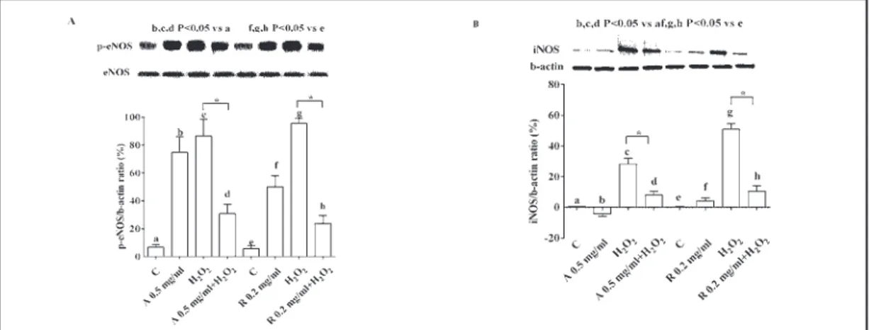
The effects of the anti-VEGF agents on NO release were accompanied by changes in eNOS/iNOS activation/expression. While in physiological conditions those kinases were activated, in RPE subjected to peroxidation, eNOS/iNOS were inhibited (Fig. 3).
Regarding NO release, similar findings were observed in PAE treated with either Aflibercept or Ranibizumab (Fig. 4).
All together those findings showed the involvement of NO in the mechanisms of action of the anti-VEGF agents.
Aflibercept and Ranibizumab increase cell proliferation and prevent the loss of cell viability and mitochondrial membrane potential caused by peroxidation in RPE. Involvement of NO and autophagy
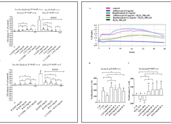
As shown in Fig. 5, the anti-VEGF agents were able to increase RPE proliferation in physiological conditions and to counteract the effects of hydrogen peroxide.
Fig. 1. Dose- and time-response effects of anti-VEGF
agents on NO release in RPE. In A, Aflibercept, in B, Ranibizumab. The values obtained correspond to the NO (μmol) produced, after each stimulation, by samples containing 1.5 μg of proteins each. They are expressed as % of control values. Reported data are means ± SD of five independent experiments.
Fig. 2. Effects of anti-VEGF agents on NO release in
RPE cultured in physiological or peroxidative condi-tions. In A, Aflibercept (A), in B, Ranibizumab (R). L-NAME= Nω-nitro-L-arginine methylester, NOS in-hibitor (10 mM). The values obtained correspond to the NO (μmol) produced, after each stimulation, by samples containing 1.5 μg of proteins each. They are expressed as % of control values (C). Reported data are means ± SD of five independent experiments. Square brackets indicate significance between groups. *P<0.05.
Fig. 3. Effects of anti-VEGF agents on activation /expression of eNOS and iNOS in RPE cultured in either
physiological or peroxidative conditions. In A and B, densitometric analysis and an example of Western Blot taken from 5 different experiments of p-eNOS and iNOS, respectively, are shown. Abbreviations are as in previous Figures. Reported data are means ± SD of five independent experiments. Square brackets indicate significance between groups. *P<0.05.
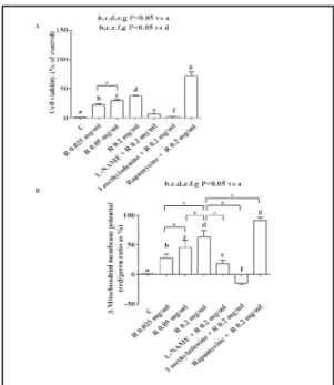
improved mitochondrial membrane potential in RPE cultured in physiological medium (Figs. 6-8). As shown in Figs. 9 and 10, both the anti-VEGF agents also prevented the effects of hydrogen peroxide.
It is to note that both in physiological and peroxidative conditions, L-NAME reduced the responses of RPE on cell viability to Aflibercept and Ranibizumab (Figs. 7-10). Moreover, rapamycin and 3 methyladenine were able to, respectively, augment and reduce the protection exerted by both the anti-VEGF agents against peroxidation (Figs. 7-10).
Thus, the protective effects elicited by Aflibercept and Ranibizumab in RPE were found to be related to the modulation of NO release and autophagy.
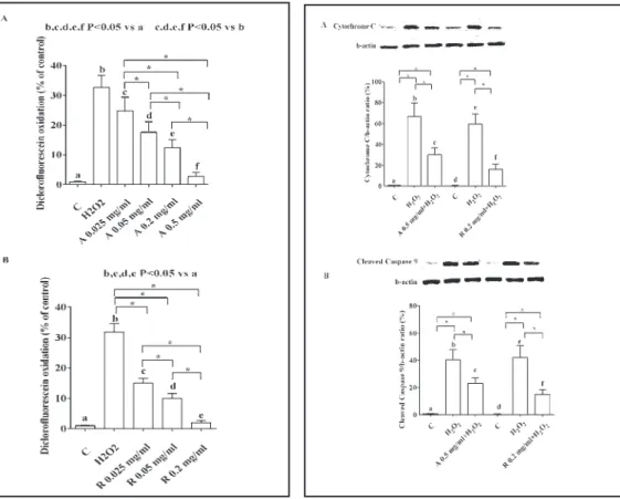
Furthermore, Aflibercept and Ranibizumab counteracted the ROS release caused by hydrogen peroxide, as well (Fig. 11).
Aflibercept and Ranibizumab inhibit apoptosis, activate autophagy, Akt and ERK1/2 in RPE
As shown in Fig. 12, the anti-VEGF agents were able to counteract the effects of hydrogen peroxide on apoptosis in RPE, as evidenced by the inhibition of the expression of the cleaved Caspase 9 and Cytochrome C. In addition, the antiapoptotic effects of both Aflibercept and Ranibizumab were accompanied by increased expression of Beclin1, an autophagic marker (Fig. 13 A). Finally, Aflibercept and Ranibizumab prevented the inhibition of survival kinases, such as ERK1/2 and Akt caused by hydrogen peroxide (Fig. 13 B and 13C).
Fig. 4. Effects of anti-VEGF agents on NO release in
PAE cultured in physiological or peroxidative condi-tions. Abbreviations and layout are as in Figure 2. Reported data are means ± SD of five independent experiments. Square brackets indicate significance between groups. *P<0.05.
Fig. 5. Effects of anti-VEGF agents on RPE
prolifera-tion in physiological and peroxidative condiprolifera-tions. In A, an example of tracing taken from xCELLigence is shown. In B and C, means ± SD of five independent experiments. C t0= control at the beginning of agents administration. Abbreviations are as in previous Fig-ures. Square brackets indicate significance between groups. *P<0.05.
Discussion
This study has shown for the first time that the anti VEGF agents, Aflibercept and Ranibizumab, play an important role in the modulation of cell viability and mitochondrial
Fig. 6. Dose-and time-response effects of anti-VEGF
agents on cell viability (A and B) and mitochondrial membrane potential (C) in RPE. Reported data are means ± SD of five independent experiments.
Fig. 7. Effects of Aflibercept (A) on cell viability and
mitochondrial membrane potential in RPE cultured in physiological conditions in presence or absence of the NOS inhibitor, the autophagy inhibitor and activator. Reported data are means ± SD of five in-dependent experiments. Square brackets indicate significance between groups. *P<0.05.
Fig. 8. Effects of Ranibizumab (R) on cell viability
and mitochondrial membrane potential in RPE cul-tured in physiological conditions in presence or ab-sence of the NOS inhibitor, the autophagy inhibitor and activator. Reported data are means ± SD of five independent experiments. Square brackets indicate significance between groups. *P<0.05.
membrane potential in RPE, cultured either in physiological and pathological conditions, by mechanisms related to NO release and apoptosis/autophagy processes
This study was performed on RPE, which are widely considered as the fulcrum of AMD pathogenesis and extensively used for in vitro studies in ophthalmology [1, 23]. Moreover, the concentration of H2O2 used for inducing peroxidation falls within the published range and is similar to that measureable in the human vitreous. [11]
In our study, Aflibercept and, at lower extent, Ranibizumab, used at doses similar to the ones achievable in humans after intravitreal injections, and previously used in RPE [24] were able to increase the NO release by RPE cultured in physiological conditions. Opposite results were obtained in peroxidation, when both Aflibercept and Ranibizumab were able to reduce NO release caused by hydrogen peroxide. It is to note that similar findings were observed in PAE, which would highlight the involvement of NO in the mechanisms of action of the anti-VEGF agents on vasculature, too.
NO is synthesized from L-arginine by three isoforms of NOS [25], which are the inducible and calcium-independent NOS (iNOS), the constitutive and calcium-dependent neuronal NOS (nNOS) and the endothelial NOS (eNOS) [4]. While the constitutive NOS may act as a regulator of physiological phenomena, the inducible NOS could be involved in longer-lasting cytotoxic and inflammatory functions [26]. The fact that NO could exert protection or damage would depend on its concentration and to the relative activity of the constitutive or inducible NOS [26].
Fig. 9. Effects of Aflibercept (A) on cell viability and
mitochondrial membrane potential in RPE cultured in peroxidative conditions in presence or absence of the NOS inhibitor, the autophagy inhibitor and acti-vator. Reported data are means ± SD of five indepen-dent experiments. Square brackets indicate signifi-cance between groups. *P<0.05.
Fig. 10. Effects of Ranibizumab (R) on cell viability
and mitochondrial membrane potential in RPE cul-tured in peroxidative conditions in presence or ab-sence of the NOS inhibitor, the autophagy inhibitor and activator. Reported data are means ± SD of five independent experiments. Square brackets indicate significance between groups. *P<0.05.
In this context, small amount of eNOS-derived NO could act as a potent vasodilator and play a key role in the physiological regulation of ocular blood flow [4, 27-29].On the other hand, the large amount of iNOS-derived NO could be partly responsible for diabetic vascular damage in the retina, as a consequence of increased peroxinitrites generation, too [26].
Hence, NO when over-secreted could increase reactive nitrose species formation and cause cellular death. Furthermore, eNOS itself has been reported to be a redox “hub”, being regulated by GSH-dependent pathways. Hence, changes of GSH have been reported to cause eNOS “uncoupling”, which would trigger ROS production from the oxygenase domain [30].
The results we obtained about NO release and eNOS/iNOS activation/expression are in agreement with previous findings. Hence, while eNOS was found to be mainly involved in NO release in physiological conditions, iNOS expression, as well as, eNOS activation were found to be increased during peroxidation. Those findings were accompanied by higher NO release by RPE. Moreover, the reduced NO release caused by the anti-VEGF agents in RPE subjected to hydrogen peroxide could be related to inhibitory effects on both NOS isoforms.
In previous studies, long-term exposure of RPE to all the anti-VEGF agents had no effect on cell viability. It was even shown by Oh et al., that the anti-VEGF agents interfered with the physiological functions of RPE cells under high-glucose conditions, by decreasing cell viability and proliferation [31].
Fig. 11. ROS release by RPE caused by hydrogen
peroxide in presence or absence of anti-VEGF agents. Abbreviations and layout are as in previous Figures. Reported data are means ± SD of five independent experiments. Square brackets indicate significance between groups. *P<0.05.
Fig. 12. Effects of anti-VEGF agents on apoptosis in
RPE in peroxidative conditions. In A, expression of Cytochrome C; in B, expression of cleaved Caspase 9. In A and B, densitometric analysis and an example of Western Blot taken from 5 different experiments, are shown. Abbreviations are as in previous Figures. Reported data are means ± SD of five independent experiments. Square brackets indicate significance between groups. *P<0.05.
Regarding mitochondria function, so far data are quite scarce and controversial. Hence, in a previous study by Malik et al., [24] neither Ranibizumab or Aflibercept produced any evidence of mitochondrial beneficial effect at clinical doses, whereas mitochondrial toxicity was observed at clinically relevant doses. In contrast, Sheu et al., [32] hypothesized that the early protective action on mitochondrial bioenergetic capacity could predict possible long-term antioxidative effects of Aflibercept and Ranibizumab in the eye.
Overall, this is the first study showing protective effects of the anti-VEGF agents on cell viability and mitochondrial membrane potential. The latter finding could be of significant clinical relevance. Hence, previous data have shown a clear association between RPE health and compromised mitochondrial function. High numbers of mitochondria are present in metabolically active cells like the RPE while their number decreases with age, particularly in AMD [33-35]. Experimental findings have also shown a link between mitochondrial impairment and RPE degeneration, which would arise as a consequence of an in-balance of the cellular redox system. In particular, mitochondrial depolarization has been reported to precede RPE cell death caused by peroxidation by the reduction of energy production, the increased Cytochrome c release and ROS generation [36-38].
It is noteworthy that in our study both anti-VEGF agents, in addition to preventing the fall of cell viability and mitochondrial membrane potential, were also able to counteract the effects of hydrogen peroxide on cell proliferation. Also this aspect could be of particular relevance in clinics. Hence, there are emerging signals that anti-VEGF treatment can potentially increase development of RPE atrophy and even macular atrophy, leading to geographic atrophy (GA) [39, 40].
In eyes with neovascular AMD undergoing multiple anti-VEGF injections, Lois et al., [40] reported progression of RPE atrophy in approximately 60% of eyes. Moreover, the number of anti-VEGF injections was significantly associated with the progression of atrophy with each additional anti-VEGF injection increasing the odds of developing atrophy by a factor of 1.35. Data from randomized clinical trials using Ranibizumab and Bevacizumab are controversial. On the one hand, CATT trial reported that after a 5-year treatment, higher rate of GA was
Fig. 13. Effects of anti-VEGF agents
on autophagy, ERK1/2 and Akt in RPE cultured in either physiological or per-oxidative conditions. In A, expression of Beclin 1; in B and C activation of ERK1/2 and Akt. In A-C, densitometric analysis and an example of Western Blot taken from 5 different experiments, are shown. Abbreviations are as in previous Figures. Reported data are means ± SD of five independent experiments. Square brackets indicate significance between groups. *P<0.05.
associated with the specific clinical characteristics of choroidal neovascularization lesion, whereas no significant difference was found between the two drugs or the dosing regimen [41].On the other hand, data from the IVAN trial reported that after a 2-year treatment, GA was not different between two drug groups but it was significantly lower when discontinuous treatment was applied [42].
Long-term data on the use of Ranibizumab in neovascular AMD documented that macular atrophy progression was major determinant of final visual acuity [43].
Furthermore, our data showing protective effects elicited by the anti-VEGF agents in RPE are in contrast with findings reported from in vivo studies, evidencing tears in the retinal pigment epithelium after Aflibercept and Ranibizumab treatment [44]. Changes in ocular microenvironment or effects of those anti-VEGF agents on retinal cells other than RPE, as well as, in their cross-talk, could be at basis of those discrepancies.
Although the exact mechanisms of anti-VEGF effect on macular atrophy has not been fully elucidated, data from the present study indicate that the keeping of mitochondria function could be hypothesized to play a key role.
Moreover, in our study Western blot analysis performed on RPE has shown that both Aflibercept and Ranibizumab not only could counteract the activation of apoptotic markers such as Cytochrome C and Caspase 9, but were also able to increase Beclin1, a marker of autophagy.
Autophagy is an intracellular process involved in protein degradation by the lysosomal pathway, and is used by cells during times of low nutrient levels and for elimination of intracellular pathogens [45].Autophagy is present at a basal level in healthy cells, among which the retinal ones, and becomes upregulated in presence of hypoxia, oxidative stress, and inflammation [46-50]. Autophagy is critical to preserve mitochondrial function, and has been recently implicated in attenuating inflammasome activation in RPE [51].When autophagic capacity reduces simultaneously with increased ROS production, as it occurs in RPE degeneration and AMD, it may activate the inflammation in retinal cells and accelerate aging process [52]. The results obtained in our study would confirm those findings and add new information about the mechanisms of action of the anti-VEGF agents. Hence, the protective effects elicited by Aflibercept and Ranibizumab on cell viability and mitochondrial membrane potential were reduced by the autophagic inhibitor, 3 methyladenine, and potentiated by the autophagic activator, rapamycin. Thus, our data would highlight for the first time the involvement of the autophagic process as intracellular mechanisms at basis of pro-survival effects of those anti-VEGF agents.
The signaling pathways downstream Akt and ERK1/2 activation are known to be involved in the regulation of cellular proliferation, differentiation, and survival processes in many cell lines, amongst which is RPE. In particular, the stimulators of PI3K/Akt could represent a promising therapeutic tool for the prevention of the degeneration of RPE, and theoretically, for the treatment of eye disorders, such as AMD [53, 54].
In our study, short-term exposure of RPE to hydrogen peroxide was able to increase Akt activation, as previously shown in the same cellular model [55]. Moreover, that effect was potentiated by pretreatment of RPE with both Aflibercept and Ranibizumab. Thus, although it was not clearly examined, the activation of intracellular signaling downstream Akt and ERK1/2 could be hypothesized to be involved in the protective effects elicited by those anti-VEGF agents against AMD.
Conclusion
This study has shown for the first time new mechanisms involving NO, mitochondria, and autophagy, in the actions of Aflibercept and Ranibizumab in RPE. Our results, showing the effects of the anti-VEGF agents in both physiologic and pathologic contexts, could not only increase knowledge about the physiology of RPE, but also could have important implication in clinical conditions. Our data may indicate that the progression of atrophy in patients with
neovascular AMD treated with Ranibizumab and or Aflibercept are not due to the treatment itself, but other local factors may play an important role. Specific subtypes of neovascular AMD have shown different natural evolution, thus with major probability to progress to the atrophy stage. Therefore, further clinical and experimental studies are needed to better understand the interaction between anti-VEGF treatment and the progression of atrophy in patients with AMD.
Aknowledgments
We thank Azienda Ospedaliero-Universitaria of Novara for its help
Disclosure Statement
The authors declare that there is no conflict of interests regarding the publication of this article.
References
1 Ambati J, Fowler BJ: Mechanisms of age-related macular degeneration. Neuron 2012;75:26-39.
2 Lim LS, Mitchell P, Seddon JM, Holz FG, Wong TY: Age-related macular degeneration. Lancet
2012;379:1728-1738.
3 Archer DB, Gardiner TA: Electron microscopic features of experimental choroidal neovascularization. Am J
Ophthalmol 1981;91:433-457.
4 Bhutto IA, Baba T, Merges C, McLeod DS, Lutty GA: Low nitric oxide synthases (NOSs) in eyes with
age-related macular degeneration (AMD). Exp Eye Res 2010;90:155-167.
5 Keles S, Ates O, Kartal B, Alp HH, Ekinci M, Ceylan E, Ondas O, Arpali E, Dogan S, Yildirim K, Keles MS: Evaluation of cardiovascular biomarkers in patients with age-related wet macular degeneration. Clin Ophthalmol 2014;8:1573-1578.
6 Blasiak J, Petrovski G, Veréb Z, Facskó A, Kaarniranta K: Oxidative stress, hypoxia, and autophagy in the neovascular processes of age-related macular degeneration. Biomed Res Int DOI: 10.1155/2014/768026.
7 Ranjbar M, Brinkmann MP, Zapf D, Miura Y, Rudolf M, Grisanti S: Fc Receptor Inhibition Reduces
Susceptibility to Oxidative Stress in Human RPE Cells Treated with Bevacizumab, but not Aflibercept. Cell Physiol Biochem 2016;38:737-747.
8 Jarrett SG, Boulton ME: Consequences of oxidative stress in age-related macular degeneration. Mol Aspects
Med 2012;33:399-417.
9 Karunadharma PP, Nordgaard CL, Olsen TW, Ferrington DA: Mitochondrial DNA damage as a potential
mechanism for age-related macular degeneration. Investigat Ophthalmol Visual Sci 2010; 51:5470-5479. 10 Lee J, Giordano S, Zhang J: Autophagy, mitochondria and oxidative stress: cross-talk and redox signalling.
Biochem J 2012;441:523-540.
11 Mitter SK, Rao HV, Qi X, Cai J, Sugrue A, Dunn WA Jr, Grant MB, Boulton ME: Autophagy in the retina: a potential role in age-related macular degeneration. Adv Exp Med Biol 2012;723:83-90.
12 Mitter SK, Song C, Qi X, Mao H, Rao H, Akin D, Lewin A, Grant M, Dunn W Jr, Ding J, Bowes Rickman C, Boulton M: Dysregulated autophagy in the RPE is associated with increased susceptibility to oxidative stress and AMD. Autophagy 2014;10:1989-2005.
13 Sreekumar PG, Kannan R, de Silva AT, Burton R, Ryan SJ, Hinton DR: Thiol regulation of vascular endothelial growth factor-A and its receptors in human retinal pigment epithelial cells. Biochem Biophys Res Commun 2006;346:1200-1206.
14 Kannan R, Zhang N, Sreekumar PG, Spee CK, Rodriguez A, Barron E, Hinton DR: Stimulation of apical and basolateral VEGF-A and VEGF-C secretion by oxidative stress in polarized retinal pigment epithelial cells. Mol Vis 2006;12:1649-1659.
15 Scott AW, Bressler SB: Long-term follow-up of vascular endothelial growth factor inhibitor therapy for neovascular age-related macular degeneration. Curr Opin Ophthalmol 2013;24:190-196.
16 Kaiser PK: Emerging therapies for neovascular age-related macular degeneration: drugs in the pipeline. Ophthalmology 2013;120:S11-15.
17 Huo X, Li Y, Jiang Y, Sun X, Gu L, Guo W, Sun D: Inhibition of ocular neovascularization by co-inhibition of VEGF-A and PLGF. Cell Physiol Biochem 2015;35:1787-1796.
18 Grossini E, Farruggio S, Qoqaiche F, Raina G, Camillo L, Sigaudo L, Mary D, Surico N, Surico D: Monomeric adiponectin modulates nitric oxide release and calcium movements in porcine aortic endothelial cells in normal/high glucose conditions. Life Sci 2016;161:1-9.
19 Surico D, Farruggio S, Marotta P, Raina G, Mary D, Surico N, Vacca G, Grossini E: Human chorionic
gonadotropin protects vascular endothelial cells from oxidative stress by apoptosis inhibition, cell survival signalling activation and mitochondrial function protection. Cell Physiol Biochem 2015;36: 2108-2120. 20 Grossini E, Bellofatto K, Farruggio S, Sigaudo L, Marotta P, Raina G, De Giuli V, Mary D, Pollesello P, Minisini
R, Pirisi M, Vacca G: Levosimendan inhibits peroxidation in hepatocytes by modulating apoptosis/ autophagy interplay. PLoS One DOI 10.1371/journal.pone.0124742.
21 Grossini E, Gramaglia C, Farruggio S, Bellofatto K, Anchisi C, Mary D, Vacca G, Zeppegno P: Asenapine increases nitric oxide release and protects porcine coronary artery endothelial cells against peroxidation. Vascul Pharmacol 2014;60:127-141.
22 Grossini E, Gramaglia C, Farruggio S, Camillo L, Mary D, Vacca G, Zeppegno P: Asenapine modulates nitric oxide release and calcium movements in cardiomyoblasts. J Pharmacol Pharmacother 2016;7:6-14. 23 Corydon TJ, Mann V, Slumstrup L, Kopp S, Sahana J, Askou AL, Magnusson NE, Echegoyen D, Bek
T, Sundaresan A, Riwaldt S, Bauer J, Infanger M, Grimm D: Reduced expression of cytoskeletal and extracellular matrix genes in human adult retinal pigment epithelium cells exposed to simulated microgravity. Cell Physiol Biochem 2016;40:1-17.
24 Malik D, Tarek M, Caceres del Carpio J, Ramirez C, Boyer D, Kenney MC, Kuppermann BD: Safety profiles of anti-VEGF drugs: bevacizumab, ranibizumab, aflibercept and ziv-aflibercept on human retinal pigment epithelium cells in culture. .Br J Ophthalmol 2014;98:i11-16.
25 Alderton WK, Cooper CE, Knowles RG: Nitric oxide synthases: structure, function and inhibition. Biochem J 2001;357:593–615.
26 Goldstein IM, Ostwald P, Roth S: Nitric oxide: a review of its role in retinal function and disease. Vision Res 1996;36:2979-2994.
27 Friedman E: A hemodynamic model of the pathogenesis of age-related macular degeneration. Am J Ophthalmol 1997;124:677–682.
28 Grunwald J, Hariprasad S, DuPont J: Effect of aging on foveolar choroidal circulation. Arch Ophthalmol 1998a;116:150–154.
29 Grunwald J, Hariprasad S, DuPont J, Maguire MG, Fine SL, Brucker AJ, Maguire AM, Ho AC: Foveolar choroidal blood flow in age-related macular degeneration. Invest Ophthalmol Vis Sci 1998b;39:385–390. 30 Crabtree MJ, Brixey R, Batchelor H, Hale AB, Channon KM: Integrated redox sensor and effector functions
for tetrahydrobiopterin- and glutathionylation-dependent endothelial nitric-oxide synthase uncoupling. J Biol Chem 2013;288:561–569.
31 Oh JR, Han JW, Kim YK, Ohn YH, Park TK: The effects of anti-vascular endothelial growth factor agents on human retinal pigment epithelial cells under high glucose conditions. Int J Ophthalmol 2017;10:203-210. 32 Sheu SJ, Chao YM, Liu NC, Chan JY: Differential effects of bevacizumab, ranibizumab and aflibercept on cell
viability, phagocytosis and mitochondrial bioenergetics of retinal pigment epithelial cell. Acta Ophthalmol 2015;93:e631-43.
33 33 Juel HB, Faber C, Svendsen SG, Vallejo AN, Nissen MH: Inflammatory cytokines protect retinal pigment epithelial cells from oxidative stress-induced death. PLoS One DOI 10.1371/journal.pone.0064619. 34 Liang FQ, Godley BF: Oxidative stress-induced mitochondrial DNA damage in human retinal pigment
epithelial cells: a possible mechanism for RPE aging and age-related macular degeneration. Exp Eye Res 2003;76:397–403.
35 Malek G, Dwyer M, McDonnell D: Exploring the potential role of the oxidant-activated transcription factor aryl hydrocarbon receptor in the pathogenesis of AMD. Adv Exp Med Biol 2012;723:51–59.
36 Barot M, Gokulgandhi MR, Mitra AK: Mitochondrial Dysfunction in Retinal Diseases. Curr Eye Res 2011;36:1069–1077.
37 Armstrong JS: Mitochondrial membrane permeabilization: the sine qua non for cell death. Bioessays 2006;28:253–260.
38 Green DR, Kroemer G: The pathophysiology of mitochondrial cell death. Science 2004;305: 626.
39 Gemenetzi M, Lotery AJ, Patel PJ: Risk of geographic atrophy in age-related macular degeneration patients treated with intravitreal anti-VEGF agents. Eye (Lond) 2017;31:1-9.
40 Lois N, McBain V, Abdelkader E, Scott NW, Kumari R: Retinal pigment epithelial atrophy in patients with exudative age-related macular degeneration undergoing anti-vascular endothelial growth factor therapy. Retina 2013;33:13-22.
41 Grunwald JE, Pistilli M, Daniel E, Ying GS, Pan W, Jaffe GJ, Toth CA, Hagstrom SA, Maguire MG, Martin DF: Comparison of Age-Related Macular Degeneration Treatments Trials Research Group: Incidence and Growth of Geographic Atrophy during 5 Years of Comparison of Age-Related Macular Degeneration Treatments Trials. Ophthalmology 2017;124:97-104.
42 Chakravarthy U, Harding SP, Rogers CA, Downes SM, Lotery AJ, Culliford LA, Reeves BC: IVAN study investigators: Alternative treatments to inhibit VEGF in age-related choroidal neovascularisation: 2-year findings of the IVAN randomised controlled trial. Lancet 2013;382: 1258–1267.
43 Bhisitkul RB, Mendes TS, Rofagha S, Enanoria W, Boyer DS, Sadda SR, Zhang K: Macular atrophy
progression and 7-year vision outcomes in subjects from the ANCHOR, MARINA, and HORIZON studies: the SEVEN-UP study. Am J Ophthalmol 2015;159:915–924.
44 Wons J, Wirth MA, Graf N, Becker MD, Michels S: Comparison of progression rate of retinal pigment epithelium loss in patients with neovascular age-related macular degeneration treated with Ranibizumab and Aflibercept. J Ophthalmol DOI 10.1155/2017/7432739.
45 Kinnunen K, Petrovski G, Moe MC, Berta A, Kaarniranta K: Molecular mechanisms of retinal pigment epithelium damage and development of age-related macular degeneration. Acta Ophthalmologica 2012;90:299–309.
46 Glick D, Barth S, Macleod KF: Autophagy: cellular and molecular mechanisms. J Pathol 2010;221:3–12 47 Hyttinen JM, Petrovski G, Salminen A, Kaarniranta K: 5′-Adenosine monophosphate-activated protein
kinase–mammalian target of rapamycin axis as therapeutic target for age-related macular degeneration. Rejuvenation Res 2011;14:651–660.
48 Kaarniranta K, Hyttinen J, Ryhanen T, Viiri J, Paimela T, Toropainen E, Sorri I, Salminen A: Mechanisms of protein aggregation in the retinal pigment epithelial cells. Front Biosci. (Elite Ed) 2010;2:1374–1384 49 Kaarniranta K: Autophagy–hot topic in AMD. Acta Ophthalmol. 2010;88:387–388.
50 Li YJ, Jiang Q, Cao GF, Yao J, Yan B: Repertoires of autophagy in the pathogenesis of ocular diseases. Cell Physiol Biochem 2015;35:1663-1676.
51 Boya P, Esteban-Martínez L, Serrano-Puebla A, Gómez-Sintes R, Villarejo-Zori B: Autophagy in the eye: Development, degeneration, and aging. Prog Retin Eye Res 2016;55:206-245.
52 Klettner A, Kauppinen A, Blasiak J, Roider J, Salminen A, Kaarniranta K: Cellular and molecular mechanisms of age-related macular degeneration: from impaired autophagy to neovascularization. Int J Biochem Cell Biol 2013;45:1457-1467.
53 Zheng W, Meng Q, Wang H, Yan F, Little PJ, Deng X, Lin S: IGF-1-Mediated Survival from Induced Death of Human Primary Cultured Retinal Pigment Epithelial Cells Is Mediated by an Akt-Dependent Signaling Pathway. Mol Neurobiol DOI 10.1007/s12035-017-0447-0.
54 Zha X, Wu G, Zhao X, Zhou L, Zhang H, Li J, Ma L, Zhang Y: PRDX6 Protects ARPE-19 Cells from Oxidative Damage via PI3K/AKT Signaling. Cell Physiol Biochem. 2015;36:2217-28.
55 Baek SM, Yu SY, Son Y, Hong HS: Substance P promotes the recovery of oxidative stress-damaged retinal pigmented epithelial cells by modulating Akt/GSK-3β signaling. Mol Vis 2016;22: 1015-1023.