“Amedeo Avogadro”
Dipartimento di Scienze del Farmaco
Dottorato di Ricerca in Biotecnologie Farmaceutiche ed Alimentari
XXVI ciclo a.a. 2010-2013
BIFUNCTIONAL CHELATING AGENTS:
CHEMOENZYMATIC APPROACH AND
APPLICATION TO DCE-MRI
Università degli Studi del Piemonte Orientale
“Amedeo Avogadro”
Dipartimento di Scienze del Farmaco
Dottorato di Ricerca in Biotecnologie Farmaceutiche ed Alimentari
XXVI ciclo a.a. 2010-2013
BIFUNCTIONAL CHELATING AGENTS:
CHEMOENZYMATIC APPROACH AND
APPLICATION TO DCE-MRI
Paolo Minazzi
Supervised by Prof. Giovanni B. Giovenzana
Contents
Chapter 1
7-60Introduction
Chapter 2
61-63Outline of the thesis
Chapter 3
64-82 “Enzymatic Approach to Bifunctional Chelating Agents”.Chapter 4
83-114“Gd(CN-AAZTA-MADEC), an excellent agent for DCE-MRI studies on mice on 1T scanners”.
Chapter 5
115-116Conclusions
List of publications
1171.1.1 Introduction
Magnetic Resonance Imaging is one of the most used imaging techniques in structural anatomic clinical diagnostics.
In 1946, Bloch and Purcell (Nobel Prizes 1952) independently discovered that the nuclei of different atoms absorbed electromagnetic radiation in the radiowaves frequency range; this phenomenon was named nuclear magnetic resonance (NMR). In 1970 Raymond Damadian discovered that the distribution of water in the human body is the key to obtain information about living organism, indeed he showed that NMR signals of tumours differed from those of normal tissues. In 1972 Lauterbur and Mansfield (Nobel Prizes in 2003) showed that NMR signals could be used to create two-dimensional maps of the density of nuclear spins in a medium, paving the way for imaging applications, immediately employed for anatomic and diagnostic purposes. This idea was further developed by Ernst (Nobel Prize in 1991): in 1975 he proposed to encode spatial information through the use of magnetic field gradients and decode them using the Fourier transform of the recorded signal. In the same year Mansfield produced the first human NMR image. The current state of the art technology uses field gradients to create sub-millimeter-thick slices, allowing the detailed acquisition of high resolution 2D and 3D images. With the fundamental technology based on NMR, MRI uses radiofrequencies as the source of excitation and magnetic fields; both of them have been shown to have no detrimental effects on patients. As a noninvasive imaging tool MRI has the potential to be less harmful compared to other imaging modalities employed to acquire diagnostic images, such as computer tomography (CT), using X-rays as well as positron emission tomography (PET) and single-photon emission computed tomography (SPECT) relying on radioactive sources. Hence, MRI analysis can be reiterated on the same patient, with insignificant side effects in contrast to other imaging modalities whose reiteration is restricted by radiation-dose safety limitations.
1.1.2 Basic Principles of NMR
1,2,3.Atomic nuclei are positively charged masses composed of protons and neutrons. They are characterized by a magnetic angular momentum, or spin. As electric charges in motion are associated with a magnetic field, the rotating atomic nucleus can be treated as a magnetic dipole. When these nuclei are placed in a magnetic field of high intensity (B0), they align along the direction of the lines of force of the field, i.e.: parallel (up - lower energy level) or antiparallel (down - higher energy level). There is a constant dynamic difference between the number of nuclei oriented up and down, which depends on the intensity of B0. The distribution of nuclei on the different energy levels is described by the Boltzmann equation:
Equation 1
Nu number of nuclei at higher energy level; N1 number of nuclei at lower
energy level; ΔE energy difference between the two levels; KB Bolzmann constant;
T absolute temperature.
In equilibrium conditions the lower energy level is slightly more populated and the sum of the overall magnetic moments give rise to a single resultant magnetic moment, the macroscopic magnetization (M0).
The nuclei do not actually align directly with the direction of the magnetic field, but rather rotate around the axis of the magnetic field with a precession motion. The precession rate is indicated as Larmor frequency and represents the electromagnetic radiation frequency allowing the nucleus to absorb energy. The absorption of energy will cause the proton to alter its alignment. The precession
exp
1=
uN
N
EκBT ∆ 8frequency is linearly dependent on the magnetic field intensity as described by the following equation:
W0 = γ x B0
Equation 2
W0 is the precession frequency, γ is the gyromagnetic ratio of the nucleus and B0 is the strength of the magnetic field.
The gyromagnetic ratio defines the ratio between the magnetic moment and the intrinsic spin angular momentum, characteristic of each nuclear species. This means that, for a given magnetic field intensity, different nuclei can be distinguished on the basis of their different resonance frequencies. If hydrogen atoms are then placed into a magnetic field (B0) the angular momentum splits in two possible orientations, either aligned with (+ ½ ) or against the magnetic field (- ½ ). This can be calculated from Equation 3:
m = (2I+1)
Equation 3
Where m is the magnetic quantum number, I = ½ for 1H nuclei. Additional nuclei used in MRI diagnostic applications include 13C, 19F and 23Na, all of them having I = ½.
The distribution of the two energy states (±½) is defined by a Boltzmann distribution, with a larger population being aligned with the magnetic field, or in a lower energy state. The difference in energy state population is defined as a vector with the macroscopic magnetization defined as M0 along the (vertical) z axis, conventionally chosen as the magnetic field direction. Irradiation at the Larmor frequency using a radiofrequency (RF) pulse will reduce the numerical difference
between the two populations of spin and causes the decrease of the resulting macroscopic magnetization along the z axis. M0 experience a deflection from the z axis towards the x or y axis of an angle (“pulse angle”), where the receiver is located to measure the energy released during the following relaxation process and recorded in terms of signal intensity as a function of time (FID, free induction decay).
The mathematical analysis of the FID by Fourier transform allows to pass from the time domain to the frequency domain, obtaining the NMR spectrum as it is normally known (signal intensity as a function of frequency).
Relaxation occurs after the RF pulse is turned off. At this point the net magnetization has been flipped onto the x or y axis now starts to relax back to the resting state. Relaxation occurs through two mechanisms:
• Relaxation leading to recovery of the longitudinal magnetization • Relaxation leading to the decay of transverse magnetization
T1 or longitudinal relaxation time, is the time required for the macroscopic magnetization along the z axis to reach 63% of its equilibrium value, immediately after the cessation of the pulse. This occurs through the interaction of the excited 1
H nucleus with its surroundings to release energy. T1 changes with the intensity of B0.
Figure 1: Effect of a 90° pulse on M0
The second mechanism is spin-spin relaxation or transverse relaxation. After the RF pulse has aligned the M0 vector onto the x or y-axis the signals are bunched together in a coherent ensemble (phase coherence).The protons start to process and interact with other protons, leading to an overall phase decoherence that reduces the transverse magnetization. The parameter T2 is a measurement of how quickly the in-phase signals become out-of-phase. T2 is independent by B0.
1.1.3 Magnetic Resonance Imaging (MRI)
1,2,3.MRI is a diagnostic scanning technique based on principles of NMR. It measures the signal from the hydrogen nuclei of water, which is modified by the chemical environment. NMR spectroscopy measures the characteristics of any hydrogen nuclei depending on their position in the molecule. Instead of obtaining information about chemical shifts and coupling constants, MRI gives spatial distribution of the intensity of the water proton signal in the volume of the body. This signal intensity depends essentially on three factors: the density of proton spins in a given volume, the longitudinal and transverse relaxation times T1 and T2 of these spins. Using different RF pulse sequences, image intensity can be weighted with respect to T1 or T2.
The data to create an MR image is obtained in a series of steps. First, the tissue magnetization is excited using an RF pulse in the presence of a slice select gradient. The other two essential elements of the sequence are phase encoding and frequency encoding/read out, which are required to spatially localize the protons in the other two dimensions. Finally, after the data has been collected, the process is repeated for a series of phase encoding steps.
The gradient echo sequence is the simplest type of MRI sequence. It consists of a series of excitation pulses, each separated by a repetition time TR. Data is
acquired at some characteristic time after the application of the excitation pulses and this is defined as the echo time TE.
The spin echo sequence is made up of a series of events : 90° pulse – 180° rephasing pulse. Following a 90° RF pulse, the magnetization vector lies in the transverse plane. Due to dephasing phenomena, some spins slow down and others speed up. A 180° pulse is then applied to ‘flip’ the spin vectors so that the previously slower vectors are effectively precessing ahead of the previously faster ones. After a further time delay (equal to TE/2), a spin echo is formed.
Figure 2: The spin-echo pulse sequence
Figure 3: Signal decay in a spin-echo sequence
If a further 180 degree pulse is applied at time TE/2 after the peak signal of the first spin echo, then a second spin echo signal will form at time TE after the first spin echo. The peak signal amplitude of each spin echo is reduced from its previous
peak amplitude due to T2 dephasing which cannot be rephased by the 180 degree pulses.
The signal intensity SI depends not only from T2, but also from the spin-lattice relaxation time T1 and the proton density ρ. In clinical diagnosis is very important to obtain images which one of these contributes is prevalent, and this can be done acting on TR and TE. According to Equations 4:
Equation 4
With a low ratio (TR-TE) in the equation, the image is weighted on the T1, while TE and TR long increase the weight of T2. The combination of TR long and TE short emphasizes the importance of ρ (Image proton-density).
In the scale of the contrast, by convention, the areas that generate a stronger is assigned to a light color, while lower intensity corresponds to a darker color. Accordingly areas of the sample with longer T1 are darker, while a transverse relaxation time longer leads to a lighter color.
The spatial position of the nucleus is fundamental to generate an image of a patient. A magnetic field gradient is a variation in the magnetic field with respect to position. Therefore, the Larmor frequency of the protons will be different and will then determine a difference in chemical shift of the signals in the NMR spectrum.
This difference in chemical shift depends on their physical separation and the strength of the field gradient, thus provides information on the spatial arrangement in the sample. If a single gradient is applied along the x axis are not obtained enough information to deduce the position in space of the nuclei, but if we rotate the gradient in three directions (x, y, z) we can locate in the three dimensions of space.
Btot = B0 + G(x,y,z) Equation 5
−
⋅
−
−
−
⋅
⋅
=
2 1exp
exp
1
T
TE
T
TE
TR
SI
κ
ρ
13Accordingly, a large number of spectra of the sample are registered, by varying the direction of the gradients, thus obtaining various projections of the object under examination. The projections obtained, are encoded in images through a technique known as “zeugmatography”.
The tomographic imaging, producing NMR images of a slice of the human body. The slice is composed of several three-dimensional elements, volume element
or voxels. While MR image are composed by bidimensional elements called picture elements or pixels. The intensity of the NMR signal of the corresponding
voxel is displayed in pixels using a gray scale. The different shades of gray of a pixel can be expressed with a number and the numerical difference between two different signals defines quantitatively the contrast. The contrast between two pixels is determinate by the follow equation:
Equation 6
Where Ia and Ib are the intensity of two adjacent pixels.
The contrast is the ability to discriminate two adjacent structures with different structural characteristics. Many pathological events determine an appreciable variation of these parameters, among which the longitudinal (T1) and transverse (T2) relaxation times of the water protons, thus allowing to recognize affected areas. Nevertheless, the difference arising from these variations may be too small and leading to a negligible contrast. To this purpose, the addition of a suitable “contrast agents” to the system analyzed is often useful to obtain an appreciable contrast.
However sometimes insufficient contrast is observed, and, thus the use of contrast agents (CAs) is of great assistance in many applications. Unlike the
b a b a I I I I C + − = 14
contrast agent used in other clinical imaging studies as PET or SPECT, these are not themselves directly imaged but rather enhance contrast indirectly by affecting the nuclear magnetic relaxation times of the water protons in surrounding tissues. Contrast agents are currently used in diagnostics to enhance the image contrast and roughly 30% of all MR images employ some form of CA administered intravenously to the patient.
1.1.4 Contrast Agents (CAs)
3,4CAs can be classified in different ways, according to their various features, such as:
1. effect on images 2. magnetic properties 3. biodistribution.
1. Magnetic resonance CAs work by reducing the T1 and T2 relaxation times of nuclei in the target tissue, and are thus described as either T1 agents or T2 agents depending on whether the relative reduction in relaxation times caused by the CA is greater for T1 or T2. According to the effect on T1 agents or T2 CAs can be divided on:
• Positive (predominant effect of T1 shortening) • Negative (predominant effect of T2 shortening)
Positive CAs have a similar effect on the T1 and T2
but, since the T1 of the tissue has a higher value of T2, at low doses the predominant effect is the decrease of the T1. Globally, CAs positive results in a better signal / noise ratio and a higher intensity of the signal.
Negative CAs act preferentially by shortening T2 and T2* (effective transverse relaxation time). T2-Weighted images give negative contrast, due to the predominant effect of T2 shortening.
The classification of MR CAs as either T1 agents or T2 agents is not always accurate, since any CA that reduces T1 also reduces T2. However, any agent that reduces T2 does not necessarily reduce T1, at least at MRI field strengths. Whether the CA functions as a ‘T1 agent’ or ‘T2 agent’ depends on the imaging sequence used, the magnetic field strength, the size of the CA, and how the CA is compartimentalized in the tissue.
2. CAs can also be classified according to their magnetic properties as paramagnetic or superparamagnetic agents.
• Metal ions with one or more unpaired electrons are paramagnetic, and therefore possess a permanent magnetic moment. Molecular motions cause random fluctuations in this dipolar magnetic interaction, reducing both the longitudinal T1 and the transverse T2 relaxation times of the water protons. However T2 in some tissues are relatively short, this effect can be almost irrelevant. Gadolinium (Gd(III)) and manganese (Mn(II)) are examples of paramagnetic ions which are used as MR CAs, because their physical properties are suitable for efficiently reducing the T1 and T2 proton relaxation times.
• Supermagnetic agents consist of materials, such as iron oxides, in the form of colloids made up of nanoparticles (5-10 nm in diameter) in suspension, which are composed of very small crystallites containing several thousand magnetic ions. The superparamagnetic materials behaves similarly to paramagnetism, except that, instead of each individual atom being independently influenced by an external magnetic field, that the magnetic moment of the entire crystallite tends to align with that magnetic field. Thus, the magnetic moments of the individual
ions do not cancel out but are mutually aligned, and the crystallites have a permanent magnetic moment, which is very large in the presence of a magnetic field, much larger than that of a single paramagnetic ion. Superparamagnetic agents were initially developed as T2-agents, producing a dark area on MRI images resulting from their negative contrast effect. However, a new generation of these agents has also been reported to have excellent T1-enhancing properties.
3. According to their biodistribution CAs can be divided as nonspecific agents and specific or target agents.
• Non specific agents are those that do not interact with any type of cells. Those agents can be extracellular fluid (ECF) agents, low molecular weight extracellular complexes that equilibrate rapidly between intravascular and interstitial space and then are mainly excreted by the kidneys. After being intravenously injected, these agents leak rapidly from the blood pool into the interstitium with a distribution half-life of about 5 minutes and are cleared mainly by the kidneys with an elimination time of about 80 minutes. ECF agents are extensively used in body imaging applications. They provide increased enhacement and visualization of lesions, such tumors. They also shown great utility in MR angiography and are used for detection of many brain pathologies. High molecular weight blood pool agents (BPA), such as high generation dendrimers, which reside within the intravascular space and are slowly excreted via the kidneys and/or the liver, can be considered as non specific agents. BPA or intravascular agents are significantly larger in size than ECF and have higher r1 relaxivities. Their high molecular weight (>20 kDa) prevents leakage into the interstitium and they remain in the intravascular system for a prolonged time compared with conventional ECF agents. The concentration of the CA in the plasma
remains stable over one hour, as its mainly renal elimination requires the previous degradation of the macromolecule. This extends the imaging window from about 1 minute to about 1 hour. The blood pool CAs can be divided into several classes, according to their mechanism of action: 1) the noncovalent binding of low molecular weight Gd(III)- based complexes to human serum albumin (HAS), which is the most abundant plasma protein, prevents immediate leakage into the interstitial space; 2) systems based on polymers or liposomes, based on an increase in the size of CA molecule, which slows down leakage through endothelial pores; and 3) systems based on particles involving a change in the route of elimination.
• The specific or target agents can be considered in two groups: those that are passively directed to a particular type of cell, and those are actively target to a molecularly specific site target with an appropriate ligand. The first group includes organ-specific agents for liver (hepatobiliary), spleen, lymph nodes, bone marrow or brain, mainly on the basis of agent size and chemical structure. The second group includes agents witch target pathological processes or state, such as inflammation, atherosclerosis, angiogenesis, apoptosis and tumors. They are cell labeling CAs, as they work through recognition of specific molecular markers of those processes at the cell surface, such as cell-specific receptor or transport proteins, accumulate at those molecular sites, usually in the intracellular space and can be used in molecular imaging application of MRI.
1.1.5 Complexes of paramagnetic ions
3,5,6,7,8.First of all, it is important introducing some fundamental parameters to better understand how a paramagnetic ion behaves as a contrast agent.
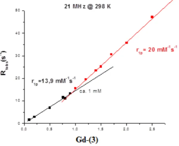
The efficacy of the CA measured as the ability of its 1 mM solution to increase the relaxation rate of water protons is called relaxivity and indicate as r1,2.
Paramagnetic CA’s increase the signal intensity of the tissue containing them by increasing the longitudinal and/or transverse relaxation rates (1/T1 or 1/T2) using their unpaired electrons to facilitate spin transfer. The diamagnetic (1/T1,2d) and paramagnetic (1/T1,2p) contributions to the relaxation rates of such solutions are additive:
1/ T1,2obs. = 1/ T1,2d + 1/ T1,2p
Equation 7
Where (1/T1,2)obs is the observed solvent relaxation rate and the subscripts “d” and “p” refer to diamagnetic and paramagnetic, respectively.
The paramagnetic contribution to the relaxation rate is linearly proportional to the concentration of the paramagnetic species:
1/ T1,2obs. = 1/ T1,2d + r1,2 [M]
Equation 8
Where M = paramagnetic substance, r1,2 = proton relaxivity (s-1mM-1). Relaxivity (r1,2) is then defined as the concentration-dependent increase in relaxation rate of the paramagnetic agent, or the slope of a plot of (1/T1,2)obs versus concentration.
The overall relaxivity can be correlated with a set of physico-chemical parameters, which characterize the complex structure and dynamics in solution, they are:
• the number of inner-sphere water molecules directly coordinated to the paramagnetic centre q,
• the residence time of the coordinated water molecule τM,
• the rotational correlation time representing the molecular tumbling time of a complex τR,
• the distance between the metal ion and the proton r, • electronic relaxation time T1e
The relaxivity enhancement of water protons in aqueous solutions of paramagnetic complexes arises from time fluctuation of the dipolar coupling between the electron magnetic moment of the metal ion and the nuclear magnetic moment of the solvent nuclei. This magnetic field around the paramagnetic center vanishes rapidly with distance. Therefore, specific chemical interactions that bring the water protons into the immediate proximity of the metal ion play an important role in transmitting the paramagnetic effect towards the bulk solvent. This interaction is expressed with a model that considers two contributions: inner sphere (IS), due to water molecules present in number q in the coordination sites of the
Figure 4
Model of Gd(III)-based contrast agent in
solution.
ion, and outer sphere (OS), which involves all the solvent molecules diffusing by the complex:
R1obs = R1pIS + R1pOS + R1W
Equation 9
Where R1obs is the measured relaxation rate relaxation and R1W the rate of the solvent without the paramagnetic complex.
The inner-sphere water protons then exchange with bulk solvent protons and this way the paramagnetic influence is propagated to the bulk. This mechanism is depicted as the inner-sphere contribution to the overall proton relaxivity. Solvent molecules of the bulk also experience the paramagnetic effect when they diffuse in the surroundings of the paramagnetic center. The effect of the random diffusion is defined as outher-sphere relaxation.
For certain agents, solvent molecules that are not directly bound in the first coordination sphere may also remain in the proximity of the paramagnetic metal for a relatively long time, this relaxivity contribution originating from these interactions is called second-sphere relaxivity.
Inner-sphere contribution
The inner-sphere contribution to the overall proton relaxation rate enhancement result from chemical exchange of the coordinated water protons with the bulk, the latter being much more populated. The observed signal correspond to that of the free water.
The relaxation of the bound water protons is governed by the dipole-dipole (DD) and scalar (SC), or contact, mechanism, where both are dependent on the magnetic field. The dipole-dipole part of the interaction is modulated by the
reorientation of the nuclear spin-electron spin vector, by changes in the orientation of the electron spin (electron spin relaxation) and by water (proton) exchange.
The relaxation rates of water protons related Ti,M are generally expressed by the modified Solomon-Blømbergen equations:
Equation 10
Equation 11
Where S is the the electronic quantic spin number (Gd= 7/2), γI is the nuclear giromagnetic ratio, g is the electron g-factor, μB is the Bohr magneton, ωS and ωI are the nuclear and electron Larmor frequencies, r is the distance between the electronic spin and nuclear spin, τc,I correlation time.
The scalar interaction remains unaffected by reorientation of the molecule and
is only modulated by electron spin relaxation and water exchange. The contact interaction is described, as dipolar interaction, by a Solomon-Blømbergen equations: Equation 12 Equation 13
+
+
+
+
=
)
1
(
7
)
1
(
3
)
1
(
15
2
1
2 2 2 2 2 1 2 1 6 2 2 2 1 S c c C I c B I DD Mr
S
S
g
T
ω
τ
τ
τ
ω
τ
µ
γ
(
)
+
+
+
+
+
=
2 1 2 2 2 2 1 2 1 6 2 2 2 24
)
1
(
13
)
1
(
3
1
15
1
1
c c S c c I c B I DD Mr
S
S
g
T
ω
τ
τ
τ
τ
ω
τ
µ
γ
(
)
+
+
=
2 2 2 2 2 11
1
3
2
1
e S e SC mh
A
S
S
T
ω
τ
τ
(
)
+
+
+
=
2 1 2 2 2 2 21
1
3
1
1
e e S e SC Mh
A
S
S
T
ω
τ
τ
τ
22Where A / h is the hyperfine or scalar coupling constant between the electron of
the paramagnetic center and the proton of the coordinated water, and τei correlation time.
Second and outher-sphere contribution
This contribution accounts for about 40% of the relaxivity of monoaquo Gd(III) complexes and arises from the modulation of the dipolar interaction by diffusion of the solvent molecules next to the paramagnetic center.
This occurs by two mechanisms: second-sphere relaxation and outer-sphere relaxation. Second-sphere relaxation occurs when water molecules in the second coordination sphere (H-bonded to lone pairs on the carboxylate oxygen atoms), are relaxed via a dipolar mechanism. This is difficult to quantify since the number of second-sphere water molecules and the metal-H distance are unknown, and τm is very short and is the likely limiting parameter in determining T1m.
Outer-sphere relaxation arises from the translational diffusion of water molecules close to the paramagnetic complex.
Experimental evidence for the occurrence of this solvent/metal complex interaction is difficult to obtain and, in most cases, its consideration may only represent a negligible correction to the inner- and outer-sphere relaxivities in the traditional model. However, in several cases, a careful analysis of the relaxation rate of the solvent as a function of magnetic field, pH and temperature allowed the evaluation of this contribution and the extraction of more useful and correct information from the data analysis.
Hydration number (q)
Inner-sphere proton relaxivity is linearly proportional to the number of water molecules directly coordinated to the paramagnetic ion.
The presence of CA influences a decrease of theoretical hydration number for the paramagnetic ion, i.e. Gd(III) aqua ion, there are eight inner-sphere water
molecules which would result in a proportionally high relaxation enhancement, but Gd(III) is toxic under physiological conditions and has a strong tendency to precipitate in the form of gadolinium-hydroxide. In fact, the thermodynamic and kinetic stabilities of the complex, which are guarantees of non-toxicity, can considerably decrease when more than one coordinated site is occupied by water.
The hydration number of a complex can be assessed from X-ray structure analysis; nevertheless, it also has to be determined in solution as the solid state structures do not always correspond to the species present in solution. There are several methods for determination such as NMR (Dysprosium Induced Shift, DIS) luminescence or EPR spectroscopy.
In all clinically utilized CAs, q = 1. Similarly, gadolinium(III) complexes of most octadentate DTPA and DOTA derivatives contain just one coordinated water molecule.
The distance between the metal ion and the proton (r)
The distance between the water proton and the unpaired electron spin, r, is a difficult parameter to measure and to control. According to modified Solomon-Blømbergen equations (equations 10-11) a decrease of 0.1 Å in the r distance corresponds to a 20% increase in inner-sphere proton relaxivity, while a decrease of 0.2 Å results in a much as 50% increase. In principle, two possibilities could be imagined to shorten the metal-H distance with the aim to increase the relaxivity.
First, higher title angles between the plane of the bound water and the metal-O bond could be induced by hydrogen bonding of the coordinated water to an appropriate side group of the chelate, which could result in a significant decrease of the metal-proton distance.
The second possibility for increasing relaxivity through changes in the metal-H distance could be the electron delocalization towards the ligand.
Nevertheless the r values used in relaxivity analyses of paramagnetic complexes are in most cases only estimations, because the metal-coordinated water hydrogen distance is a difficult parameter to obtain experimentally.
The residence time of the coordinated water molecule (τM)
The residence lifetime of protons (τM) plays a dual role in determining proton relaxivity. It modulates the efficiency of chemical exchange from the inner-sphere of the metal to the bulk, and also it is contributes to the overall correlation time (τc), that governs the dipole-dipole interaction between the electron and nuclear spin.
The exchange of coordinated water protons can occur in two ways: independently of the exchange of the entire water molecule on which it resides, or via the exchange of the water molecule itself.
Around neutral pH, the overall proton exchange rate is generally equal to the exchange rate of the entire water molecules, while, on increasing the acidity or basicity of the solution, the proton exchange may become considerably faster than the water exchange due to acid-or base-catalyzed pathways.
Generally the value of τM is in the order of 10-6 ÷ 10-9 s, which means conditions of rapid exchange about the time scale considered (τM << T1M), with a somewhat reduced dependence τM (i.e. it is not generally a limiting factor in the relaxation process).
The value of τM is very important in case of paramagnetic complexes bounded with a macromolecules, in fact this linking increase the lifetime of the coordinated water, until to obtain τM≅T1M, and then the exchange with the bulk water is slow and bringing an decrease of the relaxivity value.
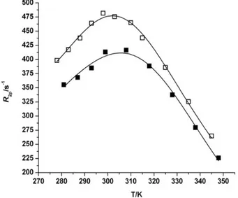
The rate of water exchange between an inner-sphere water molecule and the bulk can usually be estimated by 17O NMR by measuring the transverse relaxation rate of water in the presence and absence of a paramagnetic chelate.
It was also studied the mechanism by which water exchange occurs. There are two types of mechanisms: dissociative, it breaks the bond between the water molecule coordinated metal and later forms the bond with the new water molecule;
associative, it forms a new bond between the complex and the water molecule so as
to have a transition state that contains both the outgoing molecule that the incoming one.
Rotational correlation time (τR)
A parameter which allows a considerable increase in relaxivity for the paramagnetic complexes, is the rotational correlation time; in fact the relaxivity value increases with the increase of the rotational correlation time.
The rotational correlation time describes the molecular mobility, in particular the rotational mobility of the paramagnetic complex.
Values of short τR are associated with small molecules and with a round shape;
on the contrary a long τR is characteristic of molecules with an high molecular
weight and with irregular shapes.
In order to obtain an increase of rotational correlation time is possible to act in two different ways: leading the complex in tissues with an high microviscosity, or linking the paramagnetic complex with a macromolecule.
Electronic relaxation time T1e
The relaxivity of a paramagnetic complex depends on the electronic relaxation time, closely related to the magnetic characteristics of the metal, and at a lower rate to the structure of the ligand.
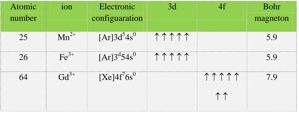
Paramagnetic metal ions show an effect which depends on the number of unpaired electrons in the ion. Paramagnetic ions of various transition metals like Fe3+, Mn2+ and rare earth metals of the lanthanide series like Gd3+, Dy3+ etc., have received great attention as magnetopharmaceuticals. The choice of Gd(III) would be expected, for no other ion has seven unpaired electrons. But there is a much
more subtle reason it performs so well. Two other lanthanide ions, dysprosium(III) and holmium(III), have larger magnetic moments (due to orbital contributions to electron angular momentum) than that of Gd(III), but the asymmetry of these electronic states leads to very rapid electron spin relaxation. The symmetric S-state of Gd(III) is a more hospitable environment for electron spins, leading to a much slower electronic relaxation rate.
This condition is best met in Mn2+, Fe3+ and Gd3+.
Atomic number ion Electronic configuaration 3d 4f Bohr magneton 25 Mn2+ [Ar]3d54s0 ↑ ↑ ↑ ↑ ↑ 5.9 26 Fe3+ [Ar]3d54s0 ↑ ↑ ↑ ↑ ↑ 5.9 64 Gd3+ [Xe]4f76s0 ↑ ↑ ↑ ↑ ↑ ↑ ↑ 7.9
Table 1: electronic configuration of some paramagnetic metals
Gadolinium(III) chelates
Gadolinium(III) chelates represent an important class of contrast agents for Magnetic Resonance Imaging (MRI). In the past two decades the use of gadolinium complexes of simple polyaminopolycarboxylic ligands as extracellular MRI contrast agents has become widespread. The main problem of the medical utilizations of heavy metal ions like the Gd(III) ion is a significant toxicity of their “free” (aqua-ion) form. Thus, for clinical use of gadolinium, it must be wrapped in a complex of high stability and, even more importantly, it must show a long-term resistance to a transmetallation/transchelation loss of the Gd(III) ion. It was proved that the endogenous metal ions Zn(II) and Ca(II) are them a in competitors and, thus, the candidate ligand has to show a higher complexation selectivity for Gd(III)
than for the two cations. The most important toxicological feature of the complex is the rate of decomplexation/transmetallation in comparison with the rate of excretion of the complex from the body. The requirements for in vivo stability suggest that kinetic stability, also called kinetic inertness, of the complexes could be as important as their thermodynamic stability.
From the structural point of view, two main families of organic ligands have been developed: twelve membered tetraazamacrocyclic cyclen derivatives (cyclen = 1,4,7,10-tetraazacyclododecane) and acyclic triamines (diethylenetriamine derivatives) with several chelating side arms, affording an octadentate model of the ligands.
The coordination number of the Gd(III) ion in these complexes is nine with the last coordination site occupied by a water molecule, which is crucial for the contrast enhancement mechanism.
Therefore, some important parameters to be taken in exam when designing a new Gd3+ contrast agent are:
• High stability. The injection of gram amounts of gadolinium into patients presents some potential toxicity problems which requires strong metal complexation by the parent ligand.
• Good water solubility and low osmolality of the solutions used clinically. Injection of relatively small volumes of a concentrated solutions of the metal complex is required.
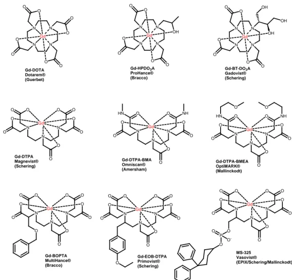
• High relaxivity. The dependence of the relaxivity on structural features of the paramagnetic compounds represent an important area of research The clinically approved Gd-based CAs are reported in Figure 5.
N N N N O O O O O O Gd OH N N N N O O O O O O Gd O O N N N N O O O O O O Gd OH OH OH N N N O O O O O O O O O O Gd N N N O O NH O O HN O O O O Gd N N N O O NH O O HN O O O O Gd O O N N N O O O O O O O O O O Gd O N N N O O O O O O O O O O Gd O N N N O O O O O O O O O O Gd O P O O O -Gd-DOTA Dotarem® (Guerbet) Gd-HPDO3A ProHance® (Bracco) Gd-BT-DO3A Gadovist® (Schering) Gd-DTPA Magnevist® (Schering) Gd-DTPA-BMA Omniscan® (Amersham) Gd-DTPA-BMEA OptiMARK® (Mallinckodt) Gd-BOPTA MultiHance® (Bracco) Gd-EOB-DTPA Primovist® (Schering) MS-325 Vasovist® (EPIX/Schering/Mallinckodt)
Figure 5: Clinically used gadolinium contrast agents
The first six complexes shown in Figure 5 act as nonspecific extracellular agents. Following intravascular injection, these compounds distribute rapidly between plasma and interstitial spaces and are ultimately eliminated through the renal route with half-lives of about 1.6 h. The remaining three DTPA derivatives, [Gd(EOB-DTPA)( H2O)]2-, MS-325, and [Gd-(BOPTA)(H2O)]2-, are designed specifically as targeted agents. The BOPTA comple is known to target the hepatobiliary system and acts as a liver imaging agent, while MS-325 interacts non-covalently with the abundant blood protein human serum albumin (HSA).
1.2.1 Bifunctional Chelating Agents (BFCAs)
The labelling of biomolecules with metal ions of diagnostic or therapeutic interest is an important tool in medicine.
Different metal ions are widely used in diagnostic and can be divided according to the techniques into they which are employed (Table 2).
Technique Metal ions Examples
MRI Gd3+, Mn2+
Gd-DTPA-BMA (Omniscan®) Approved for body imaging, MR
angiography, central nervous system imaging. PET 60,61,62,63,64 Cu2+, 66,68Ga3+, 86 Y3+, 89Zr4+ 68 Ga-DOTATOC Approved for neuroendocrine
tumor management. SPECT 67 Cu2+,67Ga3+, 90Y3+, 99mTc, 111 In3+ 99m Tc-exametazime (Ceretec®) Approved for cerebrovascular
diseases.
Table 2
Labelling of biomolecules with contrast agents is usually performed by directly linking the two moieties with a covalent linkage.
Several examples of MRI CAs directly bound to biomolecules are reported in the literature: for example a progesterone-Gd(III) chelate conjugate designed to image progesterone receptors for early detection of hormone related cancers9, or gadolinium chelates conjugated to peptides, as in the case of an arginine octamer prepared by standard peptide synthesis techniques on solid-phase resins, coupled to a Gd(III)-DTPA and used as a transporter across cellular membranes10. (Figure 6)
N N N N O O O O Gd O O N N N O O O O O O O H N O O Gd O O O Progesterone-Gd(III) chelate S S N H O O H N O OH HN H2N NH2+ 8 Gd-DTPA-SS-Arg8 5 Figure 6
A different approach can be used for MRI CAs, relying on a reversible interaction of the CA with the desired biomolecule. This strategy is exemplified by MS-325 (Figure 7)11, a Gd3+-complex containing a diphenylcyclohexyl apolar residue, bound to the metal coordination cage through a phosphodiester group. The apolar residue shows a strong affinity for human serum albumin (HSA), tightly binding to one of the hydrophobic pockets of this globular transport protein. The binding to HSA allows to modify the biodistribution of the CA while the reversible nature of the interaction warrant its excretion by glomerular filtration.
A similar approach was pursued with LipoAAZTA, where an aliphatic C17
straight chain plays the role of a fatty acid and binds strongly but reversibly to the dedicated pockets of HSA.12
N N N O O O O O O O O O O Gd O P O O O -MS-325 (Vasovist®) Gd O O O O N N N O O O O C17H35 Gd(AAZTA-C17)(H2O)2 -O H H O H H Figure 7
Complexes of positron (64Cu2+, 68Ga3+) or gamma (90Y3+, 99mTc) emitting radionuclides are largely used in positron emission tomography (PET) or single photon emission computed tomography (SPECT) respectively, a DTPA derivative (Figura 8) linkage with an analog somatostatine peptide (OC), is a SPECT imaging agent approved for routine clinical use as a diagnostic agent for neuroendocrine cancer in the US and Europe.13
N H H N O O N H S S H N O NH OH HN O NH2 H N O HO O N H O H N HO HO N N N O O O O O O O O O 111In 111In-DTPA-OC (Octreoscan®) Figure 8 32
Radionuclides are also employed in therapy, in particular in the treatment of cancer, and can be generate beta (90Y3+, 177Lu3+) or alpha (212Bi3+, 226Ac3+) particles14. Ibritumomab tiuxetan (Zevalin®) is a monoclonal antibody conjugated with the chelator tiuxetan, to which a radioactive isotope (90Y3+ or 111Y3+) is added. Tiuxetan is a modified version of DTPA whose carbon backbone contains additional isothiocyanatobenzyl and methyl groups.15
For the most part, clinical contrast agents are not specifically targeted to a protein or receptor. The linkage between these biomolecules increases the molecular specificity and allows their delivery to particular sites of human body. The biomolecules employed, usually peptides or monoclonal antibodies, are able to target particular kinds of cells with high selectivity and affinity.
For MRI agents, the detection limit for a single Gd(III) complex is in the low micromolar range, and it is often necessary to incorporate multiple complexes to enhance molecular relaxivity. Non-covalent protein binding and oligomerization often result in increased relaxivity per Gd ion, because largest molecules increase the rotational correlation time (τR). The most reliable and frequently applied method of linking the metal ion (the probe) to the biomolecule (the carrier) is by means of a bifunctional chelating agent (BFCA).
Bifunctional chelating agents are defined as molecules containing two different moieties: a strong metal chelating unit and a reactive functional group (figure 9).
Chelator Vector Gd Reactive Functional Group Gd Figure 9
The chelating unit is committed to the stable coordination of the chosen metal ion. The reactive functional group is devoted to react and give a stable covalent
bond with suitable vectors, represented by biomolecules or simple organic compounds.
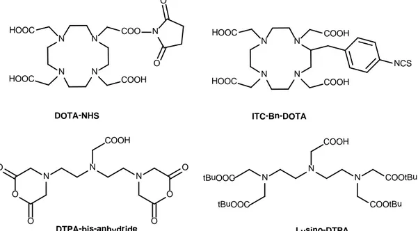
In recent years, a large number of BFCAs have appeared in the literature, those based on a polyamino polycarboxylic ligand are particularly efficient (figure 10). The main reason for this is that they form highly stable complexes with a wide variety of metal ions, this means that the undesired in vivo release of the metal ion is avoided.
BFCAs can be divided into two main categories: acyclic, such as those based on the ligands EDTA or DTPA, and macrocyclic, such as those based on the ligands DOTA or TETA. In general, the differences between these two classes are in the stability of complexes (macrocyclics are more stable than acyclics) but macrocyclic ligands are usually kinetically more inert during complexation, requiring longer reaction times and/or higher temperatures.
N N N N HOOC COO DOTA-NHS N N N O O O O O O COOH DTPA-bis-anhydride N N N COOtBu COOtBu COOH tBuOOC tBuOOC Lysino-DTPA COOH HOOC N O O N N N N HOOC COOH COOH HOOC NCS ITC-Bn-DOTA
Figure 10: Some examples of BFCAs
The choice of the right ligand is a crucial point in the design of the final conjugate and depends both on the metal ion to be complexed and the biomolecule to be labeled.
The conjugation of a BFCA to the molecule of interest (the carrier) can be performed by means of the reactive functional group. The carboxylic group is the most used, since it can be easily activated to allow attack by an amino group present in antibody or in peptide. A huge number of functional groups are reported in the literature for this aims: symmetrical or mixed anhydrides and active esters (such as pentafluorophenyl, p-nitrophenyl and NHS-esters) isothiocyanate and maleimide are frequently used reactive groups.
Another important parameter to consider in the design of new BFCAs is the linker (or spacer, the spacing unit between the probe and the functional group). The linker can increase the water solubility of the molecule (ex. PEG chain) or increase its lipophilicity (aliphatic chains, aromatic rings), affecting the pharmacokinetics and the biodistribution of the final molecule biodegradation and elimination can be increased by linkers that can be cleaved in vivo, such as particular peptide sequences, ester/amide groups and disulfide bonds.
The list of bifunctional ligands reported in the scientific literature is very long as several and different chelating agents have been modified with additional reactive functional groups depending on the metal ion to be complexed and the specific application involved. The comprehensive treatment of BFCAs is beyond the scope of this thesis and may be found in dedicated reviews.16,17
A selection of the BFCAs employed for the preparation of Gd-complexes for MRI applications will be discussed in order to introduce the experimental work performed in the PhD period, focusing on the DO3A platform (leading to DOTA-like and HP-DO3A-like BFCAs) and on the original AAZTA ligand.
1.2.2 AAZTA
(6-Amino-6-methylperhydro-1,4-diazepine-N,N’,N”,N”-tetracetic acid)
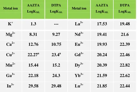
The structure of AAZTA embodies a seven-membered ring with two endocyclic and one exocyclic nitrogen atoms, carrying a total of four carboxymethyl side arms18. The corresponding Gd3+-complex is satisfactorily stable, either thermodynamically (logKf = 20.5),19 kinetically or with respect to transmetallation (table 2). The two coordinated water molecules ensure a high relaxivity (7.1 mM-1s-1), not quenched by bidentate anions such as lactate and phosphate as observed
in macrocyclic complexes. Figure 11
Table 2: Metal ion affinity of AAZTA and DTPA
Metal ion AAZTA LogKML
DTPA LogKML
Metal ion AAZTA LogKML DTPA LogKML K+ 1.3 --- La3+ 17.53 19.48 Mg2+ 8.31 9.27 Nd3+ 19.41 21.6 Ca2+ 12.76 10.75 Eu3+ 19.93 22.39 Cu2+ 22.27a 23.4a Gd3+ 20.24 22.46 Mn2+ 15.44 15.2 Dy3+ 20.39 22.82 Ga3+ 22.18 24.3 Yb3+ 21.59 22.62 In3+ 29.58 29.48 Lu3+ 21.85 22.44
Gd
O
O
O
O
N
N
N
O
O
O
O
Gd(AAZTA)(H2O)2 -r1p = 7.1 mM-1s-1 τΜ = 90ns O H H O H H 36The synthesis of AAZTA (Scheme 1) involves 4 synthetic steps with an overall 50% yield; the easy synthetic access to this ligand, combined with the interesting proprieties of its paramagnetic complexes, prompted different groups to prepare different versions of BFCA-AAZTA.
EtNO2 CH2O HN NH Ph Ph NO2 N N Ph N N N COOH COOH COOH HOOC H2 Pd/C NH2 NH NH 1) tBBA K2CO3 Ph 2) TFA
Scheme 1: Synthesis of AAZTA
The synthetic access to AAZTA allows the reactive functional group to be positioned on the ring (especially on the quaternary carbon atom bearing the iminodiacetic moiety, i.e.: C-6) or on one of carboxymethyl lateral arms.
The first BFCA of AAZTA was obtained introducing a functional group containing a hydroxyl group, spaced with one carbon atom from the ring 20. In this synthesis, nitromethane was substituted with 2-nitroethanol to give as a product of double nitro-Mannich reaction a new hydroxyl functional group (scheme 2). It has been demonstrated that is possible to obtain the product forming in situ 2-nitroethanol through the Henry reaction by using nitromethane and an excess of paraformaldehyde, but the yield is reduced to about 60%.
2 CH2O HN NH Ph Ph NO2 N N Ph N N N COOH COOH COOH HOOC H2 Pd/C NH2 NH NH 1) tBBA K2CO3 Ph 2) TFA HO NO2 HO HO HO 3 CH2O Scheme 2
The hydroxyl group was also used to introduce a carboxylic group by reaction with succinic anhydride; alternatively the protected ligand was reacted with thionyl chloride to convert the hydroxymethyl group into a 6-chloromethyl derivate. In a third strategy, this compound was reacted with 4-nitrophenyl isocyanate and after that treated with thiophosgene to give a isothiocyanate derivate (Scheme 3).
N N N COOtBu COOtBu COOtBu tBuOOC HO O O O SOCl2 N N N COOtBu COOtBu COOtBu tBuOOC O N N N COOtBu COOtBu COOtBu tBuOOC Cl O O HO NH2 NCO N N N COOtBu COOtBu COOtBu tBuOOC O O H N SCN Scheme 3
These compounds were used to prepare lipid-based paramagnetic particles used for the cell labelling or conjugated with small biomolecules. In addition the isothyocianate derivate has been employed to synthesise multimeric/dendrimeric MRI contrast agents21,22,23,24.
Other similar hydroxyl derivates were synthesized with a higher number of carbon atoms in the spacer 25, however the synthetic route is parallel to that shown in Scheme 2.
It has been reported another BFCA AAZTA-like with an amine moiety as reactive group, bounded to the ring with a little spacer of three carbon atoms (Scheme 4) 26. This synthetic approach is recalls the strategy used for the previously reported derivatives, the key step being the double nitro-Mannich reaction with 3-nitropropanamine, previously protected as N-Boc.
CH2O HN NH Ph Ph NO2 N N Ph N N N COOH COOH COOH HOOC H2 Pd/C NH2 NH NH 1) tBBA K2CO3 Ph 2) TFA NO2 NHBoc BocHN BocHN H2N Scheme 4
A further modification of AAZTA skeleton was made using 6-nitrohexanoic acid for the condensation reaction, obtaining a derivate with a free carboxylic acid moiety27 (Scheme 5) This derivative was subsequently conjugated with dextran and used for neuroanatomical connectivity studies.
CH2O HN NH Ph Ph NO2 N N Ph N N N COOH COOH COOH HOOC H2 Pd/C NH2 NH NH 1) tBBA K2CO3 Ph 2) TFA NO2 O HO HO HO O O O HO Scheme 5
All these AAZTA-like BFCAs share the common synthetic pathway, relying on the double nitro-Mannich reaction of N,N’-dibenzylethylenediamine, paraformaldheyde and a suitable nitroalkane in order to build a 7-membered ring. As a matter of fact, these agents are substituted on position 6 of the
perhydrodiazepine ring with aliphatic spacer chains of different lengths characterized by functional reactive groups located at the end and devised to be conjugated.
Different AAZTA-based BFCAs were obtained by selective alkylation at one of the secondary amine groups with tert-buthyl α-bromoester carryng a protected functional group at ω; alkylation of the remaining amine groups with tBBA (= tert-butyl bromoacetate) followed by deprotection of the reactive functional group gave corresponding AAZTA28 as shown in scheme 6.
NH2 NH NH 1) tBBA K2CO3 2) TFA COOtBu R Br NH2 N NH R COOtBu N N N R COOtBu COOH COOH HOOC R= (CH2)2COOH R= (CH2)4NH2 Scheme 6
1.2.3 DO
3A (1,4,7,10-tetraazacyclododecane-1,4,7-triacetic
acid)
DO3A is a heptadentate ligand composed by four macrocyclic nitrogen atoms and three carboxylates. This chelator can be easily derivatized on the unique nitrogen on the macrocyclic ring, to obtain several other DO3A-like BFCAs.
DO3A form stable complexes with a large number of metal ion such as transition metal ions and lanthanides, leaving two coordinated sites free for the binding of water molecules and giving neutral complexes with low osmolality.29 In the case of
N N NN N NHH N N O O O O O O O O O O O O G Gdd
G
Gd
d--D
DO
O
33A
A
q
q=
= 2
2
41lanthanides complexes and especially for the Gd3+ complex this means an improvement in the relaxivity value; nevertheless in vivo several bidentate anions such as carbonate or phosphate can displace the coordinated water molecules, quenching the relaxivity and preventing in this way its application as a MRI agent.30,31,32.
The synthesis of DO3A can be performed in different ways, firstly by direct alkylation of cyclen with bromoacetic acid in DMF or DMAc with subsequently purification, by ion exchange chromatography, from DOTA33. A recent improved method for synthesized DO3A involves the alkylation of cyclen with tBBA in dimethyl acetamide with sodium acetate as a base. After 60 h of reaction at room temperature the tri-t-butyl ester was obtained after crystallization with a 65% yield as hydrobromide.34 (Scheme 7) NH HN HN NH tBBA, DMF tBBA, DMAc CH3COONa NH N N N COOtBu COOtBu N N N N COOtBu COOtBu tBuOOC NH N N N COOtBu COOtBu NH2+ N N N COOtBu COOtBu Br -Ion exchange chromatogrphy tBuOOC tBuOOC tBuOOC tBuOOC Scheme 7 42
Another synthetic pathway involves the trivalent protection of cyclen with dimethylformamide dimethylacetal to give a tricycloderivate, subsequently hydrolysed with 50% aqueous ethanol to mono-N-formylcyclen. Subsequently the product was alkylated with tBBA followed by the combined hydrolysis of tert-butyl esters and the formyl group with H2SO4 to give DO3A in high yields.35 (Scheme 8).
Scheme 8
By alkylation of the secondary amino group of DO3A is possible to obtain a
series of new BFCA-DO3A like. A huge number of functional groups are
introduced into the DO3A skeleton, with different kind of linkers, amino groups36,37, isothiocyanate38, thiol39, bromo40, vinylsulfone41 and squarate ethylester42 are selected examples (Scheme 9).
NH HN HN NH DMF-DMA cyclohexane N N N NH H2O EtOH NH N HN NH CHO N N N N CHO tBBA aq. K2CO3 DMF/toluene COOtBu tBuOOC tBuOOC H2SO4 N HN N N COOH HOOC HOOC 43
N N N N COOH HOOC HOOC R R = R = R = R = CH2CH2NH2 NH2 CH2CH2NCS CH2CH2CH2Br CH2CH2SH CH3CH2SOCH CH2 R = R = R = O O OEt N HN N N COOH HOOC HOOC N N N N COOH HOOC HOOC R Scheme 9
All these compounds were used in a wide variety of applications, for examples the derivativee with amino moiety was conjugated to fluorescein isothyocianate in order to obtain a dual probe for MRI and Optical Imaging. The Sm(III) complex of the thiol derivative was employed in a biodistribution study, while the vinylsulfone moiety was conjugated to humanized antibodies for PET imaging. In another example the free amino group of DO3A was functionalized with a squarate ester, the latter being reactive towards amines and used for the multisite labeling of
poly-L-lysines and poly-L-ornithines.43
An interesting examples of DO3A derivative was generated using propargyl bromide as alkylating agent (Scheme 10). This compound can be easily and rapidly
conjugated to peptides by a Huisgen cycloaddition44. Copper(I) catalyzed
cycloaddition of this compound with azides, produced an octadentate complex wherein a triazole nitrogen is involved in the coordination to the lanthanide.
N N HN N tBuOOC tBuOOC Br CH3CN N N N N tBuOOC tBuOOC COOtBu 1) TFA 2) Ln(OTf)3 N N N N O O O O O O Ln COOtBu N N N N O O O O O O Ln N N N R Sodium ascorbate CuSO4, BzN3 Scheme 10
The introduction of a methylphosphinic acid moiety on the DO3A structure results in Gd(III) complexes that show a fast coordinated water exchange rate, which is close to optimal for low field application (0,5-1,5 T)45. The reason for such a fast exchange rate can be found in the steric constraint imposed by the bulky phosphinate group on the water coordination site and possibly in the structuring of the second hydration sphere brought about by the phosphinate moiety. To take advantage of this favorable water exchange property, two Bifunctional monophosphinate DO3A-Like were prepared, bearing either a free carboxyl group or an aromatic amine ready for conjugation46 as shown in figure 12.
N N N N P tBuOOC tBuOOC COOtBu O HO COOH N N N N P tBuOOC tBuOOC COOtBu O HO NH2 Figure 12 45
A DO3A-like ligand with a 2-mehylpyridine-N-oxide coordinating unit formed a Gd(III) complex with a remarkably fast water exchange rate47. A derivative with a free carboxylic available for functionalization was conjugated to a calix[4]arene platform.48 (Figure 13)
A different BFCA DO3A-like was obtained introducing an acylhydrazine moiety as shown in figure 13 and subsequently used to prepare acid labile conjugates with doxorubicin, an anthracycline anticancer drug49.
N N N N tBuOOC tBuOOC COOtBu N N N N tBuOOC tBuOOC COOtBu N+ -O O HN NH2 COOH Figure 13
An interesting example of DO3A BFCA with a aniline as a functional group was reported50. This compound was also conjugated with a galactopyranosyl moiety, as a substrate of β-galactosidase, an expression-product of the report gene Lac-Z commonly co-administrated intracellulary with the subject-gene of gene-therapy studies.The synthetic pathway of this compound (scheme 11) is composed by three different alkylation steps with (R,S)-2-bromo-4-(4-nitrophenyl)butanoate, bromoethyl-β-D-galactopyranoside tetraacetate and tBBA. Consecutive carboxyl and hydroxyl group deprotections and catalytic hydrogenation of functional group gave a corrisponding BFCA DO3A-like.
O2N COOH O2N COOtBu Br NH HN HN N NO2 NH N HN N NO2 O O OAc OAc AcO OAc tBuOOC tBuOOC N N N N NH2 HOOC O O OH OH HO OH COOH HOOC 1) SOCl2/CCl4 2) NBS/HBr aq. 3) PhN(Me)2/tBuOH Cyclen, K2CO3 O O OAc OAc AcO OAc Br 1) tBBA 2) TFA 3) NaOMe/MeOH 4) Pd/C, H2 Scheme 11
1.2.4 DOTA Monoamide
This class of chelating agents requires a separate systematic treatment. In fact, for the synthesis of DOTA monoamide BFCAs is possible to start from DOTA, for example activating one of the carboxyl groups as N-hydroxysuccinimide ester, or from DO3A by an alkylation of the secondary free amino group, or from cyclen by monoalkylation with a suitable N-substituted 2-haloacetamide as reported in Scheme 12. These approaches differ in the starting material, the number of steps, yields and ease of purification. The differences between these DOTA monoamide derivatives are related to the functional groups used to conjugated the probe to the vector of interest and to the linkers of various length, rigidity or hydrophylicity used to space the metal chelate from the biomolecule. The distance between the DOTA coordination cage and the vector may strongly affect both the conjugation reaction and the interaction of the bioconjugate interaction. Indeed, the DOTA
monoamide cage, which is often bulkier than the vector itself, may hinder the interaction considerably. NH HN HN NH N HN N N COOR1 R1OOC R1OOC N N N N COOH HOOC HOOC COOH NH HN N NH COOX N N N N COOR1 COOR2 R1OOC R1OOC N N N N COOR1 R1OOC R1OOC COOH N N N N COOR1 COONHR3 R1OOC R1OOC N N N N COOH HOOC HOOC COONHR3 X=R1 or NHR3 a b c d e f g m n h i l o
Scheme 12: Schematic representation of the various strategies used to prepare DOTA monoamides. a-b = via DOTA, c-d-e/c-l-o = via cyclen, f-g-h-i/f-m-e/f-n/f-g-o = via DO3A
The major part of DOTA monoamides are synthesized starting from DO3A by alkylation on free secondary amino group. One strategy (path f,g,h,i) involves the use of one different protecting groups on the acetic pendant arms, as benzyl esters51 or ethyl esters52,53 (Scheme 13) and after selective removal of the protecting group, it is possible to obtain a free carboxylic moiety able to be conjugated with an amine to give a monoamide derivatives in high yield.
N HN N N COOtBu N N N N COOtBu tBuOOC tBuOOC
tBuOOC tBuOOC COOR N N
N N COOtBu tBuOOC tBuOOC COOH R = Bz or Et Scheme 13
Alkylation of DO3A t-butyl ester was also used to prepare BFCAs DOTA monoamides with different functional groups as amino, maleimido, hydroxyl and formyl54 as shown in Scheme 14. The synthesis of all the adducts share the same basic strategy. DO3A t-butyl ester was alkylated with a tailored bromoacetamide bearing either a protected amine or alcohol group. After deprotection of the side arm the amino derivates were converted to the maleimido derivate by reaction with 7-exooxohimic anhydride under microwave irradiation. The alcohol derivates were converted into the corresponding aldehyde by Swern oxidation.
N N N N COOH HOOC HOOC Br H N O N H Cbz n N H O NH2 Br H N O O THP 5 N N N N COOH HOOC HOOC N H O OH O O O O N N N N COOH HOOC HOOC N H O N O O 5 n n N N N N COOH N H O O n N N N N COOtBu N H O OH n Cl H N O OH n tBuOOC tBuOOC tBuOOC tBuOOC DMSO,(COCl)2 CH2Cl2, -60°C 1) CH3CN, K2CO3 2) NaCNBH3, BF3OEt2 CH3CN 1)CH3CN, K2CO3 2)H2 Pd/C 3)TFA H2O, M.W 110°C N HN N N COOtBu tBuOOC tBuOOC Scheme 14 49
Variants with a free aromatic amine and with a thiol reactive methanethiosulfate group were prepared (Figure 14) 55,56.
N N N N COOtBu N H O NH2 N N N N COOtBu N H O S S O O tBuOOC tBuOOC tBuOOC tBuOOC Figure 14
Functionalization of DO3A t-butyl ester has been exploited introducing a spacer carrying a free terminal carboxylic group (Figure 15) which, after transformation into NHS ester, was coupled to glutamine. The resulting complex was tested “in
vitro” on different tumor cell lines and “in vivo” on cancer prone transgenic mice to
study cellular uptake through the aminoacid transporting system57.
N N N N COOH HOOC HOOC N H COOH O 4 N N N N COOH HOOC HOOC HN O 4 HN O COOH NH2 O Figure 15
Final interesting examples showed that increasing the length of the carboxyamide arm of Gd-DOTA monoamide from two to three carbons atom results in gadolinium complexes with a water exchange rate almost two orders of magnitude faster than the parent DOTA monoamide58 (Figure 16).
A DOTA monoamide with a terminal alkyne functionality, designed for Cu(I) mediated azide-alkyne cycloaddition has been prepared by alkylating DO3A t-butyl ester with N-(2-propynyl)-2-chloro- or 2-bromo-acetamide59,60 (Figure 16).
N N N N COOtBu H N tBuOOC tBuOOC NH2 O N N N N COOtBu N H O tBuOOC tBuOOC Figure 16
1.2.5 HP-DO
3A (10-(2-hydroxypropyl)-1,4,7,10
tetraaza-cyclododecane-1,4,7-triacetic acid)
HP-DO3A is an octadentate ligand composed by four macrocyclic nitrogen atoms, three carboxymethyl side armsand a
hydroxypropyl moiety. Gd-HPDO3A
(Prohance®, Bracco Imaging) is a non
specific contrast agent for MRI, commercially available and widely employed in the clinical practice. This neutral complex shows an high thermodinamic stability (Log KF= 23.8) and kinetic inertness that ensures no significant release of Gd3+ ions in vivo. The relaxivity value of this complex is 3.7 mM-1s-1 in water
at 40°C, in line with other macrocyclic Gd complexes as shown in Table 3.61,62
N N N N O O O O O O Gd
Gd-HPDO
3A
q= 1
OHr
1= 3.7 mM
-1s
-1 51Complex
q
τ
r(ps)
r
1mM
-1s
-1 Gd-HPDO3A 1.3 ±0.1 57 3.7 ±0.1 Gd-DOTA- 1.1±0.1 63 3.5 ±0.1 Gd-DTPA2- 1.1±0.1 55 3.8 ±0.1 Gd-DTPA-BMA 1.1±0.1 53 3.8 ±0.1Table 3: Comparison among some different clinically used Gd(III)-complexes.
The synthesis of HP-DO3A, reported in Scheme 15, involves the reaction of cyclen with dimethylformamide dimethylacetal to give the tricycloderivative which was hydrolysed with 50% aqueous ethanol to mono-N-formylcyclen. Subsequently the product was alkylated with cloroacetic acid and without any purification steps the basic hydrolysis of the formyl group and the reaction with propylene oxide gave HP-DO3A. NH HN HN NH DMF-DMA cyclohexane N N N NH H 2O EtOH NH N HN NH CHO N N N N CHO COOH HOOC HOOC NaOH N HN N N COOH HOOC HOOC ClCH2COOH NaOH N N N N COOH HOOC HOOC OH O Scheme 15
In literature there are only few examples of BFCAs based on HP-DO3A
structure. In a first report, two compounds with a free carboxylic group were synthesized, in order to be conjugated to amino groups of biomolecules. Two
different linkers were used to connect the coordinating cage and the functional group, in one case a diethylene glycol unit and in the other an aromatic moiety as shown in Scheme 16. These compounds were conjugated to peptide such as bombesin and oxytocin63.
In the first synthetic route the hydrophilic diethylene glycol spacer, protected as a benzyl ester, was prepared by a Williamson reaction using bromoacetic acid. The oxidation of terminal alkene gave the corresponded epoxide. (tBu)3-DO3A was alkylated with the epoxide and subsequently the hydrogenolysis of benzyl ester gave the desired ligand.
O OH O O COOBn O O COOBn O N N N N COOtBu tBuOOC tBuOOC O OH O COOBn N N N N COOtBu tBuOOC tBuOOC O OH O COOH 1) Na, THF, BrCH2COONa 2) BnBr, DBU, toluene MCPBA, CH2Cl2 tBu-DO3A, TEA, CH3CN Pd/C, H2, MeOH Scheme 16
A second HP-DO3A-based BFCA containing an aromatic spacer, was
synthesized in a three steps starting from epibromohydrin and benzyl 4-hydroxybenzoate. The resulting epoxide was reacted with (tBu)3-DO3A followed by the deprotection step to gave this BFCA HP-DO3A like in a good yield (Scheme 17).
Scheme 17
The last reported HPDO3A-based BFCA, endowed with a free amino moiety is shown in Scheme 18. This compound was synthesized by reaction of DO3A with
N-Boc-1,2-epoxy-3-propylamine followed by removal of the Boc group and
subsequent complexation with Gd(III)64. This compound was reacted with 2-iminothiolane in order to be conjugated with an activate phospholipid, and included into liposomes. COOBn OH O Br COOBn O O N N N N COOtBu tBuOOC tBuOOC O OH COOBn N N N N COOtBu tBuOOC tBuOOC O OH COOH K2CO3, DMF DO3A(tBu)3 CH3CN Pd/C, H2, MeOH 54
N HN N N HOOC COOH HOOC HN O O O N N N N HOOC COOH HOOC OH N H O O N N N N HOOC COOH HOOC OH NH2 N N N N -OOC COO --OOC OH NH2 Gd3+ N N N N -OOC COO --OOC OH NH Gd3+ +H 2N SH H2O, pH= 9-10 TFA Gd2O, H2O 2-iminothianol Scheme 18 55