1
UNIVERSITÀ DEL PIEMONTE
ORIENTALE
PhD in Medical Sciences and Biotechnologies
XXX cycle
Department of Health Sciences (DISS)
SSD: MED/08
ROLE OF HMGB1 (High Mobility Group Box-1) IN MALIGNANT
MESOTHELIOMA
PATHOGENESIS, PROGNOSIS AND AS PREDICTIVE BIOMARKER.
PhD thesis by:
RRAPAJ ELTJONA
Coordinator of PhD programme: Prof. Marisa Gariglio
Supervisor: Prof. Renzo Boldorini
2
Dedication
This thesis is dedicated to the memory of my grandfather Isuf Xhyheri, a smart man, who inspired and
supported me to pursue a career in research.
Eltjona Rrapaj, MSc
3
TABLE OF CONTENTS:
1. SUMMARY……….4
2. INTRODUCTION………...7
3. AIM OF THE STUDY………..45
4. MATERIALS AND METHODS………..47
5. RESULTS……….59
6. DISCUSSION………..85
4
1. SUMMARY ( Italian)
Il mesotelioma pleurico maligno (MPM) è un tumore aggressivo di origine mesoteliale che viene spesso diagnosticato in uno stadio avanzato. Anche per questo motivo il mesotelioma spesso presenta resistenza alla maggior parte dei trattamenti terapeutici e una prognosi infausta, esiste pertanto la necessità di sviluppare nuovi biomarcatori per la diagnosi precoce e di identificare nuovi target per trattamenti preventivi e terapeutici. Studi recenti dimostrano che la proteina High-mobility group box-1 (HMGB1) svolge un ruolo fondamentale nella carcinogenesi dell'MPM. HMGB1 è una proteina strutturale della cromatina, espressa ubiquitariamente nei nuclei delle cellule dei mammiferi. Quando trasportata nello spazio extracellulare, tra le altre funzioni, può agire sia come oncosoppressore che come proteina oncogena. Nel mesotelioma pleurico maligno, livelli sierici elevati di HMGB1 sono stati correlati a una prognosi infausta. Al contrario, il significato dell'espressione di HMGB1 nei tessuti di mesotelioma pleurico maligno deve ancora essere definito.
La discrepanza tra l'incidenza crescente di MPM e la mancanza di successo di nuove strategie terapeutiche più efficaci può essere in parte correlata a sistemi in vitro e in vivo inadeguati che imitano la tumorigenesi del MPM. Recenti scoperte hanno dimostrato che il mesotelioma maligno è un tumore policlonale e la sua formazione e crescita è influenzata dal microambiente infiammatorio. Valutare il mesotelioma come un organo completo risulta cruciale per comprenderne la biologia e sviluppare nuove strategie terapeutiche. Il nostro obiettivo è sviluppare modelli tridimensionali di mesotelioma in vitro. Gli organoidi umani derivati da paziente potranno rivelarsi di grande utilità per esaminare la sensibilità ai trattamenti farmacologici e per studiare la diafonia tra tumore e cellule immunitarie. In quest’ottica, sferoidi ottenuti da mesotelioma murino e xenotrapianti derivati da paziente (PDX) rappresentano modelli alternativi e utili per conseguire il medesimo obiettivo.
Campioni di tessuto neoplastico ottenuti mediante biopsia da 170 pazienti con MPM sono stati valutati mediante immunoistochimica e Reverse Transcription-Polymerase Chain Reaction (RT-PCR) per valutare la proteina HMGB1 e l'espressione genica. Il livello di espressione della proteina HMGB1 è stato valutato utilizzando un metodo semi-quantitativo, che somma l'intensità (0-3) e la percentuale (da 0 a 4) delle cellule con colorazione positiva, nei nuclei, nel citoplasma e in entrambi. Il punteggio finale è stato classificato come alta (> 3) o bassa (< 3) espressione proteica. I livelli di espressione genica sono stati calcolati con il metodo ΔΔCt. Livelli di espressione elevati di HMGB1 totale (p = 0,0011) e citoplasmatico (p = 0,0462), correlavano con una peggiore sopravvivenza malattia specifica (DSS) nell'intera coorte e nei sottogruppi clinico-patologici. Non è stata tuttavia osservata nessuna correlazione significativa tra espressione del gene HMGB1 e DSS.
Nella seconda parte del nostro studio, biopsie di mesotelioma umano sono state utilizzate per la generazione di organoidi seguendo procedure sperimentali precedentemente pubblicate. Gli stessi
5 frammenti tumorali sono stati trapiantati in topi immunodeficienti (NOD scid gamma, NSG). Gli organoidi ottenuti da biopsie umane presentano dimensioni ridotte (200 μm di diametro) e possono essere propagati fino a tre passaggi in vitro. La caratterizzazione morfologica degli organoidi mostrava una struttura definita con polarità interno-esterno. Gli organoidi erano inoltre positivi per i marcatori tipici del mesotelioma. Abbiamo ottenuto tre PDX; l'immunofenotipizzazione ha mostrato come i tumori propagati nei topi siano simili al tumore originale.
Questi risultati indicano che HMGB1 può essere utile come biomarcatore prognostico nel mesotelioma pleurico quando valutato mediante colorazione immunoistochimica nel tessuto neoplastico. Tuttavia, poiché è espresso anche in cellule mesoteliali normali e reattive, la valutazione dell’espressione di HMGB1 in campioni istologici di mesotelioma sembra non presentare utilità a fini diagnostici.
Siamo inoltre riusciti a generare e caratterizzare organoidi e modelli PDX di mesotelioma, ci aspettiamo che questi modelli ci aiutino a capire meglio la patogenesi del mesotelioma.
SUMMARY (English)
Malignant pleural mesothelioma (MPM) is an aggressive tumour of mesothelial origin, often diagnosed in an advanced stage, which contributes to its very poor prognosis and resistance to most of the therapeutic treatments. Therefore, the development of new biomarkers for early diagnosis and of novel targets for preventive and therapeutic treatments are needed. Recent studies show that High Mobility Group Box 1 protein (HMGB1) plays a critical role in the carcinogenesis of MPM. High-mobility group box-1 (HMGB1) is a chromatin structural protein, ubiquitously expressed in the nuclei of mammalian cells. When transported extracellularly, it, among other functions, could act as tumor suppressor and oncogenic protein. In
malignant pleural mesothelioma (MPM), high serum levels of High-mobility group box-1 (HMGB1) have been related to a poor prognosis. Conversely the significance of HMGB1 expression in malignant pleural mesothelioma (MPM) tissues is still unclear.
The discrepancy between the rising incidence of MPM and the lack of success of new more effective therapeutic strategies may be related in part to inadequate in vitro and in vivo systems that mimic MPM tumorigenesis. Recent findings showed that malignant mesothelioma is a polyclonal tumor and its
formation and outgrowth is determined by the inflammatory microenvironment. Evaluating mesothelioma as a complete organ becomes crucial to understand its biology and to develop new therapies. Our aim is to develop 3D in vitro models of mesothelioma. Patient-derived human organoids are useful to screen for drug sensitivity and to study the crosstalk between tumor and immune cells. Murine mesothelioma spheroids and patient derived xenografts (PDX) are alternative models that help us to pursue the same goal.
6 Biopsy samples from 170 patients with MPM were assessed by immunohistochemistry and Reverse
Transcription-Polymerase Chain Reaction (RT-PCR) to evaluate HMGB1 protein and gene expression. The expression level of HMGB1 protein was scored using a semi-quantitative system, that sums the intensity (0-3) and the percentage (from 0-4) of positively stained cells, in nuclei, cytoplasm and in both. The final score was considered as high (>3) or low (<3) expression. Gene expression levels were calculated with ΔΔCt method. High expression levels of HMGB1 as total (P = 0,0011) and cytoplasmic score (P = 0,0462), were related with a worse disease-specific survival (DSS) in the entire cohort and in the clinicopathologic subgroups. No significant correlation was found between HMGB1 gene expression and DSS.
Human mesothelioma biopsies were used for organoid generation following published protocols. The same tumor fragments were transplanted in immune deficient mice (NOD scid gamma, NSG mice). Organoids obtained from human biopsies are small in size (200 µm in diameter) and can be propagated up to three generations. The morphological characterization of organoids showed a defined structure with inside-outside polarity; organoids were positive for mesothelioma markers. We obtained three PDX;
immunophenotyping showed that the tumors propagated in mice are similar to the original tumor. These findings indicate that HMGB1 may be a useful prognostic biomarker in MPM when detected by immunohistochemistry. Conversely, since it is expressed also in normal and reactive mesothelial cells, HMGB1 cannot be considered a diagnostic biomarker, in histologic samples of mesothelioma.
We have the ability to generate and characterize organoids and PDX models of mesothelioma. We expect these models will help us to better understand the mesothelioma pathogenesis.
7
2. INTRODUCTION
An overview of Malignant Pleural Mesothelioma
Malignant mesothelioma (MM) is a slow-growing solid tumor originating from the mesothelial cells lining the pleural and peritoneal cavities, or less commonly the pericardium, tunica vaginalis testis and ovarian epithelium1. It is uncommon for MM cases to suffer metastasis in its early stage.2
The most frequent site of the disease presentation is the pleural surface (>70%),3 since asbestos, after inhalation in the lungs, reaches the pleura via the lymphatic system. The occurrence of MPM is related mainly with asbestos exposure; 4 when the exposure is high, further dissemination of asbestos to the peritoneum may occur. 5 Moreover, other potential carcinogenic agents, including infection by Simian Virus 40, radiation exposure, 6 germline BRCA1-associated protein 1 (BAP1) mutations 7 as well as exposure to other fibers with similar physical properties to asbestos should be considered. In the asbestos-associated MPM cases, the disease develops after a long latency period, interval between the first exposure to carcinogens and the development of the pathology, which ranges on average 30–60 years.8 The prognosis of malignant pleural mesothelioma is very poor with a median survival in no treated patients of 6–12 months. 1 Unfortunately, MPM is resistant to chemotherapy, and the efficacy of the most commonly used chemotherapy is very limited: the combination of pemetrexed and cisplatin led to an overall survival benefit of about 11 weeks.9
The incidence of MPM has a growing tendency worldwide, mainly due to the lag time after exposure to asbestos and the banning of handling and importing this product in the late twentieth century (Table 1). 10 Several studies recently conducted demonstrated that the disease incidence is likely to peak between 2015 and 2030.11, 12 In US the incidence of mesothelioma reached 3,200 cases/year in 2003 and it has remained stable since, despite the stringent regulations introduced between 1970s and 1980 to limit asbestos exposure. 12
in Italy its incidence is 2,94/100.000 for men and 1,06/100.000 for women. In the areas in which asbestos production factories are frequent like Casale Monferrato in Piedmont region, the incidence is estimated to be about 43.7/100.000 for men and 27/100.000 for woman (Centro di Riferimento per l’Epidemiologia e la Prevenzione Oncologica in Piemonte).
According to the predominance of the histomorphologic growth pattern, MPM is divided into four histologic subtypes: epithelial (50-70%), sarcomatoid (10-15%), biphasic (30%) and desmoplastic, a quite rare variant of the tumour 11, 13 associated with different prognosis (Figure 1). The epithelioid subtype is the less aggressive and most responsive to treatments, with a better prognosis than the non-epithelioid.14 The sarcomatoid subtype is associated with the worst prognosis. 15 The epithelioid MPM presented a
proliferation of oval or polygonal tumor cells, often lacking nuclear scission, being lined by vascular
8 of spindle cells which presents oval prominent nuclei and small amount of double-staining cytoplasm. In some others mesothelioma cases the morphology is fibrosarcoma-like. The biphasic subtype is the combination of both characteristics as stated above. In all of the histotypes, the malignant cells are frequently bi- or multi- nucleated, organized in clumps. MPM occurs in any part of the parietal pleura and the visceral pleura, while about 80% occurs in the visceral pleura and 20% occurs in the parietal pleura. 16
Table 1: Current incidence and predicted peak for malignant mesothelioma in various countries. 259
Figure 1. Example of histologic subtypes of MPM cases obtained from Pathologic Anatomy of Novara Hospital: A)
Sarcomatoid, B) Epithelioid. Magnification 200X
Etiology and pathogenesis
Mesothelioma is officially recognized as an occupational cancer and as a signal disease for occupational asbestos exposure. 11 The rare cases of MPM in children and young adults suggest that other factors different from asbestos exposure may be involved in the etiology of this tumor. 17 Furthermore, it has been reported that some individuals develop mesothelioma following exposure to small amounts of asbestos,
9 whereas others exposed to heavy amounts do not. Additional well-established risk factors for MM include exposure to the naturally occurring asbestos-like mineral fibers, such as germline BRCA1-associated protein 1 (BAP1) mutations, erionite, SV40 exposure and chest irradiation.18
Asbestos
Before 1950, malignant mesotheliomas were extremely rare neoplasms.19 So, the first mesothelioma case was reported in 1947 and its diagnosis relied on the current diagnostic criteria. However, the increasing use of asbestos after the second world war led to the description of a causal relationship between asbestos exposure and MPM development.4 In the 1980s, when people became aware to the risks of asbestos exposure, its use was widely abandoned in the western world. But the long latency period between
exposure to asbestos and mesothelioma development,20 meant that the mortality rates from mesothelioma have continued to rise.
Asbestos, from Greek means “inextinguishable”, is a natural silicate mineral with different
carcinogenicities.1,21 Asbestos refers to a family of six mineral fibers that were used commercially in the ‘70s, and are classified into two major subgroups: the serpentine group, consisting of chrysotile (white asbestos), and the amphiboles. The amphibole is a group of rod-like fibers, and includes crocidolite (blue asbestos), the most oncogenic type of asbestos, amosite (brown asbestos), anthophyllite, actinolite and tremolite.17 The most common and economically important form of asbestos in the Western World is represented by chrysotile.
The main asbestos mineral groups differ from each other also for their structure: the serpentine fibers are long and curly, whereas the amphibole fibers are straight, needle-like and friable. This distinction is important as the serpentine shape is more easily cleared from the respiratory tract. Furthermore, epidemiologic data suggests that the amphiboles are associated with the highest risk of mesothelioma,22 and that the serpentine fibers has the lowest. WHO confirmed that the different shapes of asbestos fibers seem to have different abilities to induce mesothelioma.23
The mechanisms at the basis of asbestos carcinogenesis are being clarified. Long and thin asbestos fibers are inhaled deeply into the lung, penetrate the pleural space and induce a chronic inflammatory response at sites of fiber deposition in the pleura that over time may lead to malignant cell transformation (Figure 2). Three main contributing mechanisms have been proposed.
1- Different studies reported that asbestos fibers are able to generate reactive oxygen species (ROS) and reactive nitrogen species, which subsequently can cause DNA damage and strand breaks into the normal mesothelial cells and macrophages at the sites of fibers deposition. Furthermore, macrophages phagocytose asbestos fibers but are unable to digest them, and produce also abundant reactive oxygen species.24,25 At the other hand, asbestos fibers are also engulfed by mesothelial cells, and into the cells can physically interfere with the mitotic process of the cell cycle
10 by disrupting mitotic spindles. This can produce abnormalities into chromosomal structures and aneuploidy of the mesothelial cells.
2- Asbestos fibers absorb a variety of proteins and chemicals, which could lead to the accumulation of harmful molecules including carcinogens. 26 Furthermore, asbestos fibers bind important cellular and functional proteins and their functional and structural deficiency may also be damaging for normal mesothelial cells.
3- Lastly, asbestos-exposed mesothelial cells and macrophages release a variety of cytokines and growth factors able to induce inflammation and tumor promotion. Those include tumor necrosis factor-α, interleukin-1β, high-mobility group box 1 (HMGB1). 27, 28 The mechanisms that describes this malignant transformation process was revealed by Yang et al.27 Asbestos caused the necrotic death of primary human mesothelial cells (HM) exposed to it, and release HMGB-1 in the extra cellular space, which cause a chronic inflammatory response. This event elicits macrophage accumulation and the secretion of TNF-alpha, which activates the NF-κB pathway. This lead to increased survival of asbestos-damaged mesothelial cells. Thus, the aberrantly activated signaling network among mesothelial cells, inflammatory cells, fibroblasts and other stromal cells may create a pool of mesothelial cells, which harbor asbestos-induced genetic damages potentially developing into cancer cells and together forming a tumor microenvironment that supports and nourishes them.1
11
Figure 2: Schematic model showing mechanisms of asbestos-induced carcinogenesis and genomic/epigenetic
changes found in mesothelioma cells, carcinogenic activities of asbestos fibers, and the relationship of the immunological effects of asbestos in regard to chronic inflammation and reduced tumor immunity.199 .
Erionite
Erionite is a fibrous form of the zeolite group of minerals, which is less widespread, but several times more carcinogenic than asbestos in causing mesothelioma.29 Wagner and colleagues showed that mice injected with erionite develop MM in almost all cases, instead mice injected with asbestos fibers has MM in a lower percentage of cases (48%).
Urban development may disturb natural outcrops of asbestos and erionite, thus leading to more
occurrences of exposure. 1,29,30 One example, during the past 2 decades, was the case of erionite exposure in North Dakota where over 300 miles of roads, playgrounds and driveways have been paved, mostly with gravel-containing erionite. More erionite-exposure is also suspected in nearby States. Furthermore, in a recent work Carbone linked erionite with endemic cases of mesothelioma. The mortality rate was 6,5% in some Turkish villages of Cappadocia where erionite is natural component of the stones of this region.30 In the North and South Dakota roads, the air concentrations of erionite were equal or exceeded of those found in the Cappadocia villages indicating that here erionite remains a serious environment pollution. Similar problems occurred in New Caledonia, where the use of antigorite (a type of serpentine) as road gravel led to mesothelioma epidemy. 31
12 In 2001, in a study conducted in some Turkish villages was reported that the development of mesothelioma has been correlated with genetic susceptibility transmitted in an autosomal dominant manner. 32 Another study has found that family members genetically susceptible to MM, when raised outside the villages, not exposed to carcinogenic minerals did not develop MM; in addition, when high-risk MM family members married into families with no history of MM, MM appeared in the descendants. 33
Initially, these findings were received with skepticism. 34 Only recently, a study conducted by Carbone et al, which was focused on two American families with high incidence of developing mesothelioma and without previous asbestos exposure, identified germline mutations in BAP1 (BRCA-1 associated protein 1) gene. 35 BAP-1 is a tumor suppressor gene located on chromosome 3p21.3 and causes the “BAP1 cancer syndrome”, characterized by the presence of benign atypical melanocytic lesions, known as melanocytic BAP1-mutated atypical intradermal tumors (MBAITs) a very high incidence of both pleural and peritoneal MMs as well as uveal melanomas (UVMs). 35 Individuals that carried germline BAP1 mutations also have an elevated risk of developing several other malignancies, such as cutaneous melanoma, clear cell renal cell carcinoma, intrahepatic cholangiocarcinoma, basal cell carcinoma, etc.
SV40 and mesothelioma
Simian virus 40 (SV40) is a DNA monkey virus that was found in contaminated polio vaccines produced from 1955 to 1978. 36 The most likely route of SV40 transmission into humans were correlated with the
contaminated forms of polio vaccines injected in millions of people worldwide in this period. SV40 is a double circle DNA virus and its oncogenic activity rests on the production of 2 proteins; the large T antigen (TAG) and small t antigen (tag), encodes respectively by early and late coding regions. The ability to induce tumor transformation in the host cells is linked to the large T antigen able to inactivate essential tumor suppressor genes, like p53 and pRb. These genes encode key proteins to the cell cycle checkpoints, and the loss of these proteins leads to uncontrolled cell proliferation. 37 However, its ability to cause tumor in humans is not clear since several conflicting data have been reported. Different preclinical studies reported that animals injected with SV40 in the pleural tissue developed MM within 6 months in 100 % of cases sustaining its probable carcinogenic role, 38 at least in animal studies. Another study conducted in hamsters indicates that SV40 alone was not able to cause mesothelioma, but infected animals exposed to lower amounts of asbestos can develop tumors in 90% of cases.36 At the other hand, it was recently shown that T antigen participates in generation of TAG-p53-pRb-p300 complex, which regulates the transcription of the insulin-like growth factor I (IGF-1) gene. An increase of IGF-1 production leads to enhanced cell growth.39 It is still not completely clear the direct carcinogenic effects of SV40 in MM in humans; however, it the role of SV40 as a co-carcinogenic player in association with asbestos in the development of MM is widely
accepted. 40
13 The pathogenesis of MPM is also linked with radiation exposure, even though these cases are rarely
observed. 41 Ionizing radiation is recognized as a carcinogen and is at the basis of development of different tumors including hematologic malignancies and solid tumors. Different evidences were reported from case reports studies and indicates the development of MM in humans previously treated with therapeutic radiation. 41 Several large-scale retrospective cohort studies investigated the occurrence of MM after exposure to therapeutic radiation for treatment of several different types of cancer. De Bruin et al found that among patients survived by Hodgkin lymphoma, the risk for developing malignant mesothelioma was almost 30-fold for patients treated with irradiation, as compared to the general population. 42 Moreover, studies in rats demonstrate that radiation is a causative co-factor of MM in combination with asbestos exposure.
In summary, the association between asbestos, erionite, SV40 infection, genetic predisposition and radiation exposure suggests a multifactorial origin for malignant mesothelioma and each factor plays a crucial role in necrosis, inflammation and genetic damage.
Diagnosis
Diagnosis of malignant mesothelioma requires the combination of careful evaluation of clinical features, examination, radiology, acquisition of pathology and accurate history of asbestos exposure. 43 Patients typically presents symptoms as shortness of breath, pain and weight loss that occur over a period of many years. It is also of great importance to be informed about the patient occupational history in a detailed way. During physical examination, unilateral effusions are often observed.
The diagnostic standard work-up includes the following steps: • Chest X-ray
• Computed tomography (CT) scan of chest and upper abdomen • Thoracentesis, with examination of the pleural effusion • General laboratory blood tests
Radiology
Radiological imaging is essential to determine the diagnosis, staging and management of mesothelioma. The main imaging diagnostic techniques used to evaluate the disease are based on: X-ray, CT, magnetic resonance imaging (MRI) and the positron tomography (PET). 43
X-ray, is the most practical method able to detect pleural thickening, pleural nodules or pleural effusion.
Because of the lack of specificity, the X-ray can’t provide a complete diagnosis for MPM.
Computer Tomography (TAC) TAC is commonly used in the preferred examination, which allow the
visualization of whole pleural surface, the diaphragm and the status of lymph nodes. 44 This method can detect different degrees of pleural effusion, a nodular or thickening of the pleura, calcification, and
14 potential thoracic invasion. However, TAC cannot determine the tumor staging and distinguish between diffuse pleural thickening from MPM. This method presents difficulty in detecting the tumor staging. The TAC scanning may help fine needle aspiration/biopsy of pleural mass.
Magnetic resonance Imaging (MRI) MRI is not used as a routine examination of malignant mesothelioma
but can do a better assessment of the individual for surgical treatment. So, MRI could allow the preoperative evaluation of the mediastinal structures, chest wall and diaphragm involvement. MRI scanning determines tumour size, the tumor area and distinguish the normal part. The imaging features and the sensitivity of MRI are similar to the chest CT. MRI also is the imaging modality of choice in those in whom intravenous iodinated contrast is contraindicated. 45
Positron emission tomography (PET) PET/CT imaging has been used for MPM diagnosis by
fluorodeoxyglucose (18FDG). PET/CT have the ability to monitor the concentration of
18F-fluorodeoxyglucose (18FDG) at different levels in lesions by a semiquantitative measure (standardized uptake value, SUV) of the metabolic activity of a lesion. It was observed that SUV is higher in mesothelioma than in other benign pleural diseases, 45 underlying its role at distinguishing benign from malignant disease. PET could be also an adjunctive tool to determine the MPM staging, and play an important role in curative evaluation, and estimating of prognosis. 46 So, there are evidences that changes in the fluorodeoxyglucose (FDG) uptake within the tumour might indicate response to treatment suggesting its role in monitoring the tumor response to different treatment alternatives. Despite the unique features of PET/CT, there are still some deficiencies such as the high expense and the false positive. 47
Thoracoscopy to obtain adequate tissue biopsy
When the occupational data of patient indicates a significant asbestos exposure, or the radiology is suggestive of mesothelioma, we need to determine a definitive diagnosis of MPM. In those patients with a pleural effusion, sampling of the fluid for cytological examination is the first step in confirming the
diagnosis. But unfortunately, pleural fluid cytology is positive for malignant cells in about a third of cases. Furthermore, many pleural effusions present cytologic atypia, papillary structures, cells with frequent cytoplasmic vacuoles and focal necrosis; features which are shared between reactive mesothelial
hyperplasia and malignant mesothelioma. 48, 49, 50 Thus, if we are able to determine a definitive diagnosis of mesothelioma by clinical, radiological and cytological results then this could be accepted. However, it is uncommon for the definitive diagnosis to be made on pleural fluid cytology alone. Therefore, the
thoracoscopic pleural biopsy for tissue diagnosis are recommended. Thoracoscopy, as a real-time imaging technique, has become the most reliable method for obtaining of tissue specimens and the diagnosis of MPM for its comprehensive observation. This method also enables clinicians to improve tumor staging, particularly in the mediastinal region, and allow pleural fluid evacuation (pleurodesis). 50 This can be performed as a pleuroscopy or as video-assisted thoracic surgery (VATS). 51 However Kao et al found that it
15 is difficult to make a definite diagnosis through the pleural biopsy alone. 52 And the accuracy of diagnosis could be improved when immunohistochemical examination is considered. In the vast majority of cases, it is necessary to have adequate tissue biopsies and to investigate a panel of tumoral markers by the mean of immunohistochemistry.
Cytokeratin (CK) has important significance in the diagnosis of mesothelioma, and a study has also shown that 92% of sarcomatoid mesothelioma is positive for CK. 53, 54 The related immunohistochemical
markers of MPM also include calretinin (CR), D2-40, CK5/6, WT-1, VIM, CD105 and so on. The sensitivity of CR in the diagnosis of epithelioid mesothelioma is 94-100%, making it a screening index of MPM.
CK5/6 is located on the plasmalemma, and mostly expressed in epithelioid mesothelioma. Vimentin (VIM) is mainly expressed in stromal cells and tumor derived cells; it also can be expressed in MPM, especially in the sarcomatoid mesothelioma and the poorly mixed mesothelioma, which can be used for differential
diagnosis with metastatic adenocarcinoma of lung. 53, 54 Thus, according to the embryologic histogenesis of mesothelial tissue, MPM shows epithelial and mesothelial markers such as cytocheratin 5/6, calretinin, thrombomodulin, mesothelin and the Wilms Tumor 1 (WT-1). The presence of at least two of positive markers in the context of a clinical and histological suspicion, is sufficient to confirm the diagnosis of MPM.55 A combination of the imaging techniques may be necessary for determining the best approach to the patient.
Staging system
Most of these staging systems had limitations, being based on small numbers of patients. The most recent system was developed in 1995; it was presented by the International Mesothelioma Interest Group (IMIG) and is approved by the Union for International Cancer Control (UICC) (Table 2). 56
16
Table 2. TNM staging according to the International Mesothelioma Interest Group (IMIG)/Union for International
Cancer Control (UICC). 56
Prognostic factors and survival time
Patients with malignant pleural mesothelioma have a poor prognosis, with estimated median survival ranging between 4 to 12 months. 57 The Cancer and Leukaemia Group B, and the European Organization for Research and Treatment of Cancer have analyzed a large number of patients enrolled in treatment trials for mesothelioma and have established the main prognostic factors. According to this analysis, the
non-epithelioid subtype, chest pain, poor performance status at the time of diagnosis (PS), male gender, age older than 75 and high tumor stage are the main predictors of a negative prognosis. 58 Most of the patients who survive more than 2 years have epithelioid histology and death from mesothelioma could be
consequence of respiratory failure.
Other prognostic factors which are mainly used for purposes of clinical research are high leucocytes counts, platelets greater than 400 000 per μL, low hemoglobin content, thrombocytosis and high LDH level.
Potential serum markers, such as soluble mesothelin or osteopontin, are now being studied but cannot currently be used for valid prognostication. 57
17
Therapy/Treatment
MPM is highly aggressive and if left untreated the median survival time of the patients is very poor. 59 So, treating MPM patients remains a challenge. Current approaches consist in treating the MPM cases by chemotherapy, or multimodal treatment. The most promising strategy up to date is the multimodality therapy including resection of visible tumor as much as possible, combined with radiotherapy,
chemotherapy and immunotherapy. 60 This strategy has been adopted because also the most complete surgical resection is associated with residual microscopic malignant tumor, and the subsequent local adjuvant treatment is able to kill residual tumor cells. Unfortunately, the treatment efficacy is very limited because of the late diagnosis. All of the current established therapies improve only the quality of life and prolong survival time of the patients. 61 The treatment options depend on the performance status, pulmonary function, stage, and age of the patient.
Surgery/Surgical treatment
The potential aims of surgery in MPM patients is to remove tumor with therapeutic intent and to relieve symptoms. 62 So far, the most commonly used operation approaches are extrapleural pneumonectomy (EPP) and pleurectomy/decortication (P/D). 63 EPP performs the complete resection of the affected visceral pleura and parietal pleura, the lungs, the diaphragm, and even part of the pericardium. The trauma caused by EPP is quite large, and the perioperative mortality is used to be as high as 32%. But, the recent
developments of the surgical methods and screening techniques reduce the mortality in perioperative period to about 4%. P/D requires the complete resection of the visceral and parietal pleura, and the retention of the lung. The trauma caused by P/D is relatively small, and the perioperative mortality rate is about 1.5-5.4%. This method has some limitations: the complete eradication of tumor by P/D has not been performed, especially when the tumor invaded other parts. 64 EPP is not able to extend the patients survival compared with P/D and increase the postoperative complications for the MPM patients at the early stage. 65 Nowdays, pleurectomy P/D is the preferred surgery and the most frequent because extrapleural pneumectomy (EPP) has higher morbidity without showing significant survival advantages.
If progressive disease is observed after neoadjuvant chemotherapy, surgery is not recommended. EEP is only recommended in the context of controlled clinical trials performed by specialized teams of
investigators. 66 So, for staging procedures, large biopsies samples can be obtained using VATS or thoracoscopy. In this way, the subsequent pathological, molecular and immunohistochemistry
assessment/evaluation will follow. During this methodology, the pleural effusions can be drained and, if required, a decortication or pleurodesis can be carried out.
18 Mesothelioma is resistant to radiotherapy, so its effect is unsatisfactory. It is also difficult to prepare the effective radiation dose, due to the ability of MPM to spread along the pleura, surrounding the lungs and other vital organs adjacent to the site of primary tumor, which may represent one of the reasons for its unsatisfactory therapeutic effect. 67 Some of the severe adverse effects of radiation therapy include pneumonitis, myocarditis, and myelopathy due to spinal cord toxicity. Nowadays, it seems that the development of three-dimensional imaging techniques has solved this problem to some extent. 67 Radiotherapy has a certain effect in relieving the symptoms, especially easing the pain. 68 Therefore, the radiation therapy is mostly used for palliative purposes or in combination with surgery. Further clinical researches at large-scale are needed, because there is still no sufficient evidence about the radiation therapy of MPM.
Chemotherapy
Chemotherapy aims at killing the tumor cells in uncontrolled proliferation, in order to extend patient survival and improve the quality of life. 69 The chemotherapeutic treatment can be used alone or in combination with surgical treatment. Chemotherapy is the preferred treatment because of the late stage diagnosis of MPM cases. A study conducted by Vogelzang et al, 2003, reported the research results from a phase III clinical trial in a large cohort (456 patients) of MPM patients comparing the pemetrexed and cisplatin treatment with cisplatin alone. Response rates were significantly better in the
pemetrexed/cisplatin arm than in the cisplatin alone arm (41.3% vs. 16.7%), and median survival time was significantly increased as well (median survival 12.1 months versus 9.3 months. 70 Afterwards, in 2004, pemetrexed (PEM) was approved by the US Food and Drug Administration (FDA) and the European Union (EU) to be used for MPM treatment. Thus, the combination of cisplatin (CDDP) and PEM become the first-line chemotherapeutic treatment for MPM. 71 Chemotherapy for MPM can be used alone or combined with surgical treatment. Treatment with vitamin B12 and folic acid could also reduce toxicity without altering survival benefit.
Recently some experts proposed in a phase II clinical study the combination of PEM and carboplatin plus bevacizumab as the first-line chemotherapeutic regimen of MPM, but further validation about the role of bevacizumab is needed. 72 Thus, the main treatment for pleural effusion remains intrathoracic
chemotherapy.
Molecular genetics and molecular therapies Germline and somatic BAP1 mutations
Recently, mounting evidence has shown that germline mutations in BAP1, a tumor suppressor gene located on chromosome 3p21.3, are the cause of the “BAP1 cancer syndrome” (Figure 3.a). 73 This cancer syndrome is accompanied by the presence of benign atypical melanocytic lesions, 74, 75 known as melanocytic
BAP1-19 mutated atypical intradermal tumors (MBAITs), 73 a very high incidence of both pleural and peritoneal MMs as well as uveal melanomas (UVMs). Furthermore, it has been recently shown that the all carriers of BAP1 germline mutations have developed one or more malignancy by age 55.76 In addition to germline
mutations, the majority (63.6%) of sporadic MMs contain somatic BAP1 mutations/inactivation. 77 Recent next generation sequencing (NGS) studies of the MPM genome revealed that in MPM biopsies various inactivating mutations occur rarely and randomly, with the exception of BAP1 that was found mutated in a high percentage (about 58%) of MPMs. 78 These data pointing at BAP1 as the putative driver mutation for a significant number of MMs.
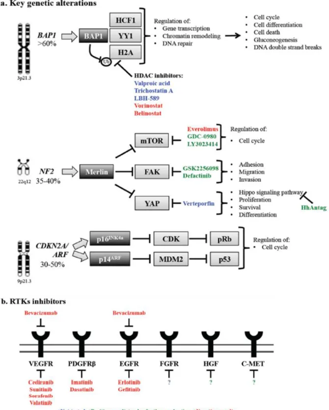
BAP1 is a member of the ubiquitin C-terminal hydrolase (UCH) subfamily of deubiquitinating enzymes (DUBs).79 BAP1 functions as a tumor suppressor because has the ability to perform the deubiquitination of histone H2A, and to remodel the chromatin, leading to transcriptional activation of genes that regulate cell growth. 80 Epigenetic regulation of tumor suppressor genes through chromatin condensation and
decondensation has emerged as an important mechanism that leads to tumorigenesis. The balance between the acetylated and deacetylated forms of histone proteins is regulated by histone
acetyltransferases (HATs) and histone deacetylases (HDACs). HATs increase acetylation promoting greater chromatin accessibility for gene expression, whereas HDAC inhibitors alter the packaging of the DNA around histones, impacting the expression of various genes. Different preclinical studies were conducted by testing in vitro- effect of various HDAC inhibitors, such as valproic acid, trichostatin A, LBH-589, and
suberoylanilide hydroxamic acid (vorinostat). In vitro data studying the role of HDAC inhibitors in MM showed increased apoptosis in MM cell lines after treatment with inhibitors, either alone or in combination with conventional chemotherapy. 81- 86 Furthermore, the combination of valproic acid and chemotherapy completely suppress the tumor generated in a mouse xenograft model of MM. 86
Vorinostat has been approved by FDA for the treatment of cutaneous T-cell lymphoma. But a Phase III trial (VANTAGE 014) study including 660 pre-treated advanced MPM patients used Vorinostat as a second-line or third-line therapy and reported that this treatment modality did not improve overall survival. Therefore, Vorinostat was not recommended as a therapy in MPM patients. 87
Loss of the tumor suppressor gene NF2, encoding Merlin
Mounting evidence has shown that NF2 plays an important role in MM pathogenesis (Figure 3.a). Recent data indicates that asbestos-treated NF2+/− mice accelerate MM tumor formation compared to the wild-types. 88 The NF2 gene is located on chromosome 22q12 and encodes merlin, a tumor suppressor protein. 89 Merlin can interact with various proteins, and in this way modulates multiple signal transduction cascades, including mTOR, focal adhesion kinase (FAK) and Hippo signaling pathways (Figure 3). In MM cells, loss of merlin causes activation of mTOR signaling, 90 therefore in merlin-silenced tumors there is an upregulation of mitogenic signaling and increased cell proliferation. As expected, merlin-negative MM cells
20 were more sensitive to the mTOR inhibitor rapamycin, compared to merlin-positive cells. 90 Therefore, in the large fraction of MMs that carry NF2 mutations, mTOR could be a therapeutic target and provide the rationale for testing mTOR inhibitors in MM.Unfortunately, the oral mTOR inhibitor everolimus (RAD001) had limited clinical activity when tested in a phase II trial S0722 (NCT 00770120) as second- and third-line treatment agent in unselected pre-treated MPM patients. 91 It was concluded that additional studies of single-agent everolimus in advanced MPM were not warranted. 91 The inhibition of mTOR can generate a compensatory mechanism of resistance, that consist in upregulation of PI3KCA and the restoration of the downstream AKT signaling pathway. 92 To address this mechanism of mTOR resistance, a selective dual inhibitor (GDC-0980) of class I PI3K and mTOR was tested. This inhibitor demonstrated broad activity in various xenograft cancer models, including MPM, 93 but also pulmonary toxicity. However, despite these disappointing results, the role of PI3K/AKT/mTOR survival pathway in MM is being further evaluated in clinical trials (NCT01655225, NCT01991938).
Deficiency in the CDKN2A /ARF locus
Previous genetic analysis performed into MM biopsies has revealed that cyclin-dependent kinase inhibitor 2A (CDKN2A)/alternative reading frame (ARF) and neurofibromatosis type 2 (NF2) were the most
commonly mutated tumor suppressor genes in MM. 94, 95 The CDKN2A/ARF is a tumor suppressor gene located at chromosome 9p21.3. 96 CDKN2A encodes p16INK4a, whereas ARF encodes p14ARF. p16INK4a inhibits the cyclin-dependent kinase (CDK)-mediate hyperphosphorylation that leads to retinoblastoma protein (pRb) inactivation. So, loss of p16INK4a results in inactivation of pRb and, consequently, failure of cell cycle arrest. The p14ARF protein promotes degradation of the human ortholog of mouse double minute 2 (MDM2), leading to stabilization of p53. A particularly high frequency of homozygous deletion of CDKN2A/ARF has been detected in mesothelioma samples, causing loss of function of both p53 and pRb tumor suppressors, with a consequent breakdown of cell cycle control mechanisms (Figure 3.a). Otherwise, only a limited number of MM biopsies contain TP53 mutations, the tumor suppressor gene that encodes p53. Different in vivo studies indicate that the inactivation of both CDKN2A and ARF gene cooperates to accelerate asbestos-induced tumorigenesis in mice. 97 However, since genetic defects in p16INK4a/p14ARF are very common, and lead to loss of function of both p53 and Rb, the defective p53 pathways is a
potential target for MM gene therapy. 98 Direct restoration of p16INK4a using gene therapy has also been tested and it has shown some promising activity in preclinical models but is still far from clinical
development. 99, 100
Receptor Tyrosine Kinase Inhibitors
In MM was often observed an activation/upregulation of several receptor tyrosine kinases that constitute a large family of receptors and regulate the cell cycle (Figure 3.b). 101 Activation of these receptors lead to the
21 transduction of abnormal cell growth signaling pathways, which is at the basis of cancer initiation and progression. Inhibition of RTKs or of their ligands with specific antibodies or small molecules has been proven to be an effective and safe targeted approach in several malignancies. 102
Inhibition of angiogenesis was shown to produce antitumor responses and decrease pleural effusion. 103 Mesothelioma secretes pro-angiogenic factors, platelet-derived growth factor (PDGF) and vascular endothelial growth factor (VEGF), both of which are also associated with cell proliferation and pleural effusion. In pleural effusions of MPM patients were detected high levels of VEGF, associated with a worse patient survival. 104 Bevacizumab is an anti-VEGF humanized monoclonal antibody approved for use in several cancers. 105 Results obtained from three independent phase II clinical trials showed that addition of bevacizumab to the standard of care failed to increase survival of MM patients. 106–108
However, recently, results from a randomized phase III trial (IFCT-GFPC-0701 MAPS) indicate that patients treated with bevacizumab and the standard of care (pemetrexed + cisplatin) experienced a significant longer median survival (18.82 months vs. 16.07 months, p = 0.0127) (2015 ASCO Meeting, Abstract #7500). These results might translate into addition of bevacizumab as part of the first line treatment for MPM. There are different studies that reported the high expression of EGFR into MM specimens, thus several inhibitors of this pathway were tested in clinical trials. The results obtained by using erlotinib and gefitinib, inhibitors of EGFR were very disappointing. 109, 110 Negative results were obtained also using erlotinib in combination with bevacizumab after platinum-based chemotherapy. 111 In MM tissue and cell lines an overexpression of several fibroblast growth factors (FGFs) and FGF receptors (FGFRs) were observed. Furthermore, a correlation between high expression of these factors and tumor aggressiveness was also detected. 112, 113 Moreover, inhibition of FGFR1 represses MM cell growth and migration in vitro and in vivo and potentiates the effect of chemotherapeutic drug or ionizing irradiation.114 Thus, inhibition of FGF signals seems to be promising and may permit further evaluation of FGFR targeting strategies.
Other activated pathways in MM are also hepatocyte growth factor (HGF) and the receptor c-Met which are important for tumor invasion and metastasis. It has been demonstrated that inhibition of this pathway suppresses tumor infiltration into neighboring tissues. 115 So, an inhibitor of c-Met kinase is under
investigation for clinical efficacy. Other preclinical studies that inhibit HGF/c-Met pathways were conducted for MM. 116 It is worthwhile to note that in several of the mentioned clinical trials, a small percentage of patients (usually ~1–5%), did experience partial benefits from the therapy with RTK inhibitors, highlighting the need to identify predictive biomarkers to select likely responders (Figure 3.b).
22
Figure 3: Key genetic alterations in MPM and potential strategies for therapeutic intervention. 10
Immunotherapy and immune checkpoint inhibitors
It is known that an immune response is induced by mesothelioma, but it is weak (Robinson et al,
2000). 117 This knowledge has prompted a number of investigators to study different ways to consolidate that response. The intrapleural instillation of cytokines is limited by the short half-life of most cytokines,
23 necessitating repeated injections or continuous infusion via a pleural catheter. Intrapleural interferon-gamma twice weekly for 2 months was reported to induce response rate of 56% in early stage disease. 118 A continuous intrapleural infusion of interleukin-2 induced a partial response in four of 21 patients and an overall survival of 16 months. 119 In both cases, side effects were minimal and consisted primarily of fever and constitutional symptoms. Studies in animals suggest that interferons have an antiproliferative effect on mesothelioma cells and enhance the cytotoxic effect of cisplatin. The results from these studies led to the development of a Phase II trial of cisplatin-doxorubicin and interferon alpha-2 in advanced malignant mesothelioma. The overall response rate was 29% and the median survival was 9.3 months with one year survival of 45% and two year of 34%. 120 However, severe myelosuppression was seen in 60% of patients limiting the application of this treatment. One of the newest hallmark of cancer proposed recently was among others, the evasion of immune destruction, 121 by expressing endogenous immune checkpoints that normally terminate immune responses after antigen activation. This state of tumor-induced immunological anergy is associated to up-regulation in tumor-infiltrating T cells of immune checkpoint molecules, such as cytotoxic T-lymphocyte-associated protein 4 (CTLA-4), and programmed cell death protein 1 (PD-1). 122 Alternatively tumors can block immune activation by upregulating PD-1 ligands. Like many other tumors, MM express high levels of the immunosuppressive PD-1 ligand 1 (PD-L1). 123, 124 Monoclonal antibodies against CTLA4 (tremelimumab, ipilimumab), PD-1 (nivolumab, pembrolizumab), and PD-L1 (avelumab, MPDL3280A) can reactivate the immune response against cancer cells, and have shown promising clinical results in melanoma and some other cancer types. 122 The CTLA4 inhibitor, tremelimumab, at a dose of 15 mg/kg once every 12 weeks, showed clinical activity in 38% of advanced MM patients in a phase II study. 125 An intensified regiment showed a good safety profile, and clinical and immunological activity in patients with advanced MM, with more than 40% of the patients achieving disease control with a median duration of response of almost 11 months. 126 Other clinical trials with PD-1 inhibitor pembrolizumab (NCT02399371) and PD-L1 inhibitor avelumab (NCT01772004) are currently ongoing.
HMGB1
High-mobility group box 1 protein (HMGB1) is a chromatin-binding factor that bends DNA and promotes access to transcriptional protein assemblies on specific DNA targets.127, 128 This protein was isolated and characterized in calf thymus in 1973, and its name derives from its electrophoretic mobility on
polyacrylamide gels. HMGB1 has two HMG-box domains (N-terminal A and central B) able to bind DNA and an acidic C terminal tail (Figure 4.a). HMGB1 is a highly conserved nuclear protein, present in almost all metazoans and plants. 129 In most cells, HMGB1 acts as a DNA chaperone to help maintain nuclear homeostasis. Later, it was discovered that HMGB1 is also expressed on cell surface membranes, cytosol, and mitochondria, and could be released into the extracellular space. So, in addition to its nuclear function,
24 HMGB1 has also many biological functions outside the cell playing a significant role as extracellular
signaling molecule during inflammation, cell differentiation, cell migration, and tumor metastasis. 127 HMGB1 is passively released from necrotic cells and is actively secreted by inflammatory cells, binding to several receptors such as the receptor for advanced glycation end products (RAGE), Toll-like receptors (TLR)-2, TLR-4, TLR-9, and, as a negative signaling molecule, CD24. The interaction between HMGB1 and its functional receptors mediates the response to infection and injury, thereby promoting inflammation (Figure 4.b). 127, 128, 129, 130, 131, 132. As such HMGB1 is the prototypic Damage Associated Molecular Pattern Molecule, or DAMP, associated with both acute inflammatory responses and driving much of the biology of chronic inflammation and wound repair.133, 134 HMGB1 plays a significant role in many diseases, especially inflammatory diseases and cancer. 135-139 Recent evidences indicate that HMGB1 dysfunction is associated with each of the central hallmarks of cancer and contributes to cancer development and therapy.
Nuclear Function of HMGB1
HMGB1 proteins are constitutively expressed in the nucleus of cells due to the presence of two lysine-rich nuclear localization sequences (NLSs) located in the A box and in the B box (Figure 4.a). Hyperacetylation of NLSs endorses the translocation of HMGB1 from the nucleus to the cytosol, and its consequent release. The studies, which have measured the affinity of HMGB1 with different DNA structures, indicated that HMGB1 is able to binds preferentially different DNA structures such as supercoiled, single-stranded, B- and Z-DNA, DNA mini-circles, and triplex DNA. 139, 140 This ability is promoted from its HMG boxes that allow HMGB1 to bind DNA without sequence-specificity and to act as a DNA chaperone. Thus, HMGB1 is the structural protein of chromatin which regulates nuclear homeostasis and genome stability in several ways (Figure 4).
1) Nucleosome structure and dynamics. Chromatin contains nucleosome units which consist in a short
length of DNA wrapped around a core of histone proteins. HMGB1 binds to nucleosomes at the dyad axis, induces the sliding of nucleosome, relaxes nucleosome structure, and due to its ability of DNA-bending, makes chromatin more accessible. 141
2) Gene transcription. HMGB1 has been found to interact with and enhance the binding affinity of many sequence- specific transcriptional factors to their cognate DNA, such as p53, 142 p73, 143 the retinoblastoma protein, 144 members of the Rel/NF-κB family 145 and estrogen receptors. This could increase their activity as transcriptional factors implicated in cancer development. Mounting evidence has shown that HMGB1 interacts with p53 and provides the optimal DNA structure for p53 binding through its bending/binding effects. 146 p53 family members are important because participate in the regulation of cell cycle progression functioning as tumor suppressors.
3) DNA repair. HMGB1 also plays a critical role in DNA repair by being part of a nuclear protein complex involved in the cytotoxic response to DNA modified by incorporation of anticancer nucleoside analogues. In addition, loss of HMGB1 increases DNA damage and decreases DNA repair efficiency in response to
25 chemotherapy, irradiation, and oxidative stress. HMGB1 directly binds to a variety of bulky DNA lesions and allows it to participate in DNA repair pathways including nucleotide excision repair, base excision repair, mismatch repair, and double strand break repair via nonhomologous end-joining. 147
Cytosolic HMGB1
Different studies have investigated the levels and distribution of HMGB1 between the nucleus and cytoplasm in different cells and tissues. Localization of HMGB1 in the cytoplasm has been confirmed in living fibroblasts, thymocytes and several different tissues (e.g., liver, kidney, brain, heart, and lung). 148, 149 Currently, we know that HMGB1 normally is located in the nucleus and translocate from the nucleus to the cytosol, including mitochondria and lysosome, following various stressors (e.g., cytokine, chemokine, heat, hypoxia, H2O2, and oncogene). Although the function of cytosolic HMGB1 still remains poorly studied, we demonstrated that the main function of HMGB1 in cytoplasm is to function as a positive regulator of autophagy. Autophagic stimuli promote the translocation of HMGB1 to the cytosol. Cytosolic HMGB1 binds to Beclin-1 inducing autophagy for degrading damaged organelles and unused proteins. 150 HMGB1 also interacts with many apparently unrelated proteins by recognizing short amino acid sequence motifs. 151 For example, the motifs PXXPXP and WXXW (where X can be any amino acid) can interact with box A and box B of HMGB1, respectively. 151 Thus, HMGB1 may be involved in many cell processes by promoting protein protein interactions.
Another potential function for cytosolic HMGB1 is involvement in the unconventional secretory pathway, found based on mass spectrometry-mediated binding partner analysis in 2010. 152
HMGB1 release
In addition to its role inside the cell, HMGB1 also functions as a damage-associated molecular pattern (DAMP) when passively released from dead, dying, or injured cells. It is also actively secreted from immune cells or cancer cells in response to exogenous and endogenous stimuli such as endotoxin, CpG DNA, double-stranded RNA (dsRNA), tumor necrosis factor (TNF)-α, interleukin (IL)-1, interferons (IFN)-γ, hydrogen peroxide, adenosine triphosphate (ATP), and hypoxia. In addition, macrophage engulfment of
apoptotic cells may induce significant active HMGB1 release, suggesting a direct interplay between dying cells and immune cells, which also induces HMGB1 release. 153 Depending on the inducing stimulus, the mechanism of HMGB1 secretion and release could be different.
Extracellular HMGB1
Besides its nuclear and cytosolic function, HMGB1 performs a significant extracellular role in inflammation, immunity, cell growth, cell proliferation, and cell death. HMGB1 can be actively secreted by immune cells or
26 passively released by dead, dying, or injured cells. Once released, extracellular HMGB1 binds to several cell surface receptors to activate the downstream signaling pathway (e.g., NF-κB, IFN regulatory factor-3 (IRF3), and phosphatidylinositol 3-kinase [PI3K]) to produce a functional response, such as activation of innate immune cells, induction of proinflammatory cytokines and type I IFNs, stimulation of cell adhesion and migration, inhibition of phagocytosis, promotion of cell proliferation and angiogenesis, and induction of autophagy. 154, 155 In addition, extracellular HMGB1 functions as an immune adjuvant to trigger a robust response to activation or suppression of T cells, dendritic cells, and endothelial cells. Activated immune cells (e.g., macrophages, monocytes, and dendritic cells) and endothelial cells also secrete HMGB1, which in turn generates a positive feedback loop that causes the release of supplemental cytokines and chemokines following engagement of multiple receptors. Thus, HMGB1 has the ability to sustain a long-term
inflammatory state under stress. Interestingly, extracellular HMGB1 has antibacterial, cell growth, and mitotic activity. These extracellular HMGB1 activities are not only mediated by receptors, but also by its redox state and structure.156
Native HMGB1 proteins from eukaryotic sources have the same (though less pronounced) biological activity in vitro compared to recombinant HMGB1 proteins from prokaryotic sources. 157 The extracellular HMGB1 plays its function by binding to several receptors such as the receptor for advanced glycation end products (RAGE), Toll-like receptors (TLRs, such as TLR2, TLR4, and TLR9), Mac-1, syndecan-1 (CD138), phosphacan protein-tyrosine phosphatase (PPTP)- ζ/β, CD24, chemokine (C-X-C motif) ligand 4 (CXCL4), T cell
immunoglobulin mucin-3 (TIM-3), and possibly others. Of these receptors, CD24 and TIM-3 act as negative receptors and inhibit immune activity of HMGB1 in macrophages and tumor-associated dendritic cells (TADCs), respectively. 158, 159 Apart from a direct receptor interaction, HMGB1 may form heterocomplexes with other immune co-activators such as IL-1, CXCL12, DNA, nucleosome, or LPS that generate synergistic responses in inflammation and immunity. The first receptor demonstrated to bind HMGB1 was RAGE.160 Later, it was discovered that HMGB1 signaling through RAGE mediates chemotaxis and migration,
proliferation and differentiation of immune and cancer cells, and upregulation of cell surface receptors. In addition, RAGE provides a functional platform for crosstalk with other HMGB1 receptors. For example, interplay between RAGE and TLR9 is important for critical for HMGB1-DNA complex, which activates the immune responses in dendritic cells (DCs). 161 The interplay between Mac-1 and RAGE is required for HMGB1-mediated adhesive and migratory neutrophil functions. 162 whereas, the interplay between syndecan-1, PPTP-ζ/β, and RAGE is required for neurite outgrowth mediated by HMGB1.
Apart from RAGE, HMGB1 binding of TLR2 and TLR4 also results in NF-κB activation. TLR4 may be more important for HMGB1-induced macrophage activation and proinflammatory cytokine release. 163
Experimental data obtained by using TLR4-deficient animals suggest that TLR4 plays a critical role in sterile inflammation. 164 These animal models are significantly protected from ischemia-reperfusion injury to the liver, kidney, and heart.
27
Figure 4. Structure and function of HMGB1
(A) HMGB1 is structurally composed of three different domains: two homologous DNA binding domains (box A and box B) and a negatively charged C-terminal domain. Two nuclear localization signals (NLS1 and NLS2) control nuclear transport of HMGB1. In addition, HMGB1 contains three redox-sensitive cysteine residues (C23, C45 and C106), which are important for HMGB1 activity. (B) HMGB1 has multiple roles inside and outside the cell. 266
HMGB1 Redox States
Recent studies underline the importance of redox modification in the regulation of HMGB1 translocation, release, and activity in disease.164 Three cysteines are encoded within the HMGB1, two vicinal cysteines in
28 box A (C23 and C45) and a single one in box B (C106). Replacement of Cys23 and/or 45 with serine did not affect the nuclear distribution of the mutant proteins. Whereas, C106S and triple cysteine mutations impaired the nuclear localization of HMGB1, allowing entry of some of the protein into the cytosol. Moreover, increased endogenous and exogenous ROS promotes HMGB1 translocation and release. 165 The redox status of HMGB1 promotes to distinguish between its cytokine and chemokine activity.166 Initial studies suggest that reduced C106 is necessary for the binding of HMGB1 to TLR4 and promotes cytokine release and inflammation. A recent study suggests that a disulfide bond between C23 and C45 is also required for HMGB1 cytokine activity. Mutations of C45 or C23 abolish the cytokine activity of HMGB1. In contrast, all-cysteine-reduced HMGB1 does not have TLR4-dependent cytokine activity, but binds to CXCL4 to induce inflammatory cell recruitment and chemotaxis by the CXCL12 receptor. 167 ROS oxidizes the HMGB1 at C106 released from apoptotic cells, thereby neutralizing its cytokine-inducing activity and promoting tolerance in DCs. Finally, all-cysteine oxidized HMGB1 impairs HMGB1’s cytokine or chemotactic activity.168 Thus, redox modifications are crucial for HMGB1 functionality as a mediator during infection and sterile inflammation.
Figure 5: The redox status of
HMGB1 regulates its cytokine-inducing and chemokine activities.200
HMGB1’s Roles in Tumorigenesis
Mounting evidences has shown that disfunction of HMGB1 is associated with tumorigenesis and contributes to cancer development and therapy (Figure 6).169 For this purpose, it will be important to understand HMGB1 regulation and its function in the mechanism of cancer biology. Furthermore, the understanding of its role in tumorigenesis will influences the strategies of a HMGB1 targeted therapy for prevention and treatment. The inflammatory tumor microenvironment (TME) is able to support the neoplastic transformation, tumor growth, invasion, and metastasis. The development of the inflammatory tumor microenvironment is associated with the tumor-infiltrating leukocytes and the cytokine-related signaling pathways. Infiltrating
29 leucocytes, as well as the cancer cells themselves, have the ability to secrete HMGB1 under hypoxia, injury, inflammatory stimuli, or environmental factors.170 In turn, extracellular HMGB1 can activate proinflammatory signaling pathways, such as the NF-κB and inflammasome pathways, to induce proinflammatory cytokine release. This loop will accelerate inflammatory responses and induce tumor formation, and metastasis. Another of the most common cancer phenotypes is a high energy request by cancer cells to allow a rapid, invasive and metastatic growth of tumor. While normal cells produce ATP through a combination of oxidative and glycolytic metabolism, cancer cells effectively stimulate and reprogram their metabolism to better fit the energy demand. HMGB1 has been implicated in tumor energy metabolism. 171,172,173
Recombinant exogenous HMGB1 or endogenous HMGB1 derived from necrotic tumor cell lysates are likely to rise ATP production and pancreatic tumor cell proliferation, providing a direct link between inflammation and energy metabolism with the TME. 171 Recently it was shown that extracellular HMGB1 increases
mitochondrial RAGE expression and translocation, which in turn increases mitochondrial complex I activity and ATP production. 174 Most cancer deaths are caused by tumor invasion and metastasis rather than the primary tumor itself. In the clinic, expression of RAGE is strictly associated with cancer invasiveness and metastasis activity such as gastric cancer 175 and colorectal cancer.176 Different in vivo and in vitro studies showed that impairment of RAGE–HMGB1 interaction inhibit tumor growth and metastasis by activation of mitogen-activated protein kinases and the NF-kB pathway. The NF-kB activation results in the expression of matrix metalloproteinases (MMP), such as MMP2 and MMP9,177, 178 which degrade extracellular matrix proteins and play an important role in tumor invasion and metastasis.177 Thus, HMGB1–RAGE signaling pathway plays a major role in tumor invasion and metastasis.
The immunity surveillance of cancer is considered to be an important defense process against carcinogenesis. HMGB1, as a multifunctional cytokine, has been characterized with both immunosuppressive and immune-activation properties, which depends on receptors, targeted cells, and redox state. 179 So, HMGB1 has the ability to induce apoptosis in macrophage-derived DCs, which diminish host anti-cancer immunity. 180 In addition, HMGB1, derived from tumor cells, suppresses naturally-acquired CD8+ T cell-dependent antitumor immunity, partly by enhancing tumor-associated Treg to produce IL-10.181 Recent findings suggest that endogenous intracellular HMGB1, as a RB- associated protein, suppresses breast tumorigenesis, acting as a tumor suppressor gene (Figure 6).182 RB is a well-known tumor suppressor protein that is dysfunctional in many cancers. HMGB1 enhances RB-mediated transcription repression such as E2F and cyclin A1, and causes RB-dependent G1 arrest and apoptosis induction. In addition, overexpression of HMGB1 inhibits RB positive breast cancer growth in vitro and prevents tumorigenicity in subcutaneous tumor models in vivo. 182 In addition, HMGB1 is also an important regulator of autophagy and its lost inhibits autophagy and increases apoptosis. Several studies have indicated that defective autophagy-associated genes (e.g., Beclin1, ATG5, UVRAG, Bif-1) in mice increase genome instability, inflammation, oxidative stress, and mitochondrial injury, which contribute to tumorigenesis. 183-185 So, these findings underscore that HMGB1 inhibition lead to
30 autophagy deficiency, cause genomic instability, inflammation and induce tumorigenesis. Thus, suppression of autophagy promotes tumorigenesis and increases the effectiveness of anticancer therapy.
HMGB1’s Protective Roles in Anticancer Therapy
Immunogenic cell death (ICD), contributes to immune-mediated elimination of tumors during
chemotherapy (e.g., anthracyclines) or radiotherapy. 186-189 ICD is characterized by the release of dying cancer cells or cell surface exposure of DAMPs (e.g., calreticulin, heat shock proteins, ATP, and HMGB1). These events are useful for the maturation, antigen uptake, and presentation of DCs and works as high-powered immunological adjuvants to active cytotoxic T lymphocyte response. Several in vivo and in vitro studies indicate that blocking the HMGB1- TLR4 pathway inhibits ICD and the anticancer immune responses upon chemotherapy. 189 (Figure 6). However, HMGB1 released from necrotic cancer cells treated with chemotherapy amplifies regrowth and metastasis of residual cancer cells in a RAGE-dependent way. 190 Thus, blocking HMGB1-RAGE signaling rise the effectiveness of chemotherapy.191 These studies suggest that TLR4 in DC is important for HMGB1-mediated ICD and tumor clearance, whereas RAGE in cancer cells is critical for HMGB1-mediated survival after chemotherapy. Although both apoptotic and necrotic cells have the ability to release HMGB1, only the HMGB1 released from apoptotic cells is tolerogenic. 190 Thus, determining the role of extracellular HMGB1 in a context specific way in chemotherapy and
immunotherapy, including ICD, and the mechanisms involved will be important to optimize the therapeutic outcomes.
The negative roles of HMGB1 in Anticancer Therapy
It has been demonstrated that suppression of HMGB1 expression by RNAi increased the anticancer activity of cytotoxic agents, whereas overexpression of HMGB1 expression by gene transfection increased drug resistance. 192, 193 HMGB1 expression regulates chemotherapeutic response and resistance by interfering with autophagy and the apoptotic pathway (Figure 6). HMGB1 has the ability to increase the pro-survival autophagy in a Beclin 1- dependent way in chemotherapy, whereas HMGB1 inhibits both intrinsic and extrinsic programmed cell death/apoptosis in a caspase-dependent way in cancer cells. The crosstalk between apoptosis and autophagy regulates cell death and determines cell fate in anticancer therapy. Upregulation of apoptosis inhibits autophagy, whereas upregulation of autophagy inhibits apoptosis during chemotherapy. HMGB1 and p53 are capable of physical interaction (Figure 6), 194 and the interplay
between HMGB1 and p53 regulates apoptosis and autophagy in clone cancer cells after treatment with DNA-damaging anticancer drugs.193 DNA damage promotes interactions between p53 and HMGB1 in the nucleus and cytoplasm. Loss of p53 increases cytosolic HMGB1, leading to increased binding to Beclin 1, thereby promoting autophagy and decreasing apoptosis; In contrast, loss of HMGB1 increases cytosolic p53
31 and apoptosis and decreases autophagy. 193 These findings provide new insights into the HMGB1-p53 signaling and cancer cell response to DNA damage.
HMGB1-Targeting Therapeutic Agents
Apart from genetic inhibition or overexpression of HMGB1 expression in cancer cells, several HMGB1-targeting agents have been used in experimental cancer research. These agents including sRAGE, HMGB1 neutralizing antibody, A box protein, platinating agent, ethyl pyruvate, quercetin, and glycyrrhizin. sRAGE acts as a decoy to prevent RAGE signaling and has been used successfully in blocking the HMGB1-RAGE signaling pathway in animal tumor models. HMGB1 neutralizing antibody and A box protein can block activity of extracellular HMGB1 in tumor therapy.195 Interestingly, platinating agents such as cisplatin and oxaliplatin have the ability to retain HMGB1 within the nucleus by conformational changes in the double helix to which HMGB1 binds quite stably. 196 Ethyl pyruvate, the first HMGB1 inhibitor used in animal models of sepsis by inhibition of NF-κB pathway, inhibits liver tumor growth. 197 In addition, glycyrrhizin and quercetin, potential HMGB1 inhibitors by directly binding to HMGB1 or inhibition of PI3K, improve the effectiveness of anticancer agents in several different tumor models. 195 Further investigation is needed to evaluate these therapies and their possible role in clinical practice.