“Amedeo Avogadro”
Dipartimento di Scienze del Farmaco
Dottorato di Ricerca in Biotecnologie Farmaceutiche ed Alimentari
XXVII ciclo a.a. 2011-2014
Biochemical and structural studies on human
enzymes involved in NAD homeostasis
“Amedeo Avogadro”
Dipartimento di Scienze del Farmaco
Dottorato di Ricerca in Biotecnologie Farmaceutiche ed Alimentari
XXVII ciclo a.a. 2011-2014
Biochemical and structural studies on human
enzymes involved in NAD homeostasis
Ada Serena Marletta
Supervised by Dr. Silvia Garavaglia
“Il viaggio da Kamakura a Kyoto dura dodici giorni: se viaggi per undici giorni e ti fermi quando ne manca uno solo, come puoi ammirare la luna sopra la capitale?”
Chapter 1
1 IntroductionChapter 2
37Outline of the thesis
Chapter 3
43 “The crystal structure of human Nicotinic AcidPhosphoribosyltransferase: a key enzyme in Preiss- Handler pathway”
Chapter 4
75“Extracellular NMN sustains Intracellular NAD+ Biosynthesis through CD73-mediated Nicotinamide Riboside Production”
Chapter 5
95 ConclusionsList of publications
103Chapter 1
1
1.0 The Chemistry of NAD(P) and its
physiological roles
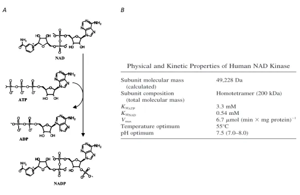
Nicotinamide adenine dinucleotide (NAD) and its phosphorylated counterpart (NADP) are ubiquitous dinucleotides, since they consist of two nucleotides joined through their phosphate groups. In particular, one nucleotide contains an adenine base and the other nicotinamide, namely adenosine monophosphate (AMP) and nicotinamide mononucleotide (NMN), respectively. Because of the presence of a pyridine ring on the NMN moiety, due to the fact that nicotinamide is a derivative of pyridine, NAD and NADP are also referred as pyridine nucleotides (Figure 1A). The presence of an additional phosphate group at the 2’ position on the ribose of the AMP moiety represents the chemical difference between NAD and NADP. Both these molecules exist in the form of a pair comprising an oxidized and a reduced form, NAD+/NADH and NADP+/NADPH, and are termed redox couples. It is usual to refer to the NAD+/NADH and NADP+/NADPH couples as NAD and NADP, respectively; all these species are collectively indicated as NAD(P). The chemical difference between the two species in each redox couple depends on the transfer of a hydride ion, consisting of two electrons and a proton, at the level of the nicotinamide ring constituting the reactive portion of NAD(P)+ (Figure 1B). This transfer is driven by numerous hydride transfer enzymes or oxidoreductases, which interconvert either NAD+ and NADH or NADP+ and NADPH to reduce or oxidize small-molecule metabolites in the biological systems. On the basis of their capacity to support the activity of enzymes catalysing oxidation-reduction reactions, as hydride acceptors and donors, NAD and NADP are defined coenzymes.
Chapter 1
2
Figure 1.A. Chemical structure of NAD+. Instead of a hydrogen atom, NADP+ has a phosphate
group at the 2’ position on the ribose of its AMP moiety. B. Bi-directional redox reaction between NAD+ and NADH.
Moreover, it is now well established that pyridine nucleotides and the balance between their oxidized and reduced forms play a wide variety of pivotal roles in cellular functions as important interfaces, beyond their coenzymatic activity. These include maintenance of redox status, cell survival and death, ion channel regulation, and cell signalling under normal and pathological conditions (1). In the role of signal transducer, NAD(P)+ is a substrate of many enzymes and need to be constantly resynthesized in the cell.
showed that NAMPT expression is regulated in a
circa-dian fashion (38, 39). The core clock components of the
circadian machinery regulate the recruitment of SIRT1 to
the NAMPT promoter to increase NAMPT expression.
This is followed by NAD
!biosynthesis, which in turn will
activate sirtuins as well as other NAD
!-dependent
en-zymes. In a negative feedback loop, SIRT1 will repress the
clock components and thereby NAMPT expression (38,
39). It is suggested that through this mechanism, NAMPT
(38, 39) and SIRT1 (40, 41) may play a crucial role in the
circadian regulation of metabolism. After the NAMPT
reaction, NMN is converted to NAD
!by NMNAT,
which is able to condense the adenylyl moiety to both
NAMN and NMN (16 –18, 42).
A third NAD
!salvage pathway, which was long
known as the only NAD
!biosynthesis pathway in certain
bacteria, is comprised of the phosphorylation of NR to
NMN by NR kinase (NRK), after which one of the
NMNAT enzymes catalyzes the formation of NAD
!.
Re-cently, this pathway was shown to be highly conserved
and also active in yeast and humans, for which two NRK
enzymes have been described (43). The NRK1 isoform
is ubiquitously expressed, whereas NRK2 localization
seems to be restricted to heart, brain, and muscle (44).
The discovery of NR as a nutrient in cow milk (43) poses
an interesting opportunity for therapeutic intervention
in NAD
!-dependent metabolism, as will be discussed in
Section IX.
C. Substrate preference for NAD
!biosynthesis
The existence of four pathways for NAD
!biosynthesis
originating from four independent precursors raises
ques-tions regarding the fluxes of metabolites. As mentioned in
Sections II.A and II.B, some of the enzymes involved in
NAD
!biosynthesis are strictly localized to a limited
number of organs. Also, some enzymes, for instance
NMNAT1-3, clearly show different subcellular
localiza-tion. To date there is no consensus with respect to the
question of the preferred substrate of NAD
!. Feeding of
rats with tryptophan, NA, or NAM showed that the first
resulted in the highest NAD
!concentration in liver,
sug-gesting that this is the preferred substrate at least in this
organ (45). These results were confirmed in isolated
hepa-tocytes, showing that NA and NAM are normally taken up
from the medium but are not used for NAD
!biosynthesis
(46). In tissues that lack the complete de novo NAD
!COOH N N O NH2 COOH N O OH HO H2 C O P O HO OH COOH N O OH HO H2 C N N NH2 N H N O HO OH H2 C O P O O OH OH P O O N O OH HO H2 C O P O HO OH O NH2 N O OH HO H2 C O NH2 N N NH2 N H N O HO OH H2 C O P O O OH P O OH O N O OH HO H2 C O NH2 HO
NA
NAMN
NAAD
NMN
NAD
NAM
NR
Quinolinic acid
QPRT
NAPT
NAMPT
NRK
NADS
de novo biosynthesis
NMNAT
NMNAT
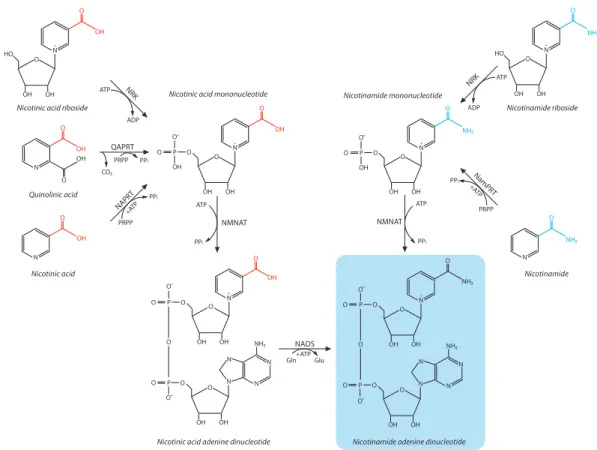
FIG. 2. Mammalian NAD! salvage pathway. NAD! is synthesized in the NAD! salvage pathway from its precursors NA, NAM, or NR. The initial
step in NAD! synthesis from NA, the so-called Preiss-Handler pathway, is catalyzed by NAPT and results in the formation of NAMN, which can also
be derived from de novo NAD! biosynthesis. In an identical reaction, but catalyzed by a different enzyme, NAM is converted by NAMPT forming
NMN, which is also the product of phosphorylation of NR by NRK. The subsequent conversion of both NAMN and NMN is catalyzed by the same enzyme, i.e., NMNAT. In the case of NAMN, this reaction is followed by amidation, finally producing NAD!. NADS, NAD synthase.
Endocrine Reviews, April 2010, 31(2):194 –223 edrv.endojournals.org 197
The Endocrine Society. Downloaded from press.endocrine.org by [${individualUser.displayName}] on 09 September 2014. at 06:12 For personal use only. No other uses without permission. . All rights reserved. NMN#
AMP#
A"
showed that NAMPT expression is regulated in a
circa-dian fashion (38, 39). The core clock components of the
circadian machinery regulate the recruitment of SIRT1 to
the NAMPT promoter to increase NAMPT expression.
This is followed by NAD
!biosynthesis, which in turn will
activate sirtuins as well as other NAD
!-dependent
en-zymes. In a negative feedback loop, SIRT1 will repress the
clock components and thereby NAMPT expression (38,
39). It is suggested that through this mechanism, NAMPT
(38, 39) and SIRT1 (40, 41) may play a crucial role in the
circadian regulation of metabolism. After the NAMPT
reaction, NMN is converted to NAD
!by NMNAT,
which is able to condense the adenylyl moiety to both
NAMN and NMN (16 –18, 42).
A third NAD
!salvage pathway, which was long
known as the only NAD
!biosynthesis pathway in certain
bacteria, is comprised of the phosphorylation of NR to
NMN by NR kinase (NRK), after which one of the
NMNAT enzymes catalyzes the formation of NAD
!.
Re-cently, this pathway was shown to be highly conserved
and also active in yeast and humans, for which two NRK
enzymes have been described (43). The NRK1 isoform
is ubiquitously expressed, whereas NRK2 localization
seems to be restricted to heart, brain, and muscle (44).
The discovery of NR as a nutrient in cow milk (43) poses
an interesting opportunity for therapeutic intervention
in NAD
!-dependent metabolism, as will be discussed in
Section IX.
C. Substrate preference for NAD
!biosynthesis
The existence of four pathways for NAD
!biosynthesis
originating from four independent precursors raises
ques-tions regarding the fluxes of metabolites. As mentioned in
Sections II.A and II.B, some of the enzymes involved in
NAD
!biosynthesis are strictly localized to a limited
number of organs. Also, some enzymes, for instance
NMNAT1-3, clearly show different subcellular
localiza-tion. To date there is no consensus with respect to the
question of the preferred substrate of NAD
!. Feeding of
rats with tryptophan, NA, or NAM showed that the first
resulted in the highest NAD
!concentration in liver,
sug-gesting that this is the preferred substrate at least in this
organ (45). These results were confirmed in isolated
hepa-tocytes, showing that NA and NAM are normally taken up
from the medium but are not used for NAD
!biosynthesis
(46). In tissues that lack the complete de novo NAD
!COOH N N O NH2 COOH N O OH HO H2 C O P O HO OH COOH N O OH HO H2 C N N NH2 N H N O HO OH H2 C O P O O OH OH P O O N O OH HO H2 C O P O HO OH O NH2 N O OH HO H2 C O NH2 N N NH2 N H N O HO OH H2 C O P O O OH P O OH O N O OH HO H2 C O NH2 HO
NA
NAMN
NAAD
NMN
NAD
NAM
NR
Quinolinic acid
QPRT
NAPT
NAMPT
NRK
NADS
de novo biosynthesis
NMNAT
NMNAT
FIG. 2. Mammalian NAD
!salvage pathway. NAD
!is synthesized in the NAD
!salvage pathway from its precursors NA, NAM, or NR. The initial
step in NAD
!synthesis from NA, the so-called Preiss-Handler pathway, is catalyzed by NAPT and results in the formation of NAMN, which can also
be derived from de novo NAD
!biosynthesis. In an identical reaction, but catalyzed by a different enzyme, NAM is converted by NAMPT forming
NMN, which is also the product of phosphorylation of NR by NRK. The subsequent conversion of both NAMN and NMN is catalyzed by the same
enzyme, i.e., NMNAT. In the case of NAMN, this reaction is followed by amidation, finally producing NAD
!. NADS, NAD synthase.
Endocrine Reviews, April 2010, 31(2):194 –223
edrv.endojournals.org
197
The Endocrine Society. Downloaded from press.endocrine.org by [${individualUser.displayName}] on 09 September 2014. at 06:12 For personal use only. No other uses without permission. . All rights reserved. R#
showed that NAMPT expression is regulated in a
circa-dian fashion (38, 39). The core clock components of the
circadian machinery regulate the recruitment of SIRT1 to
the NAMPT promoter to increase NAMPT expression.
This is followed by NAD
!biosynthesis, which in turn will
activate sirtuins as well as other NAD
!-dependent
en-zymes. In a negative feedback loop, SIRT1 will repress the
clock components and thereby NAMPT expression (38,
39). It is suggested that through this mechanism, NAMPT
(38, 39) and SIRT1 (40, 41) may play a crucial role in the
circadian regulation of metabolism. After the NAMPT
reaction, NMN is converted to NAD
!by NMNAT,
which is able to condense the adenylyl moiety to both
NAMN and NMN (16 –18, 42).
A third NAD
!salvage pathway, which was long
known as the only NAD
!biosynthesis pathway in certain
bacteria, is comprised of the phosphorylation of NR to
NMN by NR kinase (NRK), after which one of the
NMNAT enzymes catalyzes the formation of NAD
!.
Re-cently, this pathway was shown to be highly conserved
and also active in yeast and humans, for which two NRK
enzymes have been described (43). The NRK1 isoform
is ubiquitously expressed, whereas NRK2 localization
seems to be restricted to heart, brain, and muscle (44).
The discovery of NR as a nutrient in cow milk (43) poses
an interesting opportunity for therapeutic intervention
in NAD
!-dependent metabolism, as will be discussed in
Section IX.
C. Substrate preference for NAD
!biosynthesis
The existence of four pathways for NAD
!biosynthesis
originating from four independent precursors raises
ques-tions regarding the fluxes of metabolites. As mentioned in
Sections II.A and II.B, some of the enzymes involved in
NAD
!biosynthesis are strictly localized to a limited
number of organs. Also, some enzymes, for instance
NMNAT1-3, clearly show different subcellular
localiza-tion. To date there is no consensus with respect to the
question of the preferred substrate of NAD
!. Feeding of
rats with tryptophan, NA, or NAM showed that the first
resulted in the highest NAD
!concentration in liver,
sug-gesting that this is the preferred substrate at least in this
organ (45). These results were confirmed in isolated
hepa-tocytes, showing that NA and NAM are normally taken up
from the medium but are not used for NAD
!biosynthesis
(46). In tissues that lack the complete de novo NAD
!COOH N N O NH2 COOH N O OH HO H2 C O P O HO OH COOH N O OH HO H2 C N N NH2 N H N O HO OH H2 C O P O O OH OH P O O N O OH HO H2 C O P O HO OH O NH2 N O OH HO H2 C O NH2 N N NH2 N H N O HO OH H2 C O P O O OH P O OH O N O OH HO H2 C O NH2 HO
NA
NAMN
NAAD
NMN
NAD
NAM
NR
Quinolinic acid
QPRT
NAPT
NAMPT
NRK
NADS
de novo biosynthesis
NMNAT
NMNAT
FIG. 2. Mammalian NAD
!salvage pathway. NAD
!is synthesized in the NAD
!salvage pathway from its precursors NA, NAM, or NR. The initial
step in NAD
!synthesis from NA, the so-called Preiss-Handler pathway, is catalyzed by NAPT and results in the formation of NAMN, which can also
be derived from de novo NAD
!biosynthesis. In an identical reaction, but catalyzed by a different enzyme, NAM is converted by NAMPT forming
NMN, which is also the product of phosphorylation of NR by NRK. The subsequent conversion of both NAMN and NMN is catalyzed by the same
enzyme, i.e., NMNAT. In the case of NAMN, this reaction is followed by amidation, finally producing NAD
!. NADS, NAD synthase.
Endocrine Reviews, April 2010, 31(2):194 –223
edrv.endojournals.org
197
The Endocrine Society. Downloaded from press.endocrine.org by [${individualUser.displayName}] on 09 September 2014. at 06:12 For personal use only. No other uses without permission. . All rights reserved.
showed
that
NAMPT
expression
is
regulated
in
a
circa-dian
fashion
(38,
39).
The
core
clock
components
of
the
circadian
machinery
regulate
the
recruitment
of
SIRT1
to
the
NAMPT
promoter
to
increase
NAMPT
expression.
This
is
followed
by
NAD
!biosynthesis,
which
in
turn
will
activate
sirtuins
as
well
as
other
NAD
!-dependent
en
-zymes.
In
a
negative
feedback
loop,
SIRT1
will
repress
the
clock
components
and
thereby
NAMPT
expression
(38,
39).
It
is
suggested
that
through
this
mechanism,
N
AMPT
(38,
39)
and
SIRT1
(40,
41)
may
play
a
crucial
role
in
the
circadian
regulation
of
metabolism.
After
the
NAMPT
reaction,
NMN
is
converted
to
NAD
!by
NMNAT,
which
is
able
to
condense
the
adenylyl
moiety
to
both
NAMN
and
NMN
(16
–18,
42).
A
third
NAD
!salvage
pathway,
which
was
long
known
as
the
only
NAD
!biosynthesis
pathway
in
certain
bacteria,
is
comprised
of
the
phosphorylation
of
NR
to
NMN
by
NR
kinase
(NRK),
after
which
one
of
the
NMNAT
enzymes
catalyzes
the
formation
of
NAD
!.Re
-cently,
this
pathway
was
shown
to
be
highly
conserved
and
also
active
in
yeast
and
humans,
for
which
two
NRK
enzymes
have
been
described
(43).
The
NRK1
isoform
is
ubiquitously
expressed,
whereas
NRK2
localization
seems
to
be
restricted
to
heart,
brain,
and
muscle
(44).
The
discovery
of
NR
as
a
nutrient
in
cow
milk
(43)
poses
an
interesting
opportunity
for
therapeutic
intervention
in
NAD
!-dependent
metabolism,
as
will
be
discussed
in
Section
IX
.
C.
Substrate
preference
for
NAD
!biosynthesis
The
existence
of
four
pathways
for
NAD
!biosynthesis
originating
from
four
independent
precursors
raises
ques-tions
regarding
the
fluxes
of
metabolites.
As
mentioned
in
Sections
II.A
and
II.B
,
some
of
the
enzymes
involved
in
NAD
!biosynthesis
are
strictly
localized
to
a
limited
nu
m
be
r
of
organs.
Also,
some
enzymes,
for
instance
NMNAT1-3,
clearly
show
different
subcellular
localiza-tion.
To
date
there
is
no
consensus
with
respect
to
the
question
of
the
preferred
substrate
of
NAD
!.Feeding
of
rats
with
tryptophan,
NA,
or
NAM
showed
that
the
first
resulted
in
the
highest
NAD
!concentration
in
liver,
sug
-gesting
that
this
is
the
preferred
substrate
at
least
in
this
organ
(45).
These
results
were
confirmed
in
isolated
hepa-tocytes,
showing
that
NA
and
NAM
are
normally
taken
up
from
the
medium
but
are
not
used
for
NAD
!biosynthesis
(46).
In
tissues
that
lack
the
complete
de
novo
NAD
! CO OH N N O NH 2 CO OH N O OH HO H2 C O P O HO OH CO OH N O OH HO H2 C N N NH 2 N H N O HO OH H2 C O P O O OH OH P O O N O OH HO H2 C O P O HO OH O NH 2 N O OH HO H2 C O NH 2 N N NH 2 N H N O HO OH H2 C O P O O OH P O OH O N O OH HO H2 C O NH 2 HONA
NAMN
NAAD
NMN
NAD
NAM
NR
Quinolinic acid
QPR
T
NAPT
NAMPT
NRK
NADS
de novo biosynthesis
NMNA
T
NMNA
T
FIG.
2.
Mammalian
NAD
!salvage
pathway.
NAD
!is
synthesized
in
the
NAD
!salvage
pathway
from
its
precursors
NA,
NAM,
or
NR.
The
initial
step
in
NAD
!synthesis
from
NA,
the
so-called
Preiss-Handler
pathway,
is
catalyzed
by
NAPT
and
results
in
the
formation
of
NAMN,
which
can
also
be
derived
from
de
novo
NAD
!biosynthesis.
In
an
identical
reaction,
but
catalyzed
by
a
different
enzyme,
NAM
is
converted
by
NAMPT
forming
NMN,
which
is
also
the
product
of
phosphorylation
of
NR
by
NRK.
The
subsequent
conversion
of
both
NAMN
and
NMN
is
catalyzed
by
the
same
enzyme,
i.e
.,
NMNAT.
In
the
case
of
NAMN,
this
reaction
is
followed
by
amidation,
finally
producing
NAD
!.NADS,
NAD
synthase.
Endocrine
Reviews,
April
2010,
31
(2):194
–223
edrv.endojournals.org
197
The Endocrine Society. Downloaded from press.endocrine.org by [${individualUser.displayName}] on 09 September 2014. at 06:12 For personal use only. No other uses without permission. . All rights reserved.
showed that NAMPT expression is regulated in a
circa-dian fashion (38, 39). The core clock components of the
circadian machinery regulate the recruitment of SIRT1 to
the NAMPT promoter to increase NAMPT expression.
This is followed by NAD
!biosynthesis, which in turn will
activate sirtuins as well as other NAD
!-dependent
en-zymes. In a negative feedback loop, SIRT1 will repress the
clock components and thereby NAMPT expression (38,
39). It is suggested that through this mechanism, NAMPT
(38, 39) and SIRT1 (40, 41) may play a crucial role in the
circadian regulation of metabolism. After the NAMPT
reaction, NMN is converted to NAD
!by NMNAT,
which is able to condense the adenylyl moiety to both
NAMN and NMN (16 –18, 42).
A third NAD
!salvage pathway, which was long
known as the only NAD
!biosynthesis pathway in certain
bacteria, is comprised of the phosphorylation of NR to
NMN by NR kinase (NRK), after which one of the
NMNAT enzymes catalyzes the formation of NAD
!.
Re-cently, this pathway was shown to be highly conserved
and also active in yeast and humans, for which two NRK
enzymes have been described (43). The NRK1 isoform
is ubiquitously expressed, whereas NRK2 localization
seems to be restricted to heart, brain, and muscle (44).
The discovery of NR as a nutrient in cow milk (43) poses
an interesting opportunity for therapeutic intervention
in NAD
!-dependent metabolism, as will be discussed in
Section IX.
C. Substrate preference for NAD
!biosynthesis
The existence of four pathways for NAD
!biosynthesis
originating from four independent precursors raises
ques-tions regarding the fluxes of metabolites. As mentioned in
Sections II.A and II.B, some of the enzymes involved in
NAD
!biosynthesis are strictly localized to a limited
number of organs. Also, some enzymes, for instance
NMNAT1-3, clearly show different subcellular
localiza-tion. To date there is no consensus with respect to the
question of the preferred substrate of NAD
!. Feeding of
rats with tryptophan, NA, or NAM showed that the first
resulted in the highest NAD
!concentration in liver,
sug-gesting that this is the preferred substrate at least in this
organ (45). These results were confirmed in isolated
hepa-tocytes, showing that NA and NAM are normally taken up
from the medium but are not used for NAD
!biosynthesis
(46). In tissues that lack the complete de novo NAD
!COOH N N O NH2 COOH N O OH HO H2 C O P O HO OH COOH N O OH HO H2 C N N NH2 N H N O HO OH H2 C O P O O OH OH P O O N O OH HO H2 C O P O HO OH O NH2 N O OH HO H2 C O NH2 N N NH2 N H N O HO OH H2 C O P O O OH P O OH O N O OH HO H2 C O NH2 HO
NA
NAMN
NAAD
NMN
NAD
NAM
NR
Quinolinic acid
QPRT
NAPT
NAMPT
NRK
NADS
de novo biosynthesis
NMNAT
NMNAT
FIG. 2. Mammalian NAD
!salvage pathway. NAD
!is synthesized in the NAD
!salvage pathway from its precursors NA, NAM, or NR. The initial
step in NAD
!synthesis from NA, the so-called Preiss-Handler pathway, is catalyzed by NAPT and results in the formation of NAMN, which can also
be derived from de novo NAD
!biosynthesis. In an identical reaction, but catalyzed by a different enzyme, NAM is converted by NAMPT forming
NMN, which is also the product of phosphorylation of NR by NRK. The subsequent conversion of both NAMN and NMN is catalyzed by the same
enzyme, i.e., NMNAT. In the case of NAMN, this reaction is followed by amidation, finally producing NAD
!. NADS, NAD synthase.
Endocrine Reviews, April 2010, 31(2):194 –223
edrv.endojournals.org
197
The Endocrine Society. Downloaded from press.endocrine.org by [${individualUser.displayName}] on 09 September 2014. at 06:12 For personal use only. No other uses without permission. . All rights reserved.
enzymes that cleave NAD+ to produce Nam and an ADP-ribosyl product. Although, historically, these enzymes have been called NAD+glycohydrolases, NAD+ -dependent ADP-ribosyl transferase is a more precise term
[5]. However, to avoid confusion with dedicated protein mono(ADP-ribosyl) transferases, we refer to the enzymes historically termed NAD+glycohydrolases as NAD+ con-sumers. As depicted inFigure 3, the three classes of NAD+ consumers are (i) ADP-ribose transferases or poly(ADP-ribose) polymerases, (ii) cADP-ribose synthases and (iii) sirtuins (type III protein lysine deacetylases).
The substantial flux through NAD+-consuming pathways explains why people require niacin supple-mentation when tryptophan is limiting. If NAD+were only a coenzyme (i.e. not consumed but merely inter-converted between oxidized and reduced forms by hydride transfer), the nutritional requirement to support ongoing synthesis in excess of that provided by the de novo pathway would be difficult to explain. Thus, we now believe that either dietary niacins or NR in conjunction with niacin and/or NR-salvage are required to maintain
NAD+in cells that are undergoing rapid NAD+ break-down (Figure 2).
ARTs and PARPs
ADP-ribose transferases (ARTs) and the more numerous, poly(ADP-ribose) polymerases (PARPs) consume NAD+to create an ADP-ribosyl protein modification and/or to form the ADP-ribose polymer, PAR (Figure 3). In a comprehen-sive review of the function of ARTs and PARPs, de Murcia describes numerous conditions that induce this type of NAD+catabolism and the consequent ADP-ribose reaction products in DNA-damage responses, epigenetic modifica-tion, transcripmodifica-tion, chromosome segregation and pro-grammed cell death[6]. Because of the roles of PARPs in cell death, there are substantial pre-clinical and inves-tigative efforts to inhibit PARP to protect against cardiac, inflammatory and neurodegenerative conditions[7]. How-ever, because PARP has complex roles in cell survival and repair signaling in addition to mediating cell death, it might be difficult to develop neuroprotective strategies that involve chronic inhibition of this essential enzyme
[7]. Moreover, much of the benefit associated with inhibi-tion of PARP might be related to protecting cellular NAD+, such that NAD+-boosting therapies targeted to tissues in which PARP is activated might be safer and as effective.
cADP-ribose synthases
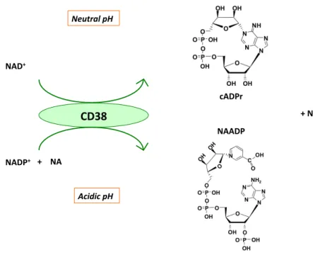
cADP-ribose synthases are a pair of ecto-enzymes also known as the lymphocyte antigens CD38 and CD157, which produce and hydrolyze the Ca2+-mobilizing second-messen-ger cADP-ribose from NAD+[8–10](Figure 3). CD38 cata-lyzes a base exchange between NADP+and Na to form Na adenine dinucleotide phosphate (NaADP)[11], which is also a hydrolytic substrate[12]. All the products of CD38 (cADP-ribose, ADP-ribose and NaADP) have distinctive roles in Ca2+mobilization.
Figure 1. NAD+as a coenzyme for reversible hydride transfer. In a typical NAD+-dependent oxidation, an alcohol is converted to the corresponding aldehyde with the production of NADH plus a proton. In the NADH-dependent direction, an aldehyde is reduced to an alcohol, which regenerates NAD+.
Box 1. History of the NAD+coenzyme
In the early 20th century, Arthur Harden and co-workers reconstituted cell-free glucose fermentation with two fractions, one termed ‘zymase’ that was heat-labile and retained by dialysis, and one termed ‘cozymase’ that was heat-stable and passed through dialysis. Zymase was not a purified enzyme but a protein fraction that contained glycolytic enzymes. The cozymase fraction contained
ATP, Mg2+and the NAD+coenzyme, the structure of which was
determined by Otto Warburg. In glucose fermentation, NAD+
func-tions as the hydride acceptor in the step catalyzed by glyceraldehyde-3-phosphate dehydrogenase, producing NADH and diphosphoglyce-rate. Similarly, NADH functions as the hydride donor for alcohol dehydrogenase, which is required for the reduction of acetaldehyde
to ethanol, regenerating NAD+. Numerous hydride transfer enzymes
or oxidoreductases interconvert either NAD+and NADH or NADP+and
NADPH to reduce or oxidize small-molecule metabolites (seeFigure 1
in the main text).
Review TRENDS in Biochemical Sciences Vol.32 No.1 13
www.sciencedirect.com
+H+,#+2e,#
3
1.0.1 NAD(P)-the coenzyme
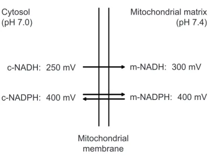
Since the beginning of the last century, seminal discoveries have identified pyridine nucleotides as the major redox carriers in all organisms. Indeed, it is beyond doubt that NAD(P) holds a key position in cellular metabolism and energy production due to its coenzymatic activity. The function of NAD as coenzyme was firstly discovered by Otto Warburg, in 1936. On the basis of his work on alcoholic fermentation, he discovered the capability of NAD, at that time known as “von Euler’s cozymase”, to transfer hydrogen from one molecule to another. We have to credit Warburg also with the discovery of NADP, the “cozymase II”, as another coenzyme with similar properties (2, 3). In the cellular metabolism, the NAD+/NADH couple primarily drives oxidation reactions (catabolic reactions), while the NADP+/NADPH couple drives reductive reactions (anabolic reactions). Fatty acid β-oxidation and biosynthesis are an example of metabolic pathways where NAD+/NADH and NADP+/NADPH are involved as coenzymes, respectively. To understand the reason why the two redox couples are generally implicated in those distinct branches of cellular metabolism, it has to be considered their redox potentials (4). The intrinsic tendency of a chemical species to acquire electrons is expressed by its reductive potential, also known as redox potential, that is measured in volt (V); the more positive the redox potential is, greater the electron affinity of the considered species will be. Both in the cytosol and the mitochondrial matrix, the NADP redox potential is higher than the NAD one
(Figure 2); that means that the ratio NADPH/NADP+ is much greater than
NADH/NAD+ ratio in the mitochondrial matrix. One process maintaining this disequilibrium is the mitochondrial proton motive force-dependent transhydrogenase (5). Moreover, it has long been recognized that the reductive potential of NAD is much higher than that in the cytosol, corresponding to a 40-fold difference in the NAD+/NADH ratio in the two compartments (Figure 2).
Chapter 1
4
Figure 2. Redox potentials of the mitochondrial and cytosolic NAD(H) and NADP(H) systems in the liver. c, cytosolic; m, mitochondrial. (modified from ref. 4).
The redox state of NAD and NADP, their different distribution in distinct cell types and tissues, together with the well-established presence of intracellular binding proteins modulating the free concentration of NAD(P) (4), all these factors contribute in determining the redox state of the cell, that represents itself an important regulatory mechanism. Indeed, the expression of several genes is linked to the redox state of pyridine nucleotides via NAD+/NADH-binding proteins that act as redox sensors (6). For example, transcriptional activity can be reciprocally regulated by direct binding of either NADH or NAD+ to transcriptional co-repressor C-terminal binding protein (7). Pyridine nucleotides have been shown to serve as electron carriers to synthesize adenosine triphosphate (ATP) in mitochondria via the electron transport chain (ETC) and oxidative phosphorylation. The participation of NAD(P) in electron transfer reactions, does not result in a net consumption of the nucleotides. Consequently, except for cell divisions, the
biosynthesis pathway, NAM is thought to be preferred
over NA as the main precursor for NAD
!biosynthesis.
This is illustrated by the fact that mice receiving an ip
injection of NA only have a transient increase in NAD
!concentration accompanied by a high excretion rate of
nicotinuric acid in urine, accounting for 36% of the
ad-ministered NA. In contrast, NAM injection results in a
more stable increase in NAD
!levels, with very limited
nicotinuric acid excretion (25). Whether or not NAM is
directly transported into the cell or is metabolized
extra-cellularly remains unknown. Because both intra- and
ex-tracellular NAMPT have been reported, as well as
circu-lating NMN (36), it seems plausible that both pathways
contribute to NAD
!biosynthesis. In addition, the lack of
NAMPT in some tissues suggests that these either use
other substrates for NAD
!biosynthesis or convert NAM
in the circulation. Recently, an NR transporter (Nrt1) has
been described in yeast (47). Nrt1 belongs to the family of
sodium-coupled transporters and was essential for
opti-mal NAD
!synthesis and for growth of yeast on NR.
NAD
!synthesis from NA and NAM was not affected by
deletion of Nrt1 (47). The human ortholog of Nrt1 is yet
unknown, although some transporters have been
identi-fied that are capable of transporting the NR analogs
ben-zamide riboside and tiazofurin (48). Despite the
signifi-cant advances in the field of NAD
!metabolism, especially
since sirtuins have come to center stage, it remains to be
established how the various pathways interact and which
precursors are preferentially used in vivo. The elucidation
of the transport pathways could advance this field of
re-search and clarify the relevant pathways for NAD
!synthesis.
III. Role of NAD
!in Cellular Redox State
Pioneering work by Warburg and co-workers in the 1930s
has led to the identification of the NAM dinucleotides
NAD(H) and NADP(H) (49, 50). Indeed, NADP
!was
discovered as the coenzyme of the glucose-6-phosphate
dehydrogenase reaction, whereas NAD
!turned out to be
the obligatory cofactor of fermentation. Later studies
re-vealed that NAD and NADP play an indispensable role in
cellular oxidation/reduction reactions, with the NAD
!/
NADH couple primarily driving oxidation reactions and
the NADP
!/NADPH couple driving reductive reactions.
The underlying basis for this remarkable difference
be-tween the NAD
!/NADH- and NADP
!/NADPH-redox
couples has to do with the fact that the redox potentials of
the two redox couples are widely different, with the
NADP(H)-redox couple being much more reduced than
the NAD(H)-redox couple (Fig. 3). The mitochondrial
en-zyme energy-linked transhydrogenase is responsible for
this phenomenon as will be discussed in Section III.B.
The contents of the NAM nucleotides in distinct cell
types and tissues differ markedly, especially if NADP plus
NADPH is concerned. For instance, in rat liver total NAD,
i.e., NAD
!plus NADH, amounts to some 800 nmoles/g
wet weight, whereas total NADP amounts to about 420
nmoles/g wet weight. For skeletal muscle, however, these
values are 590 and 30 nmoles/g wet weight, respectively.
Apart from the differences in absolute levels, there is also
the fact that NAD and NADP are heavily
compartmen-talized, with mitochondria containing high levels of
NAD(H) and NADP(H). This explains why the percentage
of NAD and NADP present in mitochondria, compared
with the total amount of tissue, is higher in heart tissue
than in liver tissue, especially if NAD is concerned (35 vs.
20%; Ref. 51), considering that mitochondria are more
abundant in heart compared with liver.
A. Cellular NAD(H) and NADP(H): binding to proteins,
and the metabolite indicator method
It has long been established that NAD(H) and
NADP(H) are predominantly bound to intracellular
pro-teins and that the free concentrations of NAD
!, NADH,
NADP
!, and NADPH are much lower than the total
con-centrations as determined in protein-free tissue extracts
obtained by acidification (NAD
!and NADP
!) and
alka-linization (NADH and NADPH). The existence of these
binding sites, which bind NADH and NADPH more
av-idly than NAD
!(and NADP
!) complicates the
determi-nation of the true redox state of the NAD(H)- and
NADP(H)-redox couples within any tissue or cell.
To circumvent this problem, the so-called metabolite
indicator method has been conceived (51). This elegant
method is based on the notion that a particular NAD
!/
NADH-ratio may be calculated from the concentrations
of a certain oxidized substrate and reduced substrate,
par-ticipating in a NAD(H)-linked dehydrogenase reaction,
provided that the selected dehydrogenase catalyzes a
near-equilibrium reaction and is localized in one particular
sub-c-NADH: -250 mV c-NADPH: -400 mV m-NADH: -300 mV m-NADPH: -400 mV Cytosol (pH 7.0) Mitochondrial matrix(pH 7.4) Mitochondrial membrane
FIG. 3. Redox potentials of the mitochondrial and cytosolic NAD(H) and NADP(H) systems in the liver. c, Cytosolic; m, mitochondrial.
198 Houtkooper et al. NAD!Metabolism in Health and Disease Endocrine Reviews, April 2010, 31(2):194 –223
5
constant requirement for pyridine nucleotide resynthesis does not arise from their function as coenzymes, but rather from their involvement in signalling reactions (8).
1.0.2 NAD(P)+- the substrate
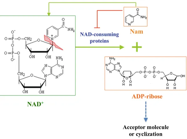
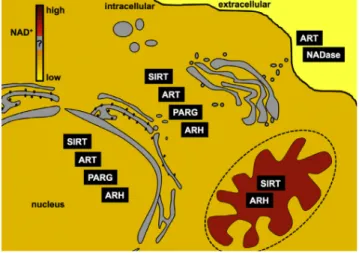
Nowadays, it is well assessed that pyridine nucleotides, besides their vital functions as electron carriers, play important roles as mediators in a plethora of fundamental cellular processes. Since many years, it has been discovered an astonishing range of enzymatic activities where NAD(P)+ is not involved as a coenzyme but rather as a substrate. These NAD(P)+-consuming enzymes get the energy to proceed in the catalysis of their reactions by the cleavage of the N-glycosidic bond between the nicotinamide ring and the ribose of the NMN moiety of NAD(P)+. Indeed, nicotinamide (Nam) and ADP-ribose (ADPr) are the common products of all these reactions; Nam can be recycled entering the NAD+ salvage pathway while the ADPr is transferred to an acceptor molecule or can undergo a cyclization (see below) (Figure 3). NAD(P)+-consuming enzymes drive essentially post-translational protein modification and messenger molecule synthesis reactions that are involved in important functions in metabolism, signalling transduction, cell survival and death as well as in gene expression regulation and aging. It is important to note that all the conversions require the oxidized form of NAD(P)+. The reduced form is not a substrate for these signalling reactions (8). Three protein families that consume NAD(P)+ in signalling reactions have been characterized on a molecular level: ADP-ribosyltransferases (ARTs and PARPs), Sirtuins (SIRTs), and NAD+ glycohydrolases (NADases) (6). NAD+-dependent ADP-ribosylation is an important post-translational reversible protein modification that regulates diverse biological processes including DNA-repair, transcriptional regulation, telomere dynamics, energy metabolism and apoptosis (10, 11).
Chapter 1
6
Figure 3. Consumption of NAD+ driven by NAD(P)+ dependent enzymes. All the reactions
catalysed by NAD+-consuming proteins use NAD(P)+ as a donor of ADPr, while generating nicotinamide (Nam) as a side product. Nicotinamide has the product inhibition effect on the enzymatic activities of most of the NAD+-consuming proteins, probably by competing for the NAD+ binding pocket (modified from ref .9).
ADP-ribosylation enzymes (ARTs) can be distinguished in mono- and poly-ribosyltransferases and catalyse, respectively, the transfer of one or more ADP-ribose moieties from NAD+ onto specific aminoacid side chains in target protein under release of nicotinamide (Figure 4). In mammals, the ART family is the largest and, perhaps, most versatile among the NAD+-converting protein families (12), and that has generated some confusion about a common shared nomenclature (13).
7
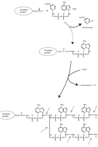
Figure 4. ADP-ribosylation reaction. Mono-ADP-ribosyltransferases catalyse the transfer of one
unit of NAD+; Poly-ADP-ribosyltransferases catalyse an adduct elongation, giving rise to polymers of up to about 200-ADP-ribosyl units. Arrows indicate the site of hydrolysis catalysed by PARG (modified from ref. 11).
Sequence and structure homology searches have identified 22 human genes encoding proteins with ADP-ribosylation activity that have been grouped into three major families: the extracellular membrane-associated ADP-ribosyltransferases (ecto-ARTs), a single member family of NAD+-dependent tRNA 2’-phosphotransferases and the poly-ADP-ribose polymerases (PARPs) (13).
Apart from the covalent modification of acceptor proteins, poly(ADP-ribose) has also been shown to interact noncovalently, yet specifically, with a wide range of proteins [3]. This interaction is mediated through a sequence motif displaying a conserved pat-tern, and it is interesting to note that DNA damage checkpoint proteins such as p53 and p21 possess such poly(ADP–ribose) interaction domains. DNA methyl-transferase-1 (DNMT1) is also able to bind long and branched ADP-ribose polymers in a noncovalent fash-ion, which leads to inhibition of DNMT1 activity [4]. Taken together, poly(ADP-ribose) can also affect the function of proteins that are not direct modification targets.
Enzymes involved in poly(ADP-ribose) metabolism
PARP-1
The prototypic enzyme of the PARP family is poly (ADP-ribose) polymerase-1 (PARP-1; EC 2.4.2.30; Table 1). Its discovery was made four decades ago by Chambon, Weill and Mandel [5] and marked the birth of a most interesting and intriguing field originally occupied by biochemists, but later on also attracting radiobiologists, toxicologists, geneticists, molecular biologists, pharmacologists, cell biologists and experts from other biological disciplines.
Nicotinamide NAD+ PAR PAR n NAD+ n Nicotinamide + n H+ Rib O O P O O P O Rib O- O -N N NH2 N N Acceptor protein CH2 C O O -Rib N N NH2 N N O O O -P O O O -P O Rib Rib N N NH2 N N O O O -P O O O -P O Rib Rib Rib N N NH2 N N O O O -P O O O -P O Rib Rib N N NH2 N N O O O -P O O O -P O Rib Rib N N NH2 N N O O O -P O O O -P O Rib Rib N+ H2NCO N H2NCO + H+ Rib N N NH2 N N O O O -P O O O -P O Rib + CH2 C O O CH2C O O Acceptor protein Acceptor protein
Fig. 1. Poly(ADP-ribose) structure. Poly(ADP-ribose) polymerases (PARPs) cleave the glycosidic bond of NAD+
between nicotinamide and ribose followed by the covalent modification of mainly glu-tamate residues of acceptor proteins with an ADP-ribosyl unit. PARPs also catalyse an adduct elongation, giving rise to linear polymers with chain lengths of up to about 200 ADP-ribosyl units, characterized by their unique ribose (1¢¢fi2¢) ribose phos-phate-phosphate backbone. At least some of the PARP family members also catalyse a branching reaction by creating ribose (1¢¢¢fi2¢¢) ribose linkages. The sites of hydrolysis catalysed by poly(ADP-ribose) glycohydrolase (PARG), the major poly(ADP-ribose)-degrading enzyme, are indicated by arrows.
A. Bu¨rkle Poly(ADP-ribosyl)ation
Chapter 1
8
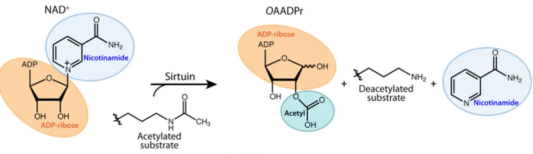
Surprisingly, all identified ecto-ARTs are mono-ADP-ribosyltransferases (2) and their activity in the extracellular compartment provides a complex regulatory mechanism for cell communication (13). Mono-ADP-ribosylation (MARylation) was originally identified as the catalityc activity of several bacterial toxins such as diphtheria, cholera and pertussis toxins, and its physiological roles are related to immune response and inflammation, transcription regulation, cell adhesion, signal and energy metabolism (14). Many functions of Poly-ADP-ribosylation (PARylation) may be explained considering the multiple roles played by PARP-1, the most studied human poly-ADP-ribosyltransferase, now considered a moonlighting protein that serves the cells both in peace (resting condition) and in war (DNA damage) (15). After the discovery of PARP-1 in the early 1960s, many other enzymes with poly ADP-ribosyltransferase activity had been discovered, highlighting a huge number of conditions inducing ADP-ribose polymer (PAR) formation (11). Both mono- and poly- ADP-ribosylation are dynamic processes because of the presence of other enzymes, ADP-ribosyl hydrolases (ARHs) and Poly-ADP-ribose glycohydrolases (PARGs), which remove mono-ADP ribose from the target and hydrolyse the O-glycosidic ribose-ribose 1-2’ bonds within PAR, respectively (16). Mono-ADP-ribosyltransferase activity was also detected in some sirtuins such as human mitochondrial SIRT4 and nuclear SIRT6 (17). Sirtuins are a family of evolutionally conserved class III histone deacylases that consume one molecule of NAD+ during each deacylation cicle (18). Even though sirtuins have been recognised mostly as deacetylases, recent studies have shown that Sirt5 is endowed of demalonylase and desuccinylase activities (19). Chemically, deacylation primarily occurs on N-ε-lysine residues and tipically regulates protein function (17). For example, sirtuin catalysed NAD+-consuming deacetylation takes place on an acetyl-lysine and results in the production of deacetylated lysine, nicotinamide and 2’-O-acetyl-ADP-ribose (OAADPr) (Figure
Chapter 1
9
Figure 5. Schematic representation of a deacetylation reaction catalysed by sirtuins (modified
from ref. 20).
In particular, OAADPr undergoes a non-enzymatic transesterification reaction and exists in solution as a mixture of 2’ and 3’ OAADPr (20). Moreover, OAADPr has been implicated as a signalling molecule and it can also be the substrate of macrodomain family enzymes that may function in the regulation of OAADPr cellular levels, hydrolysing the 2 or 3-ester bond to generate ADPr and acetate as products (16, 21). The mammalian family of sirtuins consists of seven members, Sirt1-7, and the number of their reported histone and non-histone protein targets is continually increasing. Therefore, it is not surprising that sirtuins have important roles in many biological processes such as regulation of chromatin structure and gene expression, oxidative stress reduction, glucose homeostasis and cell survival (4). The versatile functions of sirtuins are supported by their diverse cellular location allowing cells to sense changes in energy levels in the nucleus, cytoplasm, and mitochondrion (18), but it is not clear whether the link between intracellular NAD+ levels and sirtuin activity is correlative or causal (4). Alongside its consumption by sirtuins and ARTs, NAD(P)+ is also a substrate for NAD glycohydrolases enzymes, responsible for the generation of second messengers associated with calcium signalling, a universal signal trasduction mechanism involved in a wide range of physiological functions. The two major Ca2+
mobilising NAD(P)+ derivatives are the cyclic ADP ribose (cADPr) and the Nicotinic acid Adenine dinucleotide phosphate (NAADP) and, in mammals, their
mechanism (15). Several biochemical studies support the for-mation of the alkylamidate intermediate. These include
nico-tinamide base-exchange reactions (16) and18O labeling studies
that provide evidence for the direct transfer of the acetyl oxygen to the 1!-hydroxyl of OAADPr (17, 18). Utilization of acetylly-sine analogs further demonstrated the existence of the alkyl-amidate intermediate. Thioacetyllysine- and acetylazalysine-containing peptides form stalled alkylamidate intermediates when used as sirtuin substrates (19, 20). Upon formation of the
alkylamidate intermediate, the 2!-hydroxyl group of the NAD"
ribose is activated by a conserved active-site histidine (
supple-mental Fig. 1). The activated hydroxyl attacks the O-alkylami-date carbon to afford a 1!,2!-cyclic intermediate (20). Recently, the bicyclic intermediate was trapped and structurally resolved by incubating co-crystals of SIRT5 and an H3K9
thiosuccinyl-peptide with NAD", providing direct evidence
for the proposed catalytic mechanism (21). A base-activated water molecule then attacks the cyclic intermediate,
afford-ing deacetylated lysine and OAADPr (supplemental Fig. 1)
(10, 17).
Although deacylation is thought to be the primary function of human sirtuins, yeast Sir2 was first implicated as having the
capability to transfer ADP-ribose from NAD"to nucleophilic
amino acids on protein substrates (22). SIRT4 and SIRT6 have also been reported to catalyze ADP-ribosyl transfer to gluta-mate dehydrogenase and poly(ADP-ribose) polymerase 1, respectively (7, 23). This activity is not robust and has been subject to debate. Detailed kinetic characterization of the ADP-ribosyltransferase activity of a yeast and bacterial sirtuin indi-cated that ADP-ribosylation may be a low efficiency side reac-tion (#0.1% of the deacetylareac-tion reacreac-tion) of sirtuins due to the susceptibility of active site-bound ADP-ribose to nucleophilic attack (24). Understanding the mechanistic details of sirtuin-catalyzed reactions is an important step toward a complete understanding of sirtuin function and in the development of chemical tools to probe their biology.
Substrate Recognition and Acyl Group Specificity
A number of proteomics studies have greatly enhanced our understanding of lysine acetylation as a global
post-transla-FIGURE 1. Representative structure of a human sirtuin (Protein Data Bank code 3GLR) bound to acetylated peptide and NAD!. Key locations for sirtuin
modulation are highlighted. Positive regulators of sirtuin activity are indicated in green, negative regulators are indicated in red, and regulators that can activate or inhibit depending on the sirtuin are in yellow. Proposed activators include NAD"synthesis, Sirtris compound 11 (90) and other reported activators,
and phosphorylation of SIRT1. Inhibitors include cysteine nitrosylation, DBC1 binding SIRT1, pseudopeptidic inhibitors (95) and other small molecules, nicotinamide, and sumoylation of SIRT1.
FIGURE 2. Substrates and products of the sirtuin-catalyzed reaction and potential fate of the product OAADPr. The unique use of NAD"as a co-substrate
distinguishes sirtuins from other deacetylase classes and provides a direct link to energy metabolism.
42420 JOURNAL OF BIOLOGICAL CHEMISTRY VOLUME 276 • NUMBER 51 • DECEMBER 14, 2012
by guest on November 2, 2014
http://www.jbc.org/