Journal of Intercultural Ethnopharmacology
DOI: 10.5455/jice.20170327073801 www.jicep.com
INTRODUCTION
The genus Stachys comprises more than 450 species mainly distributed in warm-temperate regions of the Mediterranean basin and South-Western Asia. It represents one of the largest genera of Lamiaceae family [1], and the majority of the species belonging to this genus have been largely used in the traditional medicine. Stachys spp. are currently indicated as “Mountain tea” in several regions reflecting their principal use in infusions and decoctions. The genus Stachys is represented by annual or perennial herbs known for their interesting biological activities, i.e., anti-inflammatory [2], antispasmodic, sedative, and diuretic [3-5]. Several phytochemical components, which may be responsible of the biological activities, have been isolated from Stachys species, i.e., iridoids [6,7], phenylethanoid glycosides, flavonoids [8,9], acidic metabolites, polysaccharides, and terpenoids [10].
In Italy, the genus Stachys encompasses about 30 species and subspecies among which five species are considered endemic. Stachys alopecuros subsp. divulsa is one of these. This plant is characteristic of the mountain habitat of central Italy, being distributed only in a few regions (Umbria, Marche, Abruzzo, Lazio, and Molise) [11], on stony mountain pastures, scrubs, and screes, and preferring a calcareous, and dry soil up to 2000 m a.s.l. S. alopecuros subsp. divulsa is a perennial plant with small, hirsute, and subcylindrical ascending stem. The opposite leaves are petiolate and densely hairy on the edge and along the veins. The flowers are gathered in small verticillasters held in a dense spike, yellow-white in color and blooming from June to August-September. The fruits, which are brown colored, are constituted by 4 ovate nuculae [12]. A previous study on this subspecies revealed the presence of phenypropanoid, saponin, and iridoid glycosides [6] which
Reassessment of the polar fraction of
Stachys alopecuros
(L.) Benth. subsp.
divulsa
(Ten.) Grande (Lamiaceae)
from the Monti Sibillini National
Park: A potential source of bioactive
compounds
Alessandro Venditti
1, Claudio Frezza
2, Lorenzo M. Lorenzetti
1,
Filippo Maggi
3, Mauro Serafini
2, Armandodoriano Bianco
1ABSTRACT
Background: The phytochemical analysis of Stachys alopecuros subsp. divulsa, an endemic Italian species, has
been recently reported and has showed the presence of 8-O-acetylharpagide (2), harpagide (3), allobetonicoside (4), and 4′-O-galactopyranosyl-teuhircoside (5). In this paper, an in deep study of its glycosidic fraction with the aim to widen the knowledge on its secondary metabolites content is reported. Materials and Methods: Chromatographic techniques were used for the isolation of constituents while spectroscopic and spectrometric techniques were applied for the structures elucidation. Results: Besides the known constituents, all of them reconfirmed, ajugoside (1), reptoside (6) and 6-O-acetyl-ajugol (7) were also identified among the iridoids while the phenolic components resulted to be chlorogenic acid (8), β-arbutin (9), verbascoside (10), and stachysoside A (11), instead.
Conclusion: The iridoid pattern of S. alopecuros subsp. divulsa has been expanded with the identification of not
previously reported compounds as well as for the phenolic fraction. Except for the reconfirmed verbascoside (10), the other phenolic compounds were recognized for the first time in the studied species. The complete NMR assignment of compound (1) by means of bidimensional techniques is reported, and both the chemotaxonomic and pharmacological relevance of the isolated compounds is largely discussed.
KEY WORDS: Chemotaxonomy, chlorogenic acid, iridoid, NMR, phenylethanoid glycosides, β-arbutin
O
riginal Research
1Department of
Chemistry, Sapienza University of Rome, Rome, Italy, 2Department
of Environmental Biology, Sapienza University of Rome, Rome, Italy,
3School of Pharmacy,
Università di Camerino, Camerino, Italy
Address for correspondence: Alessandro Venditti, Dipartimento di Chimica, Sapienza Università di Roma, Roma, Italy. E-mail: alessandro.venditti@ gmail.com Received: November 17, 2016 Accepted: February 02, 2017 Published: April 12, 2017
are considered taxonomic markers for the genus and the family. In this paper, we reported a reassessment of the polar fraction constituents of this plant employing a patented method allowing the isolation of secondary metabolites of glycosidic nature.
MATERIALS AND METHODS
Plant MaterialsThe fresh plant materials (aerial parts) were collected at the flowering stage from a spontaneous population growing at the altitude of 1520 m a.s.l. in the territory of Pizzo Tre Vescovi, Marche, Italy (GPS coordinates: N 42°58′07″, and E 13°14′10″). The botanical recognition was performed by one of us (F.M.) using available literature [12], and a representative specimen has been deposited in the Herbarium Universitatis Camerinensis (CAME, included in the online edition of Index Herbariorum c/o School of Biosciences and Veterinary Medicine, University of Camerino, Italy) under the codex CAME 24825. A sample of the studied species is also stored in our laboratory (code number SAD-30062012) for any further reference.
Instruments
NMR spectra were recorded on a Varian (now Agilent Technologies) Mercury 300 MHz instrument and/or on a Bruker Avance III 400 MHz instrument using D2O or CD3OD
as deuterated solvents: The chemical shifts were expressed in ppm using the HDO signal to set the 1H-spectra acquired in
D2O, while the internal solvent signal (m5) at 3.31 ppm was set as reference for the spectra in CD3OD.
MS spectra were performed on a Q-TOF MICRO spectrometer (Micromass, now Waters, Manchester, UK) equipped with an ESI source that operated in the negative and/or positive ion mode. The flow rate of sample infusion was 10 µL/min with 100 acquisitions per spectrum. Data were analyzed using the MassLynx software developed by Waters.
Solvents having RPE purity grade were all purchased from Sigma-Aldrich or Carlo Erba Reagenti, silica gel 60 (70-230 mesh ASTM) was purchased from Fluka.
Bidimensional NMR Experiments
Bidimensional spectra were performed on a Bruker Avance III 400 MHz instrument, operating at 9.4 T at 298 °K. Heteronuclear single quantum correlation (HSQC) experiments were acquired with a spectral width of 15 and 250 ppm for the proton and carbon, respectively, an average
1J
C-H of 145 Hz, a recycle delay of 2 s and a data matrix of 4
K × 256 points. Heteronuclear multiple bond correlation (HMBC) experiments were acquired with a spectral width of 15 and 250 ppm for the proton and carbon, respectively, a long-range coupling constant of nJ
C-H of 8 Hz, a recycle delay
of 2 s and a data matrix of 4 K × 256 points.
Extraction and Isolation of Polar Compounds
A portion of 390.0 g of hair-dried plant materials was extracted with a mixture of ethanol 96% and distilled water in ratio 8:2 v/v (1.4 L). The whole was left in maceration for 5 days so that the metabolites could come into solution. The procedure was repeated three times to achieve an exhaustive extraction. The solutions, having a dark green coloration, were gathered together in a same flask and then filtered. After filtration, the organic solvent was eliminated under reduced pressure at a temperature below 45°C until a water suspension was obtained. Throughout this last part, pH was checked on normal litmus paper, and this was about 8. This step is necessary to verify that pH is not too acid, meaning not under the value of 5.5, because an extreme acidity at this step may cause secondary reactions with the formation of several artifacts coming from the hydrolysis of glycosides and esters present in the extract. The water suspension was then frozen to −20°C and later lyophilized at the same temperature to preserve temperature-sensitive compounds possibly present. The final dried crude extract obtained from this methodology weighed 86.7 g and was dark green colored.
Adsorption Chromatography
The crude extract (86.7 g) was subjected to an active charcoal treatment. It was adsorbed on a stationary phase consisting in a mixture of charcoal/celite/polyamide (10:1:1) (60.0 g) until no reaction to vanillin/HCl spray reagent occured according to a method reported in the patent by Ballero et al. [13]. The resulting suspension was then stratified on a Gooch funnel and eluted first with H2O to eliminate the mono- and disaccharides. These usually coelute with glycosidic constituents in silica gel chromatography and may interfere during the structure identification of the metabolites of interest. The desorption chromatography was conducted in steps using EtOH/H2O mixtures gradually increasing in EtOH percentages (30, 60, and 95%). In this manner, three fractions at different polarity were collected, and after freeze drying 23.9, 4.70, and 0.82 g of solids were recovered, from the 30, 60 and 96% ethanolic elutions, respectively.
Silica Gel Column Chromatography
After a preliminary screening on thin layer chromatographys and paper chromatography, the separation step on silica gel column was first conducted on the 60% eluate because this showed the whole iridoid components and in comparable amount among them in respect to the other fractions. The 30% fraction, containing the more polar compounds, was partitioned in a second step. The 95% fraction did not contain iridoid glucosides and was not even considered.
The first chromatographic separation was performed on an aliquot of the 60% ethanolic elution for the weight of 4.0 g using 120.0 g of silica gel (ratio 1:30). The eluting system was a mixture of n-BuOH/H2O (82:18 v/v). From this chromatographic
separation (Scheme 1) 11 compounds were isolated and identified by comparison with data reported in literature and/or by comparison with pure compounds available in our laboratory:
Verbascoside (10, 12.4 mg) [14] from the assembly of fractions 8-15; stachysoside A (11, 11.2 mg) [8,15] in mixture with verbascoside (1:2) in the assembly of fractions 17-24; β-arbutin (9, 3.2 mg) [16] from the assembly of fractions 30-32; reptoside (6) [17] and 6-O-acetyl-ajugol (7) [18] in mixture in ratio 1:1.5 (25.0 mg) from the assembly of fractions 43-45; ajugoside (1, 113.7 mg) [17,19] as pure compound from the assembly of fractions 53-54; 8-O-acetyl-harpagide (2, 1.0 g) [20] from the assembly of fractions 70-88; chlorogenic acid (8) [21] in mixture with 8-O-acetyl-harpagide (2) in ratio 1:2 (109.4 mg) from the assembly of fractions 95-112; harpagide (3) [22] and chlorogenic acid (8) in mixture 3:1 (48.3 mg) from the assembly of fractions 118-122; allobetonicoside (4) and chlorogenic acid (8) in mixture 10:1 (29.1 mg) from the assembly of fractions 133-135; allobetonicoside (4, 100.7 mg) [23] as a pure compound from the assembly of fractions 144-163; 4′-O-galactopyranosyl-teuhircoside (5) [6] in mixture with allobetonicoside (4) in ratio 3:1 (24.7 mg) from the assembly of fractions 166-169.
A second chromatographic column was, instead, conducted on an aliquot of the 30% desorption solids (3.5 g). Similar compounds were isolated from this chromatographic step. In particular, harpagide (3), 8-O-acetylharpagide (2) as pure compounds, allobetonicoside (4) in mixture with 4′-O-galactopyranosyl-teuhircoside (5), and ajugoside (1) occurring in traces (relative quantities not estimated). On the other hand, phenolic compounds as well as reptoside (6) and 6-O-acetyl-ajugol (7), were not found.
Spectral Data of the Isolated Compounds
Ajugoside (1): NMR data see Table 1; ESI-MS: m/z 412.80 [M+Na]+. 8-O-acetylharpagide (2): 1H NMR (300 MHz, CD 3OD) δ: 6.40 (1H, d, J = 6.4 Hz, H-3), 6.08 (1H, br s, H-1), 4.92 (1H, d, J = 6.5 Hz, H-4), 4.60 (1H, d, J = 7.8 Hz, H-1′), 3.90 (1H, d, J = 11.8 Hz, H-6′a), 3.71 (2H, m, H-6′b; H-3′), 3.44-3.29 (2H, m, H-4′; H-5′), 3.22 (1H, t like, J = 8.4 Hz, H-2′), 2.86 (1H, br s, H-9), 2.18 (1H, d, J = 15.1 Hz, H-7a), 2.02 (3H, s, CH3CO), 1.94 (1H, dd, J = 15.2, 4.4 Hz, H-7b), and 1.45 (3H, s, H-10). 13C NMR (APT) (75 MHz, CD 3OD) δ: 173.3 (CH3CO), 143.8 (C-3), 106.8 (C-4), 99.8 (C-1′), 94.5 (C-1), 88.6 (C-8), 78.0 (C-3′), 77.6 (C-5′), 77.4 (C-6), 74.4 (C-2′), 73.3 (C-5), 71.6 (C-4′), 62.8 (C-6′), 55.4 (C-9), 46.0 (C-7), 22.5 (CH3CO), and 22.2 (C-10).
ESI-MS: m/z 429.04 [M+Na]+; m/z 835.19 [2M+Na]+.
Harpagide (3): 1H NMR (300 MHz, CD 3OD) δ: 6.32 (1H, d, J = 6.3 Hz, H-3), 5.74 (1H, br s, H-1), 4.95 (1H, d, J = 6.3 Hz, H-4), 4.58 (1H, d, J = 7.9 Hz, H-1′), 3.90 (1H, d, J = 12.2 Hz, H-6′a), 3.75-3.60 (2H, m, H-6′b; H-3′), 3.44-3.33 (2H, m, H-4′; H-5′), 3.26-3.16 (1H, dd, J = 8.8, 8.2 Hz, H-2′), 2.56 (1H, br s, H-9), 1.90 (1H, dd, J = 13.8, 4.6 Hz, H-7a), 1.80 (1H, dd, J = 13.8, 3.6 Hz, H-7b), 1.25 (3H, s, H-10). ESI-MS: m/z 387.14 [M+Na]+. Allobetonicoside (4): 1H NMR (300 MHz, D 2O) δ: 6.43 (1H, d, J = 6.3 Hz, H-3), 6.11 (1H, br s, H-7), 5.94 (1H br s, H-1), 5.28 (1H, d, J = 8.3 Hz, H-1′′), 5.01 (1H, d, J = 6.3 Hz, H-4), 4.75 (1H, d, J = 7.8 Hz, H-1′), 4.19 (1H, t, J = 2.6 Hz, H-9), 3.93 (2H, m, H-6′a; H-6′′a), 3.83 (2H, m, H-6′b; H-6′′b), 3.70-3.33 (8H, m, H-3′; H-3′′; H-4′; H-4′′; H5′; H-5′′; H-2′; H-2′′), and 2.28 (3H, s, H-10). 13C NMR (75 MHz, D 2O) δ: 204.86 (C-6), 178.50 (C-8), 144.09 (C-3), 127.77 (C-7), 102.69 (C-4), 98.89 (C-1′), 96.58 (C-1′′), 92.25 (C1), 77.45 (C-5), 76.18 (C-5′), 75.60 (C-3′), 73.57 (C-2′), 72.71 (C-4′′), 71.20 (C-2′′), 69.85 (C-3′′), 69.42 (C-4′), 66.62 (C-4′′), 61.03 (C-6′′), 60.70 (C-6′), 54.85 (C-9), 17.62 (C-10). ESI-MS: m/z 528.73 [M+Na]+; m/z 544.69 [M+K]+. 4′-O-galactopyranosyl-teuhircoside (5): 1H NMR (300 MHz, D2O) δ: 6.43 (1H, d, J = 6.4 Hz, H-3), 6.13 (1H, br s, H-7), 5.94 (1H, br s, H-1), 4.98 (1H, d, J = 6.4 Hz, H-4), 4.80 (1H, partially overlapped with solvent signal, H-1″), 4.73 (1H, d, J = 8.1 Hz, H-1′), 2.29 (3H, s, H-10).
ESI-MS: m/z 529.12 [M+Na]+.
Reptoside (6): 1H NMR (300 MHz, D
2O) δ: 6.40 (1H, d,
J = 6.5 Hz, H-3), 5.90 (1H, br s, H-1), 5.02 (1H, dd, J = 6.4, 1.3 Hz, H-4), 4.75 (1H, d, partially obscured by the solvent signal, H-1′), 3.92 (1H, dd, J = 12.4, 1.9 Hz, H-6′a), 3.71 (1H, br d, J = 12.4 Hz, H-6′b), 3.26 (1H, dd, J = 8.3, 8.2 Hz, H-2′), 2.65 (1H, br s, H-9), 2.04 (3H, s, CH3CO), 1.93 (2H, m, H-6a;
H-7a), 1.71 (2H, m, H-6b; H-7b), 1.43 (3H, s, H-10).
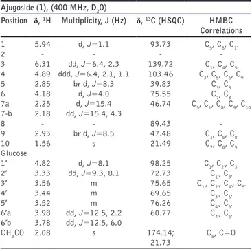
Table 1: Complete NMR assignment of ajugoside (1) in D2O
Ajugoside (1), (400 MHz, D2O) Position δ, 1H Multiplicity, J (Hz) δ, 13C (HSQC) HMBC Correlations 1 5.94 d, J=1.1 93.73 C5, C8, C1′ 2 ‑ ‑ ‑ ‑ 3 6.31 dd, J=6.4, 2.3 139.72 C1, C4, C5 4 4.89 ddd, J=6.4, 2.1, 1.1 103.46 C3, C5, C6, C9 5 2.85 br d, J=8.3 39.83 C3, C8 6 4.18 d, J=4.0 75.55 C7, C8 7a 2.25 d, J=15.4 46.74 C5, C6, C8, C9, C10 7‑b 2.18 dd, J=15.4, 4.3 8 ‑ ‑ 89.43 ‑ 9 2.93 br d, J=8.5 47.48 C1, C5, C8 10 1.56 s 21.49 C7, C8, C9 Glucose 1′ 4.82 d, J=8.1 98.25 C1, C2′, C3′ 2′ 3.33 dd, J=9.3, 8.1 72.73 C1′, C3′ 3′ 3.56 m 75.65 C1′, C2′, C4′, C5′ 4′ 3.44 m 69.65 C3′, C6′ 5′ 3.52 m 76.26 C4′, C6′ 6′a 3.98 dd, J=12.5, 2.2 60.77 C4′, C5′ 6′b 3.78 dd, J=12.5, 6.0 CH3CO 2.08 s 174.14; 21.73 C8, C=O HSQC: Heteronuclear single quantum correlation, HMBC: Heteronuclear multiple bond correlation
ESI-MS: m/z 413.04 [M+Na]+. 6-O-acetyl-ajugol (7): 1H NMR (300 MHz, D 2O) δ: 6.34 (1H, dd, J = 6.4, 1.8 Hz, H-3), 5.50 (1H, d, J = 2.8 Hz, H-1), 4.89 (1H, dd, J = 6.4, 2.8 Hz, H-4), 4.81 (1H, obscured by the solvent signal, H-6), 4.70 (1H, d, J = 8.0 Hz, H-1′), 3.90 (1H, dd, J = 12.1, 1.6 Hz, H-6′), 3.68 (1H, br d, J = 12.2 Hz, H-6′b), 3.24 (1H, m, H-2′), 2.79 (1H, br t, J = 6.5 Hz, H-5), 2.34 (1H, dd, J = 7.9, 2.7 Hz, H-9), 2.12 (1H, br dd, J = 14.0, 6.5 Hz, H-7a), 2.08 (3H, s, CH3CO), 1.93 (1H, dd, J = 13.8, 8.6 Hz, H-7b), 1.38 (3H, s, H-10). ESI-MS: m/z 413.04 [M+Na]+. Chlorogenic acid (8): 1H NMR (300 MHz, CD 3OD) δ: 7.56 (1H, d, J = 15.9 Hz, H-β′), 7.05 (1H, d, J = 1.9 Hz, H-2′), 6.96 (1H, dd, J = 8.2, 1.9 Hz, H-6′), 6.78 (1H, d, J = 8.2 Hz, H-5′), 6.26 (1H, d, J = 15.9 Hz, H-α′), 5.33 (1H, td, J = 9.1, 4.5 Hz, H-3), 4.17 (1H, dt, J = 6.2, 3.2 Hz, H-4), 3.73 (dd, J = 8.5, 3.1 Hz, H-5), 2.27-2.13 (2H, m, H-2a; H-6a), 2.13-1.98 (2H, m, H-2b;H-6b). ESI-MS: m/z 377.30 [M+Na]+. β-arbutin (9): 1H NMR (300 MHz, CD 3OD) δ: 7.07 (2H, d, J = 8.6 Hz, H-3, H-5), 6.69 (2H, d, J = 8.6 Hz, H-2, H-6), 4.95 (1H, partially obscured by HDO signal, H-1′), 3.96-3.79 (m, overlapped glucose protons), 3.77-3.54 (m, overlapped glucose protons), 3.18 (1H, br t, J = 7.9 Hz, H-2′). ESI-MS: m/z 311.22 [M+K]+. Verbascoside (10): 1H NMR (300 MHz, CD 3OD) δ: 7.59 (1H, d, J = 15.9 Hz, H-β′′), 7.05 (1H, d, J = 1.4 Hz, H-2′′), 6.96 (1H, dd, J = 8.2, 1.4 Hz, H-6′′), 6.78 (1H, d, J = 8.2 Hz, H-5′′), 6.69 (1H, br s, H-2′), 6.68 (1H, d, J = 8.0 Hz, H-5′), 6.56 (1H, dd, J = 8.0, 1.6 Hz, H-6′), 6.28 (1H, d, J = 15.9 Hz, H-α′′), 5.19 (1H, d, J = 0.9 Hz, H-1′′′), 4.38 (1H, d, J = 7.8 Hz, H-1), 2.79 (2H, br t, J = 7.3 Hz, H-β′), 1.09 (3H, d, J = 6.2 Hz, H-6′′′). ESI-MS: m/z 623.19 [M-H]-; m/z 647.12 [M+Na]+. Stachysoside A (11): 1H NMR (300 MHz, CD 3OD) δ: 7.60 (1H, d, J = 15.6 Hz, H-β′′[caff.]), 7.06 (1H, br s, H-2′′), 6.98 (1H, br d, J = 8.5 Hz, H-6′′), 6.78 (1H, d, J = 8.5 Hz, H-5′′), 6.70 (1H, br s, H-2′), 6.68 (1H, d, J = 8.0 Hz, H-5′), 6.57 (1H, br d, J = 8.1 Hz, H-6′), 6.28 (1H, d, J = 15.6 Hz, H-α′′), 5.42 (1H, br s, H-1′′′), 4.43 (1H, d, J = 7.9 Hz, H-1), 4.31 (1H, d, J = 7.2 Hz, H-1′′′′), 2.81 (2H, d, J = 7.1 Hz, H-β′), 1.16 (3H, d, J = 6.0 Hz, H-6′′′). ESI-MS: m/z 755.20 [M-H]-; m/z 779.16 [M+Na]+.
RESULTS
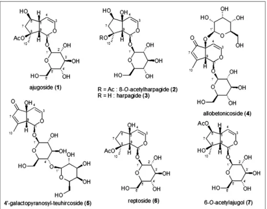
The reassessment of the glycosidic fraction of S. alopecuros subsp. divulsa allowed the isolation of several compounds. The majority of them were iridoids, followed by a phenolic glucoside, two phenylethanoid glycosides, and a caffeoylquinic acid. They were identified as ajugoside (1), 8-O-acetylharpagide (2), harpagide (3), allobetonicoside (4), 4′-O-galactopyranosyl-teuhircoside (5), reptoside (6), 6-O-acetyl-ajugol (7) [Figure 1] for the iridoid glycosides constituents, and as chlorogenic
acid (8), β-arbutin (9), verbascoside (10), and stachysoside A (11) [(2-(3,4-dihydroxyphenyl)ethyl-O-α-L-arabinopyranosyl- (1→2)-O-6-deoxy-β-L-mannopyranosyl-(1→3)-4-O- [(2E)-3-(3,4-dihydroxyphenyl)-1-oxo-2-propen-1-yl]-β-D-glucopyranoside] [Figure 2] for the phenolic compounds. The presence of previously identified iridoid glycosides (2-5) has been confirmed, together with ajugoside (1), reptoside (6), and 6-O-acetyl-ajugol (7) which resulted to be new iridoid components for this species. Among the phenolic constituents, chlorogenic acid (8), β-arbutin (9), and stachysoside A (11) were isolated for the first time from the studied species [Figure 1].
DISCUSSION
Chemosystematic Implications of Isolated Compounds
The identified iridoids have all chemosystematic relevance. In particular, 2 and 3 are considered family markers, 4 is characteristic of the section Betonica, and 5 has never been reported in other
Figure 2: Phenolics from Alopecurus alopecuros subsp. divulsa
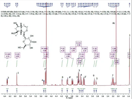
Figure 3: 1H-NMR spectrum of ajugoside (1)
Stachys species so that it may be regarded as a chemotaxonomic marker at a subspecific level. Reptoside (6) resulted to be a rare metabolite in the Stachys genus because its occurrence has been reported only in a few species, namely Betonica officinalis (syn. of Stachys officinalis) [23] and S. macrantha [24]. 6-O-acetyl-ajugol (7), is also a rare metabolite in the Lamiaceae family since it has been found only in Leonurus persicus so far [18]. It is also worth of mention the presence of ajugoside (1) and reptoside (6) which are iridoids widely distributed in the species of the Ajugoideae subfamily. In fact, 1 was mainly reported in the Ajuga genus (e.g. Ajuga tenorei and Ajuga chamaepitys) [25,26], although its presence was also signaled in a few Lamioideae species as Melittis melissophyllum subsp. melissophyllum [19] and species of the genera Stachys and Sideritis. The occurrence of 1 in the Stachys genus is limited to a few species such as B. officinalis (syn. of S. officinalis (L.) Trevis.) [2], S. macrantha [24], S. alpina [2], Stachys germanica [2,27] and Stachys recta [2], and it has been identified also in the taxonomically related Sideritis genus like for instance in Sideritis romana [28], and Sideritis perfoliata subsp. perfoliata [29].
Considering their occurrence, compounds (1) and (6) may represent chemosystematic traits of proximity between Lamioideae and Ajugoideae subfamilies. The identification of 1 has been carried out by extensive NMR analysis on both mono - [Figures 3 and 4] and bidimensional [Figures 5 and 6] experiments since the assignment of resonances was not immediate for several signals, in particular for protons in 5, 6 and 9 positions on the iridoid skeleton as well as for protons in 3′ and 5′ positions of the glucose moiety [Figures 3 and 4].
Figure 4: 13C-NMR spectrum of ajugoside (1)
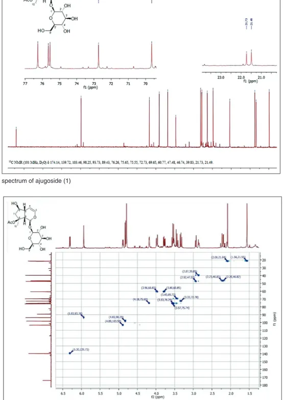
Figure 5: Heteronuclear single quantum correlation spectrum of ajugoside (1)
In particular, the resonance of H-6 was readily visible in the proton spectrum at 4.18 ppm, but the corresponding carbon signal was very near to a glucose carbon resonance and has been assigned by the direct HSQC correlation [Figure 5] with the signal resonating at 75.55 ppm. On the contrary, the assignment of protons and carbons in the position 5 and 9 was
not immediate, and several long range correlations resulted to be crucial for discriminating the resonances of these nuclei. The signals relative to the positions 5 and 9 present resonances in the proton spectrum at 2.93 and 2.85 ppm, both as broad signals, and the study of the coupling constants gave no help for the structure elucidation. In the HSQC spectrum, a direct
correlation between the proton signal at 2.93 ppm and the carbon resonating at 47.48 ppm was recorded as well as the correlation between the proton at 2.85 and the carbon at 39.83 ppm. This permitted to assign the respective carbons signals. In the HMBC experiments, [Figure 6] the only diagnostic correlation which permitted to differentiate the positions 5 and 9 was a long range correlation between H-3 (6.31 ppm) and the carbon resonating at 39.83 ppm, which could be assigned to C-5 of the iridoid skeleton, since almost all the proton signals correlated with both the carbons at 39.83 and 47.48 ppm. In fact, H-3 is relatively far from C-9, and this kind of signal could not be visualized with the coupling constant of 8 Hz set in the experiment. Using the HMBC correlations, it was also possible to differentiate the positions 3′ and 5′ on the glucopyranose unit. In particular, the resonances of the positions 1′, 2′, 4′, and 6′ (98.25, 72.73, 69.65, and 60.77, respectively) were readily assignable to the saccharidic moiety, while the signals relative to 3′ and 5′ might be differentiated on the basis of their correlation with near nuclei [Figures 5 and 6].
More specifically, from HMBC experiments the following long-range correlations were recognized: Between the proton signal at 3.33 ppm (H-2′) and the carbon at 75.65 (C-3′); the proton at 3.34 ppm (H-4′) with carbons at 76.26 (C-5′) and 75.65 (C-3′); the protons at 3.98-3.78 (H-6′) with carbon resonating at 76.26 (C-5′). The latter correlation is particularly crucial for the assignation of the C-5′ position because H-6′ cannot correlate with C-3′. The complete assignments of resonances are reported in Table 1. Chlorogenic acid (8) has been isolated for the first time from S. alopecuros subsp divulsa, although it has been already found in several Stachys species as S. tymphaea [8], S. lanata [30],
S. officinalis [31], S. recta [32], S. byzantina, S. iberica subsp. iberica var. densipilosa [33], and S. glutinosa [34]. In most of these species, the chlorogenic acid (8) resulted to be the main polyphenolic component occurring in the polar fraction just like in this case [Figure 2].
Figure 6: Heteronuclear multiple bond correlation spectrum of ajugoside (1)
Scheme 1: Chromatographic steps on 60% ethonolic fraction and
Furthermore, β-arbutin (9) was a metabolite identified for the first time in this species. It proved to be a quite rare compound in the Stachys genus since its presence has been signaled only in S. germanica subsp. salviifolia [7]. The presence of verbascoside (10), a quite common phenyletanoid glycoside (PhGs) in Lamiaceae, has been confirmed, while stachysoside A (11), also known with the name lavandulifolioside, resulted to be as a new constituent of S. alopecuros subsp. divulsa. PhGs are compounds with a chemosystematic relevance in Asterids since their cooccurrence with iridoid glycosides, as in the studied case, has been largely confirmed [35]. The presence of 11 was noteworthy since it is not an ubiquitous compound, especially in the Stachys genus where it has been recognized only in a few species such as S. lavandulifolia [36], S. sieboldii [15], and S. tymphaea [8]. This compound resulted to be a constituent of other systematically related genera such as Leonurus [37,38], Lagochilus [39], and Sideritis [40].
Bioactivities of Isolated Compounds
The isolated compounds are responsible for several interesting activities. In particular, iridoids are known to possess anti-inflammatory properties. Harpagide (3) and its derivatives are constituents of Harpagophytum procumbens D.C. (Pedaliaceae), a medicinal plant native to Namibia and commonly known as Devil’s Claw. This plant has been widely studied for its anti-inflammatory and analgesic effects which were demonstrated also in in vivo models, principally in rats [41-43]. The target of harpagide and its derivatives is represented by the inhibition of COX-2, an enzyme deputed to the production of the proinflammatory mediators prostaglandins and leukotrienes. Recent docking studies have demonstrated that harpagide is a selective inhibitor of COX-2, binding its active site with a strong interaction and being stabilized by 10 hydrogen bonds [44]. This confirms, on a molecular base, the observed anti-inflammatory and analgesic actions exerted by plants containing harpagide derivatives. Beside the anti-inflammatory action, harpagide (3) results a modulator of Tumor necrosis factor α (TNF-α) secretion and induces the expression of several proteins involved in cell migrations. It shows an immunomodulatory effect facilitating the leukocyte migration in the inflamed tissues [45]. The latter is a very important role because the inflammatory process is not only responsible of pain and loss of function but it is also involved in the onset and progression of degenerative diseases, including cardiovascular disorders and cancer. In a similar way, 8-O-acetyl harpagide (3) also showed a dose-related inhibition of leukocyte adhesion and transmigration in a system based on human umbilical vein endothelial cells, and human monocytic leukemia cell line THP-1 previously activated with TNF-α [46].
It was also studied, the protective effect of harpagide (3) against bone loss in ovariectomized mouse model. It resulted that this iridoid stimulates the differentiation and maturation processes of osteoblasts and this conducted to an overall improved bone properties with the recovery of mineral density. The reduced levels of biochemical markers of bone loss in the serum of treated mice (alkaline phosphatase, osteocalcin, and C-terminal
telopeptide) seems to be the major mode of action of harpagide (3) [47], thus demonstrating the potential role of this iridoid also in the prevention and treatment of age-dependent bone osteoporosis diseases.
From recent studies on iridoid activities, 8-O-acetylharpagide (2) and ajugoside (1) resulted to be useful compounds in the treatment of diabetes mellitus since they were able to stimulate the cellular glucose intake and lower the glucose blood level [38]. Another target of iridoids for the antidiabetic activity is represented by the inhibitory action, verified by docking studies, on glycogen phosphorylase-a [48]. In fact, several plant species rich in iridoids have been largely used in traditional medicine for the treatment of diabetes, e.g., Scrophularia deserti Del. [49] and Leonurus sibiricus L. [38].
The phenolic fraction of S. alopecuros subsp. divulsa was mainly composed by chlorogenic acid (8), which is also endowed with interesting activities [34]. This caffeoylquinic acid is a strong antioxidant [50], a good wound healer if topically applied on excisions [51] and has also antiviral action toward hepatitis B virus [52]. Furthermore, it shows antiproliferative action [34], inhibits carcinogenesis, strongly reduces the risk of cardiovascular diseases and significantly improves the metabolism of glucose. The latter is the main reason why it has recently drawn the attention from nutritionists for its high antidiabetic and anti-obesity properties [53]. The antioxidant and enzyme inhibitory activities in respect to acetylcholineserase, butrylcholinesterase, and α-amylase [33] may have a protective role in neurologic diseases such as Alzheimer’s disease, and metabolic disorders as diabetes mellitus.
The other phenolic constituents also have interesting biological activities. In fact, β-arbutin (9) possesses a strong antibacterial activity and is traditionally used for urinary tract infections [54]. It is also a tyrosinase inhibitor and is employed as a skin whitener because it inhibits the melanin synthesis [55].
The phenylethanoid glycoside verbascoside (10) shows an antibacterial action especially toward S. aureus [56] and anti-inflammatory activity [57]. More recently, in an in vivo study, it resulted to be an inhibitor of xanthine oxidase which reduced uric acid concentration in rat serum. These results may suggest its potential use to treat hyperuricemy [58].
For what concerns the activity of stachysoside A (11), in literature there are mainly studies conducted on extracts obtained from species containing this compound. In this context, it is noteworthy the neuroprotective effects observed in S. sieboldii tuber extract which resulted to improve the memory dysfunction associated with vascular dementia or Alzheimer’s disease in induced-ischaemia in mice [59]. The presence of antioxidant components resulted to have a primary role in neuroprotection and PhGs are well-known antioxidants. Species belonging to Leonurus genus, all containing (11) together with iridoids, have been largely used in the traditional Mongolian medicine for the treatment of diabetes mellitus [38] and in the treatment of neurological disorders such as anxiety, depression, and nervousness [37].
CONCLUSIONS
This phytochemical study on the endemic S. alopecuros subsp. divulsa shed light on three additional iridoid glucosides together with chlorogenic acid (8), β-arbutin (9), verbascoside (10), and stachysoside A (11) in the phenolic fraction. The presence of verbascoside has been already reported, while the other phenolics were identified for the first time in the studied species. The monoterpene glycoside pattern owned by S. alopecuros subsp. divulsa has been disclosed with respect to literature data, showing the presence of ajugoside (1), reptoside (6), and 6-O-acetyl-ajugol (7). All these compounds have chemosystematic relevance. In particular, 1 and 6 may represent a metabolic connection between Lamioideae and Ajugoideae subfamilies while 7 is a quite uncommon compound in the Lamiaceae family since it has been found only in L. persicus so far. We also took advantage of this occasion to report a complete NMR assignment for compound 1. From a therapeutic perspective, the isolated compounds are known for their antioxidant, antiproliferative, anti-inflammatory, analgesic, immunomodulatory, neuroprotective, and antidiabetic actions. Their occurrence in the studied species provides evidence of a possible natural source of bioactive compounds for extractive purposes but also of its potential as a natural remedy of medical importance.
REFERENCES
1. Govaerts R. World Checklist of Stachys L. Facilitated by the Royal Botanic Gardens, Kew, 2012. Published on the Internet. Available from: http://apps.kew.org/wcsp/namedetail.do?name_id=194757. 2. Háznagy-Radnai E, Balogh Á, Czigle S, Máthé I, Hohmann J, Blazsó G.
Antiinflammatory activities of Hungarian Stachys species and their iridoids. Phytother Res 2012;26:505-9.
3. Hartwell JL. Plants Used Against Cancer: A Survey. Massachusetts: Quarterman Publications Inc.; 1982.
4. Goren AC, Piozzi F, Akçiçek E, Kiliç T, Çarikçi S. Essential oil composition of twenty-two Stachys species (mountain tea) and their biological activities. Phytochem Lett 2011;4:448-53.
5. Lewis WH, Elvin-Lewis MP. Medical Botany: Plants Affecting Man’s Health. New York: Wiley; 1977.
6. Venditti A, Bianco A, Nicoletti M, Quassinti L, Bramucci M, Lupidi G, et al. Phytochemical analysis, biological evaluation and micromorphological study of Stachys alopecuros (L.) Benth. subsp.
divulsa (Ten.) Grande endemic to central Apennines, Italy. Fitoterapia
2013;90:94-103.
7. Venditti A, Serrilli AM, Di Cecco M, Ciaschetti G, Andrisano T, Bianco A. Phytochemical composition of polar fraction of Stachys
germanica L. subsp. salviifolia (Ten.) Gams, a typical plant of Majella
National Park. Nat Prod Res 2013;27:190-3.
8. Venditti A, Bianco A, Nicoletti M, Quassinti L, Bramucci M, Lupidi G,
et al. Characterization of secondary metabolites, biological activity
and glandular trichomes of Stachys tymphaea Hausskn. from the Monti Sibillini National Park (Central Apennines, Italy). Chem Biodivers 2014;11:245-61.
9. Venditti A, Bianco A, Quassinti L, Bramucci M, Lupidi G, Damiano S, et al. Phytochemical analysis, biological activity, and secretory structures of Stachys annua (L.) L. subsp. annua (Lamiaceae) from Central Italy. Chem Biodivers 2015;12:1172-83. 10. Piozzi F, Bruno M. Diterpenoids in the essential oils from the genus
Stachys. Rec Nat Prod 2009;3:120-5.
11. Conti F, Abbate G, Alessandrini A, Blasi C. An Annotated Checklist of the Italian Vascular Flora. Rome: Palombi Press; 2005.
12. Pignatti S. Flora d’Italia. Bologna: Edagricole; 1982.
13. Ballero M, Bianco A, Serrilli AM. Italian Patent, MI2009A000720; 2009.
14. Scarpati ML, Delle Monache F. Isolation from Verbascum sinuatum of
two new glucosides, verbascoside and isoverbascoside. Ann Chim Rome 1963;53:356-67.
15. Nishimura H, Sasaki H, Inagaki N, Chin M, Mitsuhashi H. Nine phenethyl alcohol glycosides from Stachys sieboldii. Phytochemistry 1991;30:965-9.
16. Thies H, Sulc D. Arbutus unedo L. I. Determination of arbutin in the leaves of the strawberry tree. Pharmazie 1950;5:553-5.
17. Guiso M, Marini-Bettolo R, Agostini A. Ajugoside and ajugol: Structure and configuration. Gazz Chim Ital 1974;104:25-33.
18. Tasdemir D, Scapozza L, Zerbe O, Linden A, Calis I, Sticher O. Iridoid glycosides of Leonurus persicus. J Nat Prod 1999;62:811-6. 19. Venditti A, Frezza C, Guarcini L, Maggi F, Bianco A, Serafini M.
Reassessment of Melittis melissophyllum L. subsp. melissophyllum iridoidic fraction. Nat Prod Res 2016;30:218-22.
20. Scarpati ML, Guiso M, Panizzi L. Harpagide acetate from Melittis
melissophyllum. Tetrahedron Lett 1965;6:3439-43.
21. Caprioli G, Alunno A, Beghelli D, Bianco A, Bramucci M, Frezza C,
et al. Polar constituents and biological activity of the berry-like fruits
from Hypericum androsaemum L. Front Plant Sci 2016;7:232. 22. Bobbitt JM, Segerbarth KP. In: Taylor WI, Battersby R, editors.
Cyclopentanoid Terpene Derivatives. Ch. 1. New York, NY: Marcel Dekker Inc.; 1969.
23. Jeker M, Sticher O, Çaliş İ, Rüedi P. Allobetonicoside and 6-O-acetylmioporoside: Two new iridoid glycosides from Betonica
officinalis L. Helv Chim Acta 1989;72:1787-91.
24. Çalis I, Başaran AA, Saracoǧlu I, Stcher O. Iridoid and phenylpropanoid glycosides from Stachys macrantha. Phytochemistry 1991;31:167-9. 25. Frezza C, Venditti A, Di Cecco M, Ciaschetti G, Serafini M, Bianco A. Iridoids and phenylethanoid glycosides from the aerial parts of Ajuga
tenorei, an endemic Italian species. Nat Prod Res 2017;31:218-23.
26. Venditti A, Frezza C, Maggi F, Lupidi G, Bramucci M, Quassinti L,
et al. Phytochemistry, micromorphology and bioactivities of Ajuga chamaepitys (L.) Schreb. (Lamiaceae, Ajugoideae): Two new
harpagide derivatives and an unusual iridoid glycosides pattern. Fitoterapia 2016;113:35-43.
27. Haznagy-Radnai E, Czigle S, Janicsak G, Mathe I. Iridoids of Stachys species growing in Hungary. J. Planar Chromatogr Mod TLC 2006;19:187-90.
28. Venditti A, Frezza C, Guarcini L, Foddai S, Serafini M, Bianco A. Phytochemical study of a species with ethnopharmacological interest: Sideritis romana L. Eur J Med Plants 2016;12:1-9. 29. Charami MT, Lazari D, Karioti A, Skaltsa H, Hadjipavlou-Litina D,
Souleles C. Antioxidant and antiinflammatory activities of Sideritis
perfoliata subsp. perfoliata (Lamiaceae). Phytother Res 2008;22:450-4.
30. Murata T, Endo Y, Miyase T, Yoshizaki F. Iridoid glycoside constituents of Stachys lanata. J Nat Prod 2008;71:1768-70.
31. Šliumpaite I, Venskutonis PR, Murkovic M, Ragažinskiene O. Antioxidant properties and phenolic composition of wood betony (Betonica officinalis L., syn. Stachys officinalis L.). Ind Crops Prod 2013;50:715-22.
32. Karioti A, Bolognesi L, Vincieri FF, Bilia AR. Analysis of the constituents of aqueous preparations of Stachys recta by DAD and HPLC-ESI-MS. J Pharm Biomed Anal 2010;53:15-23.
33. Sarikurkcu C, Kocak MS, Uren MC, Calapoglu M, Tepe AS. Potential sources for the management global health problems and oxidative stress: Stachys byzantina and S. iberica subsp. iberica var. densipilosa. Eur J Integr Med 2016;8:631-7.
34. Leporini L, Menghini L, Foddai M, Petretto GL, Chessa M, Tirillini B,
et al. Antioxidant and antiproliferative activity of Stachys glutinosa L.
Ethanol extract. Nat Prod Res 2015;29:899-907.
35. Jensen SR. Systematic implications of the distribution of iridoids and other chemical compounds in the Loganiaceae and other families of the Asteridae. Ann Mo Bot Gard 1992;79:284-302.
36. Başaran AA, Çalis I, Anklin C, Nishibe S, Sticher O. Lavandulifolioside: A new phenylpropanoid glycoside from Stachys lavandulifolia. Helv Chim Acta 1988;71:1483-90.
37. Rauwald HW, Savtschenko A, Merten A, Rusch C, Appel K, Kuchta K. GABAA receptor binding assays of standardized Leonurus cardiaca and Leonurus japonicus extracts as well as their isolated constituents. Planta Med 2015;81:1103-10.
38. Pitschmann A, Zehl M, Heiss E, Purevsuren S, Urban E, Dirsch VM,
et al. Quantitation of phenylpropanoids and iridoids in
insulin-sensitising extracts of Leonurus sibiricus L. (Lamiaceae). Phytochem Anal 2016;27:23-31.
J Intercult Ethnopharmacol ● 2017 ● Vol 6 ● Issue 2 153
39. Zhang CG, Lu Y, Wang ZT, Chou GX, Xu H. Chemical constituents from whole herb of Lagochilus platyacanthus. Chin Tradit Herb Drugs 2014;45:3224-9.
40. Stanoeva JP, Stefova M, Stefkov G, Kulevanova S, Alipieva K, Bankova V, et al. Chemotaxonomic contribution to the Sideritis species dilemma on the Balkans. Biochem Syst Ecol 2015;61:477-87. 41. Andersen ML, Santos EH, Seabra Mde L, da Silva AA, Tufik S.
Evaluation of acute and chronic treatments with Harpagophytum
procumbens on Freund’s adjuvant-induced arthritis in rats.
J Ethnopharmacol 2004;91:325-30.
42. Lim DW, Kim JG, Han D, Kim YT. Analgesic effect of Harpagophytum
procumbens on postoperative and neuropathic pain in rats.
Molecules 2014;19:1060-8.
43. Parenti C, Aricò G, Pennisi M, Venditti A, Scoto GM. Harpagophytum
procumbens extract potentiates morphine antinociception in
neuropathic rats. Nat Prod Res 2016;30:1248-55.
44. Rahimi A, Razmkhah K, Mehrnia M, Mohamadnia A, Sahebjamee H, Salehi S, et al. Molecular docking and binding study of harpagoside and harpagide as novel anti-inflammatory and anti-analgesic compound from Harpagophytum procumbens based on their interactions with COX-2 enzyme. Asian Pac J Trop Dis 2016;6:227-31. 45. Schopohl P, Grüneberg P, Melzig MF. The influence of harpagoside
and harpagide on TNFa-secretion and cell adhesion molecule mRNA-expression in IFNγ/LPS-stimulated THP-1 cells. Fitoterapia 2016;110:157-65.
46. You Y, Wang J, Tong Y, Hao Q, Li Y, Yang H, et al. Anti-inflammatory effect of acetylharpagide demonstrated by its influence on leukocyte adhesion and transmigration in endothelial cells under controlled shear stress. Clin Hemorheol Microcirc 2014;56:205-17.
47. Chung HJ, Kyung Kim W, Joo Park H, Cho L, Kim MR, Kim MJ,
et al. Anti-osteoporotic activity of harpagide by regulation of bone
formation in osteoblast cell culture and ovariectomy-induced bone loss mouse models. J Ethnopharmacol 2016;179:66-75.
48. Vaidya HB, Ahmed AA, Goyal RK, Cheema SK. Glycogen phosphorylase-a is a common target for anti-diabetic effect of iridoid and secoiridoid glycosides. J Pharm Pharm Sci 2013;16:530-40. 49. Perry LM, Metzger J, editors. Medicinal Plants of Southeast Asia:
Cambridge, MA and London, UK: MIT Press; 1980.
50. Ohnishi M, Morishita H, Iwahashi H, Toda S, Shirataki Y, M, et al.
Inhibitory effects of chlorogenic acids on linoleic acid peroxidation and haemolysis. Phytochemistry 1994;36:579-83.
51. Chen WC, Liou SS, Tzeng TF, Lee SL, Liu IM. Effect of topical application of chlorogenic acid on excision wound healing in rats. Planta Med 2013;79:616-21.
52. Wang GF, Shi LP, Ren YD, Liu QF, Liu HF, Zhang RJ, et al. Anti-hepatitis B virus activity of chlorogenic acid, quinic acid and caffeic acid in vivo and in vitro. Antiviral Res 2009;83:186-90.
53. Cho AS, Jeon SM, Kim MJ, Yeo J, Seo KI, Choi MS, et al. Chlorogenic acid exhibits anti-obesity property and improves lipid metabolism in high-fat diet-induced-obese mice. Food Chem Toxicol 2010;48:937-43.
54. Yarnell E. Botanical medicines for the urinary tract. World J Urol 2002;20:285-93.
55. Sugimoto K, Nishimura T, Moser H. Inhibitory effects of several types of hydroquinone glycosides on human tyrosinase and melanogenesis inhibition of alpha-arbutin. Fragr J 2005;33:60-6.
56. Avila JG, de Liverant JG, Martínez A, Martínez G, Muñoz JL, Arciniegas A, et al. Mode of action of Buddleja cordata verbascoside against Staphylococcus aureus. J Ethnopharmacol 1999;66:75-8. 57. Speranza L, Franceschelli S, Pesce M, Reale M, Menghini L,
Vinciguerra I, et al. Antiinflammatory effects in THP-1 cells treated with verbascoside. Phytother Res 2010;24:1398-404.
58. Wan Y, Zou B, Zeng H, Zhang L, Chen M, Fu G. Inhibitory effect of verbascoside on xanthine oxidase activity. Int J Biol Macromol 2016;93:609-14.
59. Harada S, Tsujita T, Ono A, Miyagi K, Mori T, Tokuyama S. Stachys
sieboldii (Labiatae, Chorogi) protects against learning and memory
dysfunction associated with ischemic brain injury. J Nutr Sci Vitaminol (Tokyo) 2015;61:167-74.
© EJManager. This is an open access article licensed under the terms of the Creative Commons Attribution Non-Commercial License (http:// creativecommons.org/licenses/by-nc/3.0/) which permits unrestricted, noncommercial use, distribution and reproduction in any medium, provided the work is properly cited.