UNIVERSITÀ
DEGLI
STUDI
DEL
MOLISE
Department of Agricultural, Environmental and Food Sciences
Ph.D. course in:
AGRICULTURE
TECHNOLOGY
AND
BIOTECHNOLOGY
(CURRICULUM:Sustainable plant production and protection)
(C
YCLEXXIX)
Ph.D. thesis
NEW
INSIGHTS
INTO
THE
BIOLOGY
AND
ECOLOGY
OF
THE
INSECT
VECTORS
OF
APPLE
PROLIFERATION
FOR
THE
DEVELOPMENT
OF
SUSTAINABLE
CONTROL
STRATEGIES
Coordinator of the Ph.D. course: Prof. Giuseppe Maiorano
Supervisor: Prof. Antonio De Cristofaro
Co-Supervisor: Dr. Claudio Ioriatti
Ph.D. student:
Tiziana Oppedisano
Matr: 151603
“Nella vita non c’è nulla da temere, c’è solo da capire.”
Index SUMMARY 5 RIASSUNTO 9 INTRODUCTION 13 Phytoplasmas 13 Taxonomy 13 Morphology 14 Symptomps 15
Transmission and spread 15
Detection 17
Phytoplasma transmission by insect vectors 17
Phytoplasma-vector relationship 18
Homoptera as vectors of phytoplasma 19
‘Candidatus Phytoplasma mali’ 21
Symptomps 21
Distribution in the tree 22
Host plant 24
Molecular characterization and diagnosis 24
Geographical distribution 25
AP in Italy 25
Transmission of AP 27
Psyllid vectors of ‘Ca. P. mali’ 28
Cacopsylla picta Förster (1848) 29
Cacopsylla melanoneura Förster (1848) 32
Other known vectors 36
Disease control 36
Aims of the research 36
References 37
CHAPTER 1: Apple proliferation in Valsugana: three years of disease and
psyllid vectors’ monitoring 49
CHAPTER 2: Evaluation of the current vectoring efficiency of Cacopsylla
melanoneura and Cacopsylla picta in Trentino 73
CHAPTER 3: The insect vector Cacopsylla picta vertically transmits the
Mobility 109 CHAPTER 4: ‘Candidatus Phytoplasma mali’ in Trentino: study of the
distribution of isolates in plants and psyllid vectors using a multilocus
sequence approach 111
CHAPTER 5: Substrate-borne vibrational communication in the psyllids
Cacopsylla picta and Cacopsylla melanoneura 137
CHAPTER 6:Searching for potential new vector of ‘Candidatus Phytoplasma
mali’ in apple orchards located in different landscapes 147
CONCLUSION 165
5 SUMMARY
Phytoplasmas are pleomorphic wall-less prokaryotes related to bacteria belonging to the Mollicutes class and are characterized by a very small genome. The lack of essential biosynthetic pathways makes them obligate parasites of plants and insect vectors. In plants, these pathogens are restricted to the phloem tissue and cause symptoms suggesting profound disorders in the normal balance of hormones. In insects, their effects on fitness can range from detrimental to beneficial, depending on the evolutionary relationships. Phytoplasmas are associated with diseases of hundreds of plant species worldwide, including many economically important crops, fruit trees, and ornamental plants. Insect vectors represent the principal means of phytoplasma transmission and interactions between phytoplasmas and insect hosts are, in some cases, very specific and involve a complex sequence of events. Only phloem-feeding insects like leafhoppers, planthoppers, and psyllids can potentially acquire and transmit these obligate parasites.
‘Candidatus Phytoplasma mali’ is the etiological agent of Apple proliferation (AP) disease, which represents one of the most severe problems in European apple orchards. In Trentino, one of the main apple producing regions in Italy, AP is the major threat for the production. The main symptoms are witches’ brooms, enlarged stipules, and early leaf reddening in autumn. The fruits of infected trees, which are smaller and have altered organoleptic properties, cannot be commercialized. So far, two psyllid species, Cacospylla picta and Cacopsylla melanoneura (Homoptera: Psyllidae), are confirmed vectors, but their actual role in the epidemiology of AP is still debated, as studies conducted in different geographical regions show different transmission efficiencies.
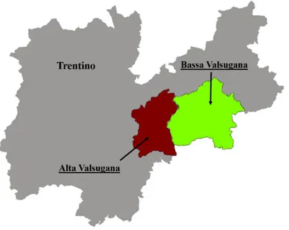
This research is part of a project launched in Trentino after a serious outbreak of the disease reported in Valsugana (southeastern Trentino) in 2011. The main objectives of this work regarded the study of epidemiological traits of the disease and biological features of the insect vectors. The first aim was monitoring the disease spread and vector populations’ dynamics, evaluating the natural infection level of psyllids and some transmission parameters, such as acquisition capacity, transmission efficiency, and the possibility of a transovarial phytoplasma transmission. The distribution of infected plants was mapped along Valsugana and the populations of the psyllid vectors were monitored in the period 2014-2016. After a three-year survey, the percentage of symptomatic apple plants drastically decreased. Regarding the psyllid vectors, C. melanoneura showed higher population levels compared to C. picta in both conventional and untreated orchards, but the percentages of infected individuals were higher in
6
the latter species. The transmission parameters were evaluated in psyllids during acquisition and transmission trials carried out with C. picta and C. melanoneura. Specific trials were conducted with C. picta to assess the vertical transmission of the phytoplasma. Experiments, conducted under semi-field and greenhouse conditions in spring and summer 2015 and 2016, involved overwintered adults of both species collected in Valsugana and nymphs and emigrant adults reared on infected apple plants. After each experiment, insects and test plants were analyzed by real-time PCR to assess the phytoplasma presence. Results confirm C. picta as a more competent ‘Ca. P. mali’ vector in Trentino, but suggest the possibility of acquiring and transmitting the phytoplasma also for C. melanoneura, even though with a low efficiency. For both species, overwintered adults were able to transmit the disease only after an acquisition period spent on infected plants and the acquiring capacity of the stages developed on infected apple plants was very high. Moreover, for the first time was demonstrated that infected C. picta individuals are able to transmit ‘Ca. P. mali’ to the progeny.
Another goal of this research was to unravel the tri-trophic relationship involving phytoplasma and its hosts by the study of the genetic diversity of phytoplasma isolates in plants and insect vectors and their geographical distribution. So, a genetic characterization of phytoplasma was conducted, and isolates obtained from insects and apple plants collected in different geographical areas were analyzed by a multilocus sequence typing method based on four phytoplasma genes. The results obtained are only partial, but the different ‘Ca. P. mali’ genotypes observed so far indicate a higher genetic diversity in insects compared to host plants and suggest the hypothesis of specific relationships between phytoplasma genotypes and insect vectors.
New insights on psyllid ethology were achieved. In particular, a research was conducted to investigate the vibrational communication involved in reproductive behavior. For the two vector species, ethological observations and laser vibrometer recordings of the vibrational signals emitted during courtship were carried out. Signals appeared to be species-specific, but they did not seem to be a prerequisite for courtship and mating. Moreover, as already seen in other psyllid species, a scanning electron microscopy investigation showed the presence of a stridulatory mechanism on thorax and wings of both species.
Finally, as other homopteran species are known to be phytoplasma vectors, a goal of this research was to look for potential new vectors of AP phytoplasma, characterizing the leafhoppers’ and planthoppers’ communities in apple orchards of Valsugana and studying the effect of surrounding landscapes on their distribution.
Samplings were conducted in apple orchards surrounded by different landscapes and in different habitats inside the orchard. The results of this study indicate that landscapes influence
7
the species richness and, regarding habitats, that grasses are visited by higher numbers of species and individuals in all landscapes considered. All insect collected were tested by real-time PCR and results indicate that three samples belonging to three different species tested positive to ‘Ca. P. mali’.
Research conducted in this thesis drew a picture of AP disease spread in Valsugana. Results obtained in all the topics described above, from epidemiological studies to phytoplasmagenetic variability, passing through vectors’ courtship behavior and agroecosystem biodiversity, represent the theoretical background that helps advisors and growers to optimize the current disease management, and researchers to develop innovative and sustainable control strategies.
9 RIASSUNTO
I fitoplasmi sono procarioti pleomorfi, privi di parete cellulare, simili a batteri ed appartenenti alla classe Mollicutes e sono caratterizzati da un genoma molto ridotto. L’assenza di vie metaboliche essenziali li rende parassiti obbligati di piante ed insetti. Nelle prime, la presenza di questi patogeni è limitata ai tessuti floematici e causa sintomi associati a profondi disordini nel normale bilancio ormonale. Negli insetti, gli effetti dei fitoplasmi sulla fitness sono vari e, a seconda delle relazioni evolutive, possono essere deleteri o benefici. I fitoplasmi sono associati a centinaia di patologie di specie vegetali distribuite in tutto il mondo, comprese colture di elevate importanza economica, fruttiferi e anche piante ornamentali. Il principale mezzo di trasmissione di questi patogeni è costituito da insetti vettori e le interazioni tra fitoplasmi ed insetti sono, in alcuni casi, molto specifiche e coinvolgono una complessa catena di eventi. Solo fitomizi floematici, come cicadomorfi, fulgoromorfi e psille hanno il potenziale per acquisire e trasmettere questi parassiti obbligati.
‘Candidatus Phytoplasma mali’ è l’agente eziologico di Apple proliferation (AP), una malattina che rappresenta uno dei più gravi problemi nei meleti europei. In Trentino, una delle principali regioni produttrici di mele in Italia, AP è una minaccia per la produzione. I sintomi più evidenti sono la presenza di scopazzi sui rami, di stipole ingrandite nelle foglie e di arrossamenti anticipati delle chiome in autunno. I frutti degli alberi infetti, che hanno dimensioni inferiori e proprietà organolettiche alterate, non possono essere commercializzati. Finora due psille, Cacospylla picta e Cacopsylla melanoneura (Homoptera: Psyllidae), sono state confermate vettori della malattia, ma il loro reale ruolo nell’epidemiologia di AP è ancora oggetto di discussione, dal momento che studi condotti in aree geografiche diverse mostrano efficienze di trasmissione differenti.
Questa ricerca si inserisce all’interno di un progetto avviato in Trentino dopo che un’esplosione della fitoplasmosi è stata segnalata in Valsugana (Trentino sud-orientale) nel 2011. I principali obiettivi di questa ricerca hanno riguardato lo studio di alcuni aspetti dell’epidemiologia di AP alcune caratteristiche biologiche degli insetti vettori. Il primo scopo è stato quello di monitorare l’avanzamento della malattia e le dinamiche di popolazione dei vettori, valutando le percentuali di infezione naturale delle psille e alcuni parametri relativi al processo di trasmissione, come la capacità di acquisizione, l’efficienza di trasmissione e la possibilità di trasmissione transovarica del fitoplasma. La distribuzione delle piante infette è stata mappata lungo la Valsugana e le popolazioni delle psille vettrici sono state monitorate tra il 2014 e il 2016. Dopo tre anni di rilievi, si è ottenuta una drastica diminuzione della percentuale di piante sintomatiche. Riguardo
10
gli insetti vettori, C. melanoneura si è rivelata essere la specie con densità di popolazione più elevate sia in frutteti coltivati, sia in frutteti abbandonati, ma la specie con percentuali di infettività maggiori è risultata essere C. picta. I parametri di trasmissione per entrambe le specie di psilla sono stati valutati attraverso prove di acquisizione e trasmissione e prove specifiche sono state condotte per verificare se vi è trasmissione transovarica del fitoplasma in C. picta. Gli esperimenti condotti in condizioni di semi-campo e serra, durante la primavera e l’estate del 2015 e del 2016, hanno riguardato adulti svernanti di entrambe le specie (campionati in Valsugana) e ninfe e adulti di nuova generazione allevati su piante di melo infette. Alla fine di ogni esperimento, piante e insetti sono stati analizzati tramite real-time PCR per verificare la presenza di fitoplasma. I risultati confermano che C. picta è un vettore molto più efficiente in Trentino, ma suggeriscono la possibilità di acquisizione e trasmissione del fitoplamsa anche in C. melanoneura, anche se con una bassa efficienza. Per entrambe le specie, gli adulti svernanti sono stati in grado di trasmettere il fitoplasma solo dopo aver trascorso un periodo di acquisizione su piante infette e la capacità di acquisizione degli stadi che si sono sviluppati su piante infette è stata molto elevata. In più, è stata dimostrato per la prima volta che individui infetti di C. picta sono capaci di trasmettere ‘Ca. P. mali’ alla progenie.
Uno degli obiettivi di questa ricerca era lo studio del rapporto tritrofico tra fitoplasma e i suoi ospiti, attraverso indagini sulla diversità genetica del fitoplasma in piante e insetti vettori e sulla loro distribuzione geografica. Quindi il fitoplasma è stato caratterizzato geneticamente e gli isolati ottenuti da piante di melo e insetti raccolti in differenti aree geografiche sono stati analizzati attraverso un metodo di multilocus sequencing typing basato su quattro geni del fitoplasma. I risultati ottenuti sono solo parziali, ma i differenti genotipi di ‘Ca. P. mali’ ottenuti finora indicano la presenza di un’elevata variabilità genetica negli insetti rispetto alle piante, che suggerisce l’ipotesi dell’esistenza di relazioni specifiche tra genotipi di fitoplasma e insetti vettori.
Nuove informazioni sono state ottenute sull’etologia delle psille, in particolare per quanto riguarda il comportamento riproduttivo. Per questo, sono state condotte osservazioni etologiche e registrazioni dei segnali vibrazionali emessi durante il corteggiamento attraverso l’uso di un laser vibrometro. Il segnale sembra essere specie-specifico, ma non sembra essere un prerequisito per il corteggiamento e l’accoppiamento. Inoltre, come già visto in altri psillidi, indagini al microscopio elettronico a scansione hanno evidenziato la presenza di strutture stridulatorie su torace e ali di entrambe le specie.
Infine, poiché è noto che altre specie di omotteri sono vettori di fitoplasmosi, un obiettivo di questo studio è stato la ricerca di potenziali nuovi vettori di ‘Ca. P. mali’, caratterizzando allo
11
stesso tempo le comunità di cicadomorfi e fulgoromorfi dei meleti della Valsugana e valutando l’effetto dei paesaggi circostanti sulla loro distribuzione.
I campionamento sono stati condotti in meleti circondati da diversi paesaggi e anche all’interno di diversi habitat nello stesso meleto. I risultati di questo studio indicano che i paesaggi influenzano la ricchezza in specie e che, riguardo gli habitat, il cotico erboso del frutteto è visitato da numeri più elevati di specie e individui in tutti i paesaggi considerati. Tutti gli individui raccolti sono stati analizzati tramite real-time PCR e i risultati indicano che almeno due specie di cicaline sono state capaci di acquisire ‘Ca. P. mali’.
Le ricerche condotte in questa tesi hanno permesso di tracciare un quadro diffusione di AP in Valsugana. I risultati ottenuti nei diversi ambiti di ricerca descritti sopra, dagli studi epidemiologici e di variabilità genetica, passando attraverso il comportamento sessuale dei vettori e la biodiversità nell’agroecosistema, rappresentano le basi teoriche per aiutare consulenti tecnici e agricoltori ad ottimizzare l’attuale gestione della malattia e i ricercatori a sviluppare strategie di controllo innovative e sostenibili.
13
INTRODUCTION
Phytoplasmas
Phytoplasmas are pathogens responsible of more than 700 plant diseases belonging to 98 families and cause different yellows, dwarf and witches’ broom diseases (Bertaccini, 2007; Hogenhout et al., 2008). Until their characterization in 1994, phytoplasmas were previously known as mycoplasma-like-organisms (MLO) because of the resemblance to mycoplasmas associated to human and animal diseases (Welliver, 1999). These organisms are distributed worldwide and, as host, include ornamental plants and many important food, vegetable and fruit crops (Lee et al., 2000; Garnier et al., 2001; Seemüller et al., 2002; Bertaccini and Duduk, 2009).
Taxonomy- Phytoplasmas represent a monophyletic group within the Mollicutes class and
belong to the Acholeplasmataceae family (Lee et al., 2000; Hogenhout et al., 2008). Currently, there is no formal taxonomy of phytoplasmas. In Mollicutes, formal recognition of species and assignment of binomial Latin names to species require description of biological properties of the species, such as growing in pure culture. As this is difficult for phytoplasmas, the convention of ‘Candidatus Phytoplasma’ species has been adopted as a means to refer to distinct phytoplasma lineages and putative species (IRPCM, 2004). In the last decades, the identification and classification of phytoplasmas were based on biological properties as the symptoms induced in infected plants, plant host range, and relationships with insect vectors (Lee et al., 2000; Duduk, 2009).
Recent molecular data on phytoplasmas have provided considerable insights into their diversity and genetic interrelationships that are the basis for several studies on phytoplasma phylogeny and taxonomy (Hogenhout et al., 2008). RFLP (restriction fragment length polymorphism) and sequence analysis of 16S rDNA have shown that phytoplasmas constitute a coherent taxon and allowed dividing the phytoplasma clade into groups and subgroups. The first complete phytoplasma classification scheme based on of PCR-amplified 16S rDNA provides a reliable means for the differentiation of a broad array of phytoplasmas and has become the most comprehensive and widely accepted phytoplasma classification system. This approach provides a simple, reliable and rapid tool for differentiation and identification of known phytoplasmas (Duduk and Bertaccini, 2011). Based on RFLP analysis, it was possible to identify 28 phytoplasma groups (Figure 1), while the sequence analysis allowed the determination of 37 ‘Candidatus Phytoplasma’ species so far (Aryan et al., 2016).
14
Figure 1. ‘Candidatus Phytoplasma’ nomenclature validity published compared with 16S rRNA classification based on Lee et al. (1998) when possible (modified from Duduk and Bertaccini, 2011).
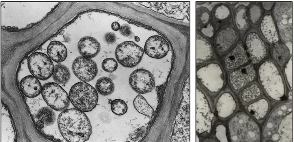
Morphology- Phytoplasmas are single-celled sub-microscopic microorganisms surrounded by
a single trilaminar membrane similar to bacteria, with smaller dimensions. Since they lack cell wall, they can change shape (Doi et al., 1967). Morphologically, by microscopic observations of phloem tissue sections, they appear as rounded, pleiomorphic bodies with an average diameter ranging from 200 to 800 µm (Figure 2) (Bertaccini et al., 2010). These organisms are obligate parasite that cannot survive apart from a host and grow and reproduce in the cytoplasm of host cell, both plants and insect vectors. Phytoplasmas reside in the phloem tissue of the
15
plants they infect, invading primarily phloem sieve tube element (Ploaie, 1981; Welliver, 1999). Since phytoplasmas’ cultivation in vitro is very problematic, the study of this group of microorganisms is complicated. Recently, a method for cultivation of various phytoplasma strains has been reported (Contaldo et al., 2012), but plant infections by cultures have not been reported yet (Aryan et al., 2016).
Figure 2. Electron micrograph of phytoplasmas in sections of sieve tubes. (A. Bertaccini, http://www.costphytoplasma.ipwgnet.org/WG1/WG1_photogallery.htm)
Symptoms- The presence of phytoplasmas is associated to plants with typical symptoms, due
to disturbances in the normal balance of plant hormones (Chang and Lee, 1995; Chang, 1998; Lee and Davis, 1992). Most common symptoms regard different apparatuses and include the proliferation of axillary shoots, phenomenon known as witches’ broom, leaf yellowing, the loss of normal flower color to green flowers or virescence, and the development of floral parts similar to leaf-like structures or phyllody. Moreover, sterility of flowers and phloem necrosis could occur (Mc Coy et al., 1989). All the symptoms produce a severe decline (Chang, 1998; Chang and Lee, 1995) and have a general negative impact on production (Figure 3). The symptoms induced in diseased plants vary with the phytoplasma and with the stage of infection (Lee et al., 2000).
Transmission and spread- Phytoplasmas responsible of ornamental and fruit tree diseases are
spread by vegetative propagation by means of cutting, storage tubers, rhizomes, or bulbs (Lee and Davis 1992). Phytoplasmas can be even transmitted through grafts, but not by inoculation with phytoplasma-containing sap or by mechanical actions. Plant viruses, mycoplasma-like and rickettsia-like organisms rely on the spreading function performed by several insect vectors, as most of them are not stable and require multiplication in the diverse plant and insect hosts (Hogenhout et al., 2008). . In particular, the ecological niche of this bacterial group is localized
16
in the sieve elements of plant hosts and some organs of sap-sucking insect vectors, belonging to the Cicadellidae and Fulgoridae families (leafhoppers and planthoppers, respectively), and Psyllidae (Bosco and Marzachì, 2016). In this three-way interaction, insect vectors appear to play an active role; their feeding behavior and preference for certain host plants probably are, in most cases, the primary factors that determine the final niches for each phytoplasma (Lee et al., 2000; Pedrazzoli, 2009).
Phytoplasmas may exhibit an overwintering behavior in insect vector or in perennial plants. Notwithstanding, no evidences of seed transmission have been recorded yet (Christensen et al., 2005), despite the revelation of phytoplasma DNA in embryo tissues, which leads to hypothesize the potential for seed transmission (Cordova et al., 2003).
Figure 3. Symptoms associated with phytoplasma presence in. A. Flavescence dorée (B. Duduk); B. Myrtle witches' broom (V. Prota); C. Pear decline (M. Cielinska); D. Purple coneflower (B. Duduk).
(http://www.costphytoplasma.ipwgnet.org/WG1/WG1_photogallery.htm)
A
B
17
Detection- Before the development of molecular diagnostic tools, the detection of phytoplasma
diseases was based on the observation of symptoms, experimental transmission to host plants, and fluorescence or electron microscopy observation of ultra-thin sections of the phloem. Serological diagnostic techniques for the detection of phytoplasmas began to emerge in the 1980’s with ELISA (enzyme-linked immunosorbent assay) based methods (Duduk, 2009). In the early 1990’s, molecular methods were developed with PCR coupled with RFLP analysis, allowing the accurate identification of different strains and species of phytoplasma (Namba et al., 1993; Lee et al., 1993; Schneider et al., 1993). From this time, diagnosis and differentiation of phytoplasma infections have therefore relied on molecular methods (Delić, 2012), targeting a wide variety of regions within the 16S-23S rRNA genes and other conserved and less-conserved non ribosomal genes or variable genes encoding surface proteins (Smart et al., 1996). The 16S-23S rRNA gene sequences have been effectively used to differentiate and classify phytoplasma strains (Maejima et al., 2014; Wei et al., 2007), resulting in the identification of at least thirty groups of phytoplasmas. More recently, several quantitative PCR (qPCR) techniques, based on different probes and dyes, have been developed to detect the phytoplasma infection and to quantify the phytoplasma titer in plants and insects (Jawhari et al., 2015; Monti et al., 2013; Christensen et al., 2013; Baric et al., 2011; Jarausch et al., 2004; Angelini et al., 2007; Nikolić et al., 1996).
Phytoplasma transmission by insect vectors
Sap-sucking insect vectors are the most important means of phytoplasma transmission in nature, as these microorganism are not sufficiently stable to spread on their own. Many arthropods are capable of vectoring diseases in an ideal (e.g., laboratory) environment, but fail to be effective vectors in the field. Some factors influencing vector competence include age of the vector and host, synchrony of vector and host, availability of alternative hosts, behavior/host preference of the vector, and the ability of the pathogen to maintain or increase its titer in the vector, environmental conditions, and host resistance.
Plant host range for a phytoplasma is dependent upon vector specificity and feeding habits. Albeit multiple hosts may increase the chance for genetic exchange and local adaptations, allowing different infection pathways (Christensen et al., 2005), the range of plant species that can be infected by a given phytoplasma in nature is determined largely by the number of insect vector species that are capable of transmitting the phytoplasma and by the feeding behaviors (monophagous, oligophagous, and polyphagous) of these vectors (Lee and Davis, 1992; Lee et al., 2000). Polyphagous vectors have the potential to inoculate a wider range of plant species,
18
depending on the resistance to infection of each host plant. Several studies (Bosco et al., 1997; Marzachì et al., 1998) have shown that even insects that normally do not feed on certain plant species can acquire and transmit phytoplasmas to those plants under laboratory conditions. Hence, in many cases, the host range of a vector, rather than lack of phytoplasma-specific cell membrane receptors, limits the spread of phytoplasmas by that species (Bosco et al., 1997; Pedrazzoli, 2009; Weintraub and Beanland, 2006).
Moreover, the capability of the host plants to harbor more than one type (or strain) of phytoplasmas depends on the susceptibility to phytoplasma infection and on the vector-phytoplama-plant interaction (Lee et al., 2000). On the other hand, the natural host ranges of phytoplasmas in insect vectors and plants vary with the phytoplasma strain (Brcák, 1979; Lee et. al., 2000; Mc Coy et al., 1989; Tsai, 1979).
Phytoplasma-vector relationship- Vector may or may not be essential for the completion of
the life cycle of the pathogenic microorganism, but its function is harboring and protecting them and creating feeding wounds and entry points into susceptible plants.
Phytoplasmas are transferred with saliva into the pierced sieve element. From here, the microorganisms spread in plant through the continuous sieve tube system. An important feature displayed by vectors is the presence of a propagative and persistent relationship with phytoplasmas (Weintraub and Beanland, 2006) (Figure 4).
Figure 4. Phytoplasma life-cycle involves two different hosts: plants and insects. Phytoplasmas are acquired during insect feeding and multiply within the vector before being transmitted to another plant
(http://www.u- tokyo.ac.jp/en/utokyo-research/feature-stories/elucidating-the-mystery-of-phytoplasmas-the-ultimate-idler-bacteria.html).
19
The term “propagative” means that the pathogen can multiply in insects; on the other hand, “persistent” means that the insect remains inoculative for life (Fletcher et al., 1998).
A specific feeding duration by insect is required to acquire a sufficient titer of phytoplasma; this phase is a part of the phytoplasma reproductive cycle and is called acquisition access period (AAP). The longer is the duration of AAP, the greater will be the chance of acquisition (Purcell, 1982). This phenomenon may also depend on the titer of phytoplasmas in the plants, even though mechanisms are not clear yet (Weintraub and Beanland, 2006). AAP is followed by a latent period (LP), consisting in the time required from initial acquisition to the actual transmission of the phytoplasmas. During this period, which is temperature-dependent, phytoplasmas move through and multiply in the vector body (Murral et al., 1996; Nagaich et al., 1974). Phytoplasmas invade the midgut passing through the epithelial cells and, multiplying within a vesicle with intracellular movement or passing between two midgut cells (Lefol et al., 1994), enter the hemocoel through the basement membrane. Through the hemolymph, they may infect other tissues such as the Malpighian tubules (Lett et al., 2001), fat bodies and brain (Lefol et al., 1994; Nakashima and Hayashi, 1995), or reproductive organs (Kawakita et al., 2000); multiplication in these tissues suggest a longer co-evolutionary relationship between host and pathogen (Weintraub and Beanland, 2006). After reaching the salivary gland cells, phytoplasmas further multiply and are transmitted in the saliva (Hogenhout et al., 2008; Kirkpatrick, 1991).
Homoptera as vectors of phytoplasmas- The order of Homoptera comprises insect groups
with a specific piercing-sucking mouthparts, which conferred a relevant effect in their adaptive radiation (Goodchild, 1966).As phloem-limited, phytoplasmas can be acquired and transmitted only by phloem-feeding insects. Homoptera feeding habits range from phytophagy (the majority of species) to predation, including ectoparasitism and haematophagy. Phytoplasma vectors must feed specifically and selectively on this particular plant tissue, where pathogens reside, with a nondestructive way. Weintraub and Beanland (2006) reviewed the features required by an insect species to be a successful phytoplasma vector and, according to the authors, Homoptera are the main elicited insect group. Insects of this order are hemimetabolous and nymphs and adults besides feeding similarly, share the same physical location; often both immatures and adults can transmit phytoplasmas. They feed specifically and selectively on certain plant tissues, which makes them efficient vectors of pathogens residing in those tissues. Furthermore, their feeding is nondestructive, promoting successful inoculation of the plant vascular system without damaging conductive tissues and eliciting defensive responses. Moreover, they have a propagative and persistent relation-ship with phytoplasmas.
20
The Homoptera is the largest exopterygote group of insects with over 80.000 described species. Their specialized structure of mouthparts are modified into concentric stylets, the mandibular enclosing the maxillary ones and together forming the food and salivary channels. Homoptera is a very diverse group comprising scale insects, aphids, psyllids and whiteflies (Sternorrhyncha), true bugs (Heteroptera), and Auchenorrhyncha. The last one have been traditionally divided into two main suborders: Cicadomorpha (leafhoppers, treehoppers, spittlebugs, and cicadas) and Fulgoromorpha (the planthopper families) (Figure 5). Leafhoppers and planthoppers are among the most abundant groups of insects. Around 20.000 leafhopper (Cicadellidae) species have been described but estimates suggest 100.000 species may exist (Dietrich, 2005). In addition, there may be around 10.000 planthopper species of which the most significant pest species occur within the family Delphacidae. Around 200 vectors of phytoplasma are already known but many more are likely to be recognized because there are many more phytoplasma diseases characterized than there are known vectors of the diseases (Wilson and Turner, 2010). More than 75% of all confirmed phytoplasma vector species are found in the subfamily Deltocephalinae (Cicadellidae). The feeding habits of species within this subfamily range from monophagous to polyphagous, and members of this group can transmit one or more different phytoplasma taxa (Weintraub and Jones, 2010).
Figure 5. Homopteran families involved in the transmission of phytoplasmas (modified from Weintraub and Beanland, 2006).
21
‘Candidatus Phytoplasma mali’
Three phytoplasma-associated diseases are known to cause serious damages to the fruit production of temperate areas: apple proliferation (AP), pear decline (PD) and European stone fruit yellows (ESFY). Although the 16S rDNA sequences of strains of these pathogens are nearly identical, indicating a close phylogenetic relationship, other criteria considered, such as molecular markers, serological comparisons, vector transmission and host-range specificity, allowed the distinction of three different putative species: ‘Candidatus Phytoplasma mali’, ‘Candidatus Phytoplasma pyri’, and ‘Candidatus Phytoplasma prunorum’ (Seemüller and Schneider, 2004). The three pathogens form, together with the peach yellow leaf roll (PYLR) phytoplasma, a cluster designated the ‘AP phytoplasma group’ (Seemüller et al., 1998) or 16SrX group (Lee et al., 2000) within the AP subclade, which is one of the major branches of the phytoplasma clade.
AP is considered one of the most important diseases of apple (EPPO/CABI, 1997), particularly in the northern areas of southern Europe, where temperatures are the most conducive to symptom expression. Where cooler or warmer growing conditions occur, the disease appears to be less impacting (Seemüller et al., 1998; Rui, 1950, Refatti and Ciferri, 1954).
Symptoms- ‘Candidatus Phytoplasma mali’ causes symptoms associated with disturbance in
the normal balance of growth regulators (Seemüller and Schneider, 2004). Late growth of terminal buds, with a delayed production of flowers in the autumn, is usually the first noticeable symptom. A rosette of terminal leaves, which often becomes infected with powdery mildew, sometimes develops late in the season in place of the normal dormant bud. However, the most reliable symptom is the premature development of axillary buds, during the first two or three years following infection, which gives rise to witches’ brooms near the apex of the main shoot. Leaves of infected plants show abnormally long stipules and rather short petioles (Figure 6). In many cases, especially with trees on calcareous soils, there is a chlorosis and reddening of the leaves. Early defoliation often occurs. Depending on soil quality, fruits are markedly reduced in size, sometimes being only 25% of the weight of healthy fruit. In addition, flavor is poor, both sugar and acidity being reduced. The peduncles are longer and thinner and the fruit takes a flattened appearance (Blumer and Bovey, 1957; Schuch, 1962; Bovey, 1963; 1972).
In general, affected trees lack vigor, shoots are thin and necrotic areas appear on the bark. Diseased trees may die but, in mild infections, they may recover after the shock symptoms of the first 2-3 years and, subsequently, produce normal fruits again, especially if adequately fertilized (Schmid, 1965). The spontaneous remission of symptoms in AP-infected plants, called “recovery”, is a natural phenomenon observed in the field in which phytoplasma
22
disappears from the aerial part of the trees and is confined to the roots (Carraro et al., 2004; Musetti et al., 2004). This phenomenon was studied in apple proliferation infected apple trees cv. Florina and in apomictic rootstocks deriving from crosses of Malus sieboldii and Malus sargentii with Malus pumila. The combination of low mortality with the elimination of the phytoplasma from the aerial part of trees opened new perspectives for the selection of resistant rootstocks suitable for controlling apple proliferation (Kartte and Seemüller, 1991).
Distribution in the tree- The individuation of AP presence in the orchard goes together with
the distribution of phytoplasmas in the tree, which is not constant over the year (Pedrazzoli et al., 2008). At the end of wintertime, the content of phytoplasmas declines in the tree due to sieve tube degeneration. They appear also to be more concentrated in the root system but, during April to May, reinvade the stem and the canopy from the roots, reaching a peak in late summer or early autumn (Baric et al. 2011; Bisognin et al., 2008; Musetti et al., 2010; Schaper and Seemüller, 1984). Normally, plants inoculated with infected buds show the first symptoms the following year, mostly on the inoculated branches. When carried in the rootstock, phytoplasmas produce symptoms on the first growth of the scion. AP phytoplasma has been observed in the phloem of leaf petioles, midribs and stipules and appears to be localized mainly in suckers and terminal shoots (Bovey, 1972; Seidl and Komarkova, 1974).
23
Figure 6. Different symptoms of AP: A. early reddening in autumn (M. Baldessari); B. branch with witches’ brooms in winter (http://www.inspection.gc.ca). C. witches’ brooms (left) compared to a healthy shoot (right) (J. Sucha); D. two leaves showing enlarged stipules (left) compared to a healthy one (right) (F. Bondaz) E. small-sized fruits with enlarged petioles (right) compared to a normal-small-sized fruit (left) (M. Cielinska).
A
B
C
24
Host plants- ‘Ca. P. mali’ occurs in a wide range of species of the genus Malus (Kartte and
Seemüller, 1991) and has been detected occasionally in plants such as Pyrus spp., Prumus spp., Corylus avellana L., Crataegus monogyna Jacq., Quercus robur L., Quercus rubra L., Carpinus betulus L., Convolvolus arvensis L. (Del Serrone et al., 1998; Lee et al., 1995; Marcone et al., 1996; Mehle et al., 2007; Schneider et al., 1997; Seemüller, 2002; Seemüller and Schneider, 2004) and also in herbaceous plants, such as dahlia (Dahlia cultorum Thorsrud et Reisaeter) and oriental hybrids of Lilium plants (Kaminska and Śliwa, 2008a, 2008b).
Molecular characterization and diagnosis- ‘Ca. P. mali’ has a linear chromosome (Kube et
al., 2008) and a very small genome averaging ~750 kb that differs from the other phytoplasmas for the law GC-content (Bai et al., 2006). Analyses of a non-ribosomal DNA fragment, composed of three putative open reading frames (ORFs), proved the existence of at least three different AP phytoplasma subtypes named AT-1, AT-2, and AP-15 (Jarausch et al., 1994, 2000). Molecular characterization of the genes coding the ribosomal proteins L22 and S3 revealed the presence of higher genetic heterogeneity within isolates of ‘Ca. P. mali’ and led to the proposal of four subtypes rpX-A, rpX-B, rpX-C, and rpX-D (Martini et al., 2008). Analyses of ribosomal and non-ribosomal DNA fragments of ‘Ca. P. mali’ populations from northwestern Italy revealed the presence of the three AP phytoplasma subtypes (AT-1, AT-2 and AP-15) and reported the identification of at least two phytoplasmal genetic lineages, designated AT-1a and AT-1b, among the AP phytoplasma isolates of the AT-1 subtype (Casati et al., 2010).
Several diagnostic tests have been developed to detect ‘Ca. P. mali’ in both plants and insects. They range from the classic biological assays, in which the tested vegetal material is grafted onto indicator plants, to serological assays using specific antibodies against AP phytoplasma,
e.g. enzyme-linked immunosorbent assays (ELISA), or DAPI staining and
immunofluorescence, in which the pathogen is directly detected in vegetal sections under a fluorescence microscope.
The development of molecular techniques based on the DNA amplification allowed the establishment of very sensitive and specific diagnostic tools. Nested PCR, a highly sensitive DNA amplification in which the sample undergoes two separated PCR runs, has been used for the detection of AP phytoplasma in plants and insects using universal primers (P1/P7 and F2n/R2) and 16SrX-group specific primers (P1/P7 and fO1/rO1) (Lee et al,. 1995; Lorenz et al., 1995). Due to the genetic closeness between AP group phytoplasma, specific identification often requires further steps, such as amplicon digestion with different restriction enzymes and subsequent RFLP analysis or sequencing (Gundersen and Lee, 1996; Jarausch et al., 2000;
25
Kison et al., 1994; Lee et al., 1995; Lorenz et al., 1995; Schneider et al., 1995; Smart et al., 1996). In recent years, different quantitative real-time PCR assays have been developed to detect and quantify AP titer in plants and insects based on SYBR Green (Galetto et al., 2005; Jarausch et al., 2004; Torres et al., 2005), TaqMan (Aldaghi et al., 2007; Baric and Dalla Via, 2004) and EvaGreen technologies (Monti et al., 2013).
Geographical distribution- Apple Proliferation has only been reported from the European and
Mediterranean Plant Protection Organization (EPPO) region (EPPO/CABI, 1997). In Europe, it has been detected in the following countries: Albania, Austria, Balkans, Belgium, Bosnia-Herzegovina, Bulgaria, Croatia, Cyprus, Czech Republic, Denmark, France, Germany, Greece, Hungary, Italy, Moldova, Netherlands, Norway, Poland, Romania, Russia, Serbia, Slovakia, Slovenia, Spain, Switzerland, Ukraine and Yugoslavia (Figure 7). The disease has also been detected in Turkey and Syria. However, there are unconfirmed reports from India and South Africa (Seemüller, 1990).
Figure 7. Geographical distribution of AP in Europe and surrounding countries (http://www.cabi.org/isc/datasheet/6502)
AP in Italy- Apple proliferation has been recorded in the main apple growing regions of Italy,
especially in the North. Rui et al. (1950) described for the first time the presence of AP in Veneto. In Piemonte, the phytoplasma was reported for the first time at the end of the ‘90s in the provinces of Torino and Cuneo. However, the spread in these areas is not a big concern (Alma et al., 2000; Minucci et al., 1996; Pinna et al., 2003; Spagnolo et al., 2005). Differently, in Valle d’Aosta AP is widespread and represents a serious threat, especially in older orchards, reaching very high percentage of infection (Tedeschi et al., 2002; Tedeschi et al., 2003).
26
Figure 8. Apple proliferation is reported in northern Italian regions and in Basilicata.
In Lombardia, the presence of ‘Ca. P. mali’ was investigated by Casati et al. in 2007. AP was recently described also in the South of Italy, in particular in Basilicata (Marcone and Seemüller, 2013) (Figure 8). In north-eastern Italy, Alto Adige represents an important apple-growing area, with more than 10% of the European apple production. Here, as in other regions, the first sporadic cases of AP were reported in the late 1950’s, but a real outbreak of the disease was reported in 1998 in Valle d’Isarco. Afterwards, AP was monitored and a massive spread of the disease was recorded in the years 2005-2006, when symptomatic trees were found in about 75% of the monitored orchards and later, in 2011, when a new increase of infestations was observed in some districts (Oettl and Schlink, 2015). In Trentino, the presence of infected apple trees was reported in the early 1950’s (Refatti and Ciferri, 1954), but the spread of the disease was rather sporadic until the beginning of the 1990’s, when an outbreak started in Val di Non and caused significant economic damage (Vindimian and Delaiti, 1996; Vindimian et al., 2002). In order to quantify the disease spread and to understand the predisposing factors, a survey of infected trees was started in Val di Non in 1999 (Figure 9). In general, higher percentages of infected trees were observed at higher altitudes and in older orchards, with more vigorous rootstocks. Surprisingly, infection levels of about 5-10% were reported in two-year-old orchards and up to 20% in some three-year-old orchards (Springhetti et al., 2002).
27
Figure 9. Representation of Trentino valleys; apples are positioned in the main areas of AP presence.
Starting from 2001, the Phytosanitary Service of Province of Trento is conducting an official monitoring, which covers the whole apple growing area of the province. A surface corresponding to 4% of the total apple growing area was chosen to evaluate the effect of differential agronomic measures, cultivars or altitudes on the disease spread (Vindimian, 2002). The infection rate rapidly decreased starting from 2006, when uprooting of infected trees became mandatory and strict chemical control measures against the insect vectors were applied. The adoption of these actions was enhanced by a subsidy for uprooting orchards older than 20 years or with more than 20% of infected trees. AP prevalence constantly decreased during the subsidized uprooting program from 2006 to 2010, when it reached the level of 0.27%. Unfortunately, the infection rate started increasing again from 2012, more significantly in the Val d’Adige and Valsugana. In these two apple districts, the average infection rate rose up to 6% in 2014, pushing up again the average infection rate of the Trentino province to 2% (Dallago, 2016).
Transmission of AP- ‘Ca. P. mali’ can be commonly transmitted by grafting: the phytoplasma
is often disseminated in scion wood and trees may yield a high proportion of apparently healthy but infected buds. According to the seasonal colonization of host plants by the phytoplasma, Pedrazzoli et al. (2008) obtained the highest percentages transmissions by grafting between June and August (12 to 30%), while grafts conducted from March to May were not very successful (0-0.08%). The authors concluded that the most suitable period for collecting scions is springtime, when the probability of AP transmission was at the lowest. Grafting of stem scions removed during dormancy prevented transmission or yielded only a low transmission
28
rate. Transmission by root grafting in winter is generally successful and for this reason it has become an established method for indexing trees (Seemuller et al., 1984). AP has been reported to spread also via natural root bridges in middle-aged and old apple orchards (Baric et al., 2008; Bliefernicht and Krczal, 1995; Ciccotti et al., 2008; Vindimian et al., 2002). There are also reports of experimental transmission to Catharanthus roseus (Madagascar periwinkle) using the parasitic plant Cuscuta spp. (dodder) (Heintz, 1986; Marwitz et al., 1974), while the transmission via seed or pollen has not been reported (Seidl and Komarkova, 1974).
Insect species such as psyllids and leafhoppers have been investigated for their ability in spreading the disease (Seemuller, 1990). So far, two psyllid species, Cacopsylla picta Förster and C. melanoneura Förster, and the leafhopper Fieberiella florii Stål were demonstrated to be vectors of AP phytoplasma (Frisinghelli et al., 2000; Krczal et al., 1989; Tedeschi and Alma, 2004; Tedeschi and Alma, 2006). Regarding Trentino, the experiments conducted so far confirmed the vectoring capability of C. picta, which is able to transmit the pathogen as neanid/nymph and new generation adult (Frisinghelli et al., 2000; Mattedi et al., 2007; Pedrazzoli, 2009). On the other hand, the role of C. melanoneura is still unclear: a very low transmission efficiency was found by Mattedi et al. (2007), even though important percentages of infected individuals have been found in the orchards of Trentino and in Alto Adige (Malagnini et al., 2010; Poggi Pollini et al., 2002).
Psyllid vectors of ‘Ca. P. mali’- Psyllids are known in agriculture as important pests of
cultivated crops but also as vectors of plant diseases (Hodkinson, 1974). The Psylloidea (Homoptera: Sternorryncha) superfamily is distributed worldwide and comprises eight families with about 3.850 species (Burckhardt and Ouvrard, 2012; Eben et al., 2015). All species of these families live on plant sap and are phloem-feeders, both as nymphs and as adults.
During their life, they are generally narrowly host-specific and are restrict almost exclusively to perennial dicotyledonous plants (Eastop, 1972). Plants may play different roles in hosting psyllid species (Conci et al., 1995). The “host plants” are the species on which insects spend time to lay eggs and develop. The “shelter plants” are the species to which adults compulsorily migrate in autumn for spending winter, reducing trophic activity. “Occasional plants” are species where insects may be accidentally transported by wind or other causes, but that normally have no importance for their biology. In relation to the host-plant range, psyllids have been divided into four categories: (1) monophagous species, where nymphs can develop exclusively on one plant species; (2) strictly oligophagous species that live on some congeneric plants; (3) widely oligophagous species that live on plants belonging to kindred genera of the same family and (4) polyphagous species that live on plants of different families (Conci et al.,
29
1995). Life-cycle in psyllids is often highly synchronized with host plant phenology (Pedrazzoli, 2009).
Cacopsylla picta Förster (1848) has a palaearctic distribution and is associated on Malus spp. (Lauterer, 1999; Jarausch et al., 2011). Previously known as C. costalis Flor (1861), it has been synonymised with C. picta by Lauterer and Burckhardt in 1997. This psyllid is a univoltine species, completing one generation per year, and overwintering as an adult on conifers (Čermák and Lauterer, 2008; Mayer et al., 2010). At the end of winter (March or April), C. picta remigrants move from their overwintering sites to their main hosts for oviposition. Larval development takes four to five weeks; the newly hatched imagines (emigrants) remain in the orchards for about two weeks before migrating to their overwintering sites in June or July. C. picta young adults are light green, with a mesothorax yellowish banded. Later their color is dirty yellow or orange-colored with more or less extensive dark brown or black markings. The abdomen is black with red segment borders (Ossiannilsson, 1992). During hibernation the body coloration changes to black-brown (Lauterer, 1999). Forewings are colorless, veins in old specimens are dark brown or black, pterostigma is fuscous. The overall length of males is 2.86-3.24 mm, of females 3.14-3.43 mm (Ossiannilsson, 1992). Fifth instar nymphs are light green, wing pads with a pale violet tinge. Abdominal margin has three pairs of sectasetae. The ocular seta is more or less simple, 0.03-0.04 mm in length. The length of the body is 1.57-2.19 mm (Figures 10 and 11) (Ossiannilsson, 1992).
The species is narrowly oligophagous on Malus domestica Borkh., Malus sylvestris Mill., Malus cv. and Prunus armeniaca L. (Conci et al., 1992; Lauterer, 1999; Ossiannilsson, 1992). According to Harisanow (1966), who studied the biology of C. picta in Bulgaria, this species overwinters as adult on Pyrus communis L., Prunus domestica L., Persica vulgaris Mill., Amygdalus communis L., Ulmus campestris L. and other plants (Lauterer, 1999; Ossiannilsson, 1992). A female may lay approximately 160 eggs and ten-fourteen days after becoming adult, the new generation moves on first to annual herbs, e.g. Brassica, Mentha, Vicia, Phaseolus, Pisum, as well as grasses, e.g. Avena; later to perennial shelter plants (Lauterer, 1999; Ossiannilsson, 1992). According to Conci et al. (1992), C. picta overwinters on conifers. These data are confirmed by Flor (1861), who collected specimens on Pinus abies L. in August. Ossiannilsson (1992) in Uppland found one male on Picea abies (L.) H. Karst. at the end of November. Recently, the complete life-cycle of C. picta was described in a permanent rearing under controlled conditions by Jarausch and Jarausch (2014), who successfully reproduced overwintered sites on pine and spruce. The host location and the migration behavior of C. picta seems to be mediated by the chemical cues emitted by plants, and the preference of the insects
30
switches between the volatiles of the host and the shelter plants during the course of the year (Gross and Mekonen, 2005).
Figure 10. Cacopsylla picta. Female: (a) head in frontal aspect; (b) left antenna in dorsal aspect. Male: (c) left forewing, (d) terminalia from the left; (e) left paramere from the left; (f) same from behind; (g) terminal part of
aedeagus from the left. Scale: 0.1 mm (modified from Ossiannilsson, 1992).
a b c d e f g
31
Figure 11. Cacopsylla picta. Female: (a) terminalia from the left; (b) proctiger from above; (c) subgenital plate from below. 5th instar nymph: (d) left antenna from above; (e) left wingpads from above; (f) abdominal dorsum
(left) and venter (right); (g) circumanal pore rings from below. Scale: 0.1 mm for (g); 0.5 mm for the rest (modified from Ossiannilsson, 1992).
a b c d e f g
32
Cacopsylla melanoneura Förster (1848) is a holopaleartic species distributed everywhere with its host plants. Young adult specimens are orange-colored, pronotum and genal cones are whitish, forewing veins are yellow. Later, they are largely dark brown with a reddish tinge, head and pronotum are partly lighter, mesonotum with pale spots and bands, forewing veins are dark brown or black. Forewings alone veins have broad spinule-free bands becoming broader apically. Overall length of males is 2.52-3.10 mm, of females is 2.95-3.30 mm (Ossiannilsson, 1992). Fifth instar nymphs are entirely light green, or green to dirty green with yellow brownish sclerites. Wing pads are often whitish. The number of marginal setae on forewing-pads is variable. On abdominal margin there are three pairs of sectasetae. The body length is 1.33-2.00 mm. Ocular seta is more or less rod-like or spine-like, length is 0.011-0.017 mm (Figures 12 and 13) (Ossiannilsson, 1992).
This species is widely oligophagous on Rosaceae Maloideae such as Crataegus spp. (Crataegus monogyna Jacq., Crataegus oxyacantha L., Crataegus maximowiczii C.K.Schneid), Malus spp. and Pyrus communis L. (Conci et al., 1992; Ossiannilsson, 1992). It is reported also on conifers and many other shelter and occasional plants of different families (Conci et al., 1992; Lauterer et al., 1999). Overwintering adults live for 9-10 months long on Pinus spp. at higher altitudes (250-1400 m asl), performing long-distance migrations between stands of pines and apple trees (Lazarev, 1974). The migrations to orchards take place during budding of the host plant. Each female lays about 200 eggs. Embryonic development lasts 7-20 days and larvae hatch at the time of maximum flowering of apple trees. The larvae develop over one month and then the new generation adults appear. After complete sclerotisation (i.e. about 5 days after their last skinning) the adults migrate to mountain elevations onto pine trees. Overwintering behavior and shelter plants of C. melanoneura were studied in Italy by Pizzinat et al. (2011). The altitudinal distribution and overwintering habitats were investigated following the direction of warm ascending currents, as proposed by Cĕrmák and Lauterer (2008), and the suitability of different conifer species as shelter plants during aestivation and overwintering periods was assessed by insect collections and observation of insect survival in outdoor trials. The results indicate that this species can potentially survive on many coniferous species.
Ossiannilsson (1992) described the life cycle of C. melanoneura on hawthorn in Sweden, where the stages are slightly delayed in time and the migration of the new generation adults to conifers does not begin before July. In Czech Republic, in Querco-Carpinetum associations and particularly in floodplain forests, however, in absence of conifers, most of the population may hibernate on other broadleaved trees, hiding under bark scales and on sprouts (Lauterer, 1999). Apparently, the long-distance seasonal migrations of young adults to mountain elevations
33
shortly after having completed sclerotisation are limited to the warmer southern parts of Europe. In the conditions of central Europe the migrations are apparently shorter (Lauterer, 1999). Mass occurrence of new generation adults in Czech Republic was observed by the author in the first decade of June, whereupon their number dropped abruptly. This early emigration from the host plants to other plants agrees with the observation of Lazarev (1974), but in Czech Republic the migration to the shelter plants seems to be gradual, and the species first migrates on occasional plants and then to conifers. Thus, for this species, three migration phases can be distinguished (Lauterer, 1999). The role of chemical signals in the migration behavior and the orientation of C. melanoneura was studied with psyllids collected from both apple and hawthorn by Gross and Mekonen (2005) and by Mayer and Gross (2007). The behavioral responses of the insects corresponded with the different phases of the migratory behavior, the overwintered adults showing strong positive responses for apple or hawthorn odors, while the newly emerged adults showing strong responses for spruce volatiles. Attempts at copulation and copulating adults were observed already during June but, apparently, fertilization does not take place until copulations after hibernation between March and May (Lauterer, 1999). About one week after the last skinning and completed sclerotisation, the adults enter dormancy of the parapause type with aestivation, later passing into a diapause during hibernation. Reactivation and development of sexual glands only occur after the cold phase in winter (Lauterer, 1999). The altitude of hibernation and aestivation places differs according to the latitude: in Moravia the majority of individuals can be found between 160 and 450 m asl, while at higher altitudes the occurrence is only sparse; the populations of the southern European regions most often hibernate and aestivate on dwarf pines in high mountain altitudes. These results are confirmed by the study on the altitudinal distribution of C. melanoneura conducted by Pizzinat et al. (2011), where the best climate conditions for aestivation and overwintering were observed between 1350 and 1650 m asl. The distribution of this species seems to be partly conditioned by its thermophily, but first of all by the composition of vegetation (especially the presence of hawthorn, which is more present in warmer biogeographical units) (Lauterer, 1999). C. melanoneura frequently hibernates together with the salicicolous psyllids (the so-called C. saliceti group) and with C. affinis Löw. In its host plants it occurs together with C. affinis, C. peregrina Förster, and the phenologically delayed C. crataegi Schrank (Lauterer, 1999). In the Crimea, members of the population which lives on apple trees will not develop if transferred to hawthorn and die within several days (Lazarev, 1974). More recently, studies on the different C. melanoneura populations collected on hawthorn and on apple were carried out to investigate the exchanges in insect populations between the two host plants and the role of hawthorn as reservoir of AP
34
phytoplasma (Tedeschi et al., 2009). Moreover, ecological trials and genetic analyses carried out by Malagnini et al. (2013) confirmed the existence of differentiated populations associated with the two host plants.
Figure 12. Cacopsylla melanoneura. Male: (a) head in frontal aspect; (b) left antenna in dorsal aspect; (c) left forewing; (d) cell m1 of forewing; (e) terminalia from the left; (f) left paramere from behind; (g) terminal part of
aedeagus from the left. Scale: 1 mm for (c); 0.5 mm for (a) and (b); 0.1 mm for (e), (f) and (g) (modified from Ossiannilsson, 1992). a b c d e f g
35
Figure 13. Cacopsylla melanoneura. Female: (a) terminalia from the left; (b) proctiger from above, (c) subgenital plate from below. 5th instar nymph: (d) left antenna from above; (e) left wing-pads from above; (f) left half of caudal part of abdominal dorsum; (g) circumanal pore rings from below. Scale: 0.1 mm for (g); 0.5
mm for the rest (modified from Ossiannilsson, 1992).
a b c d e f g
36
Other known vectors- Apart from the two psyllids, Fieberiella florii Stål (1864), a holarctic
leafhopper widely distributed in Europe and introduced also in the USA and Canada, was demonstrated to be able of transmitting AP. This univoltine leafhopper is already known in North America as one of the most important vectors of X-disease (Gold and Silvester; 1982; Van Steenwyk et al., 1990) and was successfully used in transmission trials conducted in Germany and in north-western Italy (Bliefernicht and Krczal, 1995; Krczal et al., 1989; Tedeschi and Alma, 2006). Its presence in apple orchards of Trentino is only occasional (Ioriatti and Jarausch, 2008).
Disease control- All AP symptoms cause a strong economic impact of the disease. The loss of
earnings calculated in Italy in 2001, due to lack of production of marketable apple fruits, was of about 100 million euro (Strauss, 2009). As the direct control of the phytoplasma is still unreliable unless using antibiotics, it is very important to adopt preventive strategies to fight AP spread. For instance, propagating material must be carefully selected from sources known to be free of the disease (the certification of plant material through the application of effective indexing procedures is required for new plantations). Moreover, the eradication of newly diseased trees as soon as symptoms appear in the orchards is very effective to reduce sources of inoculum. Trees must be uprooted and all the roots completely removed from the ground and destroyed. Moreover, a monitoring of insect vectors must be carried out and, in occurrence of established populations, the application of insecticide programs must be evaluated.
After a Ministry decree issued mandatory control measures against AP in 2006, a sanitation program was implemented. Trees are regularly inspected for the presence of typical symptoms or tested when no symptoms are found; infected trees are destroyed, and in case that a proportion of diseased trees higher than 20% is observed, the whole orchard has to be uprooted. As a consequence of these measures, the spread of AP has declined, but surveys have to be constantly carried out and treatments against the vectors are prescribed.
Aims of the research
Despite years of systematic control and the consequent strong reduction of psyllids population density, apple proliferation is still a major threat for apple production, especially in some apple growing areas of Trentino. C. picta and C. melanoneura are ordinarily controlled with chemicals in orchards by means of multiple treatments during springtime. After almost 20 years of research on apple proliferation disease in Trentino, some questions still remain open; in particular, the role of C. melanoneura as vector in Trentino is still unclear.
37
So, aim of this research was to deepen epidemiological, biological and ecological knowledge of the three-way system represented by the phytoplasma and its two hosts (plants and insect vectors). Valsugana (southeastern Trentino) was chosen as study area because of the sudden outbreak observed in the previous years.
The main objectives of the research activities carried out during the years 2014-2016 were: monitoring of disease spread and vectors population dynamics, with evaluation of the
infectivity of psyllids;
evaluation of transmission parameters, such as acquisition capacity and transmission efficiency, in different stages of C. picta and C. melanoneura;
study of the genetic diversity of phytoplasma strains and geographical distribution in apple plants and psyllid vectors;
investigations on psyllids’ courtship and mating behavior, with special regard to vibrational communication as basic knowledge to develop new control strategies with low impact;
research of potential new vectors of AP phytoplasma, with characterization of leafhoppers and planthoppers communities in apple orchards and effect of surrounding landscapes on their distribution.
References
Aldaghi M., Massart S., Steyer S., Lateur M., Jijakli M. H. (2007). Study on diverse grafting techniques for their capability in rapid and efficient transmission of apple proliferation disease to different host plants. Bulletin of Insectology, 60: 381-382.
Alma A., Navone P., Visentin C., Arzone A., Bosco D. (2000). Detection of “Apple proliferation” phytoplasmas in Cacopsylla melanoneura (Förster) (Homoptera Psyllidae). Petria, 10: 141-142.
Angelini E., Bianchi G. L., Filippin L., Morassutti C., Borgo M. (2007). A new TaqMan method for the identification of phytoplasmas associated with grapevine yellows by real-time PCR assay. Journal of Microbiological Methods, 68: 613-22.
Aryan A., Musetti R., Riedle-Bauer M., Brader G. (2016). Phytoplasma transmission by heterologous grafting influences viability of the scion and results in early symptom development in periwinkle rootstock. Journal of Phytopathology 164(9), DOI: 10.1111/jph.12486.
Bai X., Zhang J., Ewing A., Miller S. A., Jancso Radek A., Shevchenko D. V., Tsukerman K., Walunas T., Lapidus A., Campbell J. W., Hogenhout S. A. (2006). Living with genome instability: the adaptation of phytoplasmas to diverse environments of their insect and plant hosts. Journal of Bacteriology, 188: 3682-3696.