Department of Biomolecular Sciences
Ph.D. Course in Life Sciences, Health and Biotechnologies
Curriculum: Biochemical, Pharmacological Sciences and Biotechnologies
XXXII Cycle
Development of a real-time PCR method for the detection and/or
quantification of viable Legionella spp. and L. pneumophila in
sanitary and thermal water samples
Academic Discipline Code: BIO/10
Supervisor: Ph.D. student:
Prof. Mauro Magnani Dr. Veronica Ceppetelli
Co-Advisor: Prof. Giulia Amagliani
2
Acknowledgements
There are many people I wish to thank: my two supervisors, Professor Mauro Magnani and Professor Giulia Amagliani, Department of Biomolecular Science, and in particular Dr. Enrica Omiccioli, R&D department of Diatheva, for giving me the opportunity to work in this very interesting field of bacteriology and for giving inspiration and guidance.
Also thanks to the Dr. Anna Grottoli and Dr. Elisa Carloni of the R&D and production departments of Diatheva for practical help in the laboratory experiments.
Finally, I wish to thank my family and friends for support and fun throughout a sometimes-stressful period.
3
Table of contents
1. Introduction... 5
1.1. Taxonomy and general characteristics ... 8
1.2 Ecology and environmental sources of Legionella spp. ... 10
1.3 Clinical presentation ... 13
1.4 Risk factors and transmission ... 15
1.5 Epidemiology of Legionnaires'disease ... 19
1.6 Control measures for water risk management ... 23
1.7 Regulations and guidelines for the control and prevention of legionellosis ... 25
1.8 Standard method to detect and quantify Legionellae in water ... 27
1.9 Methods to proof the viability of Legionella spp. ... 30
Aim of the present study ... 35
Chapter 2. Evaluation of robustness of DNA extraction methods for the recovery of Legionella spp. cells ... 36
2.1 Introduction... 36
2.2. Material and Method ... 37
2.2.1 Bacterial growth condition ... 37
2.2.2 Types of water samples analysed ... 37
2.2.3 DNA extraction methods and qPCR analysis ... 38
2.2.4 Evaluation of robustness of DNA extraction methods according to ISO/TS 12869 ... 39
2.3 Results and discussion ... 41
2.4 Conclusions ... 43
Chapter 3. Development and optimization of a viability PCR method ... 44
3.1 Introduction... 44
3.2 Material and Methods ... 45
3.2.1 Bacterial strain and preparation of the viable and dead L. pneumophila cells ... 45
4
3.2.3 Evaluation of PEMAX pre-treatment directly on membrane filter or cell
suspension on artificially contaminated mineral water samples ... 46
3.2.4 Selectivity of the PEMAX-qPCR method in the quantification of live L. pneumophila cells ... 47
3.2.5. Robustness of the PEMAX-qPCR method in the quantification of live L. pneumophila cells in artificially contaminated cooling tower water ... 47
3.2.6 DNA extraction and qPCR enumeration of Legionella spp. genome units ... 47
3.2.7 Application of the developed method on field samples ... 48
3.2.8 Evaluation of alternative strategies to increase the efficacy of the developed protocol ... 48
3.3 Results and discussion ... 50
3.3.1 Optimization of PEMAX-qPCR treatment ... 50
3.3.2 Evaluation of PEMAX pre-treatment directly on membrane filter ... 52
3.3.3 Effect of PEMAX on defined ratio of viable and dead cells and on artificially contaminated water samples ... 53
3.3.4 Robustness of the PEMAX-qPCR method in the quantification of live L. pneumophila cells in artificially contaminated cooling tower water ... 55
3.3.5 Results of the application of PEMAX qPCR and alternative strategies on field samples and comparison with qPCR and culture ... 56
3.4 Conclusion ... 57
Chapter 4. Development and optimization of a nutritional stimulation method ... 58
4.1 Introduction... 58
4.2 Material and methods ... 60
4.2.1Types of water samples analysed ... 60
4.2.2 Bacteriological detection of Legionella spp. ... 60
4.2.3 Detection of living Legionella spp. and L. pneumophila by DNA stimulation method ... 60
4.3 Results and discussion ... 61
Conclusion and future directions ... 66
References... 69
5
1. Introduction
The first reported cases of infection by bacteria of Legionella genus occurred as a severe outbreak in July 1976 at a Bellevue-Stratford Hotel hosting the 58th annual convention of the American Legion in Philadelphia, Pennsylvania. Two hundred twenty-one convention attendees were infected, and 34 died. The disease was found to be caused by the bacterium Legionella pneumophila belonging to the family Legionellaceae, which was isolated in Hotel’s air conditioning system [Torrisi et al., 2012]. Subsequently, the atypical pneumonia caused by L. pneumophila was designated as Legionnaires’ disease.
Legionella spp. is also responsible for a less severe form of infection named Pontiac fever. Since then, sporadic and epidemic cases have dramatically increased in industrialized countries and this can be attributed both to the improvement of diagnostic tools and to the increase in opportunities for exposure to the causative agent [Guidelines, 2015].
In 1983, the World Health Organization (WHO) established a National Legionellosis Registry and in 1986 in Europe, the European Working Group for Legionella Infections (EWGLI) was formed with the aim to identify cases of infection in travelers, detecting epidemic outbreaks and notifying the competent authorities of the countries involved. In 1987, the Group established a surveillance scheme (EWGLINET) for the detection of the cases in people who travelled and stayed in hotels and resorts. Since 2010, the international surveillance program has been coordinated by the European Center for Disease Prevention and Control (ECDC) and called the European Legionnaires' Disease Surveillance Network (ELDSNet) [WHO, 2007].
In 2017 the ECDC reported 8 624 confirmed cases of Legionnaires’ disease in Europe with a 30% increase in the number of reported cases compared with 2016. L. pneumophila serogroup 1 was the most commonly identified species, responsible for 79% of culture-confirmed cases. Legionnaires’ disease remains an important cause of potentially preventable morbidity and mortality in Europe and there is no indication of decreasing burden [ECDC, 2019].
Some people are at higher risk including people 50 years or older, smokers and heavy drinkers, those suffering from chronic respiratory or kidney disease, and people whose
6
immune system is impaired [Farnham et al., 2014]. Nosocomial Legionnaires’ disease is an important problem as it has been estimated that 20–30% of legionellosis are nosocomial infections and that they are associated with a contamination of the health-care water networks [Tai et al., 2012]. The complexity of hospital’s water systems and the vulnerability of hospitalized patients increase the risk for Legionella spp. transmission and severe outcomes. In hospitals in addition to the water system, health practices concerning the airways (e.g. ventilation, aspiration, devices for artificial respiration and dental tools) can increase the risk of infection [Montagna et al., 2018].
Legionellae naturally occur in environmental water sources and are well adapted to man-made water systems, are often found in water system of buildings, cooling towers, evaporative condensers, and dental unit waterlines [Bonetta et al., 2017].
Legionellae are difficult to control in their natural sources due to their resistance to disinfectants, their ability to associate with biofilm and parasitism in protozoa. The risk assessment for Legionella is particularly important for public health officials and managers responsible for maintenance of water distribution systems of industrial or public buildings [Whiley et al., 2014]. Current risk assessment model is established on culture-based enumeration on selective media, that represents the reference method for Legionella control. Nevertheless, aside from the fact that this method requires up to 14 days for analysis, detection of Legionella from water samples is further confounded as the presence of disinfectants and other water treatment chemicals may render Legionella viable but not culturable (VBNC), leading to an unrealistically low number of visible colonies or false negatives, particularly in systems that are treated with monochloramine [Kirschner, 2016; Taylor et al., 2014]. This aspect has an important implication for Public Health especially in health care settings where high-risk patients may be susceptible to low concentrations of L. pneumophila in water systems: VBNC cells can be responsible for sporadic infection and outbreaks as they are able to resuscitate and preserve the virulence characteristics [Marinelli et al., 2017].
The only culture independent method that has achieved the status of a standard is quantitative real-time PCR (qPCR). This standard NF T90-471:2010 was firstly developed in France and then published as ISO/TS 12869 in 2012 [Anonymous, 2010; Anonymous, 2012]. The procedure is based on water sample filtration, DNA extraction followed by real-time PCR detection and/or quantification. DNA-based detection methods can elucidate the presence of Legionella spp. within few hours, with high sensitivity and specificity.
7
However, DNA detection can overestimate the risk of infection owing to the amplification of DNA deriving from dead cells.
A promising approach for a rapid detection of viable Legionella cells is viability-PCR, based on sample pre-treatment with photoactivable nucleic acid intercalating dyes such as Ethidium Monoazide (EMA), Propidium Monoazide (PMA) and commercial derivatives, such as PEMAX (a mix of photo-reactive azide forms of phenanthridium) prior to DNA extraction and PCR amplification [Thanh et al., 2017]. EMA and PMA are conventional dyes developed respectively in 2003 by Nogva and co-workers [Nogva et al., 2003] and in 2006 by Nocker [Nocker et al., 2016] that allow to differentiate between viable and dead cells on the basis of membrane integrity: the dyes penetrate only membrane compromised dead cells but not intact live cells. Once inside a cell, the dyes can covalently link to the nucleic acid through a photo-activation step, with the result that the amplification of the nucleic acid by PCR is inhibited. However, differentiation based on membrane integrity is not always sufficient: for example, some disinfection procedures applied to water such as UV treatment or solar disinfection do not affect primarily the cellular membranes [Cangelosi et al., 2014; Kirschner, 2016].
To overcome this limitation, it is necessary to extend the concept of viability so that cells must not only have intact membranes, but they must also be functional and active. In this case, “active” can be defined as capable of maintaining bacterial homeostasis using an active transport mechanism that requires ATP. The PEMAX dye (Geniul, Spain) is based on double-dye technology: a mixture of EMA and PMA dyes that allows to selectively amplify viable cells with both active metabolisms and intact cell membrane structure [Thanh et al., 2017; Codony et al., 2015; Augusti et al., 2017]. Several authors proposed methods for live/dead Legionella spp. differentiation based on EMA and or PMA dyes in combination with qPCR [Scaturro et al., 2016; Ditommaso et al., 2014; Mansi et al., 2014], but none was completely effective and considered validated. In 2016 a multicentre study was organized by Scaturro et al., with the aim to evaluate the efficacy of PMA-qPCR for the quantification of Legionella spp. cells. However they concluded that even if the method is easily applicable, there are some limitations linked to PMA molecule that affect the efficiency of the protocol, thus further efforts are necessary for the routine use.
On the base of our knowledge up to date, PEMAX dye has never been applied in combination with qPCR for the quantification of live Legionella spp. cells from sanitary and thermal water samples and for these reasons we focused our attention on PEMAX dye.
8
1.1. Taxonomy and general characteristics
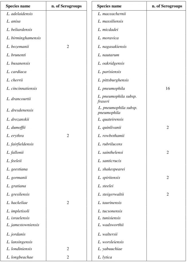
The family Legionellaceae consists of the single genus Legionella. Some investigators (Garrity et al., 1980; Brown et al., 1981) have proposed placing the legionellae in three separate genera — Legionella, Fluoribacter and Tatlockia — on the basis of low DNA hybridization values between some Legionella species [Fox et al., 1993]. However, studies based on the analysis of 16S rRNA have confirmed that the Legionellaceae family includes the single genus Legionella. To date about 61 species of Legionella are known, divided into over 70 serogroups (Table 1), [http://www.bacterio.net/legionella.htmL]. Legionella pneumophila is the species most frequently associated with human disease and includes 16 serogroups (sg) [Montagna et al., 2018; Mekkour et al., 2013; WHO, 2007]. Legionellae are facultative intracellular Gram-negative bacteria, aerobic, asporigenous bacilli, acapsulated, generally mobile due to the presence of one or more flagella, with dimensions ranging from 0.3 to 0.9 µm in width and from 1.5 to 6 µm in length. In culture Legionella forms long filamentous structures up to 20 µm [WHO, 2007]. Unlike most Gram-negative bacteria, the cell wall contains high amounts of branched-chain fatty acids and ubiquinones which make cellular staining difficult [Mekkour et al., 2013]. Concerning the biochemical properties, legionellae are urease negative, catalase positive and utilize amino acids as a source of carbon and energy rather than carbohydrates including cysteine, arginine, isoleucine and methionine. Most species produce beta-lactamases and liquefy gelatine [Fields et al., 2002]. Legionella spp. growth is stimulated by iron compounds and its cultivation in artificial media requires the addition of specific compounds such as L-cysteine, arginine, leucine, isoleucine, threonine, valine, methionine, phenylalanine, tyrosine and serine, and the addition of trace elements such as iron, calcium, cobalt, copper, magnesium, manganese, molybdenum, nickel, vanadium and zinc [Whiley et al., 2014]. Some legionellae cannot be grown on routine Legionella culture media and have been termed Legionella-like amoebal pathogens (LLAPs) [WHO, 2007]. Historically, the term Legionella-like amoebal pathogens was introduced to designate obligate intracellular parasites of free-living amoebae which were closely related to the legionellae. The term of LLAPs then has been retained for historical reasons, as most of these species have now been recognized to belong phylogenetically to the Legionella genus. Moreover, most of them are currently able to grow on BCYE agar because of the improvement in the quality
9
of media and possibly because of a progressive adaptation by successive subcultures on amoebae [Lamoth et al., 2010].
Table 1: List of Legionella species and serogroups.
Species name n. of Serogroups Species name n. of Serogroups
L. adelaidensis L. maceachernii L. anisa L. massiliensis L. beliardensis L. micdadei L. birminghamensis L. moravica L. bozemanii 2 L. nagasakiensis L. brunenti L. nautarum L. busanensis L. oakridgensis L. cardiaca L. parisiensis L. cherrii L. pittsburghensis L. cincinnatiensis L. pneumophila 16
L. drancourtii L. pneumophila subsp.
fraseri
L. dresdenensis L. pneumophila subsp. pneumophila L. drozanskii L. quateirensis L. dumoffii L. quinlivanii 2 L. erythra 2 L. rowbothamii L. fairfieldensis L. rubrilucens L. fallonii L. sainthelensi 2 L. feeleii L. santicrucis L. geestiana L. shakespearei L. gormanii L. spiritensis 2 L. gratiana L. steelei L. gresilensis L. steigerwaltii 2 L. hackeliae 2 L. taurinensis L. impletisoli L. tucsonensis L. israelensis L. tunisiensis L. jamestowniensis L. wadsworthii L. jordanis L. waltersii L. lansingensis L. worsleiensis L. londiniensis 2 L. yabuuchiae L. longbeachae 2 L. lytica
10
1.2 Ecology and environmental sources of Legionella spp.
The pathogenesis and ecology of Legionella spp. are inherently related. Legionellae are widely distributed in natural aquatic environments such as lake and river surfaces, thermal springs in which it is generally found at low concentrations. Some species have also been found in sea water and L. longbeacheae has frequently been isolated from potting soil. This species is the leading cause of legionellosis in Australia and occurs in gardeners and those exposed to commercial potting soil [Mekkour et al., 2013].
From these environments, the bacterium reaches artificial sources such as water distribution system of large buildings, humidification systems, swimming pools, decorative fountains, responsible for transmission to humans. The presence of organic sediments, rust, deposits of materials facilitates their settlement [Fields et al., 2002].
Legionella spp. can survive in a wide range of temperatures, but it prefers aquatic environments with temperatures between 25 and 42°C with optimal values around 35°C. Legionella spp. is thermotolerant and able to withstand temperatures of 50°C for several hours, while around 70°C it is rapidly destroyed. At values below 20°C Legionella spp. survives but is not more able to replicate, however when temperatures return favourable, cells begin to proliferate [Mekkour et al., 2013; WHO, 2007]. Most cases of legionellosis can be traced to human-made environments where water temperature is higher than ambient one.
The effect of pH on Legionella survival was also investigated: the bacterium has been isolated in the environment at both acid and alkaline pH values. Katz et al. demonstrated a 2 logs reduction in the number of L. pneumophila cells in mineral water after being subjected to a pH change from 4 to 7 for a month, and a 6-log decline at pH 8. Furthermore, Sheehan et al. isolated four Legionella species protected inside protozoa in the geothermal sources of the Yellowstone National Park with a pH equal to 2.7 [Katz et al., 1987; Sheehan et al., 2005].
Association with amoebae
The levels of nutrients that the legionellae require are rarely found in fresh water and may be supplied, directly or indirectly, by other species of bacteria or other associated
11
microorganisms in the form of dissolved organic constituents, through the excess of production of organic nutrients or through decay of the microorganisms. Legionellae survive in aquatic and moist soil environments as intracellular parasites of free-living protozoa: the aquatic microbial flora is generally made up of different species of bacteria, fungi and protozoa, and within these microbial communities Legionella finds an environment favourable to its development.
Legionellae have been shown to multiply in 14 species of amoebae, two species of ciliated protozoa, and one species of slime mould (Table 2) which serve as a reservoir for the multiplication and survival of legionellae and contributes to the maintenance of the pathogenetic potential and invasiveness (Figure 1) [Lau et al., 2008].
Table 2: Protozoan species found to harbour intracellular Legionella spp.
Category Species References
Amoeba Acanthamoeba castellani Rowbotham et al., 1980 Acanthamoeba culbertsoni Fields et al., 1989 Acanthamoeba hatchetti Breimanet al., 1990b Acanthamoeba polyphaga Rowbotham et al.,1980 Acanthamoeba palestinensis Rowbotham et al., 1986 Acanthamoeba royreba Tyndall et al., 1982 Amoeba proteus strain x D Park et al., 2004 Comandonia operculata Breiman et al., 1990b Echinamoeba exudans Fields et al., 1989 Filamoeba nolandi Breiman et al., 1990b Hartmannella spp. Fields et al., 1989 Hartmannella cantabrigiensis Rowbotham et al., 1986
Breiman et al., 1990b Hartmannella vermiformis Rowbotham, et al., 1986;
Fields et al., 1989; Breiman et al., 1990b Naegleria fowleri Newsome et al.,1985 Naegleria gruberi Rowbotham et al., 1980 Naegleria jadini Rowbotham et al., 1980 Naegleria lovaniensis Tyndall et al., 1982 Paratetramitus jugosis Breiman et al.,1990b Vahlkampfia spp. Breiman et al.,1990b Vahlkampfia jugosa Rowbotham et al., 1986 Vahlkampfia ustiana Breiman et al., 1990b Ciliate Tetrahymena pyriformis
Fields et al., 1984 Tetrahymena thermophila Kikuhara et al., 1994
12
Figure 1 A. polyphaga infected with L. pneumophila. The multiplication of the latter was monitored by electron microscopy after 18 (a) and 48 hours (b), [Lau et al., 2009].
Protozoa help to protect Legionella spp. from the effects of biocides used to disinfect water and it has been postulated that this can be a mechanism by which Legionella spp. is able to survive to adverse environmental conditions (such as dehydration, high temperatures, osmolarity variations and pH) and persist in heat-treated water or subjected to disinfection systems.
Biofilm
In man-made aquatic environments Legionella spp. can be found associated with biofilms (Figure 2): microbial communities that lives in close association, immersed in a polymeric matrix produced by the same microorganisms. The biofilm facilitates nutrient and gaseous exchange and protects microorganisms not only from biocides but also from periodic increases in temperature and attempts at physical removal, especially in areas where surfaces are scaled or corroded. In these artificial water systems, microbial growth is detected almost exclusively in biofilm covering the interior of pipe walls.
To date, outbreak of legionellosis have never been associated with natural aquatic environments, rather with exposure to artificial water system, such as hot water and cooling towers in which the formation of biofilms contributes to the colonization and development of Legionella spp., protecting cells from the decontamination procedure applied [Lau et al., 2009].
13
Figure 2: A scanning electron micrograph of L. pneumophila on potable water biofilms [HPSC, 2009].
1.3 Clinical presentation
Legionellosis classically presents as two distinct clinical entities, Legionnaires’ disease, a severe multisystem disease involving atypical pneumonia, and Pontiac fever, a self-limited flu-like illness. Currently, there is no consensus as to why exposure to L. pneumophila may result in either Pontiac fever or Legionnaires’ disease [Whiley et al., 2014].
Legionnaires' disease is the most severe, occurs after an incubation period ranging from 2 to 10 days with a mortality between 10-20% in healthy people and between 40-80% in hospitalized patients. It is not possible to clinically distinguish patients with Legionnaires’ disease from patients with other types of pneumonia. Signs and symptoms include fever, non-productive cough, headache, myalgias, rigors, dyspnoea, diarrhoea and delirium. About half of patients develop pus-forming sputum, and about one third develop blood-streaked sputum or cough up blood (haemoptysis) [Fields et al., 2002]. Chest X-rays often show pneumonia with consolidation in the bottom portion of both lungs.
Some patient shows gastrointestinal disorders such as diarrhoea, nausea, vomiting and abdominal pain. Moreover, almost half of patients suffer from disorders related to the nervous system, such as confusion, delirium, depression, disorientation and hallucinations. These disorders may occur in the first week of the disease. If left untreated the disease gets
14
worse during the first week and can be fatal. The most frequent complications are respiratory collapse, shock, kidney failure and multi-organ dysfunction syndrome [WHO, 2007; Mekkour et al., 2013].
Pontiac fever is a flu-like syndrome without pneumonia, characterized by fever, asthenia, headache and myalgia and manifests after a short incubation period (5-40 hours). People generally recover spontaneously after 2-5 days.
The first outbreak of Pontiac fever was caused by L. pneumophila of serogroup 1, while subsequent epidemics were attributed to L. feeleii, L. anisa and L. micdadei [Guidelines, 2015].
It has been shown by autopsy that L. pneumophila can spread from the respiratory system to the body: extrapulmonary forms are rare but have a severe and highly lethal course. Legionellae have been found in the liver, spleen, kidneys, myocardium, bones, lymph nodes and digestive tract, sporadically spreading to the nervous system [WHO, 2007]. The most common site of extrapulmonary infection is the endocardium with consequent appearance of endocarditis initially reported in the literature only in subjects with prosthetic valves but later also in native [Samuel et al., 2011].
15
1.4 Risk factors and transmission
Legionellosis infections are attributed to inhalation of contaminated water aerosols produced by infected sources (e.g. faucets, showerhead, or cooling tower) or by aspiration of contaminated water into the lungs. The ability of Legionella to access the human respiratory tract depends on the size of the aerosol: droplets with a diameter less than 10 µm stop in the nose and throat, while drops of diameter between 2-5 µm are able to enter the respiratory tract (lungs) and finally those below 2 µm reach pulmonary alveoli [Torrisi et al., 2012].Variation in the size of aerosols also affects the infectivity, which makes it difficult to determine the infectious dose and what environmental concentrations are considered acceptable [Whiley et al., 2014].
To date, the infectious dose for humans has not yet been determined, nor the reasons for which the different Legionella species have a variable virulence. However, this can be attributed to differences in surface hydrophobicity, aerosol stability and the ability to grow inside amoebas. The physiological state of Legionella spp. that causes the infection is not known either, but it can include both the stationary phase of growth and the logarithmic phase, as well as the so-called sporelike forms [Guidelines, 2015; Mekkour et al., 2013; WHO, 2007]. Infection may also occurs after inhalation of amoeba vacuoles containing Legionella spp. [Allegra et al., 2016].
Cases of legionellosis acquired by aspiration or micro-aspiration of contaminated water and cases of contagion through wound have also been reported in the literature mainly related to nosocomial Legionnaires’ disease, while human to human transmission or cases due to Legionella ingestion have never been demonstrated.
Known host risk factors for community acquired or travel associated Legionnaires’ disease (TAVLD) are smoking, chronic obstructive pulmonary disease, diabetes, alcohol abuse, older age (>50 years), and other immunosuppression. Susceptible patients for nosocomial Legionnaires’ disease include transplant recipients, other immunosuppression, surgery, cancer, diabetes, treatment with respiratory devices, chronic heart or lung disease, smoking and alcohol abuse, which are associated with higher mortality rates [WHO 2007, Whiley et al., 2014]. Environmental risk factors associated with legionellosis outbreaks are travel, residence in a health care facility, and proximity to cooling towers, whirlpool spas and decorative fountains. Any system or equipment which contains, stores, or re-circulates
non-16
sterile water that can be aerosolized is a source of legionellosis: nebulizer and humidifiers are important sources of infection, moreover the majority of legionellosis outbreaks are related to cooling towers or hot water systems of large buildings as hotel and hospitals [Farnham et al., 2014].
Environmental risk factors associated with healthcare facilities
Approximately a quarter of all reported legionnaires’ disease cases acquires their infection inside a hospital [HPSC; 2009]. Hospitals caring for immunocompromised patients such as organ or bone marrow transplant recipients are at increased risk of outbreaks of legionnaires’ disease. Hospital size may also be an important risk factor.
In hospital hot and cold water systems are the main sources of infection, factors such as water temperature, configuration, age of the water distribution systems and plumbing material encourage legionellae growth. Old components of the pipeline system, area of stagnation or low flow, dead-legs and storage tanks allow Legionella spp. survival and development [Borella et al.; 2016].
In addition, in healthcare facilities the vulnerability of hospitalized patients and health practices concerning the airways (e.g., ventilation, aspiration, devices for artificial respiration and oxygen therapy, and dental tools) significantly increase the risk for Legionella transmission and severe outcomes [Montagna et al., 2018]. In Figure 3 is shown the pathogenesis of nosocomial infection [HPSC; 2009].
17
Figure 3: The pathogenesis of nosocomial bacterial pneumonia [HPSC; 2009].
Environmental risk factors associated with recreational water
Spa-pool systems and related recreational facilities are increasingly popular and frequented by people at higher risk of infection. Such systems pose a reasonably foreseeable risk as they are a recognised source of diseases caused by infectious agents including Legionella spp. Spa pools are designed to contain water that is vigorously agitated, which leads to the formation of aerosols that can be inhaled. The water is usually maintained within the temperature range where legionellae and other infectious microorganisms can rapidly grow (20–45°C) and the high organic content of spa-pool water makes it difficult to maintain effective disinfection [HSE; 2017].
A recent review published by Leoni and co-workers summarized outbreaks and Legionella spp. cases associated with recreational aquatic environments: from 1981 to 2015, 1 079 cases of legionellosis were reported with a fatality rate of 6.3%. The most important environmental risk factors reported were inadequate water treatment and residual disinfectant below the recommended levels (Figure 4) [Leoni et al., 2018].
18
Therefore, spa-pool systems must be managed carefully to ensure a water quality level that does not encourage microbial growth and pose risks to users, people in the vicinity or passing near the spa pool. Other management strategies need to be implemented, which may include appropriate design and adequate disinfection residual and proper maintenance and cleaning of equipment as well as adequate ventilation. Features, such as water sprays, should be periodically cleaned and flushed with a level of disinfectant adequate to eliminate Legionella spp. [HSE; 2017].
Figure 4: Distribution of environmental contributing factors in 22 recreational facilities as reported by
19
1.5 Epidemiology of Legionnaires’ disease
Legionnaire’s disease remains an important cause of potentially preventable morbidity and mortality in Europe and there is no indication of decreasing burden.
Since 2010 the European surveillance of Legionnaires' disease has been carried out by ELDSNet and coordinated by the ECDC in Stockholm. The network aims to detect, control and prevent cases, clusters and outbreaks of Legionnaires’ disease in EU/European Economic Area (EEA) countries, and assist with detection and response outside these countries. The network supports the Member States and other involved countries to share information and collaborate on response actions to provide better protection from travel-associated Legionnaires’ disease, both domestically and abroad.
Data on epidemiological surveillance are collected through two schemes: the first studies all the cases reported by the 28 Member States, Iceland and Norway; the second scheme covers all travel associated cases of Legionnaires' disease including reports from countries outside Europe.
The surveillance of TALD cases is aimed at identifying clusters of cases that may not otherwise have been detected at national level, allowing a rapid adoption of corrective measures in the accommodation sites in order to prevent future infections.
Members of the ELDSNet network are appointed by their national Public Health authorities to act as national contact points for Legionnaires’ disease surveillance under the scope of ELDSNet activities. Members usually have scientific knowledge about Legionnaires’ disease or Legionella spp. bacteria and are involved in the microbiological diagnosis or epidemiological surveillance of Legionnaires' disease in their country.
At the country level, clinicians and microbiologists report individual TALD cases to their national surveillance scheme for Legionnaires’ disease. The national contact point for ELDSNet reports these cases to ECDC using the EU case definition (Table 3). With complete and rapid reporting, ELDSNet can detect clusters of cases which have a history of travel to the same accommodation site. Receipt of the information leads to specific and timely action by members in order to protect EU/EEA residents travelling in and outside of Europe. A case of legionellosis is defined travel associated if it is associated with one or more overnight stays away from home, either in the country of residence or abroad, in the 10 days before onset of illness. A case is defined nosocomial (healthcare-acquired) if occurs
20
in a patient who was in a hospital or other healthcare institution during the 10 days before onset of symptoms [ECDC, 2017].
Table 3: The current European case definition for Legionnaires’ disease stated in the Commission
Implementing Decision of 8 August 2012 [European Commission, 2012].
Legionnaire’s
disease Laboratory criteria
Clinical criteria
Confirmed cases At least one of the following three:
• Isolation of Legionella spp. from respiratory secretions or any normally sterile site
• Detection of L. pneumophila antigen in urine
• Significant rise in specific antibody level to L. pneumophila serogroup 1 in paired serum samples
Any person with pneumonia
Probable case At least one of the following four:
• Detection of L. pneumophila antigen in respiratory secretions or lung tissue e.g. by DFA staining using monoclonal-antibody derived reagents
• Detection of Legionella spp. nucleic acid in respiratory secretions, lung tissue or any normally sterile site
• Significant rise in specific antibody level to L.
pneumophila other than serogroup 1 or other Legionella spp. in paired serum samples
• Single high level of specific antibody to L. pneumophila serogroup 1 in serum
Any person with pneumonia
In 2017 the ECDC reported 9 238 cases of Legionnaires' disease, the 93% of which classified as confirmed, corresponding to 8 624 cases (Table 4). No large outbreaks contributed to the high number of reported cases: most were community acquired (69%), 21% were travel associated, 8% were associated with healthcare facilities and 2% with other settings.
L. pneumophila serogroup 1 was responsible for 801 of 1014 of culture confirmed cases (79%) [ECDC; 2019].
21
Table 4. Distribution of Legionnaires’ disease cases and rates per 100 000 population by country and year,
EU/EEA, 2014–2017.
2014 2015 2016 2017
Country Cases Rate Cases Rate Cases Rate Cases Rate Confirmed cases Austria 133 1.6 160 1.9 161 1.9 219 2.5 208 Belgium 101 0.9 118 1.1 157 1.4 235 2.1 183 Bulgaria 1 0.0 1 0.0 0 0.0 2 0.0 2 Croatia 26 0.6 48 1.1 31 0.7 33 0.8 33 Cyprus 6 0.7 2 0.2 3 0.4 1 0.1 1 Czech Republic 110 1.0 120 1.1 147 1.4 217 2.1 213 Denmark 158 2.8 185 3.3 170 3.0 278 4.8 216 Estonia 8 0.6 6 0.5 14 1.1 16 1.2 10 Finland 10 0.2 17 0.3 15 0.3 27 0.5 25 France 1 348 2.0 1 389 2.1 1 218 1.8 1 630 2.4 1 598 Germany 832 1.0 867 1.1 983 1.2 1280 1.6 1 043 Greece 27 0.2 29 0.3 31 0.3 43 0.4 43 Hungary 32 0.3 58 0.6 66 0.7 62 0.6 52 Iceland 4 1.2 1 0.3 3 0.9 3 0.9 3 Ireland 8 0.2 11 0.2 10 0.2 25 0.5 25 Italy 1 510 2.5 1 572 2.6 1 733 2.9 2 013 3.3 1 980 Latvia 38 1.9 22 1.1 24 1.2 31 1.6 24 Liechtenstein . - . - . - . - . Lithuania 8 0.3 7 0.2 11 0.4 14 0.5 11 Luxembourg 5 0.9 5 0.9 3 0.5 9 1.5 8 Malta 9 2.1 6 1.4 8 1.8 11 2.4 11 Netherlands 348 2.1 419 2.5 454 2.7 561 3.3 519 Norway 51 1.0 60 1.2 43 0.8 52 1.0 44 Poland 12 0.0 23 0.1 24 0.1 38 0.1 32 Portugal 588 5.6 145 1.4 197 1.9 232 2.3 228 Romania 1 0.0 3 0.0 2 0.0 19 0.1 15 Slovakia 14 0.3 14 0.3 14 0.3 14 0.3 12 Slovenia 59 2.9 106 5.1 93 4.5 117 5.7 117 Spain 925 2.0 1 024 2.2 951 2.0 1 363 2.9 1 349 Sweden 136 1.4 142 1.5 145 1.5 189 1.9 124 United Kingdom 370 0.6 412 0.6 383 0.6 504 0.8 331 EU/EEA 6 878 1.3 6972 1.4 7 094 1.4 9 238 1.8 5 835
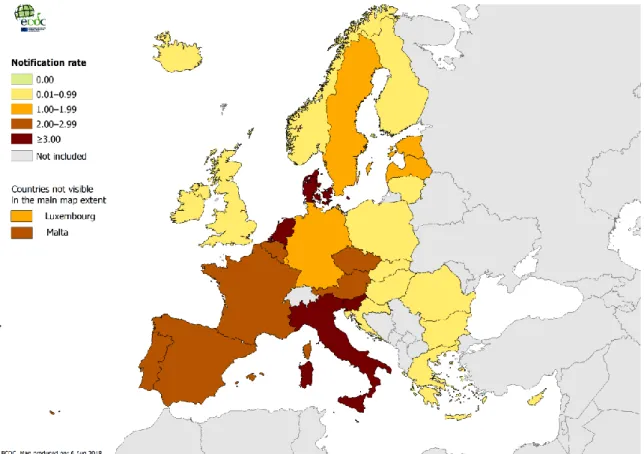
The overall notification rate per 100 000 inhabitants was 1.8 and vary a lot between each reporting country (Figure 5), which likely represents underestimation of the real incidence in all countries. Overall the notification rate continues to increase over the 2013-2017 period from 1.2 to 1.8. Respect to the previous year, there was an increment of 30% in the number of reported cases, and of 58% over the 2013-2017 period. Four countries, France, Germany, Italy and Spain, accounted for 68% of all notified cases, although their combined populations only represented approximately 50% of the EU/EEA population. The disease mostly affected male 56 years old and the overall male-to-female ratio was 2.4:1.
22
Figure 5: Distribution of Legionnaires’ disease cases per 100 000 population.
In conclusion the European situation is therefore complex, with a broad range of notification rates across countries reflecting both the quality of the national surveillance system and the local risk for Legionnaires' disease. The increasing trend is probably due to several factors, including improved surveillance systems, population aging, travel pattern and changes in climate and weather factors. Temperature, humidity and rainfall have been associated with higher incidence of disease, probably due to an effect on the bacterial ecology and/or an increased use of aerosol-producing devices or installations in the environment, such as cooling towers [ECDC, 2019].
23
1.6 Control measures for water risk management
Control measures are activities or processes applied to a system to prevent the occurrence of a hazard. Such measures are applied at control points, which are steps at which control can be applied to prevent or eliminate a water safety hazard or reduce it to an acceptable level. The strategies for preventing microbial colonization of water systems are based on technical measures implemented to make the environmental conditions in the water network unsuitable for Legionella growth.
These procedures, although they do not guarantee the complete eradication of Legionella spp., contribute considerably to reduce the risk. First, Legionella spp. prevention starts from a correct design and construction of water networks in order to make the multiplication and colonization of the bacterium unlikely.
The WHO suggests developing a water safety plan (WSP) to manage specific risks of exposure to Legionella spp. from water samples. A preliminary stage in developing a WSP is to define a team of expert with a thorough knowledge of design and operating features of the water distribution systems in order to identify hazard and risks. Any WPS would be based on a combination of different control methods.
Preventing low flow rates and stagnation of water is an essential and the system should be designed to minimize areas of stagnation. Keeping water temperature outside the ideal range for legionellae is an effective control measure for both hot and cold-water systems. Guidelines recommend that hot water should be stored above 60°C and circulated at temperature of at least 50°C while the recommended temperature for cold water is below 20°C.
When temperature controls cannot be maintained, several different disinfection procedures that can be used alone or in combination are available. Each method has advantages and disadvantages related to the ease of implementation, cost, maintenance issue, and short- and long-term effectiveness.
Water treatments are divided between physical and chemical treatments and are summarized in Table 5 [WHO, 2007; Guidelines, 2015].
24
Table 5: Advantages and disadvantages of methods for controlling Legionella in water system Treatment Type Advantages Disadvantages
Thermal shock (70-80°C)
Physical -Do not require special equipment
-Can be implemented immediately
- Difficult to maintain temperatures in old systems
-Long procedure -Short term effectiveness Heat and flush
(60°C)
Physical - Simple to use -Not completely effective -Risk of scalding
Point of use filter
Physical -Simple to use -Effective
-Require frequent replacement of filter
-Local disinfection systems UV light irradiation Physical -Easy installation
-no adverse effect on water or plumbing systems
-Effective only at point of
application; no control downstream (no residual)
-Not suitable for turbid waters -No effect on biofilm formation Hyperclorination Chemical -Effective
-Provides disinfection throughout the entire water
distribution system -Effective on biofilm formation
-Highly corrosive
-Prohibition of using water during treatment (produce potentially carcinogenic by products)
Dosing with chlorine Dioxide
Chemical - Penetrates biofilm more effectively than chlorine - Less corrosive than chlorine
- Wider pH range for activity than chlorine and Cu/Ag ionisation
-Conversion to potentially toxic chlorates and chlorites
-Corrosion of pipelines at concentration >0.4 mg/L
-Difficult to maintain the effective residual concentration
Ozone Chemical -Effective only at point of use; use can be limited to high risk areas or known contaminated taps.
-Temporary results
-Low activity on biofilm formation -Corrosive to metals
Dosing with monochloramine
Chemical - Provides a stable residual
(more stable than chlorine)
that penetrates biofilm
- Less active than chlorine.
-Low activity against protozoa (and viruses)
Copper and silver ionization
Chemical -Easy installation -Effectiveness at high water temperature -No adverse effect on water or plumbing systems
-Continuous monitoring of copper and silver ions in order to avoid that concentration exceed the limits
Dosing with hydrogenperoxide
Chemical -No adverse effect on water or plumbing systems
-There are no exhaustive tests on dynamic behaviour over time -Not suitable for galvanized steel water networks
25
1.7 Regulations and guidelines for the control and prevention of
legionellosis
In order to prevent the risk of Legionnaire's disease and ensure water safety, many international organizations have issued guidelines and/or regulations for controlling Legionella spp. in water networks which incorporate recommendations on Legionella primary prevention in the built environment, with detailed information on the appropriate prevention measures to be taken.
These organizations include both European agencies such as the WHO and the Health and Safety Executive (HSE), but also American organizations such as the Occupational Safety & Health Administration (OSHA), the Center for Disease Prevention and Control (CDC) and the Allegheny County Health Department (ACHD). There are also national guidelines in many countries that identify the necessary measures to prevent and control the risk of exposure to the Legionella spp. bacterium [HPSC, 2009; Guidelines, 2015].
Some guidelines can be applied to all types of water (HSE), while others are related to the control of legionellosis in healthcare facilities (CDC, OSHA) or to the prevention of Legionnaires' cases travel associated (EWGLI) [ACDH, 1997; WHO 2007; CDC 2003, HSE, 2000; EWGLI, 2011].
In Italy, on 7th May 2015, the State-Regions Conference sanctioned the Agreement between the Government, the Regions and the Autonomous Provinces of Trento and Bolzano, on the "Guidelines for the prevention and control of Legionnaire's disease" which brings together, updates and integrates in a single text all the indications reported in the previous national guidelines. In Italian Guidelines, it is foreseen for the first time the possibility to use a molecular method based on qPCR, developed and validated according to ISO/TS 12869, as a screening tool to quickly analyse environmental samples. qPCR can be used as a method for screening out negative samples in as quick as half a day after receipt of the sample in the laboratory, whereas positive samples must always be quantified according to culture method described in the ISO 11731:2017 [Anonymous, 2017]. The molecular approach provides information on the number of Legionella spp. genome units (GU) in the samples tested but equivalence with the number of colony forming units (CFU) has not yet been reliably established. Usually, the number of genome units is higher than the number of CFU, probably due to the presence of viable non-culturable and dead Legionella spp. cells in the samples tested [HPSC, 2009].
26
For these reasons, although the Guidelines recognize the importance of qPCR, the method cannot be considered validated. The Italian and European guidelines define the limit values for Legionella spp. concentrations in water system and suggest the corrective actions for water systems that must be immediately applied: action levels are expressed in CFU and there is no consensus on how the results obtained by one method can be compared with those obtained by the other [WHO, 2007; EWGLI, 2011; Guidelines, 2015]. Lee and co-workers conducted an international multicenter study on various types of water samples, in order to define the action thresholds of real-time PCR for the monitoring of legionellae and thereby to facilitate interpretation of environmental legionella monitoring results. They have thus proposed action and alert levels for L. pneumophila and Legionella spp. expressed as GU/L. However, the proposed levels are related to the assay used and the type of sample analysed in comparison with the culture method. As suggested by these authors, further studies are needed to derive guidelines that allow the use of qPCR in routine analyses, suggesting that in the future the cultivation method may no longer be considered the reference method [Lee et al., 2011].
27
1.8 Standard method to detect and quantify Legionellae in water
Cultivation dependent
The reference method for the detection and/or quantification of Legionella spp. in water sample is based on the cultivation of bacteria and is described in the ISO 11731-1 and ISO 11731-2 standards. The first was published in 1998 and can be apply to all types of environmental water samples, including drinking and industrial water; the ISO 11731-2 method can only be used for waters in which low bacterial contamination is suspected (eg. cold water). Following the standard method, 1000 mL of water is concentrated by filtration through 0.22 µm or 0.45 µm pore-size polycarbonate or nylon filters.
After filtration, membranes are placed into 10 mL of the original water samples and scraped to remove bacteria. Alternatively, 200 mL of samples is centrifuged, and the pellet is resuspended in 2-20 mL sterile diluent. Aliquots of the concentrate are treated with heat (30±2 min at 50±1°C) and/or acid (buffered 0.2 M HCl for 5 min) to reduce the background microflora.
The concentrate is spread on a Petri dish containing BCYE (Buffered Charcoal Yeast Extract with α-ketoglutarate, L-cysteine and ferric pyrophosphate) agar supplemented with vancomycin, polymyxin B, cycloheximide and glycine (GVPC medium).
After 7 to 10 days of incubation at 36±1°C a minimum of 5 presumptive colonies showing a greyish-white colour are streaked on both BCYE and BCYE agar without cysteine, and checked for growth after 2 days of incubation at 36±1°C. Alternative to BCYE agar without cysteine, blood agar or nutrient agar can be used.
Isolated colonies that grow only on a-BCYE agar with L-cysteine can be identified using several methods such as agglutination test or real-time PCR method complying with ISO/TS 12869 requirements. Results are expressed as CFU per litre of water samples analysed. A scheme of culture method is shown in Figure 6.
Due to the microbial complexity of environmental samples, isolating Legionella spp. by culture methods has a range of challenges: the presence of high level of contamination from background microflora can obscure the detection of Legionella spp., moreover the presence of VBNC cells can underestimate the real risk of infections. Another important shortcoming of this detection method is the long assay time that requires up to 14 days.
28
29 Cultivation independent
qPCR is an alternative method for a rapid detection and quantification of Legionella spp. from water samples. This technology amplifies and quantify a target DNA sequence, giving the number of Legionella spp. (GU) per litre of water samples analysed.
Generally, the 5S and 16S genes are used as targets for the detection of all Legionella species, while for L. pneumophila the mip gene (macrophage infectivity potentiator) is commonly used [Delgado-Viscogliosi et al., 2005].
There are available several commercial kits on the market based on qPCR technology for both Legionella spp. and L. pneumophila that have been validated according to ISO/TS 12869: the iQ-CheckTM Legionella kit (Bio-Rad, France), the mericon Quant Legionella kit (Qiagen, Germany), Aqua-Screen® L. Set Detection Kit (Minerva Biolabs, Germany), the GeneDisc® Legionella kit (Pall Corporations, France) and the Legionella spp. quantitative kit (Diatheva, Italy) [Anonymous, 2012]. The rapid turnaround time and the sensitivity of qPCR represent the main advantages when compared to culture method. However, this technology tends to overestimate due to the amplification of non-viable dead cells. Recently Legioalert (IDEXX, US) was validated according to NF148 for the detection and enumeration of L. pneumophila in drinking water and industrial water [Anonymous, 2013]. The test is based on a bacterial enzyme detection technology that signals the presence of L. pneumophila through utilization of a substrate present in the Legiolert reagent. L. pneumophila cells grow rapidly and reproduce using the rich supply of amino acids, vitamins and other nutrients present in the Legiolert reagent. Actively growing strains of L. pneumophila use the added substrate to produce a brown colour indicator. Legiolert detects L. pneumophila at 1 organism in 100 mL within 7 days [https://www.idexx.com/en/water/water-products-services/legiolert/].
30
1.9 Methods to proof the viability of Legionella spp.
The ideal scenario in most applications of microbial diagnostics is that only viable cells are detected. Theoretically, cultivation-based approach represents an excellent method as it can detect legionellae that are able to proliferate, live and infectious to humans. However, it overlooks cells that are no more culturable after stress induced by unfavourable environmental conditions (chemical disinfection, heat-treatment, UV-disinfection, limitation in nutrients). A number of techniques have been proposed; these live/dead protocols typically address one of the three aspects of microbial viability: (1) the existence of an intact, functional cell membrane, (2) the presence of cellular metabolism or energy, or (3) the possession of self-replicating DNA that can be transcribed into RNA, which, if applicable, can subsequently be translated into protein [Emerson et al., 2017].
Cell-based approach
Membrane integrity is a biomarker for viable cells because cells with compromised membranes are dead (or near). One of the most commonly used fluorescent stains to determine viability by membrane integrity is propidium iodide (PI). PI is a hydrophilic cationic molecule that can cross the damaged cell membrane and then bind to the internal nucleic acids. Because of its fluorescent properties, PI can be used to detect membrane-compromised cells via epifluorescence microscopy and flow cytometry [Alleron et al., 2008; Keserue et al., 2012]. Flow cytometry is a fast, cost-effective and potentially automatable technology: a commercial kit is available from rqmicro (Switzerland) for the quantification of live L. pneumophila cells by flow cytometry. After concentrating the water samples by filtration, the filter is resuspended in a small volume of buffer, the sample is then incubated with magnetic particles that are bound to antibodies specific for L. pneumophila sg 1-15. The target cells are isolated by immunomagnetic separation and then sample can be analysed by flow cytometry. By adding PI it is possible to quantify viable cells population.
The LIVE/DEAD BacLight Bacterial Viability Kits (ThermoFisher, US) is another commercial kit that uses PI to stain membrane-compromised cells in combination with
31
SYTO 9 (a green fluorescent total nucleic acid stain) to distinguish between dead and viable Legionella spp. cells. This approach was tested on starved [Trigui et al., 2015], heat-inactivated [Allegra et al., 2008; Nocker et al., 2011] or chemically disinfected cells [Alleron et al., 2013], but it is not a suitable technology for water samples disinfected by UV, as this treatment does not harm the cellular integrity of the cell [Kirschner, 2016]. The ScanVit Legionella® (Vermicon, Germany) is a rapid commercial system based on the VIT® Vermicon Identification Technology which consists of fluorescent in situ hybridization (FISH): DNA gene probes labelled with a fluorophore bind specifically to the sequence target site on 16S rRNA. This system allows the detection of viable cells of L. pneumophila and Legionella spp., by detecting the 16S rRNA content. The analysis procedure consists of a water sample filtration followed by treatment with acid. The filter is then placed on a plate with a selective medium and incubated for 72 h. After incubation, the membrane is transferred to a support furnished with the kit (ScanVit Reactor; Vermicon), the detection of Legionella spp. takes place on a cultivated filter brought into contact with the gene probes marked with a dye. During the ScanVIT analysis, the marked gene probes enter the bacteria and bind to the matching signatures within the cells. The membrane is then transferred to a slide and examined under a fluorescence microscope [Ditommaso et al., 2010]. However, FISH technology after UV disinfection or other scenarios such as heat or chemical treatment, is not suitable alone for monitoring viability as rRNA can be intact but cells may have compromised cell membranes [Kirschner, 2016].
Nucleic acid-based approach
An alternative approach to detect viable cells by PCR is viability PCR: concentrated water samples are pre-treated prior to DNA extraction and qPCR amplification with a nucleic acid intercalating dye (EMA, PMA, PMAxx) that selectively enters cells with compromised cell membranes, whereas an intact cell membrane presents a barrier for this molecule. Once inside a (dead) cell, the dye intercalates into the cell's DNA to which it is believed to covalently crosslink after exposure to strong visible light due to the presence of an azide group. Photolysis converts the azide group into a highly reactive nitrene radical that can react with any organic molecule in its proximity. Reaction with DNA can be assumed to occur with a high probability considering the spatial proximity of the intercalated dye and
32
this modification strongly inhibits its amplification: the nitrene group cross-links with the DNA of the membrane-compromised cells, induces a structural change in the nucleotide angle, and because of this the DNA polymerase does not bind to the DNA which leads to signal reduction (Figure 7). The remaining unbound intercalating dye is inactivated by reaction with water molecules, forming hydroxylamine that is unable to further bind to DNA molecules [Fittipaldi et al., 2012 Kumar et al., 2019].
Figure 7: Viability PCR workflow (e.g., using EMA, PMA, or similar dyes). The dye enters
compromised/dead cells, binds covalently to the DNA upon photoactivation and stops the amplification of DNA from dead cell [Kumar et al., 2019].
The first reagent that has been proposed in combination with PCR was EMA [Nogva et al., 2003], a molecule derived from ethidium bromide (EtBr), having the same basic structure as EtBr, with the addition of an azide group. In several studies EMA showed a higher cytotoxicity than PMA, due to its penetration into cells with intact membrane [Yanez et al., 2011]. This could be possible since EMA has only one positive charge and therefore more easily penetrates bacterial membranes than PMA, which has a double positive charge. However, this may also result in less efficient suppression of dead cell signals by PMA relative to EMA, because PMA may not as easily permeate cells with only slightly compromised membranes [Fittipaldi et al., 2012].
The PMA reagent is available commercially from several vendors, including Geniul (PhAST Blue), Qiagen (BLU-V PMA Viability Kit), and Biotium (PMA-Lite). In addition, a new variant on PMA, PMAxx (Biotium, Inc., Hayward, USA), has recently become
33
available, though the chemical composition of PMAxx and its relationship to PMA are proprietary [Emerson et al, 2017].
PMA has become the dye of choice as resulted to have the major specificity for dead cells [Nocker et al., 2006]: the PMA-qPCR has been applied for the quantification of viable Legionella cells from different authors [Scaturro et al., 2016; Ditommaso et al., 2014]. However, the PMA-qPCR approach is not completely efficient in the suppression in the PCR signals of dead cell’s DNA underling the need of further optimization to increase the efficacy of the protocol (Table 6) [Scaturro et al., 2016; Emerson et al.; 2017].
Table 6: Approaches to improve the efficiency of PMA-qPCR.
Parameter Description References
Amplification of longer sequences
Increasing the amplicon length increases the probability that at least one dye-binding event will have occurred, resulting in an increased suppression of signals from membrane-compromised cells
Ditommaso et al., 2015
Multiple dye treatments
Repeated sample treatment with a viability dye (i.e., the addition of dye, followed by photoactivation, then additional rounds of dye
addition and photoactivation) has been demonstrated that improve signal suppression
Kralik et al., 2010
Extending dye incubation time and concentration
Light exposure of dye treated samples is important for (i) activation of nucleic acid-bound dye and for (ii) inactivation of excess dye that has not entered cells and that could potentially bind to DNA from live cells during the DNA extraction procedure Fittipaldi et al., 2012 Incubation in the presence of facilitating substances
Co-incubation of cells with PMA and the bile salt, deoxycholate, dimethylsulfoxide (DMSO) has been shown to improve PMA to affect dye permeability through membranes
Seidel et al., 2017
Differentiation between viable and nonviable cells based on membrane integrity alone is not always sufficient as some disinfection procedures cannot be monitored by vPCR using EMA or PMA, because the damage to the cells does not directly affect membrane permeability [Nocker et al.; 2007]. A new approach has been proposed by Codony and co-workers able to discriminate between cells with an intact cell membrane and the ability to actively maintain bacterial homeostasis and cells that have an intact membrane but are metabolically inactive, extending the concept of viability so that cells must not only have intact membranes, but they must also be functional and active [Codony et al., 2015]. In this
34
case, “active” can be defined as capable of maintaining bacterial homeostasis using an active transport mechanism that requires ATP. Based on this approach the PEMAX dye was commercialized by Geniul (Spain): a double-dye reagent comprising a mix of photo-reactive azide forms of phenanthridium (Figure 8) [Codony et al., 2015; Augusti et al.; 2017].
Figure 8: Mechanism involved in v-PCR when using PEMAX dye [www.geniul.com].
As alternative to v-PCR targeting DNA, RNA could be a possible target for qPCR. Pre-rRNA synthesis in response to nutritional stimulation is exploited in a method termed Molecular Viability Testing (MVT). Pre-rRNA stimulation is very rapid and requires exposure to nutrients for 1 to 2 generation times or less (1 to 3 h for most species).
All or nearly all bacteria synthesize pre-rRNA upon nutritional stimulation, allowing the successful application of MVT to multiple diverse species. Boss and co-workers have developed a MVT method for the detection of viable L. pneumophila cells in tap water in less than 8 hours, targeting the 16S rRNA. The method proved to have a sensitivity of 91% and specificity of 97%. This procedure has the disadvantage of higher manual workload and cost compared to the culture ISO method and further research are necessary to evaluate if VBNC cells have activated their RNA synthesis during nutritional stimulation [Boss et al., 2018].
35
Aim of the present study
The aim of the present research is to develop and optimize a molecular method for the detection and/or quantification of live Legionella spp. and L. pneumophila cells in sanitary and thermal water samples based on real-time PCR technology. The first step of the research activity was focused on the evaluation of four genomic DNA extraction methods, by comparing cell lysis efficiency and quality of DNA extract from complex water samples, such as Legionella DNA extraction kit, Bacterial DNA Isolation Single Step (Diatheva, Italy) and two-in house developed methods based on chelating resin and purification by two different types of columns for ultrafiltration. Owing to the presence of organic matter and contaminants in water that can cause partial or complete inhibition of qPCR, the DNA extraction method represents a fundamental step of the molecular method. The subsequent phase was the development and optimization of the v-PCR protocol using PEMAX dye, which selectively amplifies viable cells with both active metabolisms and intact cell membrane structure. This treatment was applied after water filtration, prior to the DNA extraction and, once optimized, was tested on field samples. Results obtained highlighted the need to apply further strategies to increase the effectiveness and the selectivity of the developed protocol in the exclusive detection of live cells. For this purpose, at first an immunomagnetic separation (IMS) of L. pneumophila cells was evaluated as a purification step before v-PCR to eliminate the competing microflora. A second method involved the use of Free DNA Removal Solution (FDRS) (Biorad, France) based on DNAse I and a mixture of CMIT/MIT that inhibits microbial growth to remove free DNA from water samples prior to qPCR analysis. A final approach followed in this study, called “nutritional stimulation”, was based on the culture-enrichment of filtered water samples in Legionella specific liquid media to stimulate the growth of live Legionella cells, followed by DNA extraction and qPCR amplification. A shift between the cycle threshold (Ct) of an unstimulated and a stimulated aliquot of a sample (after incubation with liquid media) can be interpreted as the presence of viable legionella cells.
36
Chapter 2. Evaluation of robustness of DNA
extraction methods for the recovery of
Legionella spp. cells
2.1 Introduction
Environmental water samples are complex matrices that may contain PCR inhibitors that may antagonize the polymerase and decrease amplification efficiency. Moreover, PCR inhibitors present in environmental water samples may potentially lead to inaccurate target quantification or false negative results. Humic substances are the most commonly reported group of PCR inhibitors in the environmental samples and have been found to directly disturb the DNA polymerase and form colloids in water and complexes with iron ions, meaning that they could affect the ion content in PCR, probably by chelating magnesium ions. Other group of inhibitors include heavy metals, that can be present in water samples and for which the mechanisms of PCR inhibition are still not very well understood [Filion, 2012].
With the objective of applying the molecular method for quantification of Legionella spp. in water samples, the establishment of an optimal recovery is extremely important to ensure not only high sensitivity but also the consistency of results. At the same time, the quantification of the Legionella DNA must not be affected by the type and nature of water samples that are commonly analysed by laboratories. For these reasons, an accurate evaluation of DNA extraction methods was performed in order to select the best system able to recover a high amount of Legionella spp. DNA in compliance with the requirements of ISO/TS 12869.
Four DNA extraction systems were evaluated and tested for the determination of DNA extraction efficiency and the matrix effect (the possible interfering effect of some chemical components and PCR inhibitors present in water samples on DNA extraction, viability treatment or amplification) on a total of 76 water samples belonging to different water matrices (Table 7).
37
2.2. Material and Methods
2.2.1 Bacterial growth condition
L. pneumophila ATCC33152 supplied by the DSMZ (Braunschweig, Germany) were rehydrated, collected and maintained in culture as recommended by the Association Français de Normalisation (AFNOR, Paris, France) NFT90-471 and ISO/TS 12869:2012. This strain was cultured in Nutrient Broth and on Buffered Charcoal Yeast Agar supplemented with Legionella BCYE Growth Supplement (OXOID) at 37°C with 5% CO2 for 24-48 h.
2.2.2 Types of water samples analysed
During a 14-month period, a total of 75 water samples (500 mL) from different water matrixes and sampling points were collected aseptically using sterile 1 L containers containing sodium thiosulphate 20 mg/L to neutralize chlorine and transported immediately at room-temperature. Samples were categorized into 4 groups:
1. 7 cooling towers and 19 sanitary water samples collected from hot water distribution system of a local Hospital (n=26)
2. 10 domestic hot water samples (n = 10 samples),
3. 8 water samples collected from thermal pools and 7 water samples of Tabiano water (sulphurous water) (n=15)
4. 12 distilled water samples and 12 mineral water samples (Levissima) (n= 24)
All water samples were previously tested by qPCR to evaluate a natural contamination by Legionella spp. using the home method protocol A as DNA extraction system.

![Figure 1 A. polyphaga infected with L. pneumophila. The multiplication of the latter was monitored by electron microscopy after 18 (a) and 48 hours (b), [Lau et al., 2009]](https://thumb-eu.123doks.com/thumbv2/123dokorg/4775419.48115/12.892.145.781.157.417/figure-polyphaga-infected-pneumophila-multiplication-monitored-electron-microscopy.webp)
![Figure 2: A scanning electron micrograph of L. pneumophila on potable water biofilms [HPSC, 2009]](https://thumb-eu.123doks.com/thumbv2/123dokorg/4775419.48115/13.892.210.713.125.464/figure-scanning-electron-micrograph-pneumophila-potable-water-biofilms.webp)
![Figure 3: The pathogenesis of nosocomial bacterial pneumonia [HPSC; 2009].](https://thumb-eu.123doks.com/thumbv2/123dokorg/4775419.48115/17.892.141.777.107.600/figure-pathogenesis-nosocomial-bacterial-pneumonia-hpsc.webp)