Review Article
Angiogenesis Dysregulation in the Pathogenesis
of Systemic Sclerosis
Francesco Paolo Cantatore,
1Nicola Maruotti,
1Addolorata Corrado,
1and Domenico Ribatti
2,31Rheumatology Clinic, Department of Medical and Surgical Sciences, University of Foggia Medical School, Foggia, Italy
2Department of Basic Medical Sciences, Neurosciences and Sensory Organs, University of Bari Medical School, Bari, Italy
3National Cancer Institute “Giovanni Paolo II”, Bari, Italy
Correspondence should be addressed to Francesco Paolo Cantatore; [email protected] Received 16 March 2017; Revised 22 May 2017; Accepted 13 June 2017; Published 16 July 2017 Academic Editor: Toshihiro Nanki
Copyright © 2017 Francesco Paolo Cantatore et al. This is an open access article distributed under the Creative Commons Attribution License, which permits unrestricted use, distribution, and reproduction in any medium, provided the original work is properly cited.
Systemic sclerosis is a chronic autoimmune connective tissue disease characterized by vascular injury and fibrosis and by an impaired angiogenesis which cannot ensure an efficient vascular recovery. Vascular injury is responsible for hypoxia and tissual ischemia which are the primary triggers for angiogenesis and are not able to induce a compensatory angiogenesis. This review article is focused on current knowledge on the mechanisms responsible for angiogenesis dysregulation in systemic sclerosis.
1. Introduction
Systemic sclerosis (SSc) is a chronic autoimmune connective tissue disease characterized by multisystem involvement with inflammation, vasculopathy, fibrosis of the skin, and internal organs. Vascular injury is an early event in SSc pathogenesis and may be evident before the onset of fibrosis. It plays a central role in promoting Raynaud’s phenomenon, digital ulcers, renal damage, and pulmonary arterial hypertension [1–6].
The interaction between autoimmunity, vascular injury, and fibrosis promotes the progressive tissue damage. Among these factors, autoimmunity and vascular injury characterize the earliest phases of the disease and contribute to the progression of fibrosis [7–9].
Angiogenesis, the formation of newly formed capillaries from preexisting vessels via a well-programmed cascade of events, is dysregulated in SSc and cannot ensure an efficient vascular recovery. Vascular injury induces hypoxia and tis-sual ischemia which are the primary triggers for angiogenesis, but are not able to induce a compensatory angiogenesis.
2. Vascular Involvement in SSc
In early stages of SSc endothelial cell injury is followed by vascular remodeling, which is characterized by capillary enlargement, intimal proliferation, telangiectasia, and accu-mulation of proteoglycans in the arterioles. Later, during the course of the disease, loss of capillaries and small arterioles induces the formation of avascular areas [10, 11]. In larger size vessels, there is vascular occlusion and thrombosis due to endothelial proliferation, fibrotic intimal proliferation, fibrin deposition, and smooth muscle cell hypertrophy [10–12].
A typical clinical feature of SSc is the Raynaud’s phe-nomenon, which is characterized by persistent vasospasm of digital arteries with alternation of ischemia and reperfusion and an increased expression of junctional adhesion molecules (JAMs) [11]. The increase expression of JAMs promotes neutrophils and platelets attachment to endothelial cells. Neutrophils and platelets, in turn, induce the production of superoxide radicals which are responsible for endothelial cell injury [10].
Thus, in SSc an endothelial cell damage has been observed already in early phases of the disease, which probably together
Volume 2017, Article ID 5345673, 6 pages https://doi.org/10.1155/2017/5345673
with neural dysfunctions, and other intravascular alterations, is in turn involved in inducing vascular injury [3, 4, 10]. Even if etiopathogenesis of endothelial cell damage is still unclear, autoimmunity seems to be responsible for endothe-lial production of cytokines and adhesion molecules, leading to apoptosis of the endothelial cell [10, 13–15].
Moreover, an imbalance between vasodilator agents, such as nitric oxide, and vasoconstrictor agents, such as endothelin-1 (ET-1), has been observed in SSc and is involved in altered vascular permeability [15]. In fact, increased ET-1 expression is involved in inflammation, vascular fibrosis, and increased smooth muscle cell proliferation [4]. Moreover, the expression of ET-1 receptor type B, called ETB receptor, is reduced in SSc on endothelial cell surface but overexpressed on smooth muscle cells where it is responsible for fibrosis and vasoconstriction [16, 17].
Cipriani et al. [18] have demonstrated that endothelial cells in SSc, under the synergistic effect of transforming growth factor-𝛽 (TGF-𝛽) and ET-1, may transdifferenti-ate toward myofibroblasts in a process called endothelial-to-mesenchymal transition (EndoMT) by which endothe-lial cells change their morphological features and assume myofibroblast-like features. By using skin sections obtained by SSc patients, Manetti et al. [19] have recently observed that EndoMT has a key role in inducing endothelial dysfunction and dermal fibrosis in SSc. Macitentan, an ET-1 receptor antagonist, has demonstrated an inhibitory effect in vitro on EndoMT and on fibroblast activation, suggesting its potential role as new therapeutic strategy against fibrosis in SSc [18, 20]. Transdifferentiation of pericytes to myofibroblasts has also been hypothesized in SSc [21]. By considering that pericytes are cells that reside on the wall of the blood vessels and their primary function is to maintain the vessel integrity, their transdifferentiation to myofibroblasts may contribute to vessel instability.
Vascular damage may induce the onset of avascular areas and/or morphologic changes in vessel wall, such as fibrosis. These alterations play a central role in inducing tissual hypoxia which may induce digital ulcers, gangrene and amputation of the extremities, and dysfunction in several internal organs [22–26].
3. Vascular Implications and Fibrosis
Angiogenic cytokines, responsible for vessel formation and stabilization, such as vascular endothelial growth factor (VEGF), TGF-𝛽, and platelet derived growth factor (PDGF), are also involved in fibrosis. An impaired cross-talk between endothelial cells and perivascular cells, such as pericytes, may induce an abnormal expression of these angiogenic factors in SSc [27]. This is responsible for subsequent vessel instability and perivascular-myofibroblast phenotypic transdifferentia-tion, which contribute to fibrosis in the skin and internal organs and to the loss of peripheral vascularization [27].
4. The Role of Imbalance between Angiogenic
and Antiangiogenic Factors
An imbalance between angiogenic and antiangiogenic factors has been observed in several autoimmune diseases [28–30].
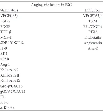
Table 1: Angiogenic and antiangiogenic agents involved in SSc: imbalance between these factors is responsible for impaired angio-genesis. Angiogenic factors in SSC Stimulators Inhibitors VEGF(165) VEGF(165)b FGF-2 TSP-1 PDGF PF4/CXCL4 TGF-𝛽 PTX3 MCP-1 Endostatin SDF-1/CXCL12 Angiostatin IL-8 Ang-2 ET-1 uPAR Ang-1 Kallikrein 9 Kallikrein 11 Kallikrein 12 Gro-𝛾/CXCL3 gGCP-2/CXCL6 Fli1 Fra-2 𝛼-Klotho
(VEGF: vascular endothelial growth factor; FGF-2: fibroblast growth facor-2; PDGF: platelet derived growth factor; TGF-𝛽: transforming growth factor-𝛽; MCP-1: monocyte chemoattractant protein-1; SDF-1: stromal cell-derived factor 1; IL-8: interleukin-8; ET-1: endothelin-1; uPAR: urokinase type plas-minogen activator receptors; Ang: Angiopoietin; Gro-𝛾: growth-regulated protein-𝛾; gGCP-2: granulocyte chemotactic protein 2; Fli1: Friend leukemia integration-1; Fra-2: Fos-related antigen 2; TSP-1: thrombospondin-1; PF4: platelet factor 4; PTX3: pentraxin 3).
In SSc a dysregulation of some angiogenic factors, such as VEGF, fibroblast growth factor-2 (FGF-2), PDGF, TGF-𝛽, monocyte chemoattractant protein-1 (MCP-1), stromal cell-derived factor 1 (SDF-1/CXCL12), interleukin (IL)-8, ET-1, and urokinase type plasminogen activator receptors (uPAR), and some antiangiogenic factors, such as angio-statin, thrombospondin-1 (TSP-1), endoangio-statin, platelet factor 4 (PF4/CXCL4), IL-4, and pentraxin 3 (PTX3), has been described (Table 1) [10, 11, 30–36].
High levels of VEGF have been demonstrated in SSc, in spite of an inadequate angiogenesis [37, 38]. Neverthe-less, by considering that previous studies did not distin-guish between proangiogenic VEGF(165) and antiangiogenic VEGF(165)b isoforms, originated by alternative splicing in the terminal exon of VEGF pre-RNA, Manetti et al. [39] have observed that a switch from proangiogenic to antiangio-genic VEGF isoforms may be responsible for the inefficient angiogenic response in SSc. Recently, increased production of VEGF(165)b has been found in platelets isolated from SSc patients, suggesting a role for platelets in insufficient angiogenesis [40].
Even if numerous angiogenic factors are overexpressed in SSc, reduced levels of Angiopoietin-1 (Ang-1) have been observed in sera of patients with SSc, whereas Ang-2, an antagonist of Ang-1, was upregulated. Moreover, reduced
levels of Kallikreins 9, 11, and 12, three serine proteases with angiogenic activity, have been observed in SSc endothelial cell [41, 42].
An increased production of antiangiogenic factors, such as endostatin and angiostatin, has been observed in SSc. Recent evidence shows that endostatin levels are increased in all phases of the disease while angiostatin levels are significantly elevated in late disease and are correlated to lung disease severity [43].
5. Reduced Expression of Receptors for
Angiogenic Factors
A possible role in the lack of response to angiogenic factors in SSc, despite their overexpression, has been suggested for the reduced expression of some receptors on cell membrane. In fact, a reduced expression of stromal cell-derived factor 1 (SDF1), an angiogenic factor also known as CXC motif chemokine 12 (CXCL12), and its receptor CXCR4 has been found in later stages of disease skin biopsy samples from SSc patients, while they were upregulated in the skin of patients with early SSc, playing probably a role in the inadequate angiogenic response [44]. Nevertheless, contrasting results for VEGF receptor-1 (VEGFR-1) and VEGFR-2 expression have been described in SSc endothelial cells [45–49]. More-over, overexpression of VEGFR-3 and chemokine receptors, such as CXCR2 (receptor of IL-8) and CXCR6 (receptor of CXCL6), has been found in endothelial cells and dermal fibroblasts isolated from SSc patients [44, 50, 51]. Recently, Tsou et al. [52] have found that increased expression of angiogenic chemokines, such as growth-regulated protein-𝛾 (Gro-protein-𝛾/CXCL3) and granulocyte chemotactic protein 2 (GCP-2/CXCL6) in serum and endothelial cells obtained from SSc patients, was unable to induce angiogenesis.
6. Impaired Expression of Angiogenic
Transcription Factors
Another hypothesis to explain the lack of response to angiogenic factors in SSc is the impaired expression of angiogenic transcription factors, such as Friend leukemia integration-1 (Fli1) and Fos-related antigen 2 (Fra-2). Fli1 acts as a suppressor of collagen transcription in human skin as demonstrated in vivo. The persistent reduced expression of Fli1 in SSc fibroblast cultures has been correlated to abnormal matrix deposition in scleroderma skin. Low Fli1 levels have been correlated to the detachment of preexisting pericytes, extracellular matrix degradation by endothelial proteinases, enhanced migration, proliferation, and cell survival. On the contrary, Fli1 deficiency plays a role in inhibiting tube formation of endothelial cells, suggesting that Fli1 deficiency is probably a consequence of both proliferative obliterative vasculopathy, characterized by occlusion of arterioles and small arteries, and destructive vasculopathy, characterized by loss of small vessels, which are the typical alterations in SSc vasculopathy [53]. On the other hand, high levels of Fra-2 have been seen in SSc patients, and its overexpression has been correlated to increased profibrotic effects of TGF-𝛽 and PDGF [54–56].
7. JAM-A
The reduced expression of JAM-A on endothelial cells surface decreases FGF-2 induced angiogenesis [57] and has been correlated to an increased cleavage of IL-8 and uPAR, two angiogenic factors which are responsible for endothelial cell proliferation, extracellular matrix degradation, and the adhe-sion of endothelial cells to the extracellular matrix, by MMPs overexpression in fibroblast and endothelial cell [58–60].
8. Genetic Polymorphisms
Genetic polymorphisms may also be involved in SSc pathogenesis. Gene polymorphism of uPAR, called UPAR rs344781, has been associated with increased risk of vascular injury in SSc, while gene polymorphism of MMP-12, named MMP-12 rs2276109, has been correlated with diffuse cuta-neous SSc and pulmonary fibrosis [61, 62]. Moreover, an increased expression of histone deacetylases-5, an enzyme involved in the control of genes associated with angiogenesis regulation, has been observed in endothelial cells from SSc patients, suggesting a potential role for epigenetic modifica-tion in impaired angiogenesis [63].
9.
𝛼-Klotho
Recently, a role for𝛼-klotho, a pleiotropic protein, originally
described as an antiaging factor, has been suggested in SSc pathogenesis by acting as a powerful proangiogenic factor. This factor plays important pleotropic effects on endothelial cells, by interacting with VEGFR-2 and transient receptor potential canonical-1 (TRPC-1) cation channel to control cellular homeostasis [64]. Mazzotta et al. [65] have found that 𝛼-klotho is significantly decreased in the microvasculature in SSc skin and that its administration may efficiently improve dermal microvascular endothelial cells from SSc patients functions in vitro.
10. A Link between Vascular and
Nervous System
Emerging evidences underline the link between vascular and nervous system. In fact, factors responsible for transmitting axonal guidance cues, such members of class III semaphorin (Sema3) family, play an antiangiogenic role in physiological and pathological vascular development. These factors are involved in reducing cell adhesion by disrupting integrin-mediated adhesive structures, resulting in a filopodial retrac-tion in endothelial cells. Recently, by using dermal microvas-cular endothelial cell cultures from SSc patients, Mazzotta et al. [66] have suggested that a member of Sema family, named Sema3E, by binding to its receptor Plexin-D1 plays probably a role in the dysregulation of angiogenesis and vascular tone control by inducing neurovascular mechanism alterations which are clinically evident above all in the early stage of the disease. A low expression of neuropilin-1, a receptor for both Sema3s and VEGF-A, has been observed in SSc, suggesting a further additional factor involved in impaired angiogenesis [67].
11. The Role of Mesenchymal Stem
Cells (MSCs) in the Vascular Alteration
during SSc: Therapeutic Implications
In SSc patients, MSCs are characterized by senescence [68]. Nevertheless, MSCs may preserve immunomodulatory abil-ity, which might have potential therapeutic implications in SSc. In fact, Cipriani et al. [68] have found increased levels of IL-6 and TGF-𝛽 in SSc-MSCs. On one hand, increased levels of IL-6 have been considered as an adaptive mechanism to senescence and are responsible for immunosuppressive effects. On the other hand, increased levels of TGF-𝛽 may be involved in determining both immunosuppressive effect on lymphocyte proliferation and immunoregulatory effects, via inducing expression of CD69 on T cells surface [68]. Moreover, MSCs may differentiate into endothelial cells [69], suggesting a potential therapeutic role in vascular alteration during SSc.
Different sclerotic conditions, including localized scle-roderma, have been effectively treated with autologous fat tissue grafting (AFTG). In patients affected by advanced SSc-related perioral thickening and mouth opening limitation, AFTG of the lips has demonstrated an improvement of mouth opening [70]. The efficacy of this treatment has been correlated to the presence of a stem cell population, called adipose-derived MSCs (ATDMSCs) in the adipose tissue. In fact, ATDMSCs may differentiate into endothelial cells and produce angiogenic factors, suggesting a potential role in promoting angiogenesis [71]. ATDMSCs exert also several immunosuppressive and anti-inflammatory effects by inhibiting both proliferation of T and B cells, and the expression of numerous proinflammatory cytokines [72]. Furthermore, adiponectin expression from adipose tissue is responsible for antifibrotic effects [73].
12. Concluding Remarks
SSc in the earliest stages is characterized by morphologic alterations in vessel walls, such as fibrosis and capillary loss. Endothelial cell injury plays a central role in promoting these changes, which are responsible for inducing hypoxia. These events lead to an increased angiogenesis. Nevertheless, in SSc patients angiogenesis is not compensatory. The reason of this inefficient angiogenesis in SSc is still unclear. Nevertheless, an imbalance between angiogenic and antiangiogenic factors and a reduced expression of some receptors or cofactors of angiogenic agents has been suggested.
Even if further studies are needed to explain the role of angiogenesis in the pathogenesis of SSc and to elucidate the mechanism responsible for angiogenesis dysregulation, endothelial cell injury and angiogenesis dysregulation seem to play a central role in the pathogenesis of SSc. This may provide a basis for a rational approach to the development of new therapeutic strategy to ensure efficient angiogenesis.
Conflicts of Interest
The authors declare that there are no conflicts of interest.
References
[1] Y. Asano and S. Sato, “Vasculopathy in scleroderma,” Seminars in Immunopathology, vol. 37, no. 5, pp. 489–500, 2015. [2] M. Matucci-Cerinic, B. Kahaleh, and F. M. Wigley, “Review:
evidence that systemic sclerosis is a vascular disease,” Arthritis & Rheumatology, vol. 65, pp. 1953–1962, 2013.
[3] M. B. Kahaleh, “Vascular involvement in systemic sclerosis (SSc),” Clinical and Experimental Rheumatology, vol. 22, pp. S19–S23, 2004.
[4] D. Abraham and O. Distler, “How does endothelial cell injury start? The role of endothelin in systemic sclerosis,” Arthritis Research & Therapy, vol. 9, no. 2, article S2, 2007.
[5] A. Gabrielli, E. V. Avvedimento, and T. N. Krieg, “Scleroderma,” The New England Journal of Medicine, vol. 360, no. 19, pp. 1989– 2003, 2009.
[6] P. Cipriani, A. Marrelli, V. Liakouli, P. Di Benedetto, and R. Giacomelli, “Cellular players in angiogenesis during the course of systemic sclerosis,” Autoimmunity Reviews, vol. 10, no. 10, pp. 641–646, 2011.
[7] D. J. Abraham and J. Varga, “Scleroderma: From cell and molec-ular mechanisms to disease models,” Trends in Immunology, vol. 26, no. 11, pp. 587–595, 2005.
[8] A. Gabrielli, S. Svegliati, G. Moroncini, and E. V. Avvedimento, “Pathogenic autoantibodies in systemic sclerosis,” Current Opinion in Immunology, vol. 19, no. 6, pp. 640–645, 2007. [9] N. Hunzelmann and J. Brinckmann, “What are the new
mile-stones in the pathogenesis of systemic sclerosis?” Annals of the Rheumatic Diseases, vol. 69, no. 1, pp. i52–i56, 2010.
[10] D. Pattanaik, M. Brown, B. C. Postlethwaite, and A. E. Postlethwaite, “Pathogenesis of systemic sclerosis,” Frontiers in Immunology, vol. 6, article no. 272, 2015.
[11] B. J. Rabquer and A. E. Koch, “Angiogenesis and vasculopathy in systemic sclerosis: Evolving concepts,” Current Rheumatology Reports, vol. 14, no. 1, pp. 56–63, 2012.
[12] S. A. Jimenez, “Role of endothelial to mesenchymal transition in the pathogenesis of the vascular alterations in systemic sclerosis,” ISRN Rheumatology, vol. 2013, Article ID 835948, 15 pages, 2013.
[13] C. Lunardi, C. Bason, R. Navone et al., “Systemic scle-rosis immunoglobulin G autoantibodies bind the human cytomegalovirus late protein UL94 and induce apoptosis in human endothelial cells,” Nature Medicine, vol. 6, no. 10, pp. 1183–1186, 2000.
[14] S. S. Ahmed, F. K. Tan, F. C. Arnett, L. Jin, and Y.-J. Geng, “Induction of apoptosis and fibrillin 1 expression in human dermal endothelial cells by scleroderma sera containing anti-endothelial cell antibodies,” Arthritis & Rheumatology, vol. 54, pp. 2250–2262, 2006.
[15] B. Kahaleh, “Vascular disease in scleroderma: mechanisms of vascular injury,” Rheumatic Disease Clinics of North America, vol. 34, no. 1, pp. 57–71, 2008.
[16] D. J. Abraham, R. Vancheeswaran, M. R. Dashwood et al., “Increased levels of endothelin-1 and differential endothelin type A and B receptor expression in scleroderma-associated fibrotic lung disease,” American Journal of Pathology, vol. 151, pp. 831–841, 1997.
[17] M. Bauer, H. Wilkens, F. Langer, S. O. Schneider, H. Lausberg, and H.-J. Sch¨afers, “Selective upregulation of endothelin B receptor gene expression in severe pulmonary hypertension,” Circulation, vol. 105, no. 9, pp. 1034–1036, 2002.
[18] P. Cipriani, P. Di Benedetto, P. Ruscitti et al., “The endothelial-mesenchymal transition in systemic sclerosis is induced by endothelin-1 and transforming growth factor-𝛽 and may be blocked by Macitentan, a dual endothelin-1 receptor antago-nist,” Journal of Rheumatology, vol. 42, no. 10, pp. 1808–1816, 2015.
[19] M. Manetti, E. Romano, I. Rosa et al., “Endothelial-to-mesen-chymal transition contributes to endothelial dysfunction and dermal fibrosis in systemic sclerosis,” Annals of the Rheumatic Diseases, vol. 76, pp. 924–934, 2017.
[20] P. Cipriani, P. Di Benedetto, P. Ruscitti et al., “Macitentan inhibits the transforming growth factor-𝛽 profibrotic action, blocking the signaling mediated by the ETR/T𝛽RI complex in systemic sclerosis dermal fibroblasts,” Arthritis Research and Therapy, vol. 17, p. 247, 2015.
[21] P. Cipriani, A. Marrelli, P. D. Benedetto et al., “Scleroderma Mesenchymal Stem Cells display a different phenotype from healthy controls; Implications for regenerative medicine,” Angi-ogenesis, vol. 16, no. 3, pp. 595–607, 2013.
[22] M. Humbert, N. W. Morrell, S. L. Archer et al., “Cellular and molecular pathobiology of pulmonary arterial hypertension,” Journal of the American College of Cardiology, vol. 43, no. 12, pp. 13S–24S, 2004.
[23] S. Guiducci, R. Giacomelli, and M. M. Cerinic, “Vascular com-plications of scleroderma,” Autoimmunity Reviews, vol. 6, no. 8, pp. 520–523, 2007.
[24] S. I. Nihtyanova, G. M. Brough, C. M. Black, and C. P. Denton, “Clinical burden of digital vasculopathy in limited and diffuse cutaneous systemic sclerosis,” Annals of the Rheumatic Diseases, vol. 67, no. 1, pp. 120–123, 2008.
[25] C. P. Denton, G. Lapadula, L. Mouthon, and U. M¨uller-Ladner, “Renal complications and scleroderma renal crisis,” Rheu-matology, Oxford, vol. 48, pp. iii32–iii35, 2009.
[26] M. Hinchcliff, C. S. Desai, J. Varga, and S. J. Shah, “Prevalence, prognosis, and factors associated with left ventricular diastolic dysfunction in systemic sclerosis,” Clinical and Experimental Rheumatology, vol. 30, pp. S30–S37, 2012.
[27] P. Cipriani, P. Di Benedetto, P. Ruscitti et al., “Impaired endothelium-mesenchymal stem cells cross-talk in systemic sclerosis: a link between vascular and fibrotic features,” Arthritis research & therapy, vol. 16, p. 442, 2014.
[28] N. Maruotti, F. P. Cantatore, E. Crivellato, A. Vacca, and D. Ribatti, “Angiogenesis in rheumatoid arthritis,” Histology and Histopathology, vol. 21, pp. 557–566, 2006.
[29] N. Maruotti, F. P. Cantatore, B. Nico, A. Vacca, and D. Rib-atti, “Angiogenesis in vasculitides,” Clinical and Experimental Rheumatology, vol. 26, pp. 476–483, 2008.
[30] V. Liakouli, P. Cipriani, A. Marrelli, S. Alvaro, P. Ruscitti, and R. Giacomelli, “Angiogenic cytokines and growth factors in systemic sclerosis,” Autoimmunity Reviews, vol. 10, no. 10, pp. 590–594, 2011.
[31] O. Distler, A. Del Rosso, and R. Giacomelli, “Angiogenic and angiostatic factors in systemic sclerosis: increased levels of vascular endothelial growth factor are a feature of the earliest disease stages and are associated with the absence of fingertip ulcers,” Arthritis Research, vol. 4, p. R11, 2002.
[32] R. F. Macko, A. C. Gelber, B. A. Young et al., “Increased circu-lating concentrations of the counteradhesive proteins SPARC and thrombospondin-1 in systemic sclerosis (scleroderma). Relationship to platelet and endothelial cell activation,” The Journal of Rheumatology, vol. 29, no. 12, pp. 2565–2570, 2002.
[33] B. Giusti, G. Fibbi, F. Margheri et al., “A model of
anti-angiogenesis: Differential transcriptosome profiling of
microvascular endothelial cells from diffuse systemic sclerosis patients,” Arthritis Research and Therapy, vol. 8, p. R115, 2006. [34] M. J. Mulligan-Kehoe, M. C. Drinane, J. Mollmark et al.,
“Antiangiogenic plasma activity in patients with systemic scle-rosis,” Arthritis & Rheumatology, vol. 56, pp. 3448–3458, 2007. [35] P. Cipriani, P. Di Benedetto, H. Dietrich et al., “Searching for
a good model for systemic sclerosis: The molecular profile and vascular changes occurring in UCD-200 chickens strongly resemble the early phase of human systemic sclerosis,” Archives of Medical Science, vol. 12, no. 4, pp. 828–843, 2016.
[36] U. ˙Ilgen, M. E. Yayla, and N. D¨uzg¨un, “Low serum fibroblast growth factor 2 levels not accompanied by increased serum pentraxin 3 levels in patients with systemic sclerosis,” Clinical Rheumatology, pp. 1–6, 2016.
[37] M. Bielecki, K. Kowal, A. Lapinska, S. Chwiesko-Minarowska, L. Chyczewski, and O. Kowal-Bielecka, “Peripheral blood mononuclear cells from patients with systemic sclerosis spon-taneously secrete increased amounts of vascular endothelial growth factor (VEGF) already in the early stage of the disease,” Advances in Medical Sciences, vol. 56, no. 2, pp. 255–263, 2011. [38] B. Maurer, A. Distler, Yossra A Suliman et al., “Vascular
endothelial growth factor aggravates fibrosis and vasculopathy in experimental models of systemic sclerosis,” Annals of the Rheumatic Diseases, 2013.
[39] M. Manetti, S. Guiducci, E. Romano et al., “Overexpression of
VEGF165b, an inhibitory splice variant of vascular endothelial
growth factor, leads to insufficient angiogenesis in patients with systemic sclerosis,” Circulation Research, vol. 109, no. 3, pp. e14– e26, 2011.
[40] D. Hirigoyen, P. I. Burgos, V. Mezzano et al., “Inhibition of angi-ogenesis by platelets in systemic sclerosis patients,” Arthritis Research & Therapy, vol. 17, p. 332, 2015.
[41] B. Giusti, S. Serrati, F. Margheri et al., “The antiangiogenic tissue kallikrein pattern of endothelial cells in systemic sclerosis,” Arthritis & Rheumatology, vol. 52, pp. 3618–3628, 2005. [42] M. Michalska-Jakubus, O. Kowal-Bielecka, G. Chodorowska,
M. Bielecki, and D. Krasowska, “Angiopoietins-1 and -2 are differentially expressed in the sera of patients with systemic sclerosis: High angiopoietin-2 levels are associated with greater severity and higher activity of the disease,” Rheumatology, vol. 50, no. 4, Article ID keq392, pp. 746–755, 2011.
[43] I. Almeida, A. O. Gomes, M. Lima, I. Silva, and C. Vasconce-los, “Different contributions of angiostatin and endostatin in angiogenesis impairment in systemic sclerosis: A cohort study,” Clinical and Experimental Rheumatology, vol. 34, pp. 37–42, 2016.
[44] P. Cipriani, A. F. Milia, V. Liakouli et al., “Differential expression of stromal cell-derived factor 1 and its receptor CXCR4 in the skin and endothelial cells of systemic sclerosis patients: Pathogenetic implications,” Arthritis and Rheumatism, vol. 54, pp. 3022–3033, 2006.
[45] Z. Mackiewicz, A. Sukura, D. Povilenait´e et al., “Increased but imbalanced expression of VEGF and its receptors has no positive effect on angiogenesis in systemic sclerosis skin,” Clinical and Experimental Rheumatology, vol. 20, pp. 641–646, 2002.
[46] O. Distler, J. H. W. Distler, A. Scheid et al., “Uncontrolled expression of vascular endothelial growth factor and its recep-tors leads to insufficient skin angiogenesis in patients with
systemic sclerosis,” Circulation Research, vol. 95, no. 1, pp. 109– 116, 2004.
[47] C. A. Davies, M. Jeziorska, A. J. Freemont, and A. L. Herrick, “The differential expression of VEGF, VEGFR-2, and GLUT-1 proteins in disease subtypes of systemic sclerosis,” Human Pathology, vol. 37, no. 2, pp. 190–197, 2006.
[48] J. Avouac, J. Wipff, O. Goldman et al., “Angiogenesis in systemic sclerosis: Impaired expression of vascular endothelial growth factor receptor 1 in endothelial progenitor-derived cells under hypoxic conditions,” Arthritis & Rheumatology, vol. 58, pp. 3550–3561, 2008.
[49] N. Higashi-Kuwata, T. Makino, Y. Inoue, and H. Ihn, “Expres-sion pattern of VEGFR-1, -2, -3 and D2-40 protein in the skin of patients with systemic sclerosis,” European Journal of Dermatology, vol. 21, pp. 490–494, 2011.
[50] M. T. Carulli, V. H. Ong, M. Ponticos et al., “Chemokine receptor CCR2 expression by systemic sclerosis fibroblasts: Evi-dence for autocrine regulation of myofibroblast differentiation,” Arthritis & Rheumatology, vol. 52, pp. 3772–3782, 2005. [51] B. J. Rabquer, P.-S. Tsou, Y. Hou et al., “Dysregulated expression
of MIG/CXCL9, IP-10/CXCL10 and CXCL16 and their receptors in systemic sclerosis,” Arthritis Research & Therapy, vol. 13, p. R18, 2011.
[52] P.-S. Tsou, B. J. Rabquer, R. A. Ohara et al., “Scleroderma dermal microvascular endothelial cells exhibit defective response to pro-angiogenic chemokines,” Rheumatology (Oxford, England), vol. 55, no. 4, pp. 745–754, 2016.
[53] T. Toyama, Y. Asano, T. Miyagawa et al., “The impact of transcriptional factor Fli1 deficiency on the regulation of angio-genesis,” Experimental Dermatology, 2017.
[54] N. Reich, B. Maurer, A. Akhmetshina et al., “The transcription factor Fra-2 regulates the production of extracellular matrix in systemic sclerosis,” Arthritis & Rheumatology, vol. 62, pp. 280– 290, 2010.
[55] M. Kubo, J. Czuwara-Ladykowska, O. Moussa et al., “Persistent down-regulation of Fli1, a suppressor of collagen transcription, in fibrotic scleroderma skin,” American Journal of Pathology, vol. 163, pp. 571–581, 2003.
[56] Y. Asano, “Epigenetic suppression of Fli1, a potential pre-disposing factor in the pathogenesis of systemic sclerosis,” International Journal of Biochemistry & Cell Biology, vol. 67, pp. 86–91, 2015.
[57] Y. Hou, B. J. Rabquer, M. L. Gerber et al., “Junctional adhesion molecule-A is abnormally expressed in diffuse cutaneous sys-temic sclerosis skin and mediates myeloid cell adhesion,” Annals of the Rheumatic Diseases, vol. 69, no. 1, pp. 249–254, 2010. [58] S. D’Alessio, G. Fibbi, M. Cinelli et al., “Matrix
metallopro-teinase 12-dependent cleavage of urokinase receptor in systemic sclerosis microvascular endothelial cells results in impaired angiogenesis,” Arthritis & Rheumatology, vol. 50, pp. 3275–3285, 2004.
[59] F. Margheri, M. Manetti, and S. Serrat`ı, “Domain 1 of the urokinase-type plasminogen activator receptor is required for
its morphologic and functional,𝛽2 integrin-mediated
connec-tion with actin cytoskeleton in human microvascular endothe-lial cells: Failure of association in systemic sclerosis endotheendothe-lial cells,” Arthritis and Rheumatism, vol. 54, pp. 3926–3938, 2006. [60] R. A. Dean, J. H. Cox, C. L. Bellac, A. Doucet, A. E. Starr, and
C. M. Overall, “Macrophage-specific metalloelastase (MMP-12) truncates and inactivates ELR + CXC chemokines and generates CCL2, -7, -8, and -13 antagonists: Potential role of
the macrophage in terminating polymorphonuclear leukocyte influx,” Blood, vol. 112, no. 8, pp. 3455–3464, 2008.
[61] M. Manetti, L. Ibba-Manneschi, C. Fatini et al., “Association of a functional polymorphism in the matrix metalloproteinase-12 promoter region with systemic sclerosis in an Italian popula-tion,” Journal of Rheumatology, vol. 37, no. 9, pp. 1852–1857, 2010. [62] M. Manetti, Y. Allanore, L. Revillod et al., “A genetic variation located in the promoter region of the UPAR (CD87) gene is associated with the vascular complications of systemic sclero-sis,” Arthritis & Rheumatology, vol. 63, pp. 247–256, 2011. [63] P.-S. Tsou, J. D. Wren, M. A. Amin et al., “Histone deacetylase
5 is overexpressed in scleroderma endothelial cells and impairs angiogenesis via repression of proangiogenic factors,” Arthritis & Rheumatology, vol. 68, pp. 2975–2985, 2016.
[64] T. Kusaba, M. Okigaki, A. Matui et al., “Klotho is associated with VEGF receptor-2 and the transient receptor potential canonical-1 Ca2+channel to maintain endothelial integrity,” Proceedings of the National Academy of Sciences of the United States of America, vol. 107, no. 45, pp. 19308–19313, 2010. [65] C. Mazzotta, M. Manetti, I. Rosa et al., “Proangiogenic effects
of soluble a-Klotho on systemic sclerosis dermal microvascular endothelial cells,” Arthritis Research & Therapy, vol. 19, p. 27, 2017.
[66] C. Mazzotta, E. Romano, C. Bruni et al., “Plexin-D1/Sema-phorin 3E pathway may contribute to dysregulation of vascular tone control and defective angiogenesis in systemic sclerosis,” Arthritis Research & Therapy, vol. 17, p. 221, 2015.
[67] E. Romano, I. Chora, M. Manetti et al., “Decreased expression of neuropilin-1 as a novel key factor contributing to peripheral microvasculopathy and defective angiogenesis in systemic scle-rosis,” Annals of the Rheumatic Diseases, 2015.
[68] P. Cipriani, P. Di Benedetto, V. Liakouli et al., “Mesenchymal stem cells (MSCs) from scleroderma patients (SSc) preserve their immunomodulatory properties although senescent and normally induce T regulatory cells (Tregs) with a functional phenotype: implications for cellular-based therapy,” Clinical and Experimental Immunology, vol. 173, no. 2, pp. 195–206, 2013. [69] J. Oswald, S. Boxberger, B. Jørgensen et al., “Mesenchymal stem
cells can be differentiated into endothelial cells in vitro,” Stem Cells, vol. 22, no. 3, pp. 377–384, 2004.
[70] N. Del Papa, F. Caviggioli, D. Sambataro et al., “Autologous fat grafting in the treatment of fibrotic perioral changes in patients with systemic sclerosis,” Cell Transplantation, vol. 24, no. 1, pp. 63–72, 2015.
[71] M. Takahashi, “Adipose tissue: - An alternative source for therapeutic angiogenesis,” Circulation Journal, vol. 76, no. 7, pp. 1597-1598, 2012.
[72] T. Yi and S. U. Song, “Immunomodulatory properties of mesen-chymal stem cells and their therapeutic applications,” Archives of Pharmacal Research, vol. 35, no. 2, pp. 213–221, 2012. [73] F. Fang, L. Liu, Y. Yang et al., “The adipokine adiponectin has
potent anti-fibrotic effects mediated via adenosine monophos-phate-activated protein kinase: novel target for fibrosis therapy,” Arthritis Research and Therapy, vol. 14, article R229, 2012.
Submit your manuscripts at
https://www.hindawi.com
Stem Cells
International
Hindawi Publishing Corporationhttp://www.hindawi.com Volume 2014
Hindawi Publishing Corporation
http://www.hindawi.com Volume 2014
INFLAMMATION
Hindawi Publishing Corporation
http://www.hindawi.com Volume 2014
Behavioural
Neurology
Endocrinology
International Journal of Hindawi Publishing Corporationhttp://www.hindawi.com Volume 2014
Hindawi Publishing Corporation
http://www.hindawi.com Volume 2014
Disease Markers
Hindawi Publishing Corporation
http://www.hindawi.com Volume 2014
BioMed
Research International
Oncology
Journal ofHindawi Publishing Corporation
http://www.hindawi.com Volume 2014
Hindawi Publishing Corporation
http://www.hindawi.com Volume 2014
Oxidative Medicine and Cellular Longevity
Hindawi Publishing Corporation
http://www.hindawi.com Volume 2014
PPAR Research
The Scientific
World Journal
Hindawi Publishing Corporation
http://www.hindawi.com Volume 2014
Immunology Research
Hindawi Publishing Corporation
http://www.hindawi.com Volume 2014
Journal of
Obesity
Journal ofHindawi Publishing Corporation
http://www.hindawi.com Volume 2014
Hindawi Publishing Corporation
http://www.hindawi.com Volume 2014
Computational and Mathematical Methods in Medicine
Ophthalmology
Journal ofHindawi Publishing Corporation
http://www.hindawi.com Volume 2014
Diabetes Research
Journal ofHindawi Publishing Corporation
http://www.hindawi.com Volume 2014
Hindawi Publishing Corporation
http://www.hindawi.com Volume 2014
Research and Treatment
AIDS
Hindawi Publishing Corporationhttp://www.hindawi.com Volume 2014
Gastroenterology Research and Practice
Hindawi Publishing Corporation
http://www.hindawi.com Volume 2014