Bridging Pharmaceutical Chemistry with Drug and
Nanoparticle Targeting to Investigate the Role of the
18-kDa Translocator Protein TSPO
Rosa Maria Iacobazzi
+,
[b]Antonio Lopalco
+,
[a]Annalisa Cutrignelli,
[a]Valentino Laquintana,
[a]Angela Lopedota,
[a]Massimo Franco,
[a]and Nunzio Denora*
[a]1. Introduction
In the last years nanotechnology has become a very important tool in the field of drug delivery.[1]Thanks to their unique
prop-erties, nanoparticles offer several important advantages over free drug administration such as higher drug concentrations at the target site, decreased side effects, improved solubility and stability, improved pharmacokinetic profiles, longer circulation times, and triggered release by several stimuli.[2] In particular,
nanoparticle surfaces can be engineered with specific targeting moieties allowing the design of highly tuned drug delivery sys-tems able to reach their biological targets with high efficiency and selectivity. Nevertheless, to exert its pharmacological effect, a drug must safely reach not just the target cell, but often subcellular compartments such as mitochondria.[3] In
fact, several diseases and therapeutic strategies (i.e., gene ther-apy, molecular imaging) have a specific subcellular organelle as final target. Therefore, nanoparticles targeting a specific sub-cellular organelle, such as the nucleus, mitochondria, cytosol, or endoplasmic reticulum,[4]must first reach the target cell, be
internalized by crossing the cell membrane, and evade
endo-somes.[5] In this scenario it is important to design
multifunc-tional nanoparticles that are able to reach intracellular targets. Among subcellular organelles that can be found in eukaryotic cells, mitochondria play a vital role by supplying cellular energy via oxidative phosphorylation and ATP synthesis and regulating apoptosis[6] by governing the translocation of
pro-apoptotic proteins from the mitochondrial intermembrane space to the cytosol.[7] Hence, it is evident that mitochondria
can regulate cell survival. Their dysfunctions, in fact, are related to many human diseases such as cancer, neurodegenerative disorders, obesity, diabetes, and ischemia-reperfusion injury. The association of mitochondria in numerous pathological con-ditions makes these organelles a potential target for the deliv-ery of therapeutics. Nevertheless, targeting mitochondria is a hard and challenging task due to their highly complex struc-ture and intracellular localization. To date mitochondrial drug delivery systems possess two important requirements: delocal-ized positive charge and lipophilicity.[8] An interesting
mito-chondrial biomarker is the 18-kDa mitomito-chondrial translocator protein (TSPO),[9] localized predominantly on the outer
chondrial membrane (OMM) such a component of the mito-chondrial permeability transition pore (MPTP).[10]TSPO plays an
important role in a wide range of cellular functions, including cholesterol transport, corticosteroids and sex steroids synthe-sis, cellular respiration that take place in mitochondria, MPTP opening, the programmed cell death and proliferation.[11]It is
worth to note TSPO overexpression in a variety of tumors,[12]
and on activated microglial cells of patients affected by neuro-degenerative or neuroinflammatory diseases such as Alzheim-er’s disease, Huntington’s disease, and multiple sclerosis.[13]
TSPO has therefore become an appealing subcellular target for An interesting mitochondrial biomarker is the 18-kDa
mito-chondrial translocator protein (TSPO). Decades of study have shown that this protein plays an important role in a wide range of cellular functions, including opening of the mitochon-drial permeability transition pore as well as programmed cell death and proliferation. Variations in TSPO expression have been correlated to different diseases, from tumors to endo-crine and neurological disorders. TSPO has therefore become an appealing target for both early diagnosis and selective mi-tochondrial drug delivery. The number of structurally different
TSPO ligands examined has increased over time, highlighting the scientific community’s growing understanding of the roles of TSPO in normal and pathological conditions. However, only few TSPO ligands are characterized by the presence of groups that are potentially derivatizable; therefore only few such li-gands are well suited for the preparation of targeted prodrugs or nanocarriers able to deliver therapeutics and/or diagnostic agents to mitochondria. This review provides an overview of the very few examples of drug delivery systems characterized by moieties that target TSPO.
[a] Dr. A. Lopalco,+Dr. A. Cutrignelli, Dr. V. Laquintana, Prof. A. Lopedota,
Prof. M. Franco, Dr. N. Denora
Dipartimento di Farmacia—Scienze del Farmaco, Universit/ degli Studi di Bari Aldo Moro, Via Orabona 4, 70125 Bari (Italy)
E-mail: [email protected] [b] Dr. R. M. Iacobazzi+
Istituto Tumori IRCCS Giovanni Paolo II, Viale O. Flacco 65, 70124 Bari (Italy)
[++] These authors contributed equally to this work.
This article is part of a Special Issue on the XXIV National Meeting in Medicinal Chemistry (NMMC 2016, Perugia, Italy). To view the complete issue, visit: http://onlinelibrary.wiley.com/doi/10.1002/cmdc.v12.16/ issuetoc.
both the early detection of disease conditions involving its overexpression and the selective mitochondrial drug delivery. The number of structurally diverse TSPO drug ligands studied has increased over time, underlining the great attention of the scientific community in understanding the functions of this translocator protein in normal and pathological conditions. Ex-tensive investigations proved that these ligands can affect ster-oidogenesis and in a range of concentrations can be consid-ered pro-apoptotic molecules potentially helpful for the treat-ment of tumors.[14] Furthermore, recently various approaches
have been proposed to visualize activated microglia using fluo-rescent probes chemically linked to TSPO ligands,[15]and in
ad-dition, novel PET imaging probes to monitor the TSPO expres-sion in pathological disorder such as neuroinflammation and cancers.[16] Moreover, different metal-based complexes
target-ing the TSPO have been realized and recently summarized in a comprehensive overview concerning their potential applica-tions in cancer diagnosis and therapy.[17]Although several new
TSPO ligands have been synthesized, only some of these are characterized by the presence of groups potentially derivatiza-ble and thus ideal for the preparation of targeted nanocarriers able to deliver therapeutics and diagnostics to mitochondria. This review provides an overview of the very few examples of conjugates and nanosystems targeting TSPO, and the potential applications in the diagnosis and therapy of disease states in which this protein is overexpressed are discussed.
2. TSPO
2.1. Structure and functions
TSPO was identified in the late 1970s and was initially known as the peripheral-type benzodiazepine receptor (PBR) to distin-guish it from the central-type benzodiazepine receptor (CBR), which mediates the classic sedative, anxiolytic, anticonvulsant, and muscle-relaxant effects of benzodiazepines.[18] TSPO is a
169-residue 18-kDa protein characterized by a channel-like structure, mainly located on the outer mitochondrial mem-brane, and is a fundamental component of the MPTP (140– 200 kDa), which results in association with two other protein subunits present on the outer and inner mitochondrial mem-branes, namely a voltage-dependent anion channel (VDAC, 32 kDa), and an adenine nucleotide translocase (ANT, 30 kDa).[19] However, TSPO has also been identified on the
plasma membrane,[20]Golgi apparatus, lysosomes, rough
endo-plasmic reticular microsomes, peroxisomes,[21] and the nuclear
membrane.[22]
TSPO plays a crucial role in the regulation of important cellu-lar functions and among these, steroidogenesis is the best characterized.[11b, 23] In fact, TSPO is abundantly expressed in
steroidogenic tissues, where it is involved in cholesterol trans-port from the outer (OMM) to the inner (IMM) mitochondrial membrane which appears to be the rate-limiting step in the synthesis of steroids, hormones and neurosteroids.[24] In
mito-chondrial matrix cholesterol is processed to pregnenolone which is further converted in two allosteric GABAA receptor
modulators with anxiolytic properties, in particular the
neuro-steroids allopregnenolone and 3a,5a-tetrahydrodeoxycorticos-terone (THDOC). Therefore, TSPO ligands could also be consid-ered for their anxiolytic potential.[25]
Specifically, neurosteroids, which are positive allosteric mod-ulators of the GABAA receptor and glutamate, are synthesized
in the glial cells of the central nervous system (CNS) and have a protective effect against neuronal disorders. Thus, it is rea-sonable that selective ligands of TSPO could be used for the pharmacological control of the synthesis of neurosteroids in the therapy of CNS diseases. In specific neurodegenerative dis-eases, such as Alzheimer’s and Parkinson’s, and in ischemic or neurotoxic brain damage, a variation of TSPO expression levels has been detected thus suggesting that this protein may be the key in the adaptation of the organism to such pathological conditions.[26]There is currently much debate about the role of
TSPO in the aforementioned functions in light of recent in vivo results obtained on TSPO knockout models, in which a disrup-tion of these processes was not found.[27] In particular, Tu
et al.[27b] demonstrated that TSPO global knock-out mice are
viable with no effects on steroid hormone biosynthesis. These findings directly refute the dogma that TSPO is indispensable for steroid hormone biosynthesis and viability.
As a component of the MPTP complex, TSPO is also involved in modulation of the cellular apoptotic process,[28]and not
sur-prisingly, many ligands for TSPO have been resulted able to induce apoptosis and cell cycle arrest in cancer cells.[29]
Apop-tosis is a process that involves a stereotyped cascade of events and culminates in death and fragmentation of the cell. Mito-chondria are also affected by the cell death program. In partic-ular, many studies have indicated a partial depolarization of the mitochondrial membrane potential in apoptotic cells,[30]
and the opening of the multimeric protein complex MPTP is especially responsible for this and for all consequential events, such as uncoupling of oxidative phosphorylation, blocking of the ATP synthesis and formation of free radicals, permeabiliza-tion of IMM and osmotic swelling of the mitochondrial matrix with release of Cytochrome c (Cyt-c) and of the apoptosis-in-ducing factor (AIF).[11b,31]In addition, the evidence of TSPO
in-volvement in modulation of apoptosis was also derived from studies of myxoma poxvirus M11L,[32] a mitochondrial
anti-apoptotic protein which can regulate MPTP by direct interac-tion with TSPO.[28b]Even though TSPO is able to induce
apop-tosis in response to toxic and harmful agents, on the other hand, Kugler et al.,[33]showed that ligands for this receptor can
exert dual effect, lethal at high concentrations and protective at low concentrations. This ability of TSPO ligands, inactive in absence of pre-existing injury, but contrasting programmed cell death when lethal agents are present, could be exploited for the treatment of brain damage and neurodegenerative dis-eases.
Furthermore, TSPO overexpression has been assessed in cancer diseases (i.e., breast, colorectal, liver, and glioma can-cers), with a probable correspondence between the degree of malignancy of the tumor and the TSPO localization in the nu-clear and perinunu-clear region.[34]In this regard, several findings
have confirmed the ability of TSPO ligands to induce in vitro inhibition of cancer cell proliferation mediated by arrest of
mi-tosis at the G2/M stage without affecting DNA synthesis.[23,34,35]
These outcomes supported the aim of achieving a TSPO tar-geting, as a tumor specific intracellular factor, in the treatment of cancer diseases.
Finally, other TSPO roles have been identified, such as the regulation of inflammatory processes,[36] ischemia-reperfusion
damage via membrane biogenesis,[37] the protection of
hema-topoietic cells against free radical oxidative damages,[38]
altera-tions in mitochondrial membrane fluidity,[39] and the
modula-tion of bronchomotor tone.[40] What has been above asserted
point out the prominence of TSPO as an ideal target for the di-agnosis and therapy of disease states overexpressing this pro-tein, including cancer.
2.2. Endogenous TSPO ligands
Cholesterol has nanomolar binding affinity for the cholesterol recognition cytosolic amino acid consensus (CRAC) segment of the carboxy-terminal chain of TSPO which mediates cholesterol transport through the mitochondrial inner-membrane for sub-sequent steroidogenesis.[41]Endozepine or DBI (diazepam
bind-ing inhibitor) is a 10-kDa peptide of 86 residues that is able to bind not selectively TSPO with binding affinity in the micromo-lar order, both present in glial cells (CNS) and in peripheral organs in particular in steroidogenic cells.[42] The
correspond-ence of the amino acid sequcorrespond-ence of DBI with that of acyl coen-zyme-A binding protein (ACBP) indicate an involvement of DBI in fatty acid metabolism.[43]Porphyrins are pigments with
tetra-pyrrolic structures originating in the heme synthetic cascade, with both high selectivity and binding affinity (in nanomolar order) for TSPO but not for the CBR.[44]
2.3. Synthetic TSPO ligands
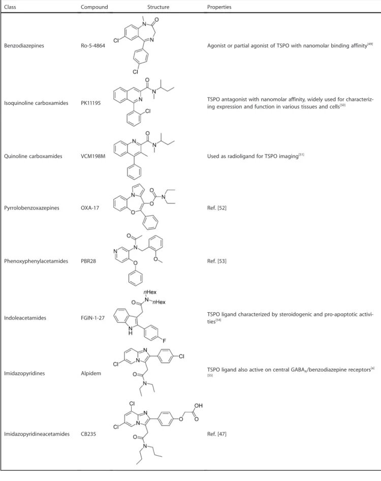
An extensive variety of specific molecules with high affinity and selectivity for TSPO have been yet classified belonging to different structural classes, in particular: benzodiazepines, iso-quinoline carboxamides, iso-quinoline carboxamides, pyrroloben-zoxazepines, phenoxyphenyl acetamides, indoleacetamides, imidazopyridines, pyrazolopyrimidine acetamides, phenylpur-ines, benzoxazphenylpur-ines, Vinca alkaloids, N,N-dialkyl-2-phenylindol-3-yl-glyoxylamide (PIGA), and the aza-isosteres of PK11195, the 4-phenylquinazoline-2-carboxamides. The leader compounds of these classes and their properties are listed in the Table 1. In particular, N,N-dialkyl-2-phenylindol-3-ylglyoxylamide deriva-tives have been designed as conformationally constrained ana-logues of indoleacetamides such as FGIN-1-27[45] (Table 1).
Most of these new molecules exhibited a nanomolar/sub-nano-molar affinity for TSPO and stimulated steroidogenesis in C6 glioma cell line from rat with a potency comparable to or higher than that of classic TSPO ligands such as the isoquino-line carboxamide PK11195. Among the imidazopyridines, alpi-dem has been shown to act on both TSPO and the central benzodiazepine receptor, with a preference toward TSPO. In an effort to improve the TSPO selectivity of alpidem analogues, some of us have designed new selective and affine TSPO li-gands by introducing several substituents on the
imidazopyri-dine nucleus (Table 2).[46] The structure–activity correlations
studies showed that substitutions at the 8-position of the imi-dazopyridine nucleus with lipophilic groups, and a para-chloro substitution on the phenyl ring at C(2) are fundamental in order to obtain high affinity and selectivity toward TSPO recep-tor (Table 2). Furthermore, the substituents on the acetoamide nitrogen on the 3-position of the imidazopyridine nucleus are accountable for variation of affinity. Moreover, substitutions with aromatic rings of carboxamide nitrogen is responsible of high affinity and selectivity, while the presence of polar sub-stituents in this region is unfavorable for affinity properties. Other investigations pointed out the effects of substitution on the 2-and 8-position of the imidazopyridine skeleton with hy-drophilic groups, polar or ionizable, to fulfill the need of a greater aqueous solubility of the 2-phenylacetamidoimida-zo[1,2-a]pyridines. In particular, the phenyl group on the 2-po-sition of the imidazopyridine nucleus has been functionalized with amino, hydroxy, and carboxylic groups. These polar sub-stituents offer in addition, the advantage of further functionali-zation with anticancer drugs, hydrophilic polymers (e.g., PEG, dendrimers) through reversible covalent bond, allowing the preparation of targeted nanocarriers and conjugates able to deliver therapeutics and diagnostics to mitochondria. The 2- (6,8-dichloro-2-(4-hydroxyphenyl)imidazo[1,2-a]pyridin-3-yl)-N,N-dipropylacetamide (CB235),[47]
2-(6,8-dichloro-2-(4-hydroxy-phenyl)imidazo[1,2-a]pyridin-3-yl)-N,N’-dipropyl acetamide (CB185),[46]
[2-(4-chlorophenyl)-8-aminoimidazo[1,2-a]pyridin-3-yl]-N,N-di-n-propylacetamide (CB86),[48] and
N,N-dipropyl-[2-(8- (2-aminoacetamido)-2-(4-chlorophenyl)imidazo[1,2-a]pyridin-3-yl)]acetamide (glycine derivative of CB86),[46]for instance,
rep-resent eligible candidates for this purpose.
3. Mitochondrial Targeting
Mitochondria dysfunctions are related to many human diseases ranging from cancer, neurodegenerative disorders, obesity, dia-betes and ischemia-reperfusion injury. Therefore, the involve-ment of mitochondria in various pathological conditions, makes these organelles a potential drug target in order to de-velop new therapeutic strategies. Nevertheless, targeting mito-chondria is a hard and challenging task due to their highly complex structure. The mitochondrion is constituted by the outer mitochondrial membrane (OMM), the intermembrane space (IMS), the inner mitochondrial membrane (IMM) and the matrix. Only molecules with a molecular weight of 5 kDa or less can cross the OMM because of the presence of a channel-forming protein VDAC (voltage-dependent anion channel).[61]
In addition, the IMM and the matrix are the headquarters of important mitochondrial functions thanks to the presence of several proteins in the IMM and enzymes and copies of the mi-tochondrial genome in the matrix. Thus a therapeutic mole-cule, in order to exert its action, has not only to reach the target cell, penetrate cellular membrane and face the intracel-lular environment but it has also to cross the mitochondrial membranes in order to reach the matrix. Moreover, the mito-chondrion has a strong negative membrane potential between @160 mV to @180 mV and, in particular, the IMM is highly
Table 1. Synthetic TSPO ligands.
Class Compound Structure Properties
Benzodiazepines Ro-5-4864 Agonist or partial agonist of TSPO with nanomolar binding affinity[49]
Isoquinoline carboxamides PK11195 TSPO antagonist with nanomolar affinity, widely used for characteriz-ing expression and function in various tissues and cells[50]
Quinoline carboxamides VCM198M Used as radioligand for TSPO imaging[51]
Pyrrolobenzoxazepines OXA-17 Ref. [52]
Phenoxyphenylacetamides PBR28 Ref. [53]
Indoleacetamides FGIN-1-27 TSPO ligand characterized by steroidogenic and pro-apoptotic activi-ties[54]
Imidazopyridines Alpidem TSPO ligand also active on central GABAA/benzodiazepine receptors[a]
[55]
dense due to the abundance of saturated phospholipids, re-sulting in impermeability against a lot of molecules. Thus, it is evident that mitochondrial drug delivery systems must possess two important requirements: delocalized positive charge and lipophilicity. Taking into account these features, several mito-chondria targeting strategies have been developed, also ex-ploiting nanoparticle-based systems such as the attachment of lipophilic cations to small molecules or nanoparticles (e.g., tri-phenylphosphonium, TPP); the combination of antioxidant with mitochondrial penetrating peptides (e.g., SS31, d-Arg-Dmt-Lys-Phe-NH2); the preparation of mitochondriotropic lipid
dequalinium chloride-containing vesicles (DQAsomes) or of MITO-porter, a mitochondrial fusogenic lipid containing lipo-somes-based carrier [DOPE/sphingomyelin/stearyl-R8 (9:2:1)];
the transport of proteins via mitochondrial protein import ma-chineries.[6]
3.1. Prodrugs characterized by TSPO moieties
Although several new TSPO ligands have been synthesized, to date very few examples of conjugates or nanosystems target-ing TSPO are counted, probably because only some of TSPO li-gands mentioned above are characterized by the presence of groups potentially derivatizable (i.e., COOH, OH, and NH) and thus ideal for the preparation of targeted drug delivery sys-tems able to deliver therapeutics and diagnostics to mitochon-dria. The first polymeric conjugate model has been proposed for the first time by Ringsdorf in 1975,[62]as a macromolecular
Table 1. (Continued)
Class Compound Structure Properties
Pyrazolopyrimidineacetamides DPA Ligand used for in vivo imaging of TSPO[56]
Phenylpurines Emapunil Ligand with rapid anxiolytic effects[57]
Benzoxazines Etifoxine Anxiolytic effects mediated by both GABAAand TSPO receptors;
neu-rodegenerative effects mediated by TSPO[58]
Vinca alkaloids Vinpocetine Ligand with neuroprotective activity that binds TSPO and other recep-tors such as adrenergic receptors[59]
N,N-Dialkyl-2-phenylindol-3-yl-glyoxylamide (PIGA) PIGA 1128 Ref. [45]
4-Phenylquinazoline-2-carbox-amides 9
Aza-isosteres of PK11195. In particular, [11C]ER176 has been identified
as a PET radioligand with sufficient sensitivity to robustly image all three TSPO affinity genotypes in human brain[60]
prodrug that would be able to modify the solubility character-istics of a drug, its distribution in the body and to determine its selective cellular action. Generally, in order to realize a con-jugate, a covalent bond must be formed between a drug and another molecule which may itself be pharmacologically active, or can selectively target the drug to a specific site of action, or be completely inactive (i.e., a backbone polymer). The drug activation occurs as a result of the rupture of the co-valent bond, following in vivo administration and cell internali-zation. For this purpose, Denora et al. in 2010[63] published
their work on a new approach for the selective delivery of the antineoplastic drugs to brain tumors and to overcome P-gp re-sistance induction observed for the majority of cytotoxic agents, based on the conjugation of 2-phenylimidazo[1,2-a]pyridine derivatives (i.e., CB86, CB185) with Ara-C [cytarabine, cytosine arabinose, 1-(b-d-arabinofuranosyl) cytosine], a pyri-midine nucleoside analogue employed for the treatment of various cancers including brain tumors. Specifically, they pre-pared novel N-imidazopyridinacetyl–Ara-C conjugates different for the specific position in which the hydrophilic Ara-C moiety was introduced, such as the 3-position of the imidazopyridine nucleus, or the 8- and the para position of the 2-phenylimida-zopyridine skeleton through appropriate spacers (Figure 1). The differences in binding affinity and selectivity observed for the conjugates investigated by these authors were coherent with the structure–affinity relationship analysis of the 2-phenyl-imidazo[1,2-a]pyridine derivatives,[46] suggesting that the
sub-stitution with two or three chlorine atoms on the imidazopyri-dine nucleus would lead to a favorable interaction with the corresponding complementary site of the receptor. In particu-lar, the conjugate 3 in Figure 1 (namely the N1-(2-(4-chloro- phenyl)-3-(2-(dipropylamino)-2-oxoethyl)imidazo[1,2-a]pyridin- 8-yl)-N6-(1-((2R,3S,4S,5R)-3,4-dihydroxy-5-(hydroxymethyl)tetra-hydrofuran-2-yl)-2-oxo-1,2-dihydropyrimidin-4-yl)adipamide) displayed very high in vitro TSPO affinity and selectivity and
was considered able to enhance the clinical potential of the nucleoside drug Ara-C. Moreover, Denora et al. in 2012[64]
pur-sued the aim to realize new co-drugs of the GABAergic agent 2-phenylimidazo[1,2-a]pyridinacetamide and dopamine or ethyl ester l-DOPA, in which dopamine and l-DOPA were linked through a carbamate bond at the para position of the phenyl group on the imidazopyridinacetamide nucleus (Figure 2). The in vitro and in vivo evaluation of compounds re-vealed that conjugation was an efficient strategy to deliver dopamine to the brain for dopamine-replacement therapy and simultaneously activate GABA receptors in the brain. Other ex-amples of prodrugs with potential applications in cancer diag-nosis and therapy are represented by metal-based complexes
Table 2. Selective TSPO ligands with imidazopyridine acetamide cores bearing conjugatable groups.[46]
Compd X Y R1 R2 K i[nm] CBR TSPO 6 Cl Cl H OH >105 1.31 12 Cl Cl H OCOCH2NH2·HCl >104 1.52 15a Cl Cl H NH2 246 2.22 19a Cl Cl NH2 Cl >105 0.33 20 Cl Cl NH2 Cl >104 0.78 21 H Cl NH2 Cl 535 1.04 22 Cl Cl NHCO(CH2)2COOH Cl 194 14.4 23 H NHCOCH2COOH H Cl >104 193.1 24 H NHCO(CH2)2COOH H Cl >104 285.3 25 H NHCO(CH2)3COOH H Cl >104 117.7 26 H NHCOCH2NH2·HCl H Cl >104 14.2
targeting the TSPO and recently summarized in a comprehen-sive overview.[17] In this regard, Choi et al. recently prepared a
novel 99mTc-tricarbonyl labelled imidazolpyridine compound
useful for SPECT imaging of TSPO-rich cancers[65] using the
2- (8-(2-(bis(pyridin-2-yl)methyl)amino)acetamido)-2-(4-chloro-phenyl)-H-imidazo[1,2-a]pyridin-3-yl)-N,N-dipropylacetamide (CB256) as TSPO ligand,[66]able to perform a bifunctional
che-late approach, (1, Figure 3).[29a] Some of us realized another
rhenium complex to be used as a model of radiopharmaceuti-cal agent targeted to TSPO[67] obtained using the
2-[6,8-di- chloro-2-(1,3-thiazol-2-yl)-H-imidazo[1,2-a]pyridin-3-yl]-N,N-di-n-propylacetamide (TZ6) as a potent and selective TSPO ligand (2, Figure 3).
In addition, some of us prepared antitumor platinum com-pounds with specific TSPO ligands,[68]in particular the PtII
com-plex cis-[PtCl2(TZ6)] (3 in Figure 3), combined the antitumor
properties of the metal core with the high TSPO affinity of TZ6. Other two Pt compounds have been prepared using a ligand
with high affinity and selectivity for the translocator protein, the [2-(4-chlorophenyl)-8-aminoimidazo[1,2-a]pyridin-3-yl]-N,N-di-n-propylacetamide (CB86) having formula cis-[PtX2(NH3)(CB86)] with X= I@ or Cl@ (4, Figure 3).[29b]
Further-more, some of us prepared the first bimetallic Re/Pt complex useful for theranostic purpose, availing the coordination po-tential of CB256 toward PtIIand ReIions (5, Figure 3).[66]Finally,
Savino et al., in order to overcome the limitations and the side effects associated with PtII complex, realized a PtIV prodrug
containing CB235 as TSPO ligand in axial position, namely the oxaliplatin derivative cis,trans,cis-[Pt(ethanedioato)Cl{2-(2-(4- (6,8-dichloro-3-(2-(dipropylamino)-2-oxoethyl)imidazo[1,2-a]pyr-idin-2-yl)phenoxy)acetate)ethanolato}(1R,2R-DACH)] (DACH = diaminocyclohexane) shown in Figure 3 (compound 6).[69]
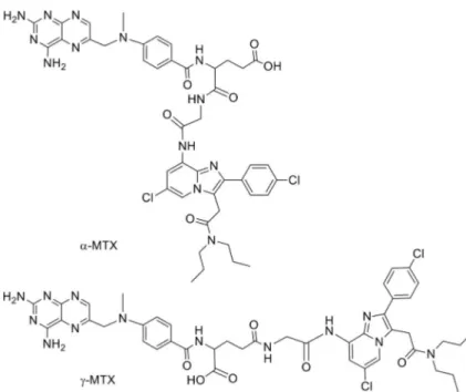
Recently, Laquintana et al.[70]described a new prodrug
strat-egy to deliver anticancer drug to brain cancers overexpressing TSPO receptor. In particular, they prepared two TSPO ligand– methotrexate conjugates (TSPO ligand a-MTX and TSPO ligand g-MTX) in order to transport the hydrophilic drug through the blood–brain barrier (BBB) and determine its accumulation in target cells overexpressing the TSPO. The TSPO ligand used was a glycine derivative of CB86 (Figure 4).
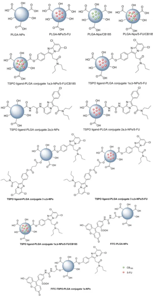
The same authors in the late 2009[48] have exploited the
polymeric conjugate strategy by chemical conjugation of two imidazopyridinacetamides (CB185 and glycine derivative of CB86), chosen as affine and selective TSPO ligands, via an ester or amide linkage to poly(d,l-lactic-co-glycolic acid) polymers having different average molecular weights (PLGA, Resomer RG502H (a) and Resomer RG503H (b)), (conjugates 1a,b, 2a,b and the bis TSPO–PLGA conjugate 3, Figure 5). They evaluated these conjugates as delivery systems of TSPO ligands endowed with apoptosis inducing activity. Moreover, for detecting the
Figure 2. Structures of co-drugs of the GABAergic agent 2-phenylimida-zo[1,2-a]pyridinacetamide and dopamine (DA) or ethyl ester l-DOPA (LD).
exact position of TSPO ligand–PLGA conjugates in tissues or cells after in vivo administration, they also prepared a fluores-cent probe (4, Figure 5) by reaction of FITC glycine[71]with the
conjugate 1. After accurate physicochemical investigations aimed at evaluating the successful conjugation between TSPO
ligands and PLGA, it was assessed the stability in an acidic environment and in the physiological medium, showing that TSPO ligand released from these conju-gates occurred in human serum and in 0.1n HCl so-lution at a faster rate than that observed in phos-phate buffer, pH 7.4. These macromolecular conju-gates showed high affinity and selectivity for TSPO similar to that of the reference ligands. Moreover, in vitro studies were conducted on C6 rat glioma cells known for their overexpression of TSPO. Cytotoxicity assay showed that TSPO ligand–PLGA polymer conju-gates 1–3, induced survival inhibition with EC50
values ranging from 1.75 to 34.29 mm; uptake and apoptosis studies conducted with fluorescence mi-croscopy demonstrated the internalization of the flu-orescent conjugate probe 4 and the induction of a mitochondrial morphology modification caused by CB185 and its PLGA conjugate 1.
These results suggested to the authors the poten-tial of the newly prepared conjugates, as well as modulators of the neurosteroid synthesis, and also as macromolecular apoptosis inducing agents and hence able to induce tumor cell death. In conclusion the authors stated that these TSPO ligand–PLGA conjugates availing of passive targeting mechanism, could have been for-mulated into micro- and nanoparticles providing a new mito-chondrial targeted approach useful for improved cancer che-motherapy.
Figure 4. Structures of TSPO ligand–methotrexate conjugates a-MTX and g-MTX.
3.2. TSPO ligands as mitochondrial targeting moieties of nanoparticles
As a direct consequence of the results obtained by Laquintana et al. in 2009,[48] the same researchers pursued their
investiga-tion through the realizainvestiga-tion of nanoparticle delivery systems (NPs), employing the TSPO ligand–PLGA conjugated (PLGA– TSPO) polymers described above. Specifically, the PLGA NPs (Figure 6) were prepared by a quasi-emulsion solvent diffusion procedure (QESD) as well as the TSPO ligand–PLGA conjugate 1a,b–Nps, the TSPO ligand–PLGA conjugate 2a,b–NPs, and the TSPO ligand–PLGA conjugate 3a,b–Nps, starting by PLGA with free hydroxy and carboxylic acid group at its terminal ends. In order to assess the ability of these NPs to be used as a drug delivery systems for cancer treatment, TSPO–PLGA–NPs were loaded with the hydrophilic anticancer drug 5-fluorouracil (5-FU) through a double emulsion solvent diffusion method (DESD). Furthermore, dual drug loaded PLGA NPs (PLGA NPs/5-FU/CB185) and dual drug loaded TSPO–PLGA–NPs (TSPO– PLGA–NPs/5-FU/CB185), with 5-FU and TSPO ligand CB185 physically included together, were prepared by the DESD method, with the aim to investigate the occurrence of syner-gistic effects. Dual drug loaded TSPO–PLGA–1a,b-NPs/5-FU/ CB185 have the benefit of delivering the TSPO ligand CB185 through a chemical conjugation as well as by physical encap-sulation. In addition, PLGA hydroxy end groups were conjugat-ed to the fluorescent probe FITC, in order to prepare fluores-cent FITC–PLGA NPs and FITC-TSPO–PLGA–NPs (Figure 6).
After a comprehensive physicochemical characterization aimed to determine particle size and size distribution (81.1– 168.6 nm, PDI 0.08–0.43), surface morphology, drug encapsula-tion efficiency and drug release kinetics of all newly prepared Nps, they were evaluated in terms of cytotoxicity on C6 glioma cells overexpressing TSPO by MTT assay and combination index (CI) calculations, as well as of internalization ability by fluorescence microscopy (FITC-PLGA–TSPO NPs). Based on those outcomes, Laquintana et al. considered these Nps, namely TSPO–FU, dual drug loaded PLGA–NPs/5-FU/CB185 and TSPO–PLGA–NPs/5-PLGA–NPs/5-FU/CB185, able to considera-bly increase toxicity on human cancer cells on account of the synergistic effect of the TSPO ligand CB185 with 5-FU.[72]
In 2009 Musacchio et al. published the results of their stud-ies regarding the use of an highly selective TSPO ligand, in par-ticular the above mentioned CB86, to exploit a tumors recep-tor-mediated drug targeting strategy.[73] They prepared
poly-ethylene glycol–phosphatidylethanolamine (PEG-PE) micelles, loaded with the anticancer drug paclitaxel and targeted to TSPO by surface modification with CB86 ligand. The average conjugation yield, as determined by the HPLC, was 40% (CB86-ligand moles of the total available p-nitrophenyloxycar- bonyl-1,2-distearoyl-sn-glycero-3-phosphoethanolamine-N-[me-thoxy(polyethylene glycol)-2000] (p-NP-PEG2k-PE moles). The obtained micelles were also characterized in terms of size and size distribution (9.5–18.8 nm and PDI 0.104: 0.028) zeta po-tential (0.18:0.931 mV), drug loading and drug release pro-files, storage stability and serum stability, showing that micelles stayed intact and without drug release even after the TSPO
ligand conjugation. In vitro experiments conducted on LN18 human glioblastoma TSPO-overexpressing cell line, confirmed the cellular uptake of the targeted micelles after 3 h of incuba-tion, and at the same time an apoptosis induction only in cells treated with the targeted empty micelles (no paclitaxel), as judged by the nuclear fragmentation, in contrast to what ob-served for the cells treated with non-targeted micelles paclitax-el loaded, thus suggesting to the authors that the TSPO ligand CB86 is able to interact with the mitochondrial target and to initiate apoptosis in cells overexpressing TSPO. Moreover, the authors verified the hypothesis of a possible synergism be-tween the TSPO ligand and paclitaxel resulting in good syner-gism when both compounds were components of the same micellar formulation. In fact, the cytotoxicity of the targeted paclitaxel loaded micelles was greater than that of non-target-ed micelles and of free paclitaxel, with IC50values of 175.1 nm,
267.0 nm, and 271.1 nm, respectively. These outcomes allowed the authors to consider these TSPO-targeted nanosystems loaded with anticancer drugs as potentially promising antitu-mor nanomedicines. Kim et al. also exploited a TSPO ligand, the PK11195, as mitochondria target moiety. In this work PK11195 was linked to chitosan-graft-PEI (polyethylenimmine) modified superparamagnetic iron oxide nanoparticles (PK-CP-SPION) and employed as a gene vector.[74] PK-CP-SPION were
synthesized via a Grignard reaction between PK11195 ligand and the Fe2O3SPIONs surface modified with chitosan graft-PEI
(CHI-g-PEI), and then were complexed with DNA at functional weight ratios. PK-CP-SPION and its complexes have been total-ly characterized and the in vitro cytotoxicity of PK-CP-SPION was evaluated on four different cell lines (A549, KB, HeLa and HepG2). In particular, the authors have established an higher transfection efficiencies of PK-CP-SPION with the application of an external magnetic field. Accumulation of PK-CP-SPION in mitochondria was demonstrated by laser scanning microscopy and confirmed by the leakage of Cyt-c and the dissipation of mitochondrial membrane potential, due to the interaction be-tween PK11195 and TSPO, thus confirming the triggering of the cells apoptotic process. In this work, Kim et al. also demon-strated the intracellular internalization of PKCP-SPION and their mitochondrial targeting ability by HR-TEM. The low cytotoxicity and the high transfection efficiency of PK-CP–SPIONs enable them as possible good mitochondria targeting gene vector.
Reflecting the need of the advancement of personalized medicine, some researchers have developed nanocarriers tar-geted to mitochondria and useful as appropriate probes to image diseases or biological processes. A first example has been reported by Bornhop in 2009,[75] concerning the
realiza-tion of a TSPO targeted imaging agent, the ClPhIQ-PAMAM-Liss, consisting of a polyamidoammine dendrimer G(4)-PAMAM functionalized with the 1-(2-chlorophenyl)isoquinoline-3-car-boxylic acid (ClPhIQ Acid)[76]as targeting moiety and with the
fluorophore Lissamine (Figure 7).
Dendrimers are emerging nanomaterials, monodisperse and globular, characterized by a great number of peripheral groups potentially functionalized with properly selected targeting moiety and fluorophores or metal chelates for optical imaging or for fluorescence, MRI, PET and SPECT, respectively.[77]These
characteristics make these nanocarriers ideal delivery vectors also to further investigate the effects of the molecular weight of the polymer, the presence of negative or positive charges and composition on biologically relevant consequences such as cytotoxicity, cellular uptake, and subcellular interactions.
With this in mind, Bornhop realized the new imaging agent dendrimer based targeted to TSPO, described above. The suc-cess of conjugation reactions was confirmed by MALDI-TOF-MS and NMR techniques, while in vitro studies conducted on two TSPO-overexpressing cell lines, that is, C6 rat glioma and MDAMB-231 human breast cancer cells, assessed the cellular internalization ability of the TSPO-targeted dendrimer. In par-ticular, the preferential mitochondrial localization of the ClPhIQ-PAMAM was evidenced by its co-localization with the mitochondrial marker MitoTracker green.
The skills of synthetic dendrimers, namely the specific cellu-lar and subcellucellu-lar internalization and targeting, make them helpful to understand biological events at the molecular level. However, Bornhop et al. concluded their work affirming the need of further investigation to explore the exact ways of in-ternalization exploited by these nanosystems. In this regard, the research project of Denora et al.[47]has given the
opportu-nity to deeper investigate these issues. Specifically, they have focused their attention on the synthesis and in vitro evaluation of new TSPO ligand characterized by a substituted imidazopyr-idine nucleus, namely the 2-(4-(6,8-dichloro-3-(2-(dipropylami-no)-2-oxoethyl)imidazo[1,2-a]pyridin-2-yl)phenoxy)acetic acid (CB235), as high-affinity conjugatable TSPO ligand linked to the amine end-group of PAMAM dendrimers, in order to obtain macromolecules targeting TSPO. The synthesis of the new TSPO targeted G(4)-PAMAM dendrimers were accom-plished using well-known synthetic methods. Further, the TSPO targeted-G(4)-PAMAM dendrimers were functionalized by reacting with the organic fluorophore fluorescein isothiocya-nate isomer 1 (FITC), giving fluorescent dendrimers able to bind the mitochondrial protein TSPO. The comprehensive physicochemical and morphological characterization of den-drimers by means of 1H NMR, dynamic light scattering (DLS),
laser Doppler velocimetry (LDV), atomic force microscopy (AFM), evidenced their monodispersity, the spherical shape
and a hydrodynamic diameter of about 24 nm. To estimate the biological activity of the imaging agents, C6 glioma cell line from rat, after incubation with the FITC dendrimers, has been visualized by both confocal and fluorescence microscopies. In particular, these authors have explored the cellular uptake be-havior of these dendrimers in the presence of various endocy-tosis inhibitors, finding that the TSPO targeted-G(4)-PAMAM– FITC dendrimer is quickly internalized by C6 cells through pi-nocytosis and that no significant exocytosis was observed. Moreover, competition studies in presence of the TSPO ligand, subcellular fractionation experiments and co-localization stud-ies performed with CAT (confocal-AFM-TIRF) microscopy have been crucial in confirming the ability of the TSPO-targeted dendrimer to co-localize in mitochondria (Figure 8). Afterward, the same authors, in order to obviate the limitations resulted from the use of organic fluorophores (e.g., FITC), such as broad emission spectra, susceptibility to photobleaching under continuous light exposure and short fluorescence (PL) lifetimes, have suitably engineered luminescent semiconductor quantum dots (QDs), based on a core–shell structure of inorganic nano-crystals (CdSe@ZnS), able to target mitochondria while keeping the colloidal stability and optical properties.[78]Qds represent a
new class of fluorophores that can be used as advanced toluminescence (PL) probes, taking advantage of the QD pho-tophysical properties and their versatile surface chemistry. Spe-cifically, Fanizza et al. have prepared multifunctional nanostruc-tures based on CdSe@ZnS Qds coated with a silica shell
func-Figure 7. The imaging agent ClPhIQ–PAMAM–Lissamine.
Figure 8. Representative confocal microscopy image of co-localization in a double-tagged C6 glioma cell (green: TSPO targeted-G(4)-PAMAM dendrim-ers grafted with FITC 1 mm; red: mitochondrion tagged with MitoTracker Red; yellow: convergence of red and green, indicating co-localization). Insert: TSPO ligand molecule and the TSPO-targeted G(4)-PAMAM–FITC den-drimer.
tionalized with amine groups and conjugated with the selec-tive and affine TSPO ligand, CB235 (Figure 9). The new result-ing nanosystem, namely TSPO-targeted Qds@SiO2, is conveyed
in a single nanostructure, the selectivity of the TSPO ligand and the photostability and high luminescence properties of in-organic QDs. For this purpose, in this work the luminescent QDs, of hydrophobic nature, have been appropriately de-signed, by controlling size, shape and surface charge, in order to guarantee aqueous dispersibility, facilitate the cellular inter-nalization and mitochondrial targeting, maintaining the colloi-dal stability and the optical properties. The silica shell has al-lowed a significant improvement of the stability of Qds in aqueous phase, because silica is an inert, biocompatible and transparent material, and therefore able to decrease the re-lease of cytotoxic ions and prevent photo-oxidation of QDs. Therefore silica-coated QDs represent an inorganic solid system, characterized by high chemical stability, intrinsic hy-drophilicity and highly versatile surface. The nanostructure pro-posed in this study provides a great advantage, as it associates the photophysical stability of the QDs and the versatility of silica shell, with the TSPO receptor recognition properties of the highly affine selective ligand. The physicochemical charac-teristics of this new nanomaterial have been extensively stud-ied from morphological, structural and optical points of view, and in vitro subcellular fractionation experiments and co-locali-zation studies conducted by means of laser scanning confocal microscopy on C6 rat glioma cells, have finally shown the suc-cess of this new nanostructure to target mitochondria thanks to the molecular recognition of TSPO (Figure 9).
The list of nanocarriers targeting the mitochondrial translo-cation protein TSPO ends with a liposomal system realized by Cerutti et al.[79] developed to incorporate a selective ligand for
TSPO complexed with gadolinium (Gd), potentially useful as MRI contrast agent (Figure 10). In particular the 2-phenylpyra-zolo[1,5-a]pyrimidineacetamide DPA-713 was successfully de-rivatized and coupled to the magnetic resonance imaging re-porter Gd-DOTA (DOTA =1,4,7,10-tetraazacyclododecane-1,4,7,10-tetraacetic acid). Because the ideal MRI targeting con-trast agent should remain in the blood circulation for a long time, this gadolinium-based TSPO-targeted complex has been
incorporated in the liposomal membrane and in the inner aqueous cavity of liposomes in order to increase the circulation lifetime and to obtain a slow release of the MRI agent. In par-ticular, liposomes were formulated with the thin-film hydration method using POPC (1-palmitoyl-2-oleoyl-sn-glycero-3-phos-phocholine), cholesterol and DSPE-PEG-methoxy-2000 (1,2-dis- tearoyl-sn-glycero-3-phosphoethanolamine-N-[methoxy(polye-thyleneglycol)-2000] (ammonium salt)). The characterization of liposomes loaded with this complex showed inspiring out-comes as slow releasing MRI targeting agents and encouraged the authors to perform further in vivo studies (Figure 10).
4. Conclusions
The involvement of mitochondria in various pathological con-ditions makes these organelles a potential drug target for the development of new therapeutic and diagnostic strategies. Nevertheless, targeting mitochondria is a difficult and challeng-ing task due to their highly complex structure. The 18-kDa mi-tochondrial translocator protein (TSPO) is an interesting mito-chondrial biomarker, and although several new TSPO ligands have been synthesized, only some of these feature the pres-ence of groups potentially derivatizable and therefore suitable for the preparation of targeted nanocarriers able to deliver therapeutics and diagnostics to mitochondria. This review overviews the very few examples of prodrugs and nanosys-tems targeting TSPO, and their potential applications in
diag-Figure 9. A) Co-localization analysis of C6 rat glioma cells stained with MitoTracker green and TSPO-targeted QD@SiO2NPs. All pixels of the co-localization
image shown in panel B are reported in the scatter diagram of panel A, in which the two image channels are compared. Region 1 of the diagram displays the co-localization pixels. B) Representative confocal image of co-localization of TSPO-targeted QD@SiO2NPs (red) and MitoTracker (green) in C6 cells. The
co-lo-calization is marked by the convergence of the red and green to yellow fluorescence; scale bar: 10 mm. C) TSPO-targeted QD@SiO2NPs.
nosis and therapy of disease states in which this protein is overexpressed.
Acknowledgements
We acknowledge the University of Bari (Italy), the Italian Minis-tero dell’Universit/ e della Ricerca (MIUR), and the Inter-University Consortium for Research on the Chemistry of Metal Ions in Bio-logical Systems (C.I.R.C.M.S.B., Bari, Italy) for support.
Conflict of interest
The authors declare no conflict of interest.
Keywords: drug targeting · imaging agents · nanoparticles · prodrugs · TSPO receptors
[1] a) A. Lopalco, A. Hazem, N. Denora, E. Rytting, Int. J. Nanomed. 2015, 10, 1985 –1996; b) A. Lopedota, A. Cutrignelli, V. Laquintana, N. Denora, R. M. Iacobazzi, M. Perrone, E. Fanizza, M. Mastrodonato, D. Mentino, A. Lopalco, N. Depalo, M. Franco, Pharm. Res. 2016, 33, 2195 – 2208; c) N. Denora, A. Lopedota, M. Perrone, V. Laquintana, R. M. Iacobazzi, A. Mile-lla, E. Fanizza, N. Depalo, A. Cutrignelli, A. Lopalco, M. Franco, Acta Bio-mater. 2016, 43, 170–184.
[2] W. H. De Jong, P. J. Borm, Int. J. Nanomed. 2008, 3, 133– 149.
[3] S. M. Moghimia, A. R. Rajabi-Siahboomib, Adv. Drug Delivery Rev. 2000, 41, 129– 133.
[4] M. Toporkiewicz, J. Meissner, L. Matusewicz, A. Czogalla, A. F. Sikorski, Int. J. Nanomed. 2015, 10, 1399 –1414.
[5] A. Jhaveri, V. Torchilin, Expert Opin. Drug Delivery 2016, 13, 49– 70. [6] S. Biswas, V. P. Torchilin, Adv. Drug Delivery Rev. 2014, 66, 26– 41. [7] a) A. Lopalco, G. Dalwadi, S. Niu, R. L. Schowen, J. Douglas, V. J. Stella, J.
Pharm. Sci. 2016, 105, 705–713; b) A. Lopalco, V. J. Stella, J. Pharm. Sci. 2016, 105, 2879 –2885; c) A. Lopalco, J. Douglas, N. Denora, V. J. Stella, J. Pharm. Sci. 2016, 105, 664– 672.
[8] R. K. Pathak, N. Kolishetti, S. Dha, Wiley Interdiscip. Rev. Nanomed. Nano-biotechnol. 2015, 7, 315– 329.
[9] V. Papadopoulos, M. Baraldi, T. R. Guilarte, T. B. Knudsen, J. J. LacapHre, P. Lindemann, M. D. Norenberg, D. Nutt, A. Weizman, M. R. Zhang, M. Gavish, Trends Pharmacol. Sci. 2006, 27, 402 –409.
[10] a) R. R. Anholt, P. L. Pedersen, E. B. De Souza, S. H. Snyder, J. Biol. Chem. 1986, 261, 576 –583; b) A. S. Basile, P. Skolnick, J. Neurochem. 1986, 46, 305– 308; c) L. Ntkiewicz-Michaluk, A. Guidotti, K. E. Krueger, Mol. Phar-macol. 1988, 34, 272– 278.
[11] a) A. Batarseh, V. Papadopoulos, Mol. Cell. Endocrinol. 2010, 327, 1– 12; b) P. Casellas, S. Galiegue, A. S. Basile, Neurochem. Int. 2002, 40, 475 – 486; c) M. Gavish, I. Bachman, R. Shoukrun, Y. Katz, L. Veenman, G. Wei-singer, A. Weizman, Pharmacol. Rev. 1999, 51, 629 –650.
[12] a) S. Batra, C. S. Iosif, Int. J. Oncol. 1998, 12, 1295 – 1298; b) I. Venturini, H. Alho, I. Podkletnova, L. Corsi, E. Rybnikova, R. Pellicci, M. Baraldi, M. Pelto-Huikko, P. Helen, M. L. Zeneroli, Life Sci. 1999, 65, 2223 –2231; c) M. Hardwick, D. Fertikh, M. Culty, H. Li, B. Vidic, V. Papadopoulos, Cancer Res. 1999, 59, 831– 842; d) K. Maaser, P. Grabowski, A. P. Sutter, M. Hopfner, H. D. Foss, H. Stein, G. Berger, M. Gavish, M. Zeitz, H. Scher-ubl, Clin. Cancer Res. 2002, 8, 3205– 3209.
[13] a) D. Diorio, S. A. Welner, R. F. Butterworth, M. J. Meaney, B. E. Suranyi-Cadotte, Neurobiol. Aging 1991, 12, 255 –258; b) E. Vowinckel, D. Reut-ens, B. Becher, G. Verge, A. Evans, T. OwReut-ens, J. P. Antel, J. Neurosci. Res. 1997, 50, 345– 353.
[14] A. Midzak, N. Denora, V. Laquintana, A. Cutrignelli, A. Lopedota, M. Franco, C. D. Altomare, V. Papadopoulos, Eur. J. Pharm. Sci. 2015, 76, 231– 237.
[15] N. Denora, V. Laquintana, A. Trapani, H. Suzuki, M. Sawada, G. Trapani, Pharm. Res. 2011, 28, 2820 –2832.
[16] a) M. Perrone, B. S. Moon, H. S. Park, V. Laquintana, J. H. Jung, A. Cu-trignelli, A. Lopedota, M. Franco, S. E. Kim, B. C. Lee, N. Denora, Sci. Rep. 2016, 6, 20422; b) K. Sekimata, K. Hatano, M. Ogawa, J. Abe, Y. Magata, G. Biggio, M. Serra, V. Laquintana, N. Denora, A. Latrofa, G. Trapani, G. Liso, K. Ito, Nucl. Med. Biol. 2008, 35, 327–334.
[17] N. Denora, R. M. Iacobazzi, G. Natile, N. Margiotta, Coord. Chem. Rev. 2017, 341, 1 – 18.
[18] R. R. H. Anholt, P. L. Pendersen, E. B. De Suoza, S. H. Snyder, J. Biol. Chem. 1986, 261, 576– 583.
[19] a) F. Delavoie, M. Hardwick, J. C. Robert, C. Giatzakis, G. P8ranzi, Z. X. Yao, J. Maccario, J. J. LacapHre, V. Papadopoulos, Biochemistry 2003, 42, 4506 –4519; b) J. J. LacapHre, V. Papadopoulos, Steroids 2003, 68, 569 – 585.
[20] J. M. Olson, B. J. Ciliax, W. R. Mancini, A. B. Young, Eur. J. Pharmacol. 1988, 152, 47 –53.
[21] G. B. O’Beirne, M. J. Wooods, D. C. Williams, Eur. J. Biochem. 1990, 188, 131– 138.
[22] M. Hardwick, D. Fertikh, M. Culty, H. Li, B. Vidic, V. Papadopoulos, Cancer Res. 1999, 59, 831 –842.
[23] a) V. Papadopoulos, A. Berkovich, K. E. Krueger, E. Costa, A. Guidotti, En-docrinology 1991, 129, 1481 –1488; b) A. Verma, S. H. Snyder, Annu. Rev. Pharmacol. Toxicol. 1989, 29, 307– 322; c) M. J. Woods, D. C. Williams, Biochem. Pharmacol. 1996, 52, 1805 – 1814; d) T. Azarashvili, O. Krestini-na, I. Yurkov, Y. Evtodienko, G. Reiser, J. Neurochem. 2005, 94, 1054 – 1062; e) J. D. Hirsch, C. F. Beyer, L. Malkowitz, B. Beer, A. J. Blume, Mol. Pharmacol. 1989, 35, 157– 163; f) I. Carmel, F. A. Fares, S. Leschiner, H. Scherubl, G. Weisinger, M. Gavish, Biochem. Pharmacol. 1999, 58, 273 – 278; g) J. K. Wang, J. I. Morgan, S. Spector, Proc. Natl. Acad. Sci. USA 1984, 81, 753– 756.
[24] a) K. E. Krueger, V. Papadopoulos, J. Biol. Chem. 1990, 265, 15015 – 15022; b) E. Kelly-Hershkovitz, R. Weizman, I. Spanier, S. Leschiner, M. Lahav, G. Weisinger, M. Gavish, J. Biol. Chem. 1998, 273, 5478 –5483; c) E. Romeo, J. Auta, A. P. Kozikowski, D. Ma, V. Papadopoulos, G. Puia, E. Costa, A. Guidotti, J. Pharmacol. Exp. Ther. 1992, 262, 971 –978. [25] C. Nothdurfter, R. Rupprecht, G. Rammes, Curr. Top. Med. Chem. 2012,
12, 360– 370.
[26] R. Rupprecht, V. Papadopoulos, G. Rammes, T. C. Baghai, J. Fan, N. Akula, G. Groyer, D. Adams, M. Schumacher, Nat. Rev. Drug Discovery 2010, 9, 971 –988.
[27] a) H. Wang, K. Zhai, Y. Xue, J. Yang, Q. Yang, Y. Fu, Y. Hu, F. Liu, W. Wang, L. Cui, H. Chen, J. Zhang, W. He, PLoS One 2016, 11, e0167307; b) L. N. Tu, K. Morohaku, P. R. Manna, S. H. Pelton, W. R. Butler, D. M. Stocco, V. Selvaraj, J. Biol. Chem. 2014, 289, 27444– 27454.
[28] a) L. Veenman, V. Papadopoulos, M. Gavish, Curr. Pharm. Des. 2007, 13, 2385 –2405; b) H. Everett, M. Barry, X. Sun, S. F. Lee, C. Frantz, L. G. Ber-thiaume, G. McFadden, R. C. Bleackley, J. Exp. Med. 2002, 196, 1127 – 1139.
[29] a) N. Denora, N. Margiotta, V. Laquintana, A. Lopedota, A. Cutrignelli, M. Losacco, M. Franco, G. Natile, ACS Med. Chem. Lett. 2014, 5, 685–689; b) N. Margiotta, N. Denora, R. Ostuni, V. Laquintana, A. Anderson, S. W. Johnson, G. Trapani, G. Natile, J. Med. Chem. 2010, 53, 5144 –5154. [30] A. Krippner, A. Matsuno-Yagi, R. A. Gottlieb, B. M. Babior, J. Biol. Chem.
1996, 271, 21629–21636.
[31] a) G. Kroemer, J. C. Reed, Nat. Med. 2000, 6, 513 –519; b) G. J. Pilkington, K. Parker, S. A. Murray, Semin. Cancer Ther. 2008, 18, 226 –235. [32] H. Everett, M. Barry, S. F. Lee, X. Sun, K. Graham, J. Stone, R. C. Bleackley,
G. McFadden, J. Exp. Med. 2000, 191, 1487 –1498.
[33] W. Kugler, L. Veenman, Y. Shandalov, S. Leschiner, I. Spanier, M. Lako-mek, M. Gavish, Cell. Oncol. 2008, 30, 435– 450.
[34] A. P. Sutter, K. Maaser, M. Hopfner, B. Barthel, P. Grabowski, S. Faiss, P. Carayon, M. Zeitz, H. Scherebl, Int. J. Cancer 2002, 102, 318 –327. [35] K. Maaser, M. Hopfner, A. Jansen, G. Weisinger, M. Gavish, A. P.
Kozikow-ski, A. Weizman, P. Carayon, E. O. Riecken, M. Zeitz, H. Scherebl, Br. J. Cancer 2001, 85, 1771 –1780.
[36] a) S. R. Torres, T. S. Frode, G. M. Nardi, N. Vita, R. Reeb, P. Ferrara, R. M. Ri-beiro-do-Vallea, R. C. Fargeset, Eur. J. Pharmacol. 2000, 408, 199 –211; b) J. D. Waterfield, E. G. McGeer, P. L. McGeer, Rheumatology 1999, 38, 1068 –1073; c) E. Bribes, B. Bourrie, M. Esclangon, S. Galiegue, H. Vidal, P. Casellas, Eur. J. Pharmacol. 2002, 452, 111– 122.
[37] a) V. Papadopoulos, Ann. Pharm. Fr. 2003, 61, 30–50; b) N. Leducq, F. Bono, T. Sulpice, V. Vin, P. Janiak, G. L. Fur, S. E. O’Connor, J. M. Herbert, J. Pharmacol. Exp. Ther. 2003, 306, 828– 837.
[38] P. Carayon, M. Portier, D. Dussossoy, A. Bord, G. Petitpretre, X. Canat, G. Le Fur, P. Casellas, Blood 1996, 87, 3170 –3178.
[39] L. Miccoli, S. Oudard, A. Beurdeley-Thomas, B. Dutrillaux, M. F. Poupon, Biochem. Pharmacol. 1999, 58, 715 –721.
[40] G. Pelaia, E. D. Di Paola, G. De Sarro, S. A. Marsico, Gen. Pharmacol. 1997, 28, 495– 498.
[41] H. Li, B. Degenhardt, D. Tobin, Z. X. Yao, K. Tasken, V. Papadopoulos, Mol. Endocrinol. 2001, 15, 2211–2228.
[42] a) H. Alho, V. Varga, K. E. Krueger, Cell Growth Differ. 1994, 5, 1005 – 1014; b) P. Bovolin, J. Schlichting, M. Miyata, C. Ferrarese, A. Guidotti, H. Alho, Regul. Pept. 1990, 29, 267– 281.
[43] J. Knudsen, Neuropharmacology 1991, 30, 1405 –1410.
[44] M. Gavish, I. Bachman, R. Shoukrun, Y. Katz, L. Veenman, G. Weisinger, A. Weizman, Pharmacol. Rev. 1999, 51, 629– 650.
[45] a) G. Primofiore, F. Da Settimo, S. Taliani, F. Simorini, M. P. Patrizi, E. Nov-ellino, G. Greco, E. Abignente, B. Costa, B. Chelli, C. Martini, J. Med. Chem. 2004, 47, 1852 –1855; b) F. Da Settimo, F. Simorini, S. Taliani, C. La Motta, A. M. Marini, S. Salerno, M. Bellandi, E. Novellino, G. Greco, B. Co-simelli, E. Da Pozzo, B. Costa, N. Simola, M. Morelli, C. Martini, J. Med. Chem. 2008, 51, 5798 – 5806; c) E. Barresi, A. Bruno, S. Taliani, S. Cosco-nati, E. da Pozzo, S. Salerno, F. Simorini, S. Daniele, C. Giacomelli, A. M. Marini, C. La Motta, L. Marinelli, B. Cosimelli, E. Novellino, G. Greco, F. Da Settimo, C. Martini, J. Med. Chem. 2015, 58, 6081 –6092.
[46] N. Denora, V. Laquintana, M. G. Pisu, R. Dore, L. Murru, A. Latrofa, G. Tra-pani, E. Sanna, J. Med. Chem. 2008, 51, 6876 –6888.
[47] N. Denora, V. Laquintana, A. Lopalco, R. M. Iacobazzi, A. Lopedota, A. Cutrignelli, G. Iacobellis, C. Annese, M. Cascione, S. Leporatti, M. Franco, J. Controlled Release 2013, 172, 1111– 1125.
[48] V. Laquintana, N. Denora, T. Musacchio, M. Lasorsa, A. Latrofa, G. Trapa-ni, J. Controlled Release 2009, 137, 185 –195.
[49] A. M. Scarf, L. M. Ittner, M. Kassiou, J. Med. Chem. 2009, 52, 581 –592. [50] M. Awad, M. Gavish, J. Neurochem. 1987, 49, 1407 –1414.
[51] A. Cappelli, M. Matarrese, R. M. Moresco, S. Valenti, M. Anzini, S. Vomero, E. A. Turolla, S. Belloli, P. Simonelli, M. A. Filannino, M. Lecchi, F. Fazio, Bioorg. Med. Chem. 2006, 14, 4055 –4066.
[52] G. Campiani, V. Nacci, I. Fiorini, M. P. De Filippis, A. Garofalo, S. M. Ciani, G. Greco, E. Novellino, D. C. Williams, D. M. Zisterer, M. J. Woods, C. Mihai, C. Manzoni, T. Mennini, J. Med. Chem. 1996, 39, 3435 –3450. [53] K. C. Probst, D. Izquierdo, J. R. Davies, J. L. E. Bird, T. D. Fryer, H. K.
Ri-chards, J. C. Clark, E. A. Warburton, P. L. Weissberg, F. I. Aigbirhio, J. La-belled Compd. Radiopharm. 2007, 50, 561– 562.
[54] J. Auta, E. Romeo, A. Kozikowski, D. Ma, E. Costa, A. Guidotti, J. Pharma-col. Exp. Ther. 1993, 265, 649 –656.
[55] M. L. James, S. Selleri, M. Kassiou, Curr. Med. Chem. 2006, 13, 1991 – 2001.
[56] F. Dolle, C. Luus, A. Reynolds, M. Kassiou, Curr. Med. Chem. 2009, 16, 2899 –2923.
[57] R. Rupprecht, G. Rammes, D. Eser, T. C. Baghai, C. Schele, C. Nothdurfter, T. Troxler, C. Gentsch, H. O. Kalkman, F. Chaperon, V. Uzunov, K. H. McAllister, V. Bertaina-Anglade, C. D. La Rochelle, D. Tuerck, A. Floesser, B. Kiese, M. Schumacher, R. Landgraf, F. Holsboer, K. Kucher, Science 2009, 325, 490 –493.
[58] C. Girard, S. Liu, F. Cadepond, D. Adams, C. Lacroix, M. Verleye, J. M. Gil-lardin, E. E. Baulieu, M. Schumacher, G. Schweizer-Groyer, Proc. Natl. Acad. Sci. USA 2008, 105, 20505 –20510.
[59] K. T#rnok, E. Kiss, P. G. Luiten, C. Nyakas, K. Tihanyi, K. Schlett, U. L. Eisel, Neurochem. Int. 2008, 53, 289– 295.
[60] a) S. Castellano, S. Taliani, C. Milite, I. Pugliesi, E. Da Pozzo, E. Rizzetto, S. Bendinelli, B. Costa, S. Cosconati, G. Greco, E. Novellino, G. Sbardella, G. Stefancich, C. Martini, F. Da Settimo, J. Med. Chem. 2012, 55, 4506 – 4510; b) S. Castellano, S. Taliani, M. Viviano, C. Milite, E. Da Pozzo, B. Costa, E. Barresi, A. Bruno, S. Cosconati, L. Marinelli, G. Greco, E. Novelli-no, G. Sbardella, F. Da Settimo, C. Martini, J. Med. Chem. 2014, 57, 2413 –2428; c) M. Ikawa, T. G. Lohith, S. Shrestha, S. Telu, S. S. Zoghbi, S. Castellano, S. Taliani, F. Da Settimo, M. Fujita, V. W. Pike, R. B. Innis, Bio-markers Consortium Radioligand Project Team, J. Nucl. Med. 2017, 58, 320– 325.
[61] a) J. J. Lemasters, E. Holmuhamedov, Biochim. Biophys. Acta Mol. Basis Dis. 2006, 1762, 181 –190; b) V. Shoshan-Barmatz, A. Israelson, D. Brdicz-ka, S. S. Sheu, Curr. Pharm. Des. 2006, 12, 2249 –2270.
[62] H. Ringsdorf, J. Polym. Sci. Polym. Symp. 1975, 51, 135 –153.
[63] N. Denora, V. Laquintana, A. Trapani, A. Lopedota, A. Latrofa, J. M. Gallo, G. Trapani, Mol. Pharm. 2010, 7, 2255 –2269.
[64] N. Denora, T. Cassano, V. Laquintana, A. Lopalco, A. Trapani, C. S. Cimmi-no, L. Laconca, A. Giuffrida, G. Trapani, Int. J. Pharm. 2012, 437, 221 – 231.
[65] J. Y. Choi, R. M. Iacobazzi, M. Perrone, N. Margiotta, A. Cutrignelli, J. H. Jung, D. D. Park, B. S. Moon, N. Denora, S. E. Kim, B. C. Lee, Int. J. Mol. Sci. 2016, 17, 1085.
[66] N. Margiotta, N. Denora, S. Piccinonna, V. Laquintana, F. M. Lasorsa, M. Franco, G. Natile, Dalton Trans. 2014, 43, 16252–16264.
[67] S. Piccinonna, N. Margiotta, N. Denora, R. M. Iacobazzi, C. Pacifico, G. Trapani, G. Natile, Dalton Trans. 2013, 42, 10112– 10115.
[68] N. Margiotta, R. Ostuni, R. Ranaldo, N. Denora, V. Laquintana, G. Trapani, G. Liso, G. Natile, J. Med. Chem. 2007, 50, 1019– 1027.
[69] S. Savino, N. Denora, R. M. Iacobazzi, L. Porcelli, A. Azzariti, G. Natile, N. Margiotta, Int. J. Mol. Sci. 2016, 17, E1010.
[70] V. Laquintana, N. Denora, A. Cutrignelli, M. Perrone, R. M. Iacobazzi, C. Annese, A. Lopalco, A. A. Lopedota, M. Franco, Int. J. Mol. Sci. 2016, 17, E967.
[71] H. Maeda, N. Ishida, H. Kawauchi, K. Tuzimura, J. Biochem. 1969, 65, 777– 783.
[72] V. Laquintana, N. Denora, A. Lopalco, A. Lopedota, A. Cutrignelli, F. M. Lasorsa, G. Agostino, M. Franco, Mol. Pharm. 2014, 11, 859–871. [73] T. Musacchio, V. Laquintana, A. Latrofa, G. Trapani, V. P. Torchilin, Mol.
Pharm. 2009, 6, 468 –479.
[74] Y. K. Kim, M. Zhang, J. J. Lu, F. Xu, B. A. Chen, L. Xing, H. L. Jiang, J. Drug Targeting 2016, 24, 457– 467.
[75] L. E. Samuelson, M. J. Dukes, C. R. Hunt, J. D. Casey, D. J. Bornhop, Bio-conjugate Chem. 2009, 20, 2082 – 2089.
[76] H. C. Manning, T. Goebel, J. N. Marx, D. J. Bornhop, Org. Lett. 2002, 4, 1075 –1078.
[77] a) P. M. Winter, K. J. Cai, J. Chen, C. R. Adair, G. E. Kiefer, P. S. Athey, P. J. Gaffney, C. E. Buff, J. D. Robertson, S. D. Caruthers, S. A. Wickline, G. M. Lanza, Magn. Reson. Med. 2006, 56, 1384 –1388; b) W. Pham, Y. D. Choi, R. Weissleder, C. H. Tung, Bioconjugate Chem. 2004, 15, 1403 –1407. [78] E. Fanizza, R. M. Iacobazzi, V. Laquintana, G. Valente, G. Caliandro, M.
Striccoli, A. Agostiano, A. Cutrignelli, A. Lopedota, M. L. Curri, M. Franco, N. Depalo, N. Denora, Nanoscale 2016, 8, 3350 –3361.
[79] E. Cerutti, A. Damont, F. Doll8, S. Aime, Magn. Reson. Chem. 2013, 51, 116–122.
Manuscript received: May 29, 2017 Revised manuscript received: July 6, 2017 Accepted manuscript online: July 6, 2017 Version of record online: August 3, 2017
![Figure 2. Structures of co-drugs of the GABAergic agent 2-phenylimida- 2-phenylimida-zo[1,2-a]pyridinacetamide and dopamine (DA) or ethyl ester l-DOPA (LD).](https://thumb-eu.123doks.com/thumbv2/123dokorg/5435568.60276/7.892.124.382.91.277/figure-structures-gabaergic-agent-phenylimida-phenylimida-pyridinacetamide-dopamine.webp)