Received: 2019.02.21 Accepted: 2019.05.24 Published: 2019.XX.XX
3809 6 3 60
Elevated Cardiac Troponin in Clinical Scenarios
Beyond Obstructive Coronary Artery Disease
EF 1 Michael Sternberg* EF 2 Evasio Pasini* EF 3 Carol Chen-Scarabelli* EF 4 Giovanni Corsetti EF 5 Hemang Patel EF 6 Daniele Linardi EF 6 Francesco Onorati EF 6 Giuseppe Faggian# EF 3 Tiziano Scarabelli# EF 7 Louis Saravolatz#* Michael Sternberg, Evasio Pasini and Carol Chen-Scarabelli equally contributed to this work
# Giuseppe Faggian, Tiziano Scarabelli and Louis Saravolatz equally contributed as senior authors of the manuscript Corresponding Author: Tiziano Scarabelli, e-mail: [email protected]
Source of support: Self financing
In this systematic review article, we aim to summarize the most up-to-date evidence regarding elevations of cardiac troponin, especially in clinical scenarios other than obstructive coronary artery disease. The accurate interpretation of raised cardiac troponin is challenging because it relies on unconfirmed postulations and dog-matic knowledge (e.g., the exclusive provenience of cardiac troponin from cardiac myocytes), based on which every troponin elevation is assumed to definitely indicate myocardial damage. Indeed, the investigation of the pathophysiologic mechanism leading to the release in the bloodstream of cardiac biomarkers should be the first step of the diagnostic process to fully understand the clinical significance of the elevated serum lev-els and identify the best management. A prominent effort should be put in place to identify the contribution of potential confounding factors, both cardiac and non-cardiac in etiology, with the ability to affect synthesis and clearance of cardiac biomarkers. Regardless of the underlying cause, it is well established that cardiovas-cular biomarkers are increasingly useful to further risk stratification and prognosticate patients. Accordingly, we sought to clarify the meaning and impact of elevated cardiac troponin in those frequently encountered re-al-world scenarios presenting clinicians with a diagnostic dilemma, with the final goal of facilitating the diag-nosis and help optimize individually tailored treatment strategies.
MeSH Keywords: Full-text PDF: https://www.medscimonit.com/abstract/index/idArt/915830 Authors’ Contribution: Study Design A Data Collection B Statistical Analysis C Data Interpretation D Manuscript Preparation E Literature Search F Funds Collection G
1 Department of Internal Medicine, Virginia Commonwealth University, Richmond, VA, U.S.A.
2 Scientific Clinical Institutes Maugeri, Cardiac Rehabilitation Lumezzane Institute, Brescia, Italy
3 Center for Heart and Vessel Preclinical Studies, Department of Internal Medicine, St. John Hospital and Medical Center, Wayne State University, Detroit, MI, U.S.A. 4 Division of Human Anatomy and Physiopathology, Department of Clinical and
Experimental Sciences, University of Brescia, Brescia, Italy
5 General Medical Education, Department of Internal Medicine, Ascension St. John Hospital, Detroit, MI, U.S.A.
6 Division of Cardiovascular Surgery, Verona University Hospital, Verona, Italy 7 Department of Medicine, Ascension St. John Hospital and Wayne State University
School of Medicine, Detroit, MI, U.S.A.
PROOF © INTERNA
TIONAL SCIENTIFIC INFORMA
Background
Since the landmark discovery of the utility of serum biomark-ers in diagnosing myocardial necrosis more than a half century ago [1], numerous advances and evolving concepts in macro-molecular biomarkers have emerged. Approximately 40 years ago, the International Society and Federation of Cardiology and World Health Organization (WHO) defined the initial diagnos-tic criteria for ischemic heart disease [2]. Subsequently, it was recognized by expert consensus opinion that acute myocardial infarction (MI) can be detected by utilization of pertinent clini-cal history, electrocardiographic (ECG) findings, and highly sen-sitive biochemical markers for myocardial injury [3]. With the advent of more accurate cardiovascular biomarkers, the term myocardial infarction was updated with a more precise and meaningful definition to assist in immediate treatment direc-tives: revascularization and reperfusion strategies. In 2018, a joint taskforce of the European Society of Cardiology/American College of Cardiology Federation/American Heart Association/ World Heart Federation revised the definition of MI as myocar-dial necrosis secondary to ischemia [4]. It is now a standard practice to first categorize MI, based on ECG findings, as ST-segment elevation myocardial infarction (STEMI) versus non-ST-segment elevation myocardial infarction (NSTEMI), and then further distinguish MI into 5 groups based on clinical, patho-logical, and prognostic variations, as well as treatment dictums (Table 1). Acute coronary syndrome (ACS) includes unstable
angina, NSTEMI, and STEMI; it is a continuum of myocardial ischemia due to an abrupt decrease of oxygen-rich blood flow through the coronary arteries [4]. The measurement of serum cardiac troponin (cTn) levels has become the criterion stan-dard biomarker in diagnosing myocardial ischemia and infarc-tion [5,6]. Indeed, as cardio-specific contractile proteins, cardiac troponin T (cTnT) and I (cTnI) have been shown to be supe-rior to creatine kinase MB (CK-MB) in the detection of myo-cardial injury, especially in instances of minor damage, due to their high sensitivity and favorable kinetics (Tables 2–4) [7,8]. Conventionally, the term myocardial injury is used in a broad fashion to describe a disorder in which cardiac troponin is el-evated in the serum, with at least 1 value above the 99th
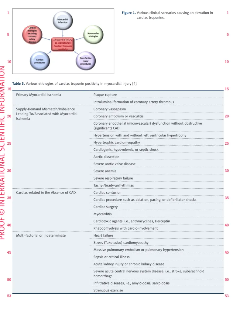
per-centile upper reference limit (URL). Such injury is classified as acute if characterized by a rise and/or fall in cardiac troponin values (Figure 1). Elevated cardiac troponin can also be ob-served in the absence of acute myocardial ischemia or obstruc-tive CAD in a host of clinical conditions (reviewed in Table 5), confirming that cardiac biomarkers are specific for the heart but not for ACS.
Slight elevations in serum cTn often lead to extensive and cost-ly cardiovascular workups, which may sidetrack clinicians, lead-ing them astray from the true cause of cTn elevation. The pres-ent article reviews a) the pathophysiology of cTn release from the heart and different organs, and b) the clinical scenarios
Type 1: Spontaneous MI
Related to atherosclerotic plaque rupture, ulceration, fissuring, erosion, or dissection leading to intraluminal thrombus in one or more coronary arteries leading to ischemia and resultant myocyte necrosis
Type 2: MI secondary to oxygen supply and demand mismatch/ischemia imbalance
Ischemia imbalance leading to myocyte necrosis and is resultant from a condition other than CAD Type 3: MI secondary to sudden cardiac death (biomarker values unavailable)
Cardiac death associated with symptoms and ECG findings suggestive of ischemia in the absence of cardiac biomarkers Type 4a: MI secondary to percutaneous coronary intervention (PCI)
PCI-associated MI defined arbitrarily as an elevation in cTn >5×99th percentile of the upper limit of normal with normal baseline
values or a rise of cTn values >20% of previously elevated baseline values that are stable or falling in association with the presence of at least one of the following: symptoms suggestive of cardiac ischemia, new ischemic ECG findings or new LBBB, angiographic loss of patency of a major coronary vessel or a side branch or persistent slow- or no-flow or embolization, or imaging demonstrating new loss of viable myocardium or new regional wall motion abnormality
Type 4b: MI secondary to stent thrombosis
MI associated with stent thrombosis as detected by coronary angiography or autopsy with a combination of myocardial ischemia and rise and/or fall of cardiac biomarkers with at least one value ³99th percentile of the upper limit of normal
Type 5: MI secondary to coronary artery bypass grafting (CABG)
MI associated with CABG defined arbitrarily as an elevation in cardiac biomarker levels >10×99th percentile of the upper limit
of normal in patients with normal baseline values in addition to at least one of the following: new pathological Q waves or new LBBB, new graft or new native coronary artery occlusion, or imaging demonstrating new loss of viable myocardium or new regional wall motion abnormality
Table 1. Classification of myocardial infarction [4].
PROOF © INTERNA
TIONAL SCIENTIFIC INFORMA
TION
1 5 10 15 20 25 30 35 40 45 50 53 1 5 10 15 20 25 30 35 40 45 50 53leading to cTn elevations, with a special focus on those unre-lated to obstructive coronary artery disease.
Troponins are integral regulatory proteins located on the thin actin filament within the myocytes of striated heart muscle, and are released when cardiac myocyte injury occurs. These proteins are responsible for the intricate contraction-relax-ation cycle of myocytes (Figure 2). The cTn complex is com-posed of 3 different subunits: cardiac troponin-C (cTnC, the
calcium-binding component), cTnT (the tropomyosin-binding component), and cTnI (the inhibitory-activity regulator of the myosin-binding sites on actin thin filaments) [9]. During de-polarization of the cardiac myocyte, calcium enters the sarco-plasm, binds to cTnC, and induces structural changes in the cTn complex. The resulting shift of tropomyosin away from the ac-tive site of actin allows the myosin heads of the thick filament to interact with the now-exposed myosin-binding site of the actin filaments, thereby producing contraction of the sarco-mere and, consequently, the myocardium as a syncytium [9,10]. The existence of several isoforms of troponin depends upon
medical attention
Table 2. Features of an “ideal” serum cardiac biomarker [5,6].
Modern markers in use Cardiac Troponin T
Cardiac Troponin I
CK and CK-MB with relative index
Point-of-care Troponin I, CK-MB and myoglobin panel
Obsolete markers Aspartate transaminase
Lactate dehydrogenase Markers in development or undergoing further study High-sensitivity troponin assay
High-sensitivity C-reactive protein Urocortin
CK-MB isoforms B-type natriuretic peptide
Table 3. Overview of molecular biomarkers of myocardial injury [7,8].
Marker Onset Peak Return to baseline
Troponin T 4–9 h 12–24 h 7–14 d
Troponin I 4–9 h 12–24 h 7–14 d
CK/CK-MB 4–9 h 24 h 2–3 d
Myoglobin 1 h 4–12 h 24 h
Table 4. Comparison of cardiac biomarker kinetics during myocardial injury [5–8].
PROOF © INTERNA
TIONAL SCIENTIFIC INFORMA
TION
1 5 10 15 20 25 30 35 40 45 50 53 1 5 10 15 20 25 30 35 40 45 50 53Figure 1. Various clinical scenarios causing an elevation in cardiac troponins.
Primary Myocardial Ischemia Plaque rupture
Intraluminal formation of coronary artery thrombus Supply-Demand Mismatch/Imbalance
Leading To/Associated with Myocardial Ischemia
Coronary vasospasm
Coronary embolism or vasculitis
Coronary endothelial (microvascular) dysfunction without obstructive (significant) CAD
Hypertension with and without left ventricular hypertrophy Hypertrophic cardiomyopathy
Cardiogenic, hypovolemic, or septic shock Aortic dissection
Severe aortic valve disease Severe anemia
Severe respiratory failure Tachy-/brady-arrhythmias Cardiac-related in the Absence of CAD Cardiac contusion
Cardiac procedure such as ablation, pacing, or defibrillator shocks Cardiac surgery
Myocarditis
Cardiotoxic agents, i.e., anthracyclines, Herceptin Rhabdomyolysis with cardio-involvement Multi-factorial or Indeterminate Heart failure
Stress (Takotsubo) cardiomyopathy
Massive pulmonary embolism or pulmonary hypertension Sepsis or critical illness
Acute kidney injury or chronic kidney disease
Severe acute central nervous system disease, i.e., stroke, subarachnoid hemorrhage
Infiltrative diseases, i.e., amyloidosis, sarcoidosis Strenuous exercise
Table 5. Various etiologies of cardiac troponin positivity in myocardial injury [4].
PROOF © INTERNA
TIONAL SCIENTIFIC INFORMA
TION
1 5 10 15 20 25 30 35 40 45 50 53 1 5 10 15 20 25 30 35 40 45 50 53differential gene expression in particular tissues. For example, expression of cTnC occurs in both cardiac and skeletal mus-cle, making it a poor indicator of myocardial injury. In contrast, cTnT and cTnI are expressed almost exclusively in myocardial tissue, making them ideal markers of myocardial damage [10]. Both cTnT and cTnI possess unique N-terminal amino acid se-quences, ideal targets for modern serum assays [11], which were developed using antibodies directed against these se-quences [12]. Utilization of these assays to detect cTn positiv-ity in serum has proven both highly sensitive and specific for myocardial injury [13]. Prior to their development, clinical de-cision-making relied on the detection of other molecular bio-markers such as CK-MB, lactate dehydrogenase, and myoglo-bin, which all lacked specificity, due to their expression in the musculoskeletal tissue (Table 3) [14]. The various biomarkers have different properties, with troponins having the most ide-al kinetics, ide-along with the highest sensitivity and specificity, for acute coronary syndromes (Table 4) [1],
Accurate interpretation of cTn positivity can be challenging be-cause it relies on unverified assumptions, such as the exclu-sive provenience of cTn from cardiac myocytes, the freedom from laboratory error, the appropriate time of acquisition of blood samples, and the cognizance of the patient baseline cTn values [15]. Based on these postulations, every incremental change in cTnT, cTnI, and CK-MB, even the smallest, is seen as persuasive evidence for myocardial injury [5,6,15,16]. However, such a dogma seems to be challenged by a fresher revisitation
of old findings. It is known that elevated cTn levels can re-flect not only the detection of intact cTn, but also of its pro-teolytic degradation products, generated by the calcium-acti-vated protease, calpain. Such products, due to their reduced size, may egress from cells more easily and rapidly than their parent structural proteins. Calpain activation, eventuating in generation of proteolytic products, may explain the apparent inconsistency of modest, persistent elevations of cTn associ-ated with negative CK-MB, which can occur in some patients suffering from recurrent bouts of short-lasting sublethal myo-cardial ischemia, or in the setting of systemic inflammatory processes [15]. Furthermore, recent findings indicate that car-diac troponin T (cTnT) is also expressed in human smooth mus-cle cells of different organs and apparatuses, including aorta, trachea, gut, and urinary bladder [17]. Hence, calpain activa-tion at these sites would result, at least theoretically, in mild troponinemia (i.e., a slight troponin increase), which is totally unrelated to myocardial ischemia or any other cardiac injury.
Disruption of myocyte cell membrane integrity (occurring in any type of myocardial injury) results in leakage of cytoplas-mic proteins into the extracellular serum. The majority of cTn is bound to actin thin filaments; however, a small proportion of approximately 3–8% exists in an unbound state, free in the cytosol [18]. During myocardial injury, this unbound troponin is released first and detected as the initial rise of troponin as-says [19]. Troponin elevations from this pool, in cases of short-lasting myocardial ischemia or injury, would be expected to quickly rise and fall over the course of hours due to the rela-tively short half-life (2–4 hours) of cTnI and cTnT [20]. However, if there is significant injury causing extensive and progressive necrosis, a continued release of the myofibril-bound troponin pool results in elevations of cTnI and cTnT for up to 7–14 days. From a pathophysiological standpoint, release of cTn into the circulation can be caused by 2 major categories of events: 1) myocardial injury due to ischemia (including ACS), and 2) myo-cardial injury in the absence of ischemia.
Ischemic myocardial injury
The majority of ischemic myocardial injury is caused by ob-structive coronary artery disease, which may result in ACS. Ischemia, defined as reduced oxygen availability due to lack of blood flow, causes a series of crucial biochemical and met-abolic changes, which decrease mitochondrial oxidative phos-phorylation and deplete cellular adenosine triphosphate (ATP). If ischemia is prolonged, metabolism shifts to anaerobic gly-colysis with lactate accumulation and consequent intracellu-lar acidosis, which activates the Na+-H+ exchanger. The
ensu-ing acidosis and lack of ATP alters Na+-K+ ATPase function and
causes accumulation of cytosolic Na+, which is exchanged with Myosin
Tropomyosin Troponin Actin complex
Myosin binding sites become exposed once calcium binds to troponin complex Myosin binding sites on the actin filaments are blocked by tropomyosin
Ca2+ Myosin
binding site
Figure 2. Physiology of contraction-relaxation cycle in cardiac
myocytes and role of troponin [10].
PROOF © INTERNA
TIONAL SCIENTIFIC INFORMA
TION
1 5 10 15 20 25 30 35 40 45 50 53 1 5 10 15 20 25 30 35 40 45 50 53Ca2+ from the sarcoplasmic reticulum, resulting in overload of
intracellular Ca2+ [21]. Such alterations in cellular metabolism
lead to osmotic cell stress, lysosomal activation, reactive oxy-gen species production, and infiltration of inflammatory cells, which actively release mediators, promoting both inflamma-tion and increased catabolism [22]. Accordingly, the metabol-ic state arising from ischemmetabol-ic myocardial injury results in cat-abolic protein degradation and compromised cell membrane integrity, allowing for leakage of cytoplasmic proteins into the extracellular serum, and subsequent detection of cTn on se-rum assays.
Clinical encounters associated with ischemic myocardial injury
Besides obstructive CAD and ACS, typically occurring in the setting of thrombotic coronary artery obstruction, elevation of cTn may be caused by a number of other disease states that do not compromise coronary artery patency. Elevation of cTn in the absence of obstructive CAD is often caused by ischemia secondary to an imbalance between demand and supply for oxygen-rich blood flow (demand ischemia). Thus, in the 2018 Joint ESC/ACCF/AHA/WHF Fourth Universal Definition of MI, demand ischemia is referred to as type-2 myocardial infarc-tion, which is cardiac ischemia due to either increased oxygen demand or decreased supply, occurring in the absence of an acute primary coronary thrombotic event [4]. The conditions leading to demand ischemia are very broad and diverse. The most commonly observed disorders in clinical practice include fixed coronary artery atherosclerosis, coronary spasm, coronary embolism caused by thrombi, calcium, or vegetation originat-ed from atria or ventricles, coronary artery dissection with or without intracoronary hematoma, tachyarrhythmias, bradyar-rhythmias, severe hypertension with or without left ventricu-lar hypertrophy, hypovolemic shock and other types of hypo-tension, and severe anemia (Table 5).
Myocardial injury in the absence of ischemia
Numerous clinical conditions other than ACS and ischemic myocardial injury have been recognized to cause elevated cTn, with several occurring independent of myocardial ne-crosis [23–25]. Different mechanisms for troponin release in non-ACS disorders have been postulated, including, but not limited to, increased cell membrane permeability due to re-versible injury or myocardial stretch [26] and increased rates of cell turnover, whether physiologic or pathologic [27]. Such mechanisms seem to have a common denominator in the hy-percatabolic state associated with the generation of inflam-matory and/or hormonal catabolic molecules.
Indeed, it is well known that inflammatory molecules, like those produced from reperfusion injury, can cause the formation of
reactive oxygen species (ROS) via altered mitochondrial oxi-dation and function. ROS interact with cell membrane phos-pholipids, which alters membrane permeability and favors the extracellular leakage of cytosolic proteins [28]. Cell membrane dysfunction also causes cytosolic Ca+ overload, which is partic-ularly important because Ca2+ activates specific proteases able
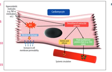
to reinforce myofibril degradation [28,29]. Moreover, levels of circulating catecholamines and inflammatory cytokines, like tu-mor necrosis factor-alpha (TNF-alpha), interleukin-1 (IL-1), and interleukin-6 (IL-6), are known to increase in catabolic condi-tions such as sepsis, immunological diseases, trauma, and/or chronic diseases. It was recently postulated that such molecules stimulate muscular fiber proteolysis, most notably in skeletal muscle, but also in cardiac myocytes, contributing to the hy-percatabolic state [30]. Furthermore, animal studies showed that IL-6, TNF-alpha, and circulating catabolic molecules bind specific cardiomyocyte membrane receptors, engaging an intra-cellular cascade (likely via the phosphatidylinositol (PI3K)/Aky pathway), whose activation results in protein disarrangement. Protein Kinase B (or Ak strain transforming/AKT), a key mol-ecule in such a pathway, decreases the phosphorylation (and therefore the activity level) of mammalian target of rapamy-cin (mTOR), which exerts pro-catabolic effects by inhibiting the synthesis of survival proteins such as eukaryotic initiation fac-tor binding protein-1 (4E-BP1) and S6 ribosomal protein, and activating the ubiquitin proteasome system, with ensuing in-tracellular protein breakdown [31]. The adverse effects of cy-tokines on cardiac cells and sarcomeres are compounded by other molecular mechanisms responsible for proteolysis and myocyte loss. Indeed, binding of TNF-alpha to its membrane receptor results in direct myocyte apoptosis (elicited by acti-vation of the death receptor, caspase 8-mediated pathway), and activation of other pro-apoptotic molecules such as Bcl-2-associated X protein (BAX) [32]. Figure 3 illustrates the po-tential mechanisms of contractile protein breakdown associ-ated with hypercatabolic states.
Clinical encounters of elevated cardiac biomarkers in the absence of ischemia
Numerous conditions, varied in presentation and nature, can engender myocardial injury in the absence of ischemia. Such conditions include: sepsis, cerebrovascular accidents, cardio-toxic medications, renal failure, infiltrative diseases, acute re-spiratory failure, extreme exertion, cardiac contusion, burns affecting >30% of the body surface area, pacing and electrical shock in the setting of cardioversion, carbon monoxide expo-sure, Takotsubo cardiomyopathy, peripartum cardiomyopathy, heart failure, endocarditis, myocarditis, myopericarditis, rhab-domyolysis, and sequelae of malignancy. Among these, the most studied are sepsis, neurogenic diseases such as stroke and subarachnoid hemorrhage, and cardiotoxic medications such as anthracyclines and trastuzumab.
PROOF © INTERNA
TIONAL SCIENTIFIC INFORMA
TION
1 5 10 15 20 25 30 35 40 45 50 53 1 5 10 15 20 25 30 35 40 45 50 53Despite major advances in an organized treatment approach to sepsis over the last 2 decades, sepsis remains the most com-mon cause of death in critically ill patients [33]. Sepsis-induced cardiomyopathy is a common complication of sepsis, defined by left ventricular dilation with normal or low filling pressures, a decrease in LVEF, and normalization of cardiac function, typi-cally within 7–10 days [34]. However, sepsis-induced cardiomy-opathy has also been described in the setting of normal LVEF due to complex hemodynamic changes during sepsis, includ-ing decreased systemic vascular resistance, tachycardia, and significant alterations in intravascular volume during resusci-tation [35]. Septic cardiomyopathy commonly results in ele-vated serum levels of cardiac troponin, which has significant prognostic value. The pathophysiologic mechanisms underly-ing septic cardiomyopathy involve the release of cardiac-de-pressive factors like endotoxin and endogenous inflammatory cytokines (TNF-alpha, IL-1, and IL-6), thereby increasing nitric oxide production. Excess nitric oxide depresses myofibril re-sponse to calcium, downregulates beta-adrenergic receptors, and leads to global mitochondrial dysfunction [34]. These cel-lular alterations cause protein demolition, impair the contrac-tile apparatus, and increase the microvascular permeability of cardiac myocytes [35,36], which can facilitate muscle contrac-tile protein breakdown and troponin release. Inflammatory cytokines like TNF-alpha, IL-1, and IL-6 have been shown to cause degradation of proteins, including free troponin, to low-er molecular-weight fragments, which are released into sys-temic circulation through the highly permeable membranes of cardiac myocytes [30]. The detection of these small troponin fragments by conventional assays would explain troponinemia in the absence of myocyte necrosis. Although this hypothe-sis is highly contentious and not supported by definitive evi-dence, the clinical observation that sepsis-related myocardial depression resolves without residual ventricular wall motion
abnormalities [34] suggests that cardiac enzymes can be re-leased in the absence of significant cardiomyocyte cell death. Septic cardiomyopathy is commonly accompanied by an ele-vation of serum cTn, which is highly correlated with left ven-tricular dysfunction and increased mortality [34,35,37–39]. In one study of 58 consecutive patients admitted to the medical intensive care unit with sepsis, all patients with LVEF <45% had detectable serum levels of cardiac troponin. The degree of troponin elevation demonstrated a significant inverse cor-relation with LVEF, and 30-day mortality was 4 times great-er in patients with elevated troponin levels [38]. Regardless of the mechanism of troponin leakage, septic cardiomyopa-thy demonstrates that elevated serum troponin, especially in critically ill patients, should not be assumed to indicate ACS or the presence of obstructive coronary artery disease. Critically ill patients often have multiple non-coronary etiologies of se-rum cTn elevation, which may be deceivingly heightened or prolonged by concomitant renal impairment. Unfortunately, no specific treatment exists for septic cardiomyopathy except for implementing evidence-based, guideline-directed therapy for the treatment of sepsis [35]. While it is not recommended by guideline-directed therapy, physicians may utilize cardiac enzymes as an adjunctive tool to risk-stratify septic patients in the intensive care unit in an effort to optimize treatment strategies and improve outcomes.
An elevation of cTn, often associated with electrocardiogram abnormalities and clinical evidence of left ventricular dysfunc-tion, is detected in up to 20% of patients after stroke, particu-larly subarachnoid hemorrhage [40]. Besides preexisting CAD, another postulated mechanism for cardiac damage in this spe-cific patient population seems to be myocardial injury elicit-ed by centrally melicit-ediatelicit-ed release of catabolic molecules like
Systemic circulation Amino acid release Membrane damage Demolition of contractile proteins ROS Hypercatabolic molecules (e.g. TNF-α, interleukines, ect.) Cardiomyocyte Increase in cell membrane permeability P eak myofilament protein chain (e.g. cTnl)
relase
Figure 3. Postulated biochemical mechanisms
responsible for hypercatabolic protein breakdown.
PROOF © INTERNA
TIONAL SCIENTIFIC INFORMA
TION
1 5 10 15 20 25 30 35 40 45 50 53 1 5 10 15 20 25 30 35 40 45 50 53catecholamines in response to hypoperfusion of the posteri-or hypothalamus. A powerful catecholamine surge may be an alternative mechanism of cardiac damage, because troponin-emia is often detected even in young individuals with very low pretest probability for an acute coronary syndrome. Cardiac myofibrillar degeneration, which was described in patients who died due to acute stroke, occurs in the proximity of the cardi-ac nerves and is histologically indistinguishable from myocyte death caused by catecholamine infusion or reperfusion of tran-siently ischemic myocardium [41], being characterized by con-traction bands necrosis and associated with mononuclear in-filtration and early calcification. Mounting evidence suggests that stroke affecting the insular cortex, which plays a central role in the autonomic control of cardiovascular function, is as-sociated with increased risk of adverse cardiac outcomes, in-cluding neurogenic cardiac damage. Brain regions associated with cTnT elevation include the right posterior, superior, and medial insula, and the right inferior parietal lobule.
Medication-induced troponin release
Many medications, especially chemotherapeutics and anti-neoplastic agents, have adverse effects on cardiac myocytes and contribute to the development of cardiovascular compli-cations such as hypertension, heart failure, arrhythmias, and ischemia, all of which may be accompanied by elevated serum cardiac biomarkers [42]. Advances in cancer treatment provide a unique opportunity for cardiologists and oncologists to col-laborate in an effort to improve treatment outcomes for pa-tients with concomitant heart disease and malignancy [43]. One way to mitigate the cardiovascular complications of can-cer treatment is to expand the current guidelines regarding cardiac monitoring during the administration of cardiotoxic chemotherapeutic agents. Besides cardiac imaging, physicians have also discussed the routine monitoring of serum cardiac troponin before and during treatment with cardiotoxic med-ications, in an effort to diagnose cardiotoxicity at its earliest stages, when it is more amenable to medical treatment [44]. While a comprehensive review of cardiotoxic medications is beyond the scope of this discussion, Table 6 reports some of the medications commonly implicated in the causality of car-diac damage. A brief review of the most studied cardiotoxic medications is presented below.
Anthracyclines injure cardiac myocytes primarily by inhibiting topoisomerase 2-beta, which increases breaks in double-strand-ed DNA [45] in addition to increasing free radical production via iron deposition in mitochondria [46]. These mechanisms can lead to apoptosis, necrosis, and permanent cardiomyop-athy [45,46]. Anthracyclines also increase intracellular calci-um levels and cause direct cellular injury through drug me-tabolism, oxidative damage, and influx of pro-inflammatory
molecules. A direct correlation exists between the cumula-tive dose of anthracycline, the magnitude of troponin eleva-tion, and the subsequent degree of left ventricular dysfunc-tion, which is often permanent [47].
Trastuzumab is a monoclonal antibody that antagonizes the human epidermal growth factor receptor 2 (HER-2), which is expressed in 20–25% of breast cancers. It has reduced disease recurrence and increased survival in patients with breast can-cers that express HER-2, especially when it is combined with anthracyclines like doxorubicin [48]. Unfortunately, this com-bination results in cardiac dysfunction in approximately 25% of patients [49]. Anthracycline-induced oxidative damage to cardiomyocytes results in upregulation of HER-2 receptors as part of cellular repair. While trastuzumab inhibition of HER-2-mediated cell repair plays a role in the loss of cardiac my-ocytes, the precise mechanism of trastuzumab-induced car-diomyopathy is still unclear [49]. Such carcar-diomyopathy differs from that caused by anthracyclines in that the cardiac dys-function is not dose-dependent, and it is reversible in approx-imately 60% of patients [50]. Elevated cardiac biomarkers in the setting of trastuzumab regimen decrease the chance of cardiac recovery and increase the risk for major adverse car-diac events (MACE) [50]. Patients with elevated troponin who were treated with trastuzumab had a greater risk of develop-ing cardiac dysfunction and a 25-fold increase in MACE [49]. Patients at highest risk were those treated with both anthra-cyclines and trastuzumab [51], requiring cardiac monitoring with serial echocardiograms or multigated acquisition scans (MUGA), due to the risk of cardiac injury and left ventricular dysfunction. High-sensitivity cTn measured prior to and dur-ing the administration of trastuzumab seems to be useful in the early identification of patients at higher risk of developing drug-induced cardiotoxicity, as well as of those whose cardi-ac function will remain impaired despite an appropriate med-ical regimen [50].
While anthracyclines and trastuzumab are the most stud-ied cardiotoxic medications used in cancer treatment, a host of anti-neoplastic agents are known to produce cardiac dys-function and have the potential to cause an elevation of car-diac biomarkers. The major classes of anti-neoplastic agents most commonly associated with troponin elevations are list-ed in Table 6. As the field of oncology continues to advance, more malignancies are being managed with a variety of nov-el therapeutics. The evaluation of cardiac biomarkers before and during cancer treatment, in conjunction with serial imag-ing studies, is currently the earliest and most accurate way to identify cardiotoxicity, and has the potential to be integrated into future cardio-oncology guidelines.
PROOF © INTERNA
TIONAL SCIENTIFIC INFORMA
TION
1 5 10 15 20 25 30 35 40 45 50 53 1 5 10 15 20 25 30 35 40 45 50 53Conclusions
In this review, we described the different pathophysiological mechanisms of cTn release, and appraised a variety of clinical conditions presenting with elevated cTn, giving special attention to those cases of troponinemia unrelated to obstructive CAD. Ischemic myocardial injury is most commonly caused by ob-structive coronary artery disease and ACS, which typically oc-cur in the setting of thrombotic coronary artery occlusion. The 2018 Joint ESC/ACCF/AHA/WHF Fourth Universal Definition of MI placed greater emphasis on the measurement of cardi-ac enzymes, specifically high-sensitivity cTn, in the diagnos-tic criteria of acute myocardial infarction [4]. Testing of car-diac enzymes is ordered in 16.9% of patients presenting to emergency departments in the United States, but chest pain only accounts for approximately 5.3% of chief complaints [52]. Current international guidelines regarding chest pain recom-mend trending serial cTn and EKGs to assess for a potentially evolving ACS, thereby subjecting many patients to prolonged ED observation or hospital admission, and increasing the number
of false-positive troponin results in patients with low pretest probability for coronary artery disease [53–55].
The term myocardial infarction in the absence of obstructive coronary artery disease (MINOCA) is an increasingly recognized diagnosis used to describe ischemic myocardial injury resulting in elevated cTn, despite insignificant coronary artery disease on angiography [56]. Mild troponinemia can also reflect the de-tection of small proteolytic products of cTn, which are released in the circulation following calpain activation in the setting of recurrent bouts of short-lasting sublethal myocardial ischemia or systemic inflammatory processes [16]. Likewise, calpain ac-tivation in organs other than the heart (such as smooth mus-cle cells of the aorta, trachea, gut, and urinary bladder, where cTnT is also expressed) might explain the occurrence of tro-poninemia in medical conditions totally unrelated to myocar-dial ischemia or any cardiac injury.
Scoring systems incorporating the first cTn, such as a low HEART score (0–3) or low TIMI score (0–1), are the most helpful in identifying patients less likely to have ACS [57]. In patients
Anti-metabolites Decitabine
Clofarabine
Alkylating agents Cyclophosphamide
Iphosphamide Melphalan
Small molecule tyrosine kinase inhibitors Sunitinib and sorafenib Pazopanib
Dabrafenib and dasatinib Lapatinib and trametinib Microtubule polymerization inhibitors Paclitaxel
Docetaxel
Anthracyclines Doxorubicin
Daunorubicin Epirubicin Idarubicin
Proteasome inhibitors Carfilzomib and bortezomib
Monoclonal antibody- based tyrosine kinase inhibitors Trastuzumab Bevacizumab
Adotrastuzumab emtansine Pertuzumab
Table 6. List of cardiotoxic medications [42,43,47].
PROOF © INTERNA
TIONAL SCIENTIFIC INFORMA
TION
1 5 10 15 20 25 30 35 40 45 50 53 1 5 10 15 20 25 30 35 40 45 50 53with a low pretest probability for ACS, an elevation in cardi-ac biomarkers can become a confounding fcardi-actor, delaying the formulation of the correct diagnosis and thereby postponing initiation of adequate medical management.
Regardless of the clinical situation, elevated serum tropo-nin is associated with increased mortality and adverse out-comes [58–60], and may be used to risk-stratify patients. Awareness of the multiple conditions associated with positive
troponin, as well as understanding of the pathophysiology of its release, are essential preconditions for minimizing unnec-essary, costly, and potentially risky interventions, providing timely and appropriate medical care.
None.
1. Karmen A, Wroblewski F, Ladue JS: Transaminase activity in human blood. J Clin Invest, 1955; 34: 126–31
2. Nomenclature and criteria for diagnosis of ischemic heart disease. Report of the Joint International Society and Federation of Cardiology/World Health Organization task force on standardization of clinical nomencla-ture. Circulation, 1979; 59: 607–9
3. Amsterdam EA, Wenger NK, Brindis RG et al: 2014 AHA/ACC Guideline for the Management of Patients With Non–ST-Elevation Acute Coronary Syndromes. J Am Coll Cardiol, 2014, 64(24): e139–228
4. Thygesen K, Alpert JS, Jaffe AS et al: Executive Group on behalf of the Joint European Society of Cardiology (ESC)/American College of Cardiology (ACC)/ American Heart Association (AHA)/World Heart Federation (WHF) Task Force for the Universal Definition of Myocardial Infarction. Fourth Universal Definition of Myocardial Infarction (2018). J Am Coll Cardiol, 2018; 72(18): 2231–64
5. Newby LK, Jesse RL, Babb JD et al: ACCF 2012 expert consensus document on practical clinical considerations in the interpretation of troponin ele-vations: A report of the American College of Cardiology Foundation task force on Clinical Expert Consensus Documents. J Am Coll Cardiol, 2012; 60: 2427–63
6. Babuin L, Jaffe AS: Troponin: The biomarker of choice for the detection of cardiac injury. CMAJ, 2005; 173: 1191–202
7. Park KC, Gaze DC, Collinson PO, Marber MS: Cardiac troponins: From myocar-dial infarction to chronic disease. Cardiovasc Res, 2017; 113(14): 1708–18 8. Hoff J, Wehner W, Nambi V: Troponin in cardiovascular disease prevention:
Updates and future direction. Curr Atheroscler Rep, 2016; 18(3): 12 9. Lewandrowski K, Chen A, Januzzi J: Cardiac markers for myocardial
infarc-tion. A brief review. Am J Clin Pathol, 2002; 118(Suppl.): S93–99 10. Parmacek MS, Solaro RJ: Biology of the troponin complex in cardiac
myo-cytes. Prog Cardiovasc Dis, 2004; 47: 159–76
11. Wilkinson JM, Grand RJ: Comparison of amino acid sequence of troponin I from different striated muscles. Nature, 1978; 271: 31–35
12. Melanson SE, Conrad MJ, Mosammaparast N, Jarolim P: Implementation of a highly sensitive cardiac troponin I assay: Test volumes, positivity rates and interpretation of results. Clin Chim Acta, 2008; 395: 57–61 13. Apple FS, Wu AH, Jaffe AS et al: National Academy of Clinical Biochemistry
and IFCC Committee for Standardization of Markers of Cardiac Damage Laboratory Medicine practice guidelines: Analytical issues for biomarkers of heart failure. Circulation, 2007; 116: e95–98
14. Halim SA, Newby LK, Ohman EM: Biomarkers in cardiovascular clinical tri-als: Past, present, future. Clin Chem, 2012; 58: 45–53
15. Sandoval Y, Jaffe AS: Type 2 myocardial infarction: JACC Review Topic of the Week. J Am Coll Cardiol, 2019; 73(14): 1846–60
16. Morrow DA, Cannon CP, Jesse RL et al: National Academy of Clinical Biochemistry Laboratory Medicine Practice Guidelines: Clinical character-istics and utilization of biochemical markers in acute coronary syndromes. Circulation, 2007; 115: e356–75
17. Kajioka S, Takahashi-Yanaga F, Shahab N et al: Endogenous cardiac tropo-nin T modulates Ca2+-mediated smooth muscle contraction. Sci Rep, 2012; 2: 979
18. Bleier J, Vorderwinkler KP, Falkensammer J et al: Different intracellular com-partmentations of cardiac troponins and myosin heavy chains: A causal connection to their different early release after myocardial damage. Clin Chem, 1998; 44: 1912–18
19. Katus HA, Remppis A, Neumann FJ et al: Diagnostic efficiency of troponin T measurements in acute myocardial infarction. Circulation, 1991; 83: 902–12 20. Gerhardt W, Katus H, Ravkilde J et al: S-troponin T in suspected ischemic
myocardial injury compared with mass and catalytic concentrations of S-creatine kinase isoenzyme MB. Clin Chem, 1991; 37: 1405–11 21. Consolini AE, Ragone MI, Bonazzola P, Colareda GA: Mitochondrial
bioen-ergetics during ischemia and reperfusion. Adv Exp Med Biol, 2017; 982: 141–67
22. McDougal AD, Dewey CF Jr.: Modeling oxygen requirements in ischemic car-diomyocytes. J Biol Chem, 2017; 292(28): 11760–76
23. Eggers KM, Lindahl B: Application of cardiac troponin in cardiovascular dis-eases other than acute coronary syndrome. Clin Chem, 2017; 63(1): 223–35 24. White HD: Pathobiology of troponin elevations: Do elevations occur with
myocardial ischemia as well as necrosis? J Am Coll Cardiol, 2011; 57: 2406–8 25. Yang CW, Li H, Thomas L et al: Retrospective cause analysis of troponin
I elevation in non-CAD patients: Special emphasis on sepsis. Medicine (Baltimore), 2017; 96(37): e8027
26. Hessel MH, Atsma DE, van der Valk EJ et al: Release of cardiac troponin I from viable cardiomyocytes is mediated by integrin stimulation. Pflugers Arch, 2008; 455: 979–86
27. Hammarsten O, Mair J, Möckel M et al: Possible mechanisms behind car-diac troponin elevations. Biomarkers, 2018; 23(8): 725–34
28. Mittal M, Siddiqui MR, Tran K et al: Reactive oxygen species in inflamma-tion and tissue injury. Antioxid Redox Signal, 2014; 20(7): 1126–67 29. Forrester SJ, Kikuchi DS, Hernandes MS et al: Reactive oxygen species in
metabolic and inflammatory signaling. Circ Res, 2018; 122(6): 877–902 30. Pasini E, Corsetti G, Aquilani R et al: Protein-amino acid metabolism
disar-rangements: The hidden enemy of chronic age-related conditions. Nutrients, 2018; 10(4): pii: E391
31. Flati V, Pasini E, D’Antona G et al: Intracellular mechanisms of metabolism regulation: The role of signaling via the mammalian target of rapamycin pathway and other routes. Am J Cardiol, 2008; 101(11A): 16E–21E 32. Scarabelli TM, Gottlieb RA: Functional and clinical repercussions of
myo-cyte apoptosis in the multifaceted damage by ischemia/reperfusion inju-ry: Old and new concepts after 10 years of contributions. Cell Death and Differ, 2004; 11(Suppl. 2): S144–52
33. Dellinger RP, Levy MM, Rhodes A et al: Surviving Sepsis Campaign: International guidelines for management of severe sepsis and septic shock: 2012. Crit Care Med, 2013; 41: 580–637
34. Sato R, Nasu M: A Review of sepsis-induced cardiomyopathy. J Intensive Care, 2015: 3; 48
35. Antonucci E, Fiaccadori E, Donadello K et al: Myocardial depression in sep-sis: From pathogenesis to clinical manifestations and treatment. J Crit Care, 2014; 29(4): 500–11
36. Celes MR, Torres-Dueñas D, Malvestio LM et al: Disruption of sarcolemmal dystrophin and beta-dystroglycan may be a potential mechanism for myo-cardial dysfunction in severe sepsis. Lab Invest, 2010; 90: 531–42 37. Kim JS, Kim M, Kim YJ et al: Troponin testing for assessing sepsis-induced
myocardial dysfunction in patients with septic shock. J Clin Med, 2019; 8(2): pii: E239
38. Vallabhajosyula S, Sakhuja A, Geske JB et al: Role of admission troponin-T and serial troponin-T testing in predicting outcomes in severe sepsis and septic shock. J Am Heart Assoc, 2017; 6(9): pii: e005930
PROOF © INTERNA
TIONAL SCIENTIFIC INFORMA
TION
1 5 10 15 20 25 30 35 40 45 50 53 1 5 10 15 20 25 30 35 40 45 50 5339. Bessiere F, Khenifer S, Dubourg J et al: Prognostic value of troponins in sep-sis: A meta-analysis. Intensive Care Med, 2013; 39(7): 1181–89 40. Zhang L, Wang Z, Qi S: Cardiac troponin elevation and outcome after
sub-arachnoid hemorrhage: A systematic review and meta-analysis. J Stroke Cerebrovasc Dis, 2015; 24(10): 2375–84
41. Krishnamoorthy V, Mackensen GB, Gibbons EF, Vavilala MS: Cardiac dys-function after neurologic injury: What do we know and where are we go-ing? Chest. 2016; 149(5): 1325–31
42. Chang HM, Okwuosa TM, Scarabelli T et al: Best practices in cardio-oncol-ogy: Part 2. JACC, 2017; 70: 2552–65
43. Chen-Scarabelli C, McRee C et al: Comprehensive review on cardio-oncol-ogy: Role of multimodality imaging. J Nucl Cardiol, 2017; 24(3): 906–35 44. Cardinale D, Sandri MT, Colombo A et al: Prognostic value of troponin I in
cardiac risk stratification of cancer patients undergoing high-dose chemo-therapy. Circulation, 2004; 109(22): 2749–54
45. Zhang S, Liu X, Bawa-Khalfe T et al: Identification of the molecular basis of doxorubicin-induced cardiotoxicity. Nat Med, 2012; 18(11): 1639–42 46. Ichikawa Y, Ghanefar M, Bayeva M et al: Cardiotoxicity of doxorubicin is
mediated through mitochondrial iron accumulation. J Clin Invest, 2014; 124(2): 617–30
47. Chang HM, Moudgil R, Scarabelli T et al: Best practices in cardio-oncology: Part 1. JACC, 2017; 70: 2536–51
48. Cardinale D, Sandri MT, Martinoni A et al: Myocardial injury revealed by plasma troponin I in breast cancer treated with high-dose chemotherapy. Ann Oncol, 2002; 13(5): 710–15
49. Ewer MS, Ewer SM: Troponin I provides insight into cardiotoxicity and the anthracycline-trastuzumab interaction. J Clin Oncol, 2010; 28(25): 3910–19 50. Cardinale D, Colombo A, Torrisi R et al: Trastuzumab-induced cardiotoxicity:
Clinical and prognostic implications of troponin I evaluation. J Clin Oncol, 2010; 28(25): 3910–16
51. Seidman A, Hudis C, Pierri MK et al: Cardiac dysfunction in the trastuzum-ab clinical trials experience. J Clin Oncol, 2002; 20: 1215–21
52. Mackam AM, Nguyen OK: Use of cardiac biomarker testing in the Emergency Department. JAMA Intern Med, 2015; 175(1): 67–75
53. Fihn SD, Gardin JM, Abrams J et al: 2012 ACCF/AHA/ACP/AATS/PCNA/ SCAI/STS Guideline for the Diagnosis and Management of Patients with Stable Ischemic Heart Disease: Executive Summary: A Report of the American College of Cardiology Foundation/American Heart Association Task Force on Practice Guidelines, and the American College of Physicians, American Association for Thoracic Surgery, Preventive Cardiovascular Nurses Association, Society for Cardiovascular Angiography and Interventions, and Society of Thoracic Surgeons. J Am Coll Cardiol, 2012; 60(24): 2564–603 54. Newby LK, Jesse RL, Babb JD et al: ACCF 2012 expert consensus document
on practical clinical considerations in the interpretation of troponin eleva-tions: A report of the American College of Cardiology Foundation task force on Clinical Expert Consensus Documents. J Am Coll Cardiol, 2012; 60(23): 2427–63
55. Penumetsa SC, Mallidi J, Friderici JL et al: Outcomes of patients admitted for observation of chest pain. Arch Intern Med, 2012; 172: 873–77 56. Tamis-Holland JE, Jneid H, Reynolds HR et al: Contemporary diagnosis and
management of patients with myocardial infarction in the absence of ob-structive coronary artery disease. Circulation, 2019; 139: e891–908 57. Fanaroff AC, Rymer JA, Goldstein SA et al: Does this patient with chest pain
have acute coronary syndrome?: The rational clinical examination system-atic review. JAMA, 2015; 314: 1955–65
58. Gallagher S, Jones DA, Anand V, Mohiddin S: Diagnosis and management of patients with acute cardiac symptoms, troponin elevation and culprit-free angiograms. Heart, 2012; 98(13): 974–81
59. Ahmed AN, Blonde K, Hackam D et al: Prognostic significance of elevat-ed troponin in non-cardiac hospitalizelevat-ed patients: A systematic review and meta-analysis. Ann Med, 2014; 46(8): 653–63
60. Sara JD, Holmes DR Jr., Jaffe AS: Fundamental concepts of effective troponin use: important principles for internists. Am J Med, 2015; 128(2): 111–19
![Table 1. Classification of myocardial infarction [4].](https://thumb-eu.123doks.com/thumbv2/123dokorg/5530669.64845/2.918.74.824.667.1101/table-classification-of-myocardial-infarction.webp)
![Table 4. Comparison of cardiac biomarker kinetics during myocardial injury [5–8].](https://thumb-eu.123doks.com/thumbv2/123dokorg/5530669.64845/3.918.87.843.422.697/table-comparison-cardiac-biomarker-kinetics-myocardial-injury.webp)

![Table 6. List of cardiotoxic medications [42,43,47].](https://thumb-eu.123doks.com/thumbv2/123dokorg/5530669.64845/9.918.96.852.166.702/table-list-of-cardiotoxic-medications.webp)
