Università degli Studi di Salerno
Dipartimento di Chimica e Biologia
“A. Zambelli”
Corso di Dottorato di Ricerca in Chimica
XIV CICLO NUOVA SERIETesi di dottorato in:
“Synthesis of calixarene derivatives active
towards proteic targets involved in tumor
pathologies”
Tutor: Candidato:
Prof. Carmine Gaeta Stefano Tommasone Matr.: 8880700199
Coordinatore:
Prof. Gaetano Guerra
Index
Chapter 1
1. Introduction on calix[n]arenes ... 1
1.1 Applications in host-guest chemistry ... 3
1.2 Biological applications of calix[n]arenes ... 8
Chapter 2 2. DNA intercalators in anticancer therapy ... 17
2.1 Calixarene based DNA intercalators – Results and discussion ... 23
2.1.1 Synthesis of the calixarene conjugates ... 25
2.1.2 Biological studies ... 34
2.1.3 Circular Dichroism ... 36
2.1.4 Molecular Modeling ... 38
2.2 Conclusions ... 46
Chapter 3 3. Chemical proteomics and drug discovery ... 47
3.1 Application of chemical proteomics ... 51
3.2 Biomolecular recognition abilities of calixarenes through the chemoproteomic approach - Results and discussion ... 55
3.2.1 Synthesis of compound 13 ... 57
3.2.2 Chemical proteomics experiment ... 63
3.2.3 Molecular Docking ... 74
3.2.4 Biological activity studies, ... 78
3.2.5 Synthesis of an advanced intermediate for a new calixarene ligand ... 82
3.3 Conclusions ... 89
Chapter 4 4. Multivalent systems in glycosidase inhibition ... 91
inhibition ... 94 4.2 Calix[n]arenes as scaffolds for multivalent systems
………...98 4.3 Multivalent iminosugar calix[8]arene conjugates – Results and discussion ... 101
4.3.1 Synthesis of the multivalent iminosugar-calix[8]arene conjugates ... 102 4.3.2 Glycosidase inhibition studies ... 111 4.3.3 Higher valency iminosugar clusters ... 113 4.4 A new ligation strategy for the synthesis of glycoconjugates... 116
4.4.1 Results and discussion ... 118 4.5 Conclusions ... 125 Chapter 5
1
Chapter 1
1. Introduction on calix[n]arenes
Calix[n]arenes1 are macrocycles belonging to the [1.1.1.1.]metacyclophane family, in which p-substituted phenolic units are linked together via methylene bridges. The calixarene family consists of 17 cyclooligomers, ranging from the tetramer to the eicosomer.
Figure 1.1
1
(a) Vicens, J.; Harrowfield, J. Calixarenes in the Nanoworld, Springer: Dordrecht,
2007; (b) Gutsche, C. D. Calixarenes Revisited, Royal Society of Chemistry:
Cambridge, 1998; (c) Bohmer, V. The Chemistry of Phenols, Wiley: Chichester,
2003; (d) Gutsche, C. D. Calixarenes, An Introduction, Royal Society of Chemistry:
2
Regarding the calix[n]arene nomenclature, the word “calixarene” was coined by C. David Gutsche, because the
p-t-butylcalix[4]arene resembled the shape of a Greek vase
called calyx krater (Figure 1.2), while "n" between square brackets indicates the number of phenolic components of the macrocycle. (Figure 1.1).
calyx krater p-t-butylcalix[4]arene
Figure 1.2
The calixarenes can exist in several conformations because the phenolic units freely rotate around the methylene bridge. Regarding the p-tert-butylcalix[4]arene, it adopts both in solution and in the solid state the cone conformation, with the four phenolic hydroxyl groups engaged in a cyclic network of hydrogen bonds. In this case it is possible to distinguish two regions, the Exo or Upper Rim and the Endo or Lower Rim (Figure 1.2).
Exo or Upper Rim
Endo or Lower Rim
Exo or Upper Rim
3
Both the lower and upper rim can be easily functionalized, allowing the introduction of several functional groups such as ethers and esters by alkylation of the hydroxyl groups, and the introduction of halogens, -NO2, -SO3H, -CHO and -COOH
by electrophilic substitution.2
1.1 Applications in host-guest chemistry
Together with cyclodextrins, cucurbiturils and crown ethers, calixarenes constitute the major classes of supramolecular hosts. They are bowl-shaped macrocycle and their cavity is able to host small molecules or ions. The synthetic versatility of the calixarenes enables them to act as a framework that can support a broad range of binding, signaling or other functionalities. Thus, currently many examples have been reported of calixarene-based hosts for the recognition of cations, anions and neutral molecules with the aim to develop new materials with potential applications as sensors3 or membranes for the removal of pollutants.4
2
(a)Gutsche, C. D.; Pagoria, P. F. J. Org. Chem. 1985, 50, 5795-5802; (b) Zhang, W. C.; Zheng, Y. A.; Huang, Z. T. Synthetic Commun, 1997, 3763-3767; (c) Smith, W. E. J. Org. Chem. 1972, 37, 3972-3973. (d) Rieche, A.; Gross, H. Chem. Ber.
1960, 93, 88-94. 3
(a) Czarnik, A. W.; Desvergne, J. P. Chemosensors for Ion and Molecule Recognition. Kluwer: Dordrecht, 1997; (b) Beer, P. D.; Gale, P. A. Angew.Chem. Int. Ed. 2001, 40, 486–516.
4
(a) Kumar, P. L.; Saxena, C.; Dubey, V. J. Membr. Sci. 2003, 227, 173–182; (b) Uragami, T.; Meotoiwa, T.; Miyata, T. Macromolecules 2003, 36, 2041–2048.
4
Regarding the cationic recognition, an interesting example is the p-sulfonatocalix[7]arene, a water soluble compound, that shows good affinity toward some organic quaternary ammonium salts of environmental relevance, such as Diquat and Paraquat.5 Interestingly, in the solid state the dicationic Diquat guest is encapsulated in a molecular capsule constituted by two molecule of p-sulfonatocalix[7]arene (Figure 1.3).6
Figure 1.3
5
Caeta, C.; Caruso, T.; Mincolelli, M.; Troisi, F.; Vasca, E.; Neri, P. Tetrahedron,
2008, 64, 5370-5378. 6
Erra, L.; Tedesco, C.; Vaughan, G.; Brunelli, M.; Troisi, F.; Gaeta, C.; Neri, P. CrystEngComm, 2010, 12, 3463-3466.
5 N H O NH NH HN O O O O O O NH HN HN O NH R R R R N H O NH NH HN O O O O O O NH HN HN O NH R R R R O O R = H or NO2 C5H11 C5H11 C 5H11C5H11
The coordination chemistry of anions has gained more interest in the last twenty years. The design of anion receptors is really challenging. Anions are larger then isoelectronic cations, thus electrostatic binding interactions are less effective. Moreover anion species have different geometries, thus a high degree of design is required for the synthesis of suitable complementary receptors for anionic guests. Functional groups like amides, ureas, thioureas and pyrroles are very attractive for this purpose and they have recently been incorporated into macrocycle scaffolds, like calixarenes.7
An example is the study of the binding properties toward anionic guest by pyrrolamidocalix[4]arene hosts (Figure 1.4).8
Figure 1.4
7
Matthews, S. E.; Beer, P. D. Supramol. Chem. 2005, 17, 411-435.
8
6
The derivatives shown in Figure 1.4 are able to bind anions such as dihydrogen phosphate, fluoride, acetate and benzoate. Particularly, these derivatives are able to discriminate efficiently the H2PO4- anion, with a selectivity more than
38-fold higher than those for the acetate and benzoate anions. This selectivity can be explained by the planar trigonal arrangement of NHs groups of three amidopyrrole units, that are complementary to three H-bonding acceptor sites of H2PO4- anion.
Calixarenes have the characteristic to organize themselves in the solid state through weak interactions. They form supramolecular frameworks with lattice voids that can be occupied by guest molecules.9 Thus, p-H-calix[4]arene (Figure 1.5a) is able to form crystal structures in which three calixarene units adopt a cyclic arrangement, with an approximately spherical shape, held together only by van der Walls interactions (Figure 1.5b).10
9
Atwood, J. L.; Barbour, L. J.; Jerga, A. Chem. Comm. 2001, 22, 2376-2377.
10
7
a) b)
c)
Figure 1.5 a) H-calix[4]arene; b) trimeric arrangement of calix[4]arene; c) space filling representation of
p-H-calix[4]arene@CF3Br.
These trimers form a hexagonal close-packed (hcp) lattice with interstitial voids that are occupied by gases such as CCl4,
8
freons and also methane (Figure 1.5c). The gases are retained even at relatively high temperatures and low pressures. Thus, this solid-state self-assembly of calix[4]arenes reported in 2002 by Atwood and coworkers,9 results in a supramolecular system with potential application in gas storage.
1.2 Biological applications of calix[n]arenes
Over the last 30 years a growing interest has been direct toward the biomolecular recognition of calixarene derivatives and more in particular to the interaction with druggable target(s).11,12 At this regard one of the first report by Cornforth and coworkers in 1955 showed that the p-octyl-calix[8]arene bearing polyoxyethylene units at the lower rim was active as anti-tuberculosis agent.13
More recently, p-sulfonatocalix[n]arenes have interesting antiviral activity and they find potential use in the treatment
11
(a) de Fátima, A.; Fernandes, S. A.; Sabino, A. A. Curr. Drug. Discov. Technol.
2009, 6, 151-170. (b) Giuliani, M.; Morbioli, I.; Sansone, F.; Casnati, A. Chem.
Commun. 2015, 51, 14140-14159; (c) Park, H. S.; Lin, Q.; Hamilton, A. D. Proc. Natl. Acad. Sci. USA 2002, 99, 5105-5109; (d) Baldini, L.; Casnati, A.; Sansone, F.; Ungaro, R. Chem. Soc. Rev. 2007, 36, 254-266; (e) Schrader, T. Nat. Chem. 2012, 4, 41529-41542.
12
Da Silva, E.; Lazar, A. N.; Coleman, A. W. J. Drug. Del. Sci. Tech. 2004, 14, 3-20.
13
(a) Cornforth, J. W.; D’Arcy Hart, P.; Nicholls, G. A.; Rees, R. J. W.; Stock, J. A. Brit. J. Pharmacol. 1955, 10, 73-86; (b) D’Arcy Hart, P.; Armstrong, J. A.; Brodaty, E. Infetc. Immun. 1996, 64, 1491-1493.
9
of viruses such as AIDS and Herpes.14,15
Some of the most fascinating results in the biological activity of calixarenes have been achieved by Hamilton and his co-workers, that described a series of peptidocalix[4]arene derivatives active as antiangiogenic and anticancer agents.16 In this report Hamilton and coworkers studied a series of protein surface binders constituted by a calix[4]arene backbone adorned to the upper rim with four peptide loops (Figure 1.6).
Figure 1.6 growth factor binders functionalized with: a) peptide loops; b) isophthalate groups.
14
Hwang, K. M.; Qi, Y. M.; Liu, S. Y; Choy, W.; Chen, J.: WO9403164 (1994); (b) Harris, S. J.: WO200244141 (2002); (c) Coveney, D.; Costello, B.: US2005113454 (2005); (d) Wang, K.; Guo, D. S.; Zhang, H. Q.; Li, D.; Zheng, X. L.; Liu, Y. J. Med. Chem. 2009, 52, 6402-6412
15
Motornaya, A. E.; Alimbarova, L. M.; Shokova, E. A.; Kovalev, V. V. Pharm. Chem. J. 2006, 40, 68-72.
16
(a) Sebti, S. M.; Hamilton, A. D. Oncogene, 2000, 19, 6566-6573; (b) Blaskovich, M. A.; Lin, Q.; Delarue, F. L.; Sun, J.; Park, H. S.; Coppola, D.; Hamilton, A. D.; Sebti, S. M. Nat. Biotechnol. 2000, 18, 1065-1070; (c) Zhou, H.; Wang, D.; Baldini, L.; Ennis, E.; Jain, R.; Carie, A.; Sebti, S. M.; Hamilton, A. D. Org. Biomol. Chem. 2006, 4, 2376-2386.
10
These compounds were able to bind the surface of the platelet-derived growth factor (PDGF), which is a potent inducer of growth and mobility in a large number of cell types such as endothelium, fibroblasts and smooth muscle.17 This growth factor plays a central role in cell proliferation, angiogenesis and apoptosis inhibition.18 PDGF or PDGF receptors are implicated in the development of many malignant diseases such as cancer, and their overexpression is responsible for the uncontrolled cell proliferation and tumor growth.19
The growth factor binders (GFBs in Figure 1.6) reported by Hamilton are able to disrupt the interactions between PDGF and its cell surface receptor, a tyrosine kinase. Particularly, GFB-111 (Figure 1.6) binds the regions of PDGF that are involved in binding to its receptor (IC50 = 250 nM). The
treatment of nude mice bearing human tumors with GFB-111 resulted in significant inhibition of tumor growth and angiogenesis. Human glioblastoma (U87MG)-implanted in nude mice treated with GFB-111 at 50, 100 and 200 mg/kg per day were subject to a tumor growth inhibition of 56, 81 and 88% respectively after a few weeks (Figure 1.7).
17
Hannink, M.; Donoghue, D. J. Biochim. Biophys. Acta, 1989, 989, 1-10.
18
(a) Jones, S. M.; Kazlauskas, A. FEBS Lett. 2001, 490, 110-116; (b) Heldin, C. H.; Ostman, A.; Ronnstrand, L. Biochim. Biophys. Acta 1998, 1378, F79-F113.
19
(a) Seymour, L.; Dajee, D.; Bezwoda, W. R. Breast Cancer Res. Treat. 1993, 26, 247-252 ; (b) Dunn, I. F.; Heese, O.; Black. P. M. J. Neuro-Oncol. 2000, 50, 121-137. .
11
Figure 1.7 Inhibition of tumor growth in nude mice by GFB-111.
However GFB-111 has some limitations due to the difficulty of their synthesis and their low solubility that causes aggregation in water. These drawbacks were overcome with the synthesis of a second generation of GFBs in which the peptide loops at the upper rim of the calix[4]arene were replaced by acyclic isophthalate groups (Figure 1.6). With these modifications the molecular weight was significantly reduced without compromising the biological activity (IC50 =
190 nM).
The derivatives previously reported prevents the binding of PDGF to its receptor and consequently the biological activity of the growth factor (PDGF) is disrupted, resulting in a significant inhibition of the tumor growth. Hence, these
12
results suggest that the disruption of protein-protein interactions can be considered a promising approach for anticancer therapy.
There have been several studies on the biomolecular recognition abilities of calixarenes towards drugguble enzymes, such as α-chimotripsin20, cholinesterase21 and alkaline phosphatases22.
A series of N-linked tetrapeptidocalix[4]arenes (Figure 1.8) were found to be able to inhibit the activity of transglutaminases.23 These enzymes catalyze an acyl transfer reaction between the -carboxamide group of the protein-bound glutamine residue and the primary amino group of the protein-bound lysine residue or biogenic polyamines.24
20
Park, H. S.; Lin, Q.; Hamilton, A. D. J. Am. Chem. Soc. 1999, 121, 8-13.
21
Stoikova, E. E.; Evtugyn, G. A.; Belyakova, S. V.; Khrustalev, A. A.; Stoikov, I. I.; Antipin, I. S.; Budnikov, H. C.; Konovalov, A. I. J. Inclusion Phenom. Macrocycl. Chem. 2001, 39, 339-346.
22
Vovk, A. I.; Kalchenko, V. I.; Cherenok, S. A.; Kukhar, V. P.; Muzychka, O. V.; Lozynsky, M. O. Org. Biomol. Chem. 2004, 2, 3162-3166.
23
Francese, S.; Cozzolino, A.; Caputo, I.; Esposito, C.; Martino, M.; Gaeta, C.; Troisi, F.; Neri, P. Tetrahedron Letters 2005, 46, 1611-1615.
24
13
Figure 1.8 Tetrapeptidocalix[4]arenes
When overexpressed, transglutaminases may be involved in a series of pathologies such as celiac disorders and neurological Huntington diseases.25
The tetrapeptidocalix[4]arenes showed in vitro inhibition activity against tissue and microbial transglutaminase by means of protein surface recognition on a region noncomprising the enzyme active site.
Hystone deacetylases are enzymes involved in the epigenetic regulation of gene expression as they are responsible for the removal of acetyl groups from lysine residues in hystones. Their dysfunctions or overexpressions are involved in
25
14
carcinogenesis and tumor progression.26 Thus HDAC inhibition could be a promising approach in anticancer therapy.
Alkyl and arylamidocalix[4]arenes were synthesized and tested as hystone deacetylase inhibitors (Figure 1.9).27
Figure 1.9 Calixarene-based hystone deacetylase inhibitors.
They are in a cone conformation, with three hydrophobic residues at the exo rim as well as a carbon aliphatic chain with a terminal carboxylic acid. Moreover the hydrophobic groups at the exo rim (especially the aromatic ones) can establish hydrophobic interactions with the pockets on the enzyme surface (Figure 1.10).
26
Baylin, S. B.; Ohm, J. E. Nat. Rev. Cancer 2006, 6, 107-116.
27
Chini, M. G.; Terracciano, S.; Riccio, R.; Bifulco, G.; Ciao, R.; Gaeta, C.; Troisi, F.; Neri, P. Org. Lett. 2010, 12, 5382-5385.
15
Figure 1.10 3D model of the binding mode of amidocalixarenes and the enzyme binding site.
The inhibition of HDAC activity was evaluated in vitro. Particularly the derivative bearing 2-naphtyl residues showed a high inhibitory activity (IC50 = 0.14 μM), a value that is
comparable (only seven-fold higher) to the activity of Trichostatin A, a well-known HDAC inhibitor (IC50 = 0.02
16
All the studies reported above show the potential and actual applications of calix[n]arenes in the biomolecular recognition. The synthetic and conformational versatility, together with the ability of establishing multiple interactions with biomolecules, make calixarenes a class of compounds suitable for potential applications in the field of biopharmaceutical science.
17
Chapter 2
2.
DNA intercalators in anticancer therapy
DNA-Intercalators28 are molecules able to interact reversibly with the DNA double helix. Many of them are currently used as drug for the treatment of ovarian and breast cancers.29 Regarding the structure of the DNA-intercalating agents they show common structural features such as the presence of planar polyaromatic systems30 (Figure 2.1) which intercalate between DNA base-pairs, with a selectivity for 5'-pyrimidine-purine-3' steps.31
Currently it is well known that the antitumor activity of the intercalator-based drugs is due to their ability to stabilize the DNA-intercalator-topoisomerase II ternary complex,32 causing thus the inhibition of cellular DNA-dependent replication and transcription processes.
Nucleic acids interact reversibly with a broad range of chemical species that include, metal ions and their complexes,
28
Martinez, R.; Chacon-Garcia, L. Curr. Med. Chem. 2005, 12, 127-151.
29
Zhang, N.; Yang, Q. Med. Hypotheses 2009, 73, 1058-1059.
30
Lown, J. W. Anthracyclines and Anthracenedienone-Based Anticancer Agents, Elsevier Science: Amsterdam 1988.
31
Neidle, S. Principles of Nucleic Acid Structure, Academic Press, Elsevier Inc.: London, 2008.
32
Ioanoviciu A., Antony S., Pommier Y., Staker B. L., Stewart L., Cushman M. J. Med. Chem. 2005, 48, 4803-4814.
18
small organic molecules and proteins. There are generally three mechanism for DNA interaction: a) binding along the exterior of the helix through non specific and primarily electrostatic interactions;33 b) specific groove-binding interactions of the bound molecule with the edges of the base-pairs in the major or minor grooves of nucleic acids;34 c) intercalation of planar or approximately planar aromatic ring systems between the base-pairs.35
This last mode of binding is very common for a large number of polycyclic aromatic hydrocarbons (PAHs). Intercalation requires changes in the sugar-phosphate torsional angles in order to accommodate the aromatic compound.36 The creation of an intercalation site causes the separation of the base-pairs with a lengthening of the double helix and a decrease in the helical twist at the intercalation site (unwinding).
Initially all the spaces between the base-pairs are potential binding sites. However intercalators can bind at alternate base-pairs sites, thus when an intercalator binds at one particular site, the binding of another molecule at the adjacent site is inhibited.
33
Such, D.; Chaires, J. B. Bioorg. Med. Chem. 1995, 3, 723-728.
34
Wilson, W. D. Nucleic Acids in Chemistry and Biology, IRL Press: Oxford, 1990
35
Braña, M. F.; Cacho, M.; Gradillas, A.; de Pascual-Teresa, B.; Ramos, A. Current Pharmaceutical Design, 2001, 7, 1745-1780.
36
19
In the last years many intercalation compounds have been developed, such as doxorubicin, mitoxantrone and camptothecin (Figure 2.1).37
Figure 2.1 Examples of intercalators.
37
(a) Henry, D. W Cancer Treat. Rep.1979, 63, 845-854; (b) Hsiang, Y. H.; Liu, L. F.; Wall, M. C.; Nicholas, A. W.; Manikumar, G.; Kirshenbaum, S.; Silber, R.; Potmesil, M. Cancer Res., 1989, 49, 4385-4389.
20
Many classical intercalators such as those described above are inhibitors of topoisomerase II.
Topoisomerases are enzymes responsible for the unwinding
and overwinding of DNA during the replication and transcription processes.38 Topoisomerase II is highly expressed in rapidly proliferating cells and is therefore an attractive target for antitumor drugs.
The antitumor activity of intercalator agents is closely related to the ability of interacting with the DNA-topoisomerase II complex. Upon intercalation with DNA they form a stable ternary complex drug-DNA-topoisomerase, preventing the reconnection of the DNA strands.39 As a consequence the cell replication process is inhibited, causing cell death.
Several studies have focused on bisintercalating compounds,40 which generally consist of two aromatic units connected together by a linker which can vary in length and rigidity in order to modify the binding affinity or specificity.35 Bisintercalators show higher DNA binding affinities and slower dissociation rates, as well as a greater potential for sequence selectivity. Several bisintercalators have been
38
Champoux, J. J. Annu. Rev. Biochem. 2001, 70, 369-413.
39
(a) Ross, W. E.; Bradley, M. O. Biochim. Biophys. Acta, 1981, 654, 129-134; (b) Tewey, K. M.; Rowe, T. C.; Yang, L.; Halligan, B. D.; Liu, L. F. Science 1984, 226, 466-468.
40
(a) Braña, M. F.; Castellano, J. M.; Moran, M.; Perez de Vega, M. J.; Romerdahl, C. A.; Qian, X.D.; Bousquet, P. F.; Emling, F.; Schlick, E.; Keilhauer, G. Anticancer Drug Des. 1993, 8, 257-268; (b) Braña, M. F.; Castellano, J. M.; Moran, M.; Perez de Vega, M. J.; Perron, D.; Conlon, D.; Bousquet, P. F.; Romerdhal, C. A.; Robinson, S. P. Anticancer Drug Des. 1996, 11, 297-309.
21
developed as potential anticancer drugs, such as bispnaphtalimides, bisacridinecarboxamides and bisimidazoacridones (Figure 2.2).41
Figure 2.2 Examples of bisintercalators.
41
(a) Cholody, W. M.; Hernández, L.; Hassner, L.; Scudiero, D. A.; Djurickovic, D. B.; Michejda, C. J. J. Med. Chem. 1995, 38, 3043-3052; (b) Wakelin, L. P. G. Med. Res. Rev. 1986, 6, 275-340; (c) Bousquet, P. F.; Braña, M. F.; Conlon, D.; Fitzgerald, K. M.; Perron, D.; Cocchiaro, C.; Miller, R.; Moran, M.; George, J.; Qian, X.-D.; Keilhauer, G.; Romerdahl, C. A. Cancer Res. 1995, 55, 1176-1180.
bispnaphtalimide
bisacridinecarboxamide
22
Recently Rescifina and his co-workers reported the synthesis of isoxazolidinyl polycyclic aromatic hydrocarbons (isoxazolidinyl-PAH) as potential DNA intercalators.42 These derivatives have been obtained by 1,3-dipolar cycloaddition between an appropriate nitrone and allyl alcohol.
The cycloaddition reactions were performed by microwave irradiation and gave the desired products in good yields (Figure 2.3).
Figure 2.3
The ability of isoxazolidinyl-PAHs to interact with DNA was then investigated. Binding studies have shown that all the derivatives bind to some extent to DNA by intercalation and have IC50 values in the low micromolar range (Table 2.1).
42
(a) Rescifina, A.; Zagni, C.; Romeo, G.; Sortino, S. Bioorg. Med. Chem. 2012, 20, 4978-4984; (b) Rescifina, A.; Chiacchio, U.; Corsaro, A.; Piperno, A.; Romeo, R. Eur. J. Med. Chem. 2011, 46, 129-136; (c) Rescifina, A.; Chiacchio, M. A.; Corsaro, A.; De Clercq, E.; Iannazzo, D.; Mastino, A.; Piperno, A.; Romeo, G.; Romeo, R.; Valveri, C. J. Med. Chem. 2006, 49, 709-715.
23 IC50 (μM)
Cell Line R1=Me
R2=9-phenant cis R1=Me R2=9-phenant trans R1=Me R2=1-pyr cis R1=Bn R2=1-pyr cis Molt-3 (leukemia) 125 128 112 127 THP-1 (leukemia) 169 148 107 192 U937 (lymphoma) 153 154 111 193
Table 2.1 Evaluation of cytotoxicity by MTS Assay.
2.1 Calixarene based DNA intercalators –
Results and discussion
The first topic of this PhD thesis has been inspired by the results reported by Rescifina and co-workers. In particular, we have designed and synthesized the calixarene based DNA-intercalators in Figure 2.4 bearing isoxazolidinyl-polycyclic aromatic hydrocarbon units at the exo rim.
Several calixarene conjugates presenting polycyclic aromatic hydrocarbons (PAH) can be designed. A schematic representation is shown in Figure 2.4, in which a
IC50 is the concentration of the drug required to cause 50% reduction in
24
calix[4]arene scaffold presents isoxazolidinyl-PAH units at the exo rim. The nature of the bond between the calixarene and the isoxazolidinyl unit can be of different types, such as an ester, urethane or even a direct bond.
Figure 2.4 Designed isoxazolidinyl-PAH-calixarene conjugates.
Among the different possibilities, a first class of derivatives was synthesized by esterification of a cone-shaped calix[4]arene-diacyl chloride 1 with trans- or cis-pyrenylisoxazolidinyl alcohols 2a-b (Figure 2.5).43 These compounds could act as potential DNA intercalators. Their
43
Rescifina, A.; Zagni, C.; Mineo, P. G.; Giofrè, S. V.; Chiacchio, U.; Tommasone, S.; Talotta, C.; Gaeta, C.; Neri, P. Eur. J. Org. Chem. 2014, 7605-7613.
25
synthesis, binding studies and biological activity will be described.
Figure 2.5
2.1.1 Synthesis of the calixarene conjugates
The diacyl chloride calixarene 1 was obtained by conversion of the calix[4]arene-dicarboxylic acid44 with thionyl chloride. The synthesis of 1 is illustrated in Scheme 2.1.
44
Sansone, F.; Barboso, S.; Casnati, A.; Fabbi, M.; Pochini, A.; Ugozzoli, F, Ungaro, R. Eur. J. Org. Chem. 1998, 897-905.
26
Scheme 2.1 Synthesis of diacyl chloride calix[4]arene 1.45
Starting from p-t-Bu-calix[4]arene 3, the tert-butyl groups at the exo rim were removed via retro Friedel-Crafts reaction, in
45
Casnati, A.; Di Modugno, E.; Fabbi, M.; Pelizzi, N.; Pochini, A.; Sansone, F.; Tarzia, G.; Ungaro, R. Bioorg. Med. Chem. Lett. 1996, 6, 2699–2704.
27
presence of AlCl3 and phenol.46 Then the lower rim was
selectively alkylated with K2CO3 and n-PrI in acetonitrile at
reflux.47 This was possible thanks to the different acidity of the hydroxyl groups. Indeed, after the first alkylation, the removal of the second phenolic proton takes place at the distal position, because the resulting anion is stabilized by two hydrogen bonds with the proximal hydroxyl groups. (Figure 2.6).
Figure 2.6 Selective dialkylation of 4.
46
Böhmer, V.; Rathay, D.; Kämmerer, H. Org. Prep. Proced. Int. 1978, 10, 113.
47
Groenen, L. C.; Ruël, H. M.; Casnati, A.; Verboom, W.; Pochini, A.; Ungaro, R.; Reinhoudt, D. N. Tetrahedron 1991, 47, 8379-8384.
28
Following a procedure reported by Ungaro and coworkers,48 two formyl groups were then introduced at the calixarene upper rim by Gross formylation reaction,2d affording product 6. In this case the formylation occurs on the aromatic rings bearing the free hydroxyl groups. To perform an exhaustive alkylation of the lower rim a protection of the formyl groups with trimethyl orthoformate was necessary. After removal of the protective groups via acid treatment, the aldehyde groups of 8 were oxidized to carboxylic acids with NaClO and NH2SO3H in CHCl3/Acetone.44 Finally the treatment of 8 with
thionyl chloride in dry CH2Cl2 led to the desired product 1.45
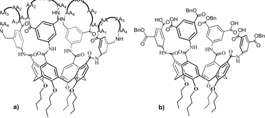
PAH-calixarenes derivatives 10-12 in Figure 2.7 were finally obtained by coupling of the diacyl chloride 1 with the corresponding racemic alcohols (±)-2a or (±)-2b using pyridine or triethylamine as base.43
After a standard work-up the calixarene/pyrenylisoxazolidinyl conjugates were isolated by silica gel column chromatography. In particular, starting by the alcohol (±)-2a, a monosubstituted derivative (±)-10a the derivative with C2 symmetry (±)-11a, and a meso
compound 12a were obtained in 15%, 10% and 8% yields respectively. Starting by the alcohol (±)-2b, then a monosubstituted derivative (±)-10b and the derivative with C2
48
Arduini, A.; Fanni, S.; Manfredi, G.; Pochini, A:; Ungaro, R.; Sicuri, A. R.; Ugozzoli, F. J. Org. Chem. 1995, 60, 1448-1453.
29
symmetry (±)-11b, were isolated in 10% and 11% yields respectively (Figure 2.7).
Figure 2.7 Pyrenylisoxazolidinylcalix[4]arene conjugates 10-12.
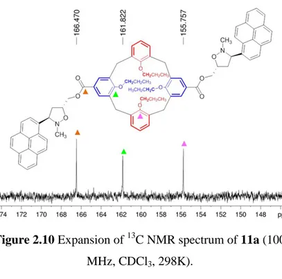
30
Concerning the stereochemistry of the bis-derivatives 11-12, the symmetry was assigned on the basis of the number of NMR resonances. For instance the 1H NMR spectrum of 11a showed two triplets at δ = 3.69 (J = 6.3 Hz, 4H) and 4.04 ppm (J = 7.6 Hz, 4H) and two triplets at δ = 0.91 and 1.07 ppm regarding the OCH2 and terminal CH3 of the propyl chains
respectively (Figure 2.8).
Figure 2.8 1H NMR spectrum of 11a (400 MHz, CDCl3,
298K).
Besides, the 2D HSQC spectrum showed two cross-peaks at δ = 3.69/76.9 and 4.04/76.7 ppm relative to a direct C-H
31
correlation between the OCH2 protons and their respective
carbons (Figure 2.9). At last, to support the C2 symmetry, the
13
C NMR spectrum showed only one signal related to the carbonyl group of the ester bond and two signals for the oxygenated quaternary carbons of the calixarene (Figure 2.10).
Figure 2.9 Expansion of 2D HSQC spectrum of 11a (400 MHz, CDCl3, 298 K).
32
Figure 2.10 Expansion of 13C NMR spectrum of 11a (100 MHz, CDCl3, 298K).
Similarly, the stereochemistry of 12a was evident from the 1H NMR spectrum in C6D6, in which were found three OCH2
signals at δ = 3.86, 3.70 and 3.69 ppm (4H, 2H and 2H respectively). These observations were characteristic of the presence of a σh symmetry plane bisecting the unsubstituted
aromatic rings, confirming that 12a is a meso compound (Figure 2.11).
33
Figure 2.11 1H NMR spectrum of 12a (400 MHz, C6D6,
298K).
As expected, the 1H NMR spectra of the monosubstituted derivatives (±)10a and (±)10b were complex due to the lack of any symmetry element. For instance, the 1H NMR spectrum of 10b showed four AX systems for the methylene bridges in the region between 3.10-3.18 ppm and 4.40-4.53 ppm. A similar set of signals was found for 10a.
The characterization of all these compounds was confirmed by ESI(+)-MS analysis.
34 2.1.2 Biological studies49
At this point the ability of compounds 10-12 to inhibit in vitro the proliferation of cancer cells was evaluated.49 These studies were performed by Rescifina’s group. Compounds 10-12 were tested against three different human tumor cell lines: FTC133 (follicular thyroid carcinoma), 8305C (undifferentiated thyroid carcinomas) and U87MG (glioblastoma). The cell cultures were incubated with 10-12 for 24h and the cell growth rates were evaluated by MTT reduction assays. The calixarenes were prepared as dimethyl sulfoxide stock solutions at 10 and 100 μM concentrations. The results of the inhibitory activities of 10-12 are shown in Table 2.2 and are compared to the activities of the pyrenylisoxazolidinyl alcohols 2a-b.
49 In collaboration with Prof. Rescifina, Dipartimento di Scienze del
35 Compound FTC133 8305C U87MG 2a 5.88 - 5.55 2b 5.22 - 5.36 10a >100 >100 >100 10b 0.095 0.130 57 11a >100 >100 >100 11b 55 55 >100 12a >100 >100 >100
Table 2.2 The results are expressed as IC50 [μM] (the
concentration of compound at which 50% of cells are viable) and determined by MTT assay.
The most cytotoxic compound was the monosubstituted calixarene with cis stereochemistry 10b, which exhibited an IC50 value of 95 nM toward FTC133 cells (for this cell line
Doxorubicin shows IC50 = 9650 nm)50. The bis-substituted
compound 11b showed an IC50 value of 55 μM, which was ca.
580-fold less cytotoxic than 10b. The other monosubstituted calixarene 10a showed a low cytotoxicity, with IC50 values
higher than 100 μM, thus stereochemistry plays a fundamental role in the activity of these compounds, as the cis derivative 10b was more active than its trans isomer 10a.
50
36
A lack of activity was found for the bis-substituted compounds 11a and 12a. These unexpected results could be explained considering the low solubility of the derivatives in the culture medium. The IC50 values probably result from
lower ligand concentrations with respect to those actually used. In contrast, the deprotonation of the carboxylic group of 10b under physiological conditions could increase its solubility. However the hydrolysis of the carboxylic acid cannot be the only explanation for the high cytotoxcity of 10b. The calixarene derivative is 58-fold more potent than the precursor 2b, thus the calixarene core must clearly have a prominent implication in the biological activity.
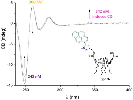
2.1.3 Circular Dichroism49
The pyrenylisoxazolidinylcalix[4]arene conjugates above described were designed as potential DNA intercalators. To prove the intercalation between base pairs some studies of circular dichroism (CD) were performed. Particularly the attention was focused on the behavior of 10b and 11b which, as previously described, turned out to be the most cytotoxic compounds. The studies were performed on a poly(dA)-poly(dT) duplex, which is a duplex of poly(deoxyadenylic-deoxythymidinic) acid, a repetitive synthetic double-stranded
37
DNA sequence and a synthetic analog of B-DNA.43
The CD spectrum of poly(dA)-poly(dT) displayed a negative peak at λ = 248 and a positive one at λ = 260 nm, due to the right-handed helicity and to base stacking respectively.51 In Figure 2.12 the CD tritation spectra of poly(dA)-poly(dT) duplex in the presence of increasing amount of 10b are shown.
Figure 2.12 CD tritation spectra at 25 °C:
[poly(dA)–poly(dT)] = 19.7 μm in base pair; [10b] = 0–32 μm
Increasing concentration of 10b led to decreases in the
51
Palumbo, M.; Capasso, L.; Palù, G.; Marcianimagno, S. Proc. Int. Symp. Biomol. Struct. Interactions, Suppl. J. Biosci. 1985, 8, 689-697.
38
intensities of the signals at λ = 248 and 260 nm, together with the appearance of a positive induced CD signal at 342 nm. These observations were typical of an intercalative binding mode. The changes in the intensities of the CD spectra were due to helix unwinding as a result of the distortion of DNA upon intercalation. The small positive induced CD signal at λ = 342 nm confirmed the intercalation phenomenon, with the pyrene moiety perpendicular to the DNA axis.
Similar CD spectra were observed with the 11b-poly(dA)-poly(dT) system, but with reduced changes in intensity.
2.1.4 Molecular Modeling49
To gain a deeper comprehension of the interaction of the calixarene derivatives 10-12 and DNA, a study of molecular modeling was performed by Prof. Rescifina and co-workers.49 The compounds with a (3R) configuration for the isoxazolidine ring possess the best intercalating properties42, thus, although the calixarene conjugates are racemates, all molecular docking calculations were performed for the (3R) stereisomers. Poly(dA–dT)2, poly(dG–dC)2, and poly(dA)–
poly(dT) were simulated as double-stranded dodecamer fragments, (dA–dT)2, (dG–dC)2 and poly(dA)–poly(dT),
39
conformation with the nucleic acids macro implemented in the YASARA software52 and minimized with the Amber03 force field.53 The simulations of the intercalation were performed by the docking methodology.54 Each intercalator was manually inserted into the middle base-step of each fragment from the minor or major groove. Particularly for monointercalations the compounds were inserted between the sixth and seventh base pairs, while for bisintercalation between the fifth and sixth as well as the seventh and eighth base pairs, simultaneously. The atom positions of each compound were fixed, however the remaining molecules were minimized to suitably accommodate the ligand. To obtain the best and most reliable docking results, a coarse docking simulation was first performed for each complex by applying the Lamarckian Genetic Algorithm (LGA) implemented in AutoDock 4.2.5.1,55 which has been recently demonstrated to accurately reproduce the complex crystallographic structures of a collection of DNA-binding small ligands.56 The best ligand position was further subjected to an MD simulation of 5 ns in a physiological environment (pH = 7.2, H2O, NaCl
52
Krieger, E. YASARA, 13.9.8 ed., YASARA Biosciences GmbH: Vienna, 2013.
53
Duan, Y.; Wu, C.; Chowdhury, S.; Lee, M. C.; Xiong, G. M.; Zhang, W.; Yang, R.; Cieplak, P.; Luo, R.; Lee, T.; Caldwell, J.; Wang, J. M.; Kollman, P. J. Comput. Chem.
2003, 24, 1999-2012. 54
Santos-Filho, O. A.; Figueroa-Villar, J. D.; Araujo, M. T. Bioorg. Med. Chem. Lett.
1997,7, 1797-1802. 55
Morris, G. M.; Huey, R.; Lindstorm, W.; Sanner, M. F.; Belew, R. K.; Goodsell, D. S.; Olson, A. J. J. Comput. Chem. 2009, 30, 2785-2791.
56
40
0.9%) to allow the ligand to be better accommodated in the pocket and model the interactions with the groove. At last, each ligand was well docked by the LGA with the system obtained by MD. Under physiological conditions the carboxylic acid moiety in compounds 10 is completely dissociated, thus, only the carboxylate form was considered. The possibility that the complexation between 10-11 with DNA may occur by binding along the grooves was also taken into account. The calculated binding energies after 5 ns of MD simulation are reported in Table 2.3.
41
Compound (dA-dT)2 dodecamer
from major groove
(dA-dT)2 dodecamer
from minor groove
2a (intercalated) -9.11 -9.03
2b (intercalated) -9.68 -9.39
10a (intercalated) -6.53 -8.83
10b (intercalated) -6.69 -10.19
11a (intercalated) -9.22 after 1 ns, -7.36 -9.81 11b (intercalated) -11.16 after 1 ns, -8.13 -15.51
10a (groove-bound) -4.66 -7.79
10b (groove-bound) -4.35 -8.16
11b (groove-bound) -8.35 -8.79
(dG-dC)2 dodecamer
from major groove
(dG-dC)2 dodecamer
from minor groove
2a (intercalated) -9.28 -9.09 2b (intercalated) -9.35 -9.23 10a (intercalated) -6.76 -7.33 10b (intercalated) -8.91 -10.88 11a (intercalated) -8.48 -9.05 11b (intercalated) -10.55 -13.09 10a (groove-bound) -5.13 -7.54 10b (groove-bound) -4.41 -8.65 11b (groove-bound) -8.15 -8.98 poly(dA)-poly(dT) from major groove
poly(dA)-poly(dT) from major groove
10b (intercalated) -6.43 -9.92
10b (groove-bound) -5.72 -6.57
Table 2.3 Calculated binding energies [Kcal/mol] after 5 ns of MD simulation.
Intercalation from the major groove and both minor and major groove binding can be ruled out on the basis of their lower binding energies. The compounds can act as mono or bisintercalators that interact preferentially with AT base pairs,
42
penetrating into the DNA double helix from the minor groove. The lower cytotoxic activity of the trans stereoisomer 10a compared to the cis one 10b could be ascribable to the steric hindrance of the N-Me group of the isoxazolidine ring. This hypothesis is supported by the lower binding energies of 10a compared to 10b (see Table 2.3).
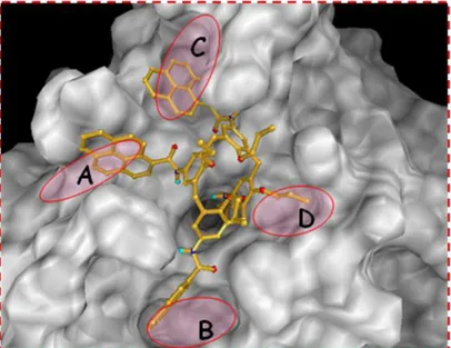
A graphical representation of 10a and 10b intercalated from the minor groove in the (dA-dT)2 dodecamer is shown in
Figure 2.13. The N-methyl group in the trans compound 10a (Figure 2.13, left) is oriented towards the base pairs of the DNA fragment. Conversely in the cis isomer 10b the methyl points outward, producing a lower steric hindrance (Figure 2.13, right).
Figure 2.13 10a (left) and 10b (right) intercalated from the minor groove in the (dA-dT)2 dodecamer.
43
According to the binding energies in Table 2.3, compounds 11a and 11b could behave as bisintercalators. In particular, the calculate binding energy of -15.51 Kcal/mol for 11b presupposes a strong interaction with the DNA, which should lead to a biological activity in the submicromolar range. Probably the low activity measured (IC50 = 55 μM for
FTC133) could be ascribable to the low solubility of the compound.
An interesting behavior was observed for both 11a and 11b when intercalated from the major groove of poly(dA-dT)2. In
fact, the examination of the MD simulation trajectories revealed that they can act only as monointercalators. After 1 ns both compounds 11 are bisintercalated into the dodecamer. Subsequently one pyrene moiety deintercalates and becomes perpendicular to the calixarene exo rim. After 5 ns simulation 11 is uniquely monointercalated. A representation of the intercalation of 11b at 1 and 5 ns are shown in Figure 2.14.
44
Figure 2.14 Representation of 11b intercalated into the (dA-dT)2 dodecamer from the major groove. After 1 ns of MD
simulation the compound is bisintercalated (left); after 5 ns only one pyrene unit is intercalated and the other is
completely deintercalated.
Nevertheless, the binding energies for these two MD periods are smaller than those observed for the intercalation of the compounds from the minor groove. Conversely, for the interaction with the (dG-dC)2 dodecamer, the bisintercalate
45
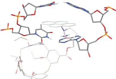
Finally, in Figure 2.15 is shown the arrangement of 10b into the pocket of poly(dA)-poly(dT) from the minor groove.
Figure 2.15 Plot representing 10b intercalated into the poly(dA)–poly(dT) dodecamer from the minor groove.
The pyrenyl unit of 10b is parallel to the long axis of the base pairs. This arrangement is in good agreement with the induced CD signal at λ = 342 nm observed (Figure 2.12), thus confirming the intercalation binding mode.
46
2.2 Conclusions
In this work we have obtained new PAH-presenting calix[4]arene conjugates bearing pyrenylisoxazolidine moieties at the exo rim.43 The in vitro cytotoxic activity against three different human tumor cell lines was tested and the most active compound 10b showed an IC50 of 95 nM
toward FTC133 cell lines (follicular thyroid carcinoma).43 Compared to the pyrenylisoxazolidinyl alcohols 2a-b a 58-fold increase in activity was observed. Both CD and docking studies indicated that these calixarene derivatives are able to intercalate DNA from its minor groove.43
Unfortunately the bis-substituted compounds did not show the expected results, probably because of their low solubility in the cell culture medium. With no doubt the results herein reported and the information obtained during these studies can be used to design and synthesize new DNA-intercalating agents with improved antitumor activity.
47
Chapter 3
3.
Chemical proteomics and drug discovery
Drug discovery is a research process that consists of the identification and development of a molecule that produces a desired effect in a living organism. Understanding the mechanism of action is a main requirement to find a new drug. However this request can only be satisfied provided that the biomolecular targets of the new potential drug are known.57 In the last decades the identification of calixarenes able to bind a known biomolecular target, has been carried out by using two principal strategies: the screening of a library of potential candidates11(c,d),16b and the virtual screening with molecular docking.27,58
Recently mass spectrometry-based chemical proteomics has gained relevance as a novel strategy for the identification of possible biological targets.59 The interest in this emerging approach has grown rapidly thanks to the remarkable
57
Lindsay, M. A. Nat. Rev. Drug Discovery 2003, 2, 831-838.
58
(a) Gordo, S.; Martos, V.; Santos, E.; Mendez, M.; Bo, C.; Giralt, E.; de Mendoza, J. Proc. Natl. Acad. Sci. USA 2008, 105, 16426-16431; (b) Martos, V.; Bell, S. C.; Isacoff, E. Y.; Trauner, D.; de Mendoza, J. Proc. Natl. Acad. Sci. USA 2009, 106, 10482-10486.
59
a) Rix, U.; Superti-Furga, G. Nat. Chem. Biol. 2009, 5, 616-624; (b) Veenstra, T. D. Drug Discovery Today: Technologies 2006, 4, 433-440.
48
technological advances in mass spectrometry.60 One of the principal chemical-proteomics approaches uses immobilized compounds and combines drug affinity chromatography with high-resolution mass spectrometry analysis and bioinformatics.59
A typical chemical proteomics experiment starts with the immobilization of a potential bioactive compound on a solid support. There are several commercially available activated resins, usually based on sepharose or agarose, that allow the attachment of chemical groups such as amino, hydroxyl or carboxyl groups.
A cell extract is then prepared either from cells or tissue and this lysate is incubated with the affinity matrix and washed extensively before the elution. For nonspecific elution, detergents, salts or denaturing agents are used. The specific elution can be achieved via the specific cleavage of an engineered linker. Subsequently, processing by SDS-PAGE and protein digestion with a protease (generally trypsin) generates a complex peptide mix, which is analyzed by nanoHPLC coupled to nanoESI/MS.
Finally the results are searched against a protein database to identify the proteins bound to the bioactive compound.
The whole chemical proteomics experiment is described in Figure 3.1.
60
49
50
A great advantage of this technique is that it can be used to probe the whole proteome. Proteins are encountered in their natural state, which includes endogenous abundance levels and post translational modifications. Besides it is possible to use any cell type or tissue, from microorganism to humans. Unfortunately chemical proteomics does not provide information about functional implications, as there is no correlation with the IC50. Thus, although it can give indication
about specific physical interactions between a compound and its target, the biological relevance must be validated by other techniques.
Figure 3.2 Advantages/disadvantages of chemical proteomics.
51
3.1 Application of chemical proteomics
The use of this technique has led to the discovery of a variety of compound targets, particularly the kinase inhibitors based on natural products. The identification of kinase targets has been investigated in various diseases such as cancer and autoimmune disorders.61 Protein kinases are among the most investigated drug target classes, so that several kinase inhibitors have entered clinical trials in the recent years. Chemical proteomics is widely applied to identify possible targets of kinase inhibitors through affinity purification from cellular extracts. In fact, the advantage of performing the experiment using disease-relevant cells enables to study more directly the mechanism of action of a particular kinase inhibitor.62
Interestingly chemical proteomics is widely applied for the identification of the biomolecular targets of natural products. Due to the low abundance, the testing for the biological activity of most of the newly discovered natural products is limited. Most of these tests are focused on the antibacterial and anticancer activity, thus the potential of most natural products as drugs and their ability to interfere with biological
61
(a) Bach, S. et al. J. Biol. Chem. 2005, 280, 31208-31219; (b) Rix, U. et al. Blood
2007, 110, 4055-4063. 62
(a) Godl, K. Et al. Proc. Natl. Acad. Sci. 2003, 100, 15434-15439; (b) Wissing, J. et al. Mol. Cell. Proteomics 2004, 3, 1181-1193.
52
pathways is unexplored. Therefore the use of natural products in chemical proteomics experiments is highly worthwhile.63 For instance, this approach was used to investigate the molecular mechanism of action of the antimitotic compound diazonamide A, a marine natural product. Chemical proteomics led to the discovery that its target, ornithine δ-amino transferase, plays a fundamental role in mitotic spindle assembly and cell division.64
Casapullo and co-workers paid much attention to the identification of the macromolecular targets of small bioactive compounds.65 They disclosed the proteasome inhibitory activity of Petrosaspongiolide M (PM), an anti-inflammatory marine sesterpene.65a The bioactive compound was covalently linked to an activated agarose matrix, which was modified with a spacer to avoid steric interference between the ligand-bearing matrix and the targets (Figure 3.3).
63
Piggott, A. M.; Karuso, P. Comb. Chem. High Throughput Screen 2004, 7, 607-630.
64
Wang, G.; Shang, L.; Burgett, A. W.; Harran, P. G.; Wang, X. Proc. Natl. Acad. Sci.
2007, 104, 2068-2073. 65
(a) Margarucci, L.; Monti, M. C.; Tosco, A.; Riccio, R.; Casapullo, A. Angew. Chem. Int. Ed. 2010, 49, 3960-3963; (b) Margarucci, L.; Monti, M. C.; Cassiano, C.; Mozzicafreddo, M.; Angeletti, M.; Riccio, R.; Tosco, A.; Casapullo, A. Chem. Commun. 2013, 49, 5844-5846; (c) Margarucci, L.; Monti, M. C.; Mencarelli, A.; Cassiano, C.; Fiorucci, S.; Riccio, R.; Zampella, A.; Casapullo, A. Mol. BioSyst. 2012, 8, 1412-1417; (d) Cassiano, C.; Monti, M. C.; Festa, C.; Zampella, A.; Riccio, R.; Casapullo, A. ChemBioChem 2012, 13, 1953-1958.
53
Figure 3.3 Preparation of PM-modified beads.
The PM-modified matrix was then incubated with human-macrophage-derived THP-1 cell extracts. Chemical proteomics revealed the ability of Petrosaspongiolide M to interact with several components of the proteasome enzymatic machinery. Thus the activity of PM on the 20S proteasome was tested in vitro by detecting its ability to modulate the proteasome-mediated proteolysis of three fluorogenic peptide substrates. PM was found to inhibit the proteasomal activity, particularly in the chymotrypsin-like site, with an effectiveness comparable to MG132, a well-known
54
proteasome inhibitor. Two proteasome inhibitors, Bortezomib66 and MLN-51967 have already entered clinical trials for the treatment of cancer and stroke patients respectively. The proteasome activity of PM, disclosed thanks to chemical proteomics, suggests that this molecule could be considered as a new lead compound.
Although its limitations, there is no doubt that chemical proteomics is a very powerful approach for the investigation of new drug targets and the elucidation of the mechanism of action of potential drugs. This is particularly relevant when the drug candidate is a low abundant compound, such as a natural product. Instead of performing a screening of several targets for the evaluation of the biological activity of a given compound, through a chemical proteomics experiment the potential partners can be identified simultaneously.
Chemical proteomics, in combination with other techniques and the recent and continuing technological improvements, will find more applications in the coming future and will be a even more powerful tool in drug discovery.
66
Crawford, L. J.; Walker, B.; Ovaa, H.; Chaunhan, D.; Anderson, K. C.; Morris, T. C. Cancer Res. 2006, 66, 6379-6386.
67
Shah, I. M.; Di Napoli, M. Cardiovasc. Hematol. Disord. Drug Targets 2007, 7, 250-273.
55
3.2 Biomolecular
recognition
abilities
of
calixarenes through the chemoproteomic
approach - Results and discussion
During this PhD research project, chemical proteomics was used to study the biomolecular recognition abilities of calix[4]arene derivative bearing acetamido groups at the exo rim. This study was performed in collaboration with Prof. Casapullo (DIFARMA, University of Salerno) and his research group.
Calix[4]arenes bearing amido, urea or thiourea functionalities have the potential to establish intermolecular interactions with biological compounds, due to their hydrogen-bond donor acceptor groups. To evaluate the biomolecular recognition abilities through a chemical proteomics approach, the calixarene must be functionalized to introduce a suitable linker for the subsequent immobilization on the solid support. The first derivative to be investigated through this approach was the p-acetamidocalix[4]arene 13 (Figure 3.4).68 This compound combines the hydrogen-bond donor/acceptor
68
Tommasone, S.; Talotta, C.; Gaeta, C.; Margarucci, L.; Monti, M. C.; Csapullo, A.; Macchi, B.; Prete, S.; De Araujo, A. L.; Neri, P. Angew. Chem. Int. Ed. 2015, 54, 15405-15409.
56
abilities of the acetamido groups at the exo rim and the hydrophobicity of the calixarene backbone, which should promote the formation of protein-calixarene interactions.
Figure 3.4 p-acetamidocalix[4]arene 13.
The p-acetamidocalix[4]arene 13 also presents a spacer arm at the endo rim, 2-(2-aminoethyldisulfanyl)ethylamine, for the immobilization on the solid support. The spacer is necessary to minimize the steric hindrance between the calixarene on the matrix and its potential targets during the affinity purification step.
57 3.2.1 Synthesis of compound 13
The synthesis of the p-acetamidocalix[4]arene 13 (pAC 13) is outlined in Scheme 3.1a-b.
The derivative 14 bearing three propyl chains at the endo rim was obtained by alkylation of p-t-Bu-calix[4]arene 3 with 1-iodopropane, BaO and Ba(OH)2.69 The free phenol ring was
alkylated with α-bromo-ethylacetate, using NaH as base in dry DMF.70 The ester group of 15 was then hydrolyzed with NaOH in a mixture of THF/H2O at reflux, affording to
derivative 16.71 Derivative 16 was exhaustively nitrated to the exo rim by treatment with HNO3 in presence of CH3COOH in
dry CH2Cl2 as solvent. Compound 1771 was purified by flash
column chromatography. The four nitro groups of 17 were then reduced to amino groups via catalytic hydrogenation with Nickel Raney in DMF.71 Subsequently the tetracetamido calix[4]arene 19 was obtained by treatment of the tetramino compound 1871 with acetyl chloride in dry THF.
The structure of 19 was assigned by spectral analysis. In particular, the presence of a molecular ion peak at m/z 859.5 (M+Na+) in the ESI(+) mass spectrum confirmed the
69
Gutsche, C. D.; Dhawan, B.; Levine, J. A.; No, K. H.; Bauer, L. J. Tetrahedron,
1983, 39, 409-426. 70
Geraci, C.; Consoli, G. M. L.; Galante, E.; Bousquet, E.; Pappalardo, M.; Spadaro, A. Bioconjugate Chem. 2008, 19, 751-758.
71
Mattiuzzi, A.; Jabin, I.; Mangeney, C.; Roux, C.; Reinaud, O.; Santos, L.; Bergamini, J. F.; Hapiot, P.; Lagrost, C. Nat. Commun. 2012, 3, 2121/1-2121/8.
58
molecular formula. The 1H NMR spectrum of 19 DMSO-d6
showed the –NH signals of the amido moieties between δ = 9.30-9.59 ppm as well as the three singlets for the aromatic protons at δ = 6.68 (2H), 6.75 (2H) and 7.04 (4H) ppm. (Figure 3.5) The protons of the methylene bridges showed signals at δ = 3.06 (d, ArCH2Ar, J = 12.8 Hz, 4H), and
4.29-4.34 (overlapped, 4H) ppm, while the 13C NMR spectrum (100 MHz, MeOD, 298K) showed a signal at δ = 154.82 ppm for the COOH.
59
60
61
Figure 3.5 1H NMR spectrum of 19 (400 MHz, DMSO-d6,
298 K)
Before the coupling reaction between 19 and the spacer arm, a mono-Boc protection of the diamino derivative 20 was necessary. This was performed following a quick and simple procedure in which the desired product 21 was isolated after two extractions.72 At first the crude was washed with diethyl ether and 1M NaH2PO4 solution (pH = 4.2). Then the aqueous
layer was basified to pH = 9.0 with 1M NaOH solution and washed with ethyl acetate. Finally the organic layer was concentrated affording the mono-Boc protected spacer arm 21. Afterwards the derivative 19 was coupled with the spacer
72
Niu, J.; Liu, Z.; Fu, L.; Shi, F.; Ma, H.; Ozaki, Y.; Zhang, X. Langmuir 2008, 24, 11988-11994.
62
arm 21 with N,N’-dicyclohexylcarbodiimide (DCC) and hydroxybenzotriazole (HOBt) in dry DMF, affording compound 22. The treatment of 22 with trifluoroacetic acid (TFA) in dichloromethane, followed by a short column chromatography purification led to the isolation of the desired ligand pAC-13, which was characterized by 1H and 13C NMR spectroscopy and ESI(+) MS. The presence of a molecular ion peak at m/z 971.4 (M+H+) in the ESI(+) mass spectrum confirmed the molecular formula. The 1H-NMR of the final product 13 showed the signals of the spacer arm at δ = 2.86 (t, CH2NH2, J = 6.4 Hz, 2H), 3.02 (t, CH2S, J = 6.6 Hz, 4H)
ppm, and the singlet for OCH2C(O) at δ = 4.78 ppm. The
protons of the methylene bridges showed overlapped signals at δ = 3.13-3.24 and 4.40-4.49 ppm (Figure 3.6).
Figure 3.6 1H NMR spectrum of 13 (300 MHz, MeOD, 298K).
63
The treatment with trifluoroacetic acid is a well established procedure for the removal of Boc protective groups, almost quantitative. Surprisingly in this case compound 13 was obtained with only a 50% yield. Probably this low yield is ascribable to the degradation of the disulfide bond of the spacer arm.
3.2.2 Chemical proteomics experiment73
The chemical proteomics experiment (or “fishing” experiment) was performed by Prof. Casapullo and co-workers. Compound 13 was covalently linked to the agarose-gel activated with 1,1’-carbonyldiimidazole (CDI). Thus 13 was dissolved in acetonitrile containing 30% NaHCO3 and
1.5% Et3N and incubated with the CDI-agarose beads for 16h.
The final concentration of 13 was 3μmol per milliliter of resin (assessed by reverse-phase HPLC analysis).
73 In collaboration with Prof. Casapullo, Dipartimento di Farmacia,
64
Figure 3.7 Immobilization of 13 on agarose beads.
Then samples of crude HeLa cells were incubated with the
pAC 13-modified beads. A calix-free matrix was used as a
control experiment to discriminate between specific and aspecific interactions. The beads were washed three times with Phosphate-Buffered Saline (PBS) solution to remove aspecific bound proteins. The beads were then treated with dithiothretiol (DTT) to break the disulfide bridge of 13 and to release in solution the pAC-proteins complexes. The protein mixture was purified by SDS-PAGE, digested with trypsin and analyzed by mass spectrometry through nanoflow reversed-phase HPLC MS/MS. The ten most intense doubly and triply charged peptide ions were fragmented and the MS spectra were subjected to a Mascot database search. The results were compared with those of the free-matrix control experiment. Usually this kind of “fishing” experiment gives a huge number of potential partners, which is reduced by applying a filter of the Mascot scores and the Pep(sig) values.
65
The Mascot score is the probability that an observed match is not a random event, while the Pep is the Posterior Error Probability, defined as the probability of a random match being classed as significant. In this case only proteins with Mascot score > 30 and Pep(sig) > 1 were considered, in order to obtain results with high trustworthiness. In addition keratins were also excluded, as their presence is due to accidental operator contamination. To establish which proteins were the best potential partners for pAC 13, the chemical proteomics experiment was performed three times, giving three sets of proteins (Table 3.1).
Table 3.1 Proteins identified in each of the three different fishing experiments and not detected in the corresponding control experiments (Mascot score > 30, Pep(sig) value >1).
66
The intersection of these sets gave only a single protein as best partner (Figure 3.8).
Figure 3.8 Venn diagram showing the intersection of the three sets of all the protein partners identified in the three
different fishing experiments.
The protein identified was the Protein Disulfide Isomerase (PDI), a 57 kDa chaperone protein located in the endoplasmic reticulum.74 PDI acts as a thiol oxidoreductase, it is able to catalyze the formation, breakage and rearrangement of disulfide bonds, and thus it is responsible for the regulation of oxidative protein folding as well as cell viability.75 Several studies report that increased PDI levels were found in a
74
(a) Goldberger, R. F.; Epstein, C. J.; Anfinsen, C. B. J. Biol. Chem. 1964, 239, 1406-1410; (b) Wilkinson, B.; Gilbert, H. F. Biochim. Biophys. Acta Proteins Proteomics 2004, 1699, 35-44.
75
(a) Lovat, P. E.; Corazzari, M.; Armstrong, J. L. Cancer Res. 2008, 68, 5363-5369; (b) Frand, A. R.; Cuozzo, J. W.; Kaiser, C. A. Trends Cell Biol. 2000, 10, 203-210.

![Figure 1.8 Tetrapeptidocalix[4]arenes](https://thumb-eu.123doks.com/thumbv2/123dokorg/7218759.77189/17.616.102.491.97.353/figure-tetrapeptidocalix-arenes.webp)
![Table 2.3 Calculated binding energies [Kcal/mol] after 5 ns of MD simulation.](https://thumb-eu.123doks.com/thumbv2/123dokorg/7218759.77189/45.616.80.545.74.613/table-calculated-binding-energies-kcal-mol-md-simulation.webp)

![Figure 3.4 p-acetamidocalix[4]arene 13.](https://thumb-eu.123doks.com/thumbv2/123dokorg/7218759.77189/60.616.108.507.205.461/figure-p-acetamidocalix-arene.webp)
