and
UNIVERSITY OF BASILICATA
DEPARTMENT OF SCIENCEPh.D. in Chemistry - XXXI Cycle
BIO/10 - Biochemistry
Doctoral Thesis in
“The protein related to Pseudoxanthoma Elasticum
regulates the purinergic system.
New insight on the ABCC6 transporter”
Ph.D. Coordinator:Prof. Gaetano Guerra
Ph.D. Student: Dr. Fabio Martinelli Tutor:
Prof. Faustino Bisaccia
Co-Tutor:
Prof. Magnus Monné
To my parents,
to my brother,
to my grandfather,
in particular to my nieces Alessia and Sofia
“The scientist is not the man
who provides the true answers;
that’s what asks the real questions.”
Claude Lévi-Strauss
7
Abbreviations:
2(-ΔΔCt) Fold Change
5’UTR 5’ Untranslated region
1609dupA Duplication of adenine in position 1609
A Alanine (Ala)
A1, A2a, A2b, A3 Adenosine receptor A1, A2a, A2b, A3
ABC ATP-binding cassette
ABCB ATP-binding cassette, sub-family B
ABCC ATP-binding cassette, sub-family C
ABCC6 ATP-binding cassette, sub-family C,
member 6
ABCC6-Flag-pcDNA Flag-pcDNA containing the sequence coding
for ABCC6
ABCC6-Ѱ Pseudogene of ABCC6
ABCC6 Δ19Δ24 ABCC6 mRNA without exons 19 and 24
8 of CD73
ADP Adenosine diphosphate
AMP Adenosine monophosphate
AOPCP α,β-methyleneadenosine-5’-diphosphate
APS Persulfate ammonium
ATCC American Type Culture Collection
ATP Adenosine triphosphate
BCRP Breast cancer resistance protein
BLAST Basic Local Alignment Search Tool
BMP-2 Bone morphogenetic protein-2
bp Base pairs
BSA Bovine serum albumin
C Cysteine (Cys)
Calcein-AM Calcein-acetoxymethyl ester
CALJA Calcification of joints and arteries
cAMP Cyclic adenosine monophosphate
9 CD73L Long CD73 isoform
CD73S Short CD73 isoform
CDKs Cyclin-dependent kinases
cDNA Complementary DNA
CFTR Cystic fibrosis transmembrane conductance
regulator
cGMP Cyclic guanosine monophosphate Co2+ Cobalt ion
CSCs Cancer stem cells
Ct Threshold cycle
D Aspartic acid (Asp)
DCF Dichlorofluorescein
DCFH Dichlorodihydrofluorescein
DCFH-DA 2’,7’-dichlorofluorescein diacetate
DMEM Dulbecco’s Modified Eagle’s Medium
DMSO Dimethyl sulfoxide
10 dNTP Deoxynucleotide
E Glutamic acid (Glu)
ECM Extracellular matrix
EDTA Ethylenediaminetetraacetic acid
em. Emission wavelength
ENPP1 Ecto-nucleotide pyrophosphate/
phosphodiesterase type I ex. Excitation wavelength
EX23_29del Alu-mediated deletion from exon 23 to exon
29
FBS Fetal bovine serum
Fetuin A Alpha 2-Heremans-Schmid glycoprotein
Flag DYKDDDDK tag
Flag-pcDNA pcDNA vector that codifies for Flag tag
G Glycine (Gly)
G418 Geneticin
11 GBM Glioblastoma multiform
Gi/o G proteinwith α inhibitory subunit
GPI Glycosylphosphatidylinositol
Gs G proteinwith α stimulating subunit
HCC Hepatocellular carcinoma
HEK293 Human embryonic kidney cells 293
HepG2 Human hepatocellular carcinoma cells HNF4 Hepatocyte nuclear factor 4
HSCs Hemapoietic stem cells
HuH-7 Human hepatocellular carcinoma well
differentiated cells K Lysine (Lys) kb Kilobases kDa Kilodalton Ki Inhibition constant L Leucine (Leu) L0 Connecting loop
12
MDA-MB-231 Triple-negative human breast cancer poorly
differentiated cells MDR Multidrug resistance
Mg2+ Magnesium ion
MGP Matrix gla protein
mRNA Messenger RNA
MRP Multidrug-resistance associated protein
MRP6 Multidrug-resistance associated protein,
member 6
MTT 3-(4,5-dimethylthiazol-2-yl)-2,5- diphenyltetrazolim bromide
NBD Nucleotide-binding domain
NCBI National Center for Biotechnology
Information
NK-cells Natural killer-cells
13
NT5E-1 Long transcript of ecto-5’-nucleotidase NT5E-2 Short transcript of ecto-5’-nucleotidase
OC Osteocalcin
OMIM Online Mendelian Inheritance in Man
OPN Osteopontin
p/q Short/long arm of a chromosome
p p-value
PBS Phosphate buffered saline
PBS-T PBS with 0.05% of Tween-20
PCR Polymerase chain reaction
PGE2 Prostaglandin E2
P-gp P-glycoprotein Pi Phosphate
PPi Pyrophosphate
Primer for Primer forward
Primer rev Primer reverse
14 PXE Pseudoxhantoma Elasticum
Q Glutamine (Gln)
R Arginine (Arg)
RNA Ribonucleic acid
ROS Reactive oxygen species
rpm Revolutions for minute S Serine (Ser)
SD Standard deviation
SDS Sodium dodecyl sulfate
SDS-PAGE Sodium dodecyl sulfate - polyacrylamide
gel electrophoresis
siRNA Small interfering RNA
SO4-. Sulfate radical
SUR Sulfonylurea receptor T Threonine (Thr)
TBS Tris buffered saline
15 TGF-β Transforming growth factor-β
Tm Melting temperature
TM Transmembrane
TMD Transmembrane domain
TNAP Tissue non-specific alkaline phosphatase
TNF-α Tumor necrosis factor-α
U Units
URG7 Up-regulated gene 7
Y Tyrosine (Tyr)
Zn2+ Zinc ion
ΔCt Difference between Ct of target gene and Ct
of housekeeping gene
ΔΔCt Difference between ΔCt of a target gene in a
sample and ΔCt of the same gene in the
17
Index:
Abbreviations: ... 7 Index: ... 17 Abstract: ... 21 Introduction: ... 23 1.1 ABC transporters ... 241.2 Human ABC transporters ... 28
1.3 ABCC subfamily ... 33
1.4 ABCC6 gene and MRP6 protein ... 35
1.5 NT5E gene and CD73 protein ... 39
1.6 Ectopic mineralization ... 45
1.7 Pseudoxanthoma Elasticum (PXE)... 49
1.8 Arterial calcification due to CD73 deficiency (ACDC) and generalized arterial calcification of infancy (GACI)……….... 54
1.9 Probenecid ... 57
Aims: ... 59
Materials and Methods: ... 61
2.1 Mammalian cellular culture ... 62
2.2 Treatment with Probenecid ... 64
2.3 Treatment with adenosine ... 65
2.4 Treatment with ATP ... 66
2.5 Treatment with Quercetin ... 67
2.6 Treatment with Doxorubicin ... 67
18
2.8 Doxorubicin efflux assay ... 70
2.9 Viability assay ... 71
2.10 Measurement of intracellular Reactive Oxygen Species (ROS) .. 72
2.11 Calcein assay ... 74
2.12 RNA extraction from cellular lines ... 75
2.13 cDNA synthesis (RT-PCR) ... 78
2.14 Primers design for Real-Time PCR ... 79
2.15 Real-Time PCR ... 80
2.16 Western blotting ... 86
2.17 Migration assay ... 90
2.18 Cellular cycle analysis ... 91
2.19 Imaging assay using confocal microscopy ... 92
2.20 Statistical analysis ... 93
Results and Discussion: ... 95
3.1 Inhibition of ABCC6 activity by Probenecid in HEK293 cells ... 96
3.2 Effects of Probenecid in HepG2 cells ... 98
3.2.1 Evaluation of cytotoxicity and morphology... 98
3.2.2 Evaluation of intracellular ROS ... 100
3.2.3 Evaluation of inhibition of calcein transport ... 101
3.2.4 Evaluation of cellular density ... 102
3.2.5 Effects of treatment with Probenecid for 24 hours ... 104
3.2.6 Effects of treatment with Probenecid for 48 hours ... 105
3.2.7 Effects of adenosine and ATP on the modulation of ABCC6 and CD73 expression ... 110
19 3.2.9 Evaluation of the mechanism promoting the decrease of
HepG2 cells migration rate ... 117
3.3 Effects of Quercetin and Doxorubicin in HepG2 cells ... 123
3.3.1 Effects of Quercetin ... 123
3.3.2 Effects of Doxorubicin... 126
3.4 Effects of Probenecid in other cellular lines ... 128
3.4.1 Effects of Probenecid in HuH-7 cells ... 128
3.4.2 Effects of Probenecid in MDA-MB-231 cells ... 135
3.4.3 Effects of Probenecid in HEK293 cells ... 142
Conclusions and future perspectives: ... 147
Bibliography: ... 153
Published Papers: ... 177
Other communications: ... 179
21
Abstract:
ABC (ATP-binding cassette) transporters are the largest superfamily of membrane proteins present in all organisms and they are particularly involved in transport of nutrients and drugs. Among these transporters
we find ABCC6, belonging to sub-family C, which is an ATP-dependent transporter mainly present in the basolateral plasma
membrane of hepatic and kidney cells. Mutations in the ABCC6 gene are associated to the Pseudoxanthoma Elasticum (PXE), an autosomal recessive disease characterized by progressive ectopic mineralization processes at level of the skin, the retina and the vascular wall. It has been reported that PXE is caused in peripheral tissues by decreased levels of PPi, a strong inhibitor of mineralization processes. In fact,
the administration of PPi reducing the effects of PXE is considered an
efficient drug. It is known that the over-expression of ABCC6 in HEK293 cells results in the outflow of ATP and that is converted
into AMP and PPi by ENPP1 protein. Then, AMP is transformed into
adenosine and Pi by CD73 protein; so ABCC6 protein could be
involved both in providing extracellular adenosine and in the regulation of the purinergic system. Previous studies show that in ABCC6-silenced HepG2 cells there is a dysregulation of some genes involved in the mineralization processes. We performed experiments in order to evaluate the mechanism by which ABCC6 is able to promote this genic dysregulation. For this purpose, the ABCC6 transport activity was inhibited with an aspecific inhibitor of ABC transporters. The main
22 results obtained in HepG2 cells are partially similar to those ones obtained in ABCC6-silenced HepG2 cells. The greatest effects were obtained on NT5E and on ABCC6. As an inhibitor, probenecid was used to reduce the transport activity of ABCC6 into HEK293 cells. In order to confirm that the observed effect is closely related to the inhibition of ATP transport, and not to the presence of probenecid, the HepG2 cells were also treated with adenosine and ATP. The results show that in the presence of 10 and 100 µM of adenosine or 50 and 500 µM of ATP, the effects of probenecid are reversed. In order to confirm the effects of probenecid on the transport activity in these cells was tested the quercetin, another ABC inhibitor; the data confirm those ones previously obtained with probenecid on the expression of ABCC6 and NT5E. In HepG2 cells treated with doxorubicin, whose presence increases the expression of ABCC6, we observed a proportional increase of NT5E. Experiments with probenecid, adenosine and ATP were also performed in cells expressing ABCC6 at different levels. The results show that in the presence of probenecid in MDA-MB-231 cells there is no effect while in the HEK293 and HuH-7 cells there are poor effects. The effect of probenecid is obtained only in cells that over-express ABCC6; in all these cells, the addition of adenosine and ATP improves the expression of CD73. It was also analysed the cellular phenotype after the treatment with probenecid. Considering the role of CD73 in cellular migration processes, motility assays confirmed that HepG2 cells migrate more slowly, after this treatment.
24
1.1 ABC transporters
Membrane transporters constitute a significant fraction of all proteins encoded in the genome of simple and complex organisms. They are divided into channels and carriers, two very large classes of proteins, within which there are superfamilies defined by both particular
sequences of amino acids and specific secondary structures. The transporters bind their substrates with high stereo-specificity;
among them there are passive transporters (which transport substrates according to a concentration gradient) and active transporters (which transport substrates through the membranes against concentration gradient). Some of these active transporters exploit the supplied energy by a chemical reaction, other of them by coupling the gradient versus transport with the gradient transport of another substrates. Among these active transporters, which perform their function using the energy deriving from the hydrolysis of ATP, one of the most important families is ABC (ATP-binding cassette) transporters. ABC transporters are the largest superfamily of membrane proteins [1] present in all living beings, from microbes to plants and animals [2-4]; they are able to couple the transport of a substrate across the membranes to the hydrolysis of the phosphate bond between the γ- and the β-phosphate of ATP [5-7]; they are codified by ABC genes that are highly conserved among species [8]. ABC transporters can work as either importers or exporters [9], and are involved in nutrient uptake, in transport of drugs, peptides and saccharides [10], hormonal and xenobiotic secretion,
25 ion and lipid homeostasis, antibiotic and multidrug resistance; for these reasons they are the main candidates for cellular regulation and pharmacological intervention [11-14]. The proteins of this group are mainly classified according to the sequence and organization of their cytoplasmic nucleotide-binding domains (NBDs) [15]. All ABC transporters show a common basic organization; their functional core consists of two NBDs and two transmembrane domains (TMDs) (Fig. 1.1).
Fig. 1.1: Basic organization of ABC transporter with two NBDs (red) and two TMDs
(blue) [8].
The TMDs domains are constituted by 6-11 transmembrane α-helicals that form a cavity within the membrane through which the substrate can be transported. The low sequence similarity among these transmembrane domains of the various ABC proteins reflects the wide variety of substrates carried [16-18]. The NBDs domains, cytoplasmic
domains, that bind and hydrolyze ATP, need magnesium (Mg2+) for
their activity; they are formed by 200-300 amino acids and contain the following motifs: A-loop involved in ATP positioning; Walker A, GXXGXGK(S/T) (where X is any amino acid), involved in phosphate
26 binding; Walker B, ɸɸɸɸD (where ɸ is an hydrophobic amino acid
residue), involved in coordination of Mg2+; D-loop, H-loop, Q-loop the
main sites of interaction with TMDs, and the signature C motif (LSGGQ) located upstream of the Walker B and implicated in binding
and hydrolysis of Mg2+-ATP [6] [15-16] [19] (Fig. 1.2).
Fig. 1.2: General representation of the functional organization of the ABC proteins: A) ABC “full transporters” with 12 TM regions, divided into 2 TMDs, each with an
NBD domain; B) ABC transporters with 5 extra TM regions, which form the TMD0 at the N-terminal; C) ABC “half-transporters” with 6 TM regions (one TMD) and
27 Prokaryotic transporters are located in plasma membrane and hydrolyze ATP in the cytoplasm, whereas eukaryotic ABC transporters are present both in plasma membrane and in organelle membranes and hydrolyze ATP in the cytoplasm (except for mitochondria and
chloroplasts transporters). Eukaryotic ABC transporters can either be
encoded as a single protein (“full transporters”) or can be considered as a result of homo- or hetero-dimerization (“half-transporters”) with only one TMD and one NBD [8] (see Fig. 1.2). Moreover, some ABC transporters contain an extension to the N-terminal consisting of five α-helicals that form the TMD0 domain [18] [21] (see Fig. 1.2). The transport mechanism of these proteins is based on the conformational change of the TMD domains, induced by the binding of the ATP to the NBD sites, which determines the opening to the outside and the release of the substrates. After the hydrolysis of the ATP the dimer NBD is deactivated and the conformation of the transporter returns to its original, facing inward, which represents the orientation with high affinity for the substrate [6] (Fig. 1.3).
28
Fig. 1.3: Transport mechanism of ABC proteins [21].
In literature there are structural studies of bacterial ABC transporters; for eukaryotic ABC transporters there are no high resolution structures
[17], but some medium resolution structures, such as P-glycoprotein
(P-gp) [17] [22].
1.2 Human ABC transporters
Human ABC proteins are encoded by 48 genes [23-24], and have been classified into seven subfamilies from ABC-A to ABC-G based on the similarity in the gene structure, order of the domains, and sequence homology in the NBDs and TMDs [23-26]. These transporters are
29 expressed ubiquitously in non-tumor cells or in a tissue-specific manner and play a role in metabolic and/or detoxification processes [1]. In tissues there is a different distribution of ABC proteins and the sub-cellular localization of these proteins can be a discriminating feature [26]; moreover, the ABC transporters are highly expressed in many tumor cells, for example in leukemic and in breast cancer cells
[26]. In Table 1.1 we can find a list of all human ABC genes known
with their chromosomal location, their subfamily, their expression pattern and their function (if it is known) [8].
30 Mutations of ABC genes are involved in several and various human
inherited disorders (Table 1.2) including cystic fibrosis,
Pseudoxanthoma Elasticum (PXE), tangier disease, alteration in metabolism and drug response [6] [25] [27].
31 Moreover, the high expression of some ABC proteins in hematopoietic stem cells (HSCs) and in cancer stem cells (CSCs), compared to their more differentiated counterpart, could suggest the hypothesis that these proteins play a role in cellular differentiation [28]. Some ABC transporters are involved in the transport of xenobiotics and drugs (Table 1.3), and in particular the two subfamilies mainly involved in the development of the drug resistance are the subfamily B (known as MDR, Multi-Drug Resistance) and the subfamily C (known as MRP, Multi-drug Resistance-associated Proteins) [20]. The most important and pharmacologically investigated protein is ABCB1 (known as P-gp or MDR1), for its implications in drug resistance; the tumor cells that over-express this protein show MDR phenotype, and are resistant to the transport of many antineoplastic drugs (such as doxorubicin and vinblastine) [29-30].
32
33
1.3 ABCC subfamily
The “C” subfamily of these transporters is one of the largest ABC subfamilies [15]. Its proteins are full transporters with various functions, such as ion transport, toxin secretion [8], and in particular drugs resistance (multidrug resistance-associated proteins (MRPs))
[31-32]. In this subfamily there are 12 members divided into three
classes: the first one involved in regulation of potassium channels (ABCC8 and ABCC9, called SURs, sulfonylurea receptors), the second one involved in chloride ion channel (cystic fibrosis transmembrane conductance regulator (CFTR)), and the third one, with the other nine members, involved in drugs resistance (MRPs) [8] [32-35]. ABCC13, the thirteenth member of this family is a truncated protein that encodes a non-functional protein [19] [36]. Proteins codified by ABCC genes are divided into two groups: “long” transporters including MRP1, MRP2, MRP3, MRP6, MRP7, SUR1 and SUR2, and “short” transporters including MRP4, MRP5, MRP8, CFTR and probably MRP9; the difference between the two groups is the presence in “long”
transporters of an extra NH2-terminal TMD named as TMD0 and
connected to the rest of the protein through a cytoplasmic loop indicated as L0 or as code CL3 (cytoplasmic loop 3) [37-41], with five TMs and an extracellular N-terminus [15]. In “short” transporters there is only L0 linker without TMD0 domain [42] (Fig. 1.4).
34
Fig. 1.4: Membrane topology of MRP proteins [43].
TMD0 sequence is poorly conserved compared to the other two homologous TMDs, but the N-terminal region of L0 is relatively conserved both in ABCC “long” proteins and in ABCC “short” proteins
[10] [44]. L0 is predicted to contain two amphipathic α-helicals in all
35 of a TMD0 domain in the “long” ABCC transporters is unique and its
eventual general function has to be clearly established [39]. For example, TMD0 of MRP1 does not play a crucial role either in
transport activity of the protein, or in its proper routing into the basolateral membrane compartment, while the presence of L0 and of other domains is necessary for MRP1 transport activity and for the proper intracellular routing of this protein [39] [46]. Moreover, in this transporter the L0 region forms a distinct structural and functional domain, which interacts with the membrane and with the core region of the transporter [39]. TMD0 domain is important for trafficking and stabilization of MRP2 in the apical membrane [47], and for the activity of SURs [38] [48-49]; L0 is essential for the localization of MPR2 [45] and for the regulation of trafficking of SURs [49].
1.4 ABCC6 gene and MRP6 protein
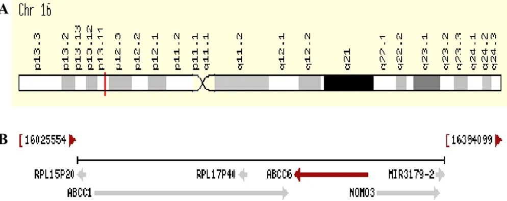
The human ABCC6 gene belongs to the large ABCC subfamily and is mapped on chromosome 16 in position p13.1 [50], between its two almost identical pseudogenes [51]. ABCC6 gene is immediately near to
ABCC1 (that codifies MRP1 protein), but these gene are in opposite
orientation, and the 3’ ends are only 9 kb apart [19] [35] (Fig. 1.5).
ABCC6 gene spans about 73 kb of genomic DNA [52], its mRNA
36 (equal to the number of exons of ABCC1) [35] [53] that codify for the MRP6 protein (or ABCC6 protein) [52].
Fig. 1.5: Position of ABCC6 gene on chromosome 16 (A). The region of chromosome
16 containing the ABCC1 and ABCC6 genes (B) [43].
There is a splicing variant of ABCC6 mRNA without exons 19 and 24 (ABCC6 Δ19Δ24), and, the product of the translation is a truncated protein that is suggested to be a half transporter [54]. ABCC6 gene has
about 45% of homology with the ABCC1 gene [35] [55]. From mutational analysis, they have been found sequence variants of
ABCC6 gene deriving from duplication of the p13.1 region of the
chromosome 16; these variants are ABCC6 pseudogenes [56]. Two pseudogenes were found in the chromosome 16, ABCC6-Ѱ1 and
ABCC6-Ѱ2, at the terminal 5’ of the ABCC6 gene [56]. In particular,
a polypeptide with 99 amino acids indicated as up-regulated gene 7 (URG7) [57] shows a high homology with the protein codified by
ABCC6-Ѱ2 [58]. This protein, URG7, has the first 74 amino acids
in common with MRP6 protein and their expression patterns are highly
A
37 similar because there is high degree of homology between ABCC6 promoter and 5’UTR of ABCC6-Ѱ2 [58]. Expression tissue-specific of
ABCC6 gene is finely regulated by the presence on its promoter of
sequences that bind specific transcription factors such as hepatocyte nuclear factor 4 (HNF4) [59] and tumor necrosis factor-α (TNF-α) which up-regulate and down-regulate, respectively, the promoter [60]. The protein MRP6 is codified by the ABCC6 gene, as reported before, and has 1503 amino acids and a molecular weight of about 165 kDa
[35] [60-61]. MRP6 is a “long” MRP transporter and it is formed by 17
TMs (grouped in three TMDs) and two cytoplasmic NBDs
[TMD0(TMD-NBD)2] [62-63] (Fig. 1.4), with conserved Walker A,
Walker B and motif C [52]. It is mainly expressed in liver and kidney, in particular on the basolateral surface of hepatocytes [41] [64-67] and poorly expressed in salivary glands, in colon, in lungs and in the thyroid
[35]. It is also expressed in acute myeloid leukemia [68]. Two possible
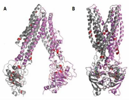
functional models for transport of molecules through ABC transporters in general were proposed: the first one suggests that each NBD hydrolyzes an ATP molecule and that this reaction promotes the transport of a molecule of substrate [19]; the second one suggests that hydrolysis of an ATP molecule promote the substrate transport through the shift from a high- to low-affinity substrate binding state and hydrolysis of the other ATP molecule restores the high affinity substrate binding state [15] [19] (Fig. 1.6).
38
Figure 1.6: Three-dimensional model of MRP6: (A) nucleotide saturated
conformational state and (B) nucleotide-free conformation. Missense mutations are indicated in red (modified from [59]).
In vitro experiments on chemically synthesized peptide E748-A785
(that correspond to a part of NBD1) [69], on NBD1 [70] and NBD2 [71] produced in Escherichia coli by recombinant DNA technologies show the ability of these fragments in ATP binding. In particular, studies on NBD1 (D627-L851) show that the A- loop of this domain is important for affinity to ATP, while, NBD2 of MRP6 (T1252-V1503) is structured in order to bind and hydrolyze ATP with a lower efficiency [71]. In vitro transport assays on membrane vesicles enriched with
39 MRP6 show that this transporter is associated with ATP-dependent transport of glutathione S-conjugates; the transport of conjugates is a feature of MRPs that have the third TMD. MRP6 is also associated to low level of resistance to agents such as doxorubicin and teniposide
[15] [19] [23] [72-73]: its natural substrate is not defined [74], and
MRP6 transport is inhibited by organic anions such as probenecid [75]. Finally, the MRP6 is associated to the metabolism of vitamin K [59], and, recent studies in HepG2 cells show that low levels of expression of MRP6 induce a dysregulation of expression of some genes and a senescent-like phenotype (cellular cycle alteration) [76-77].
1.5 NT5E gene and CD73 protein
The human NT5E gene is situated on chromosome 6 in position q14-q21 [78]; this gene spans 46 kb of genomic DNA, and it has a coding sequence of 1725 bp and is made of 9 exons that codify for CD73 protein [78-79]. Two transcripts of this gene have been identified as
NT5E-1 (3548 bp) and NT5E-2 (3384 bp), and obtained by alternative
splicing. The NT5E-2 is a new splicing variant of NT5E gene expressed less than NT5E-1 in normal tissues of human body but up-regulated in some pathological contexts such as in cirrhosis and in hepatocellular carcinoma (HCC) [79]. The difference between the two transcripts is exon 7 that is present in the longer transcript (NT5E-1) and absent in the
40 shorter transcript (NT5E-2); the two transcripts have the same N- and C- terminal [79] (Fig. 1.7).
Fig. 1.7: Transcripts of NT5E gene: NT5E-1 and NT5E-2. The black arrow indicates
the position of exon 7 [79].
In literature is reported that the expression of this gene is under the control of hypoxic conditions [80-81] and of several pro-inflammatory mediators such as transforming growth factor-β (TGF-β) or prostaglandin E2 (PGE2) [82-83]. It has also been reported that the increase of NT5E transcript, and of its correspondent CD73 protein in neoplastic tissues can play a key role in the onset and progression of neoplasia [82]. There is also a similar positive correlation between triple-negative breast cancer and NT5E levels associated to a poor
prognosis [84]. The protein CD73, cluster of differentiation 73, is codified by the NT5E gene, as reported before, and has 574 amino
acids and a molecular weight of about 70 kDa. NT5E-1 encodes for CD73 long isoform (CD73L, 574 amino acids), the enzymatically active isoform, while, NT5E-2 encodes for CD73 short isoform (CD73S, 524 amino acids), the enzymatically non-active isoform.
41 The mature CD73 form is a glycosylated dimeric enzyme located on the outer portion of the plasma membrane [85-87], and it is present in different organs (such as colon, kidney, liver, heart [88-89]) and cells (such as lymphocytes, macrophages and epithelial cells [88]). This protein is anchored to the external face of plasma membrane through glycosylphosphatidylinositol (GPI) situated on C-terminal [86]
[90-91], and, consists of two protein subunits united through
non-covalent links. N-terminal domain coordinates the link of two divalent metal ions (Zn2+ and Co2+) and is characterized by α/β-β-α-β
secondary structure. C-terminal domain provides the linking pocket for AMP [91], and has a sequence of non-charged and hydrophobic amino acids substituted with the GPI anchor linked to serine-523 [92]. The active site of the enzyme is located at the interface between the N- and C-terminal domains and shape among the residuals of both domains. Have been described different crystalline structures for CD73: if the conformation of CD73 is open it is characterized by a rather compact structure, if the conformation is closed it is characterized by a dimeric structure (Fig. 1.8).
42
Fig. 1.8: Crystalline structure of CD73 in open and closed conformations [90].
The two isoforms show different functions: CD73L is the isoform enzymatically active, an ecto-5’-nucleotidase, able to catalyze the conversion of extracellular AMP, produced by ENPP1 protein, to
adenosine and Pi [85]; CD73S is the isoform catalytically inactive,
unable to dimerize, but able to link both with the calnexin and with CD73L promoting proteasome-dependent CD73L degradation [79]. For its role in the production of extracellular adenosine CD73 can be considered a fundamental actor in the regulation of immunity response and in other pathways (for example: inflammation, ischemia, tissue fibrosis [93]). CD73-adenosine pathway is an important immunosuppressive pathway and this property is linked to its ability of
43 The adenosine is a strong immune-suppressor, in particular, of antitumor T-cells response [95]. Increasing evidence shows that CD73 participates in tumor immune-escape by inhibiting the activation, clonal expansion, and homing of tumor-specific T cells [96]; the adenosine decreases the ability of natural killer cells (NK-cells) to produce TNF-α, to promote lytic activity [97], and to decrease the activity of macrophages [98] and dendritic cells [99-100] (Fig. 1.9).
Fig. 1.9: The effects of adenosine (produced in cancer cells by CD73) on immunity
44 Furthermore, CD73 is involved in drug resistance [102], in tumor growth [103], tumor metastasis (through binding to glycoproteins of the extracellular matrix (ECM), such as fibronectin and laminin) [104-106] and tumor angiogenesis [107]. In particular, recent studies show that in some types of cancer CD73 could promote the migration and invasion of cancer cells through regulation of EGFR expression, in others type of cancer, it could be an important regulator of epithelial-mesenchymal transition [99]. Different strategies are studied to control this protein using inhibitors, such as AOPCP, α,β-methyleneadenosine-5’-diphosphate, or using antibodies. CD73 is related to the metabolism of nucleotides and nucleosides (cellular messengers), and it can control different cellular processes in purinergic system. The purinergic system is a system deputed to cellular proliferation and transformation, cellular cycle progression, metabolic changes, cell-to-cell interactions, cytokine and chemokine secretion, inhibition of lipolysis, promotion of fatty liver insulin resistance, intracellular pathogen removal and generation of reactive oxygen species (ROS) [108]. Extracellular adenosine regulates a variety of physiological processes (previously mentioned)
using a family of four receptors related to G-protein (A1, A2a, A2b, A3)
each of which is characterized by a single pharmacological profile and tissue distribution. A1, A2a and A2b protein sequences are highly
conserved across mammalian species, while, A3 is more variable.
In human, A1, A2a and A3 are considered as high affinity receptors for
adenosine (Ki about 100-300 nM), while A2b receptor has a lower
45 show a structure with seven transmembrane α-helicals, with an extracellular N-terminus and an intracellular C-terminus. Finally, these receptors perform their specific functions in different tissues because they regulate the cAMP levels differently (in particular, A1 and A3
receptors are coupled to inhibitory Gi/o proteins, while, A2a and A2b
receptors are coupled with to stimulating Gs proteins [111]).
1.6 Ectopic mineralization
The physiological process that provides for the formation of bone matrix and the functionality of skeletal tissue is known as mineralization (or calcification). This phenomenon, which is tightly controlled and restricted to specific body regions [51], can also occur in soft tissues and it is known as ectopic mineralization. This type of pathological calcification occurs when calcium and phosphate complexes form hydroxyapatite crystals and they settle in the soft tissues causing serious consequences at the clinical level. Moreover, this complex multifactorial metabolic process is associated with different clinical conditions such as aging and diabetes [112]. There have been many advances in the study of ectopic mineralization processes both in terms of molecular mechanisms and of single gene disorders characterized by phenotypic traits typical of calcification processes; the following table shows the main diseases belonging to
46 ectopic mineralization (Table 1.4). In particular, Pseudoxantoma Elasticum (PXE) and Arterial calcification due to CD73 deficiency (ACDC) are related to ABCC6 and NT5E gene mutations [112].
Table 1.4: Diseases characterized by ectopic mineralization [112].
Calcium and phosphate precipitation is highly regulated by calcification inducers and inhibitors [113-114]. In particular, inorganic
pyrophosphate (PPi) [115] acts as a powerful inhibitor of
mineralization; phosphate (Pi) is a pro-mineralization factor, and an
47 mineralization under homeostatic conditions [112] [116]. Alterations of the metabolic pathway of extracellular ATP can be considered among the main causes of the manifestation of these pathological forms of
mineralization. ATP, a precursor of PPi and Pi, is released by the liver
cells [115] [117]: its passage in the extracellular environment is facilitated by membrane transporters such as ABCC6 (other
mechanisms are vesicular exocytosis, hemichannel connexion and pannexin channel [118]), and, recently, it has been suggested that, in pathological conditions, the loss of the ABCC6-dependent outflow of ATP would cause an alteration of the extracellular PPi/Pi ratio
[119-120]. Then, ATP is converted, in the vascular system of the liver,
into PPi and AMP, by ENPP1 protein, an ecto-nucleotide
pyrophosphatase/phosphodiesterase 1, encoded by ENPP1 gene [121]. AMP is converted into adenosine (an activator of the purinergic
pathway) and Pi by CD73 [85] [121]. ABCC6 protein is thus involved
in the production of extracellular adenosine and therefore could be involved in the activation of the purinergic pathway; however, the mechanism through which ABCC6 dysfunction causes diminished
ATP release remains an enigma [122]. Finally, PPi is converted in two
molecules of Pi by TNAP protein, a tissue non-specific alkaline
phosphatase, encoded by TNAP gene. For its ability to produce Pi,
TNAP can be considered another important factor that controls the mineralization processes [85], while, adenosine is considered an inhibitor of TNAP [86]. The reactions above mentioned are well schematized in the Fig. 1.10. Moreover, in recent years some studies
48 have hypothesized that adenosine could control the phenomenon of ectopic calcification not only by acting on TNAP expression, but also
by activating the A2a receptor and consequently the membrane
translocation of the protein ENPP1, whose expression is crucial in the regulation of the processes of mineralization of soft tissues [123].
Fig. 1.10: Pathway of extracellular ATP [85].
At the moment, it is not known the mechanism through which ABCC6 promotes the release of ATP in extracellular space. It is not defined whether ABCC6 directly or indirectly transports ATP [119]. Moreover, it is not clear whether ABCC6 may favor the activation of the
49 purinergic system, and therefore regulate some fundamental physiological processes such as proteic, glucidic, and lipidic metabolisms, immunity and mineralization processes [124-126]. Other important actors of mineralization processes in soft tissues are: matrix gla protein (MGP), osteocalcin (OC), osteopontin (OPN), bone morphogenetic protein-2 (BMP-2) and alpha 2-Heremans-Schmid glycoprotein (Fetuin-A) [113]. In particular, MGP acts through the repression of BMP-2 and consequently is an inhibitor of formation of calcium crystals [113]. Fetuin-A is an important serum carrier of calcium and phosphate and is an inhibitory factor of calcification of soft tissues [113]. OPN, an acidic secreted phosphoprotein, is involved in blocking hydroxyapatite deposition and promoting crystal resorption
[127].
1.7 Pseudoxanthoma Elasticum (PXE)
Alterations in relationships of factors involved in mineralization of soft tissues promote an ectopic mineralization that causes different diseases such as Pseudoxanthoma Elasticum (PXE). PXE (OMIM, online Mendelian inheritance in man, 264800) is a disease caused by mutations of ABCC6 gene. There have been found more than of 300 mutations in ABCC6 NBD domains such as deletions, insertions or substitutions [52] [128-130], as reported in Fig. 1.11. R1141X (where X
50 is a stop codon), is the most frequent mutation (about 30%); EX23_29del deletion is also recurrent (about 10%) in European patients suffering from PXE [52] [130-131]. Furthermore, some mutations cause truncated forms of the protein and/or ABCC6 incorrect localization.
Fig. 1.11: Schematic representation of ABCC6 protein with the positions related to
51 This disease is an autosomal recessive disorder characterized by calcified and fragmented elastic fibers at level of the skin (plaques), the retina (peau d’orange, retinal angioid streaks, break of Bruch’s membrane [129]) and the vascular wall (atherosclerosis, angina pectoris) [122] [132-133], as reported in Fig. 1.12.
Fig. 1.12: Typical manifestations of PXE [134].
The prevalence of PXE is unknown. It is estimated its presence in about 1:50,000-1:100,000 people [132] [135], with a slight female predominance [136]. This pathology is sometimes underdiagnosed for its rarity and for its highly variable phenotype [132]. Heterogeneity phenotype of PXE suggests the influence in the development of the pathology of different environmental factors such as nutrition [132], and, in particular, mutations in other genes [132]. At the moment there
52 are no pharmacological care for this pathology. In PXE patients all organs of the body show extracellular matrix alterations, but eyes and skin are more severely involved independently of the type of mutation
[131]. Usually the onset of the PXE occurs in late childhood or
adolescence [59] and clinical manifestations progress with age [131]. Among the first manifestations of PXE we can find the appearance of yellow-ivory soft papules with a reticular pattern [132] and of plaques in the skin causing the lost of elasticity at level of antecubital fossae or sides of the neck [52] [129]; later these signs appear at the back of knees and at level of oral mucosa and anogenital skin [132]. In PXE patients we also find alterations of Fetuin-A, BMP-2 and vitamin K
[59], but, we don’t find alterations in levels of calcium, phosphate or in
the inflammation processes [114]. There is no satisfactory correlation between genotype and phenotype [137], and diagnosis of PXE comes from the individuation of fragmented calcified elastic fibers thanks to a skin biopsy using von Kossa staining [59]. The different manifestations of PXE were standardized in 2007 and the system that groups them into
various classes is called Phenodex (PXE phenotype index). The molecular mechanism that induces onset of PXE (when ABCC6 is
not functional) is not clear [138]. The presence of low levels of ABCC6 protein in tissues of patients suffering from PXE (Fig. 1.13) suggests that PXE is a metabolic disorder [139], resulting in the alteration of transport of substances from liver and from kidney to blood [140].
53
Fig. 1.13: Tissues in which ABCC6 protein is expressed and tissues affected by PXE
disease [52].
It is known that hepatic ATP is excreted with a mechanism related to
the ABCC6 protein. Reducing the activity of ABCC6 decreases PPi
at the extracellular level [120]; it was, also, observed an alteration of metabolites related to ATP in PXE patients [141]. In PXE patients was observed a reduced concentration of vitamin K, cofactor of γ-carboxylation, a fundamental reaction activating
54
In stable ABCC6-silenced HepG2 cells, we find an increase of pro-mineralization factors and a decrease of anti-mineralization factors:
TNAP is up-regulated at transcript, protein and activity levels;
NT5E/CD73 are downregulated at transcript and protein levels; Fetuin-A and OPN result down-regulated at transcript level [76].
On the basis of these information for the treatment of PXE there are
experimented the following treatments: oral administration of PPi [142],
oral administration of bisphosphonates, a stable form of PPi [143], oral
administration of phosphate binders [144], modifiers of protein conformation (such as sodium 4-phenylbutyrate) that target the protein to the plasma membrane [145].
1.8 Arterial calcification due to CD73 deficiency
(ACDC) and generalized arterial calcification of
infancy (GACI)
Other important diseases related to ectopic mineralization are arterial calcification due to CD73 deficiency (ACDC) and generalized arterial calcification of infancy (GACI); these diseases, together with PXE, are
characterized by similar phenotypic traits, and by alterations of some factors involved in balancing pro-mineralization and anti-mineralization processes (Fig. 1.14).
55
Fig. 1.14: Homeostasis of calcification processes depends on the functioning of some
enzymes. Some diseases caused by altered functions of these proteins are: PXE, GACI and ACDC [146].
ACDC (OMIM, 211800) is a disease caused by mutations in NT5E gene, and it is a rare autosomal recessive disease characterized by the calcification of the large arteries of upper and lower limbs and by the calcification of coronary circulation [85] [121] [139]. For these clinical manifestations ACDC is also known as CALJA (calcification of joints and arteries) (OMIM, 211800) [112]. In literature it is related to new mutations of NT5E gene [147]. The most frequent mutations observed
56 in patients affected by ACDC are S221X (where X is a stop codon), C358Y and a truncated protein caused by 1609dupA [85]. In patients affected by ACDC we find a decrease of NT5E gene and a decrease of protein and activity of CD73 protein [85], with low levels of adenosine, but normal serum levels of calcium, phosphate and vitamin D. The reduction of extracellular adenosine, caused by alterations of CD73 activity, seems to increase the activity of TNAP protein, and,
consequently, the hydrolysis of PPi increases the ratio between Pi and
PPi favoring the calcification processes [85-86] [148]. The addition of
adenosine reverses these effects both in presence of ACDC or in presence of PXE fibroblasts [114]. GACI (OMIM, 208000) is an autosomal recessive disease caused by mutations of ENPP1 gene, that is mapped on chromosome 6 in position q22-q23 [86] [149]. In patients affected by GACI we find a severe calcification of the elastic lamina inside the large and medium-sized arteries producing an arterial stenosis and others cardiac diseases [150-151], and a decrease of PPi levels [149]. GACI is a rare condition (reported in about 180
individuals [152-153]) whose prognosis is extremely poor (85% of
affected infants die within the first six months of life [154]). As reported in literature, mutations in ENPP1 gene could be associated
with a PXE phenotype [155], but also mutations in ABCC6 gene could
be associated to GACI disease [64] [155]. The formation of PPi as the
result of the sequential action of ABCC6 and ENPP1 proteins could
explain the correlation between PXE and GACI, while low levels of PPi
57
1.9 Probenecid
Probenecid, 4-dipropylsulfamoylbenzoic acid, is a uricosuric drug used for the treatment of gout. In gout an excessive presence of uric acid, produced by the metabolism of purine nitrogenous bases, could promote the precipitation of uric acid in the form of crystals. Probenecid is able to increase the excretion of uric acid and block the reuptake at kidney level [156]. Probenecid, generally, is administered orally; its absorption is rapid and complete and it is metabolised in the liver and eliminated by kidneys. The inhibition of the renal tubular transporter thanks to probenecid is also clinically exploited to increase the effective concentrations of antibiotics, chemotherapeutics, and other medications [157]. Side effects can be nausea, vomiting, fatigue and skin rash. The block of cAMP or cGMP release from erythrocytes
[158-159], and, ATP release from glia cells [160] by probenecid show
its involvement in the efflux of these substrates. For these reasons experiments have been carried out to investigate the correlation between probenecid and the mechanisms involved in the transport of ATP (such as pannexins, connexins and ABC transporters). The results of a study, in particular, show that probenecid is a powerful inhibitor of pannexin 1 channels, but it has no effects on connexin channels [157]. Probenecid is, also, a known ABC transporters inhibitor [157] [161]. In fact, a study show that probenecid is an effective chemosensitizer for MDR cells that overexpress MRPs, and, is a potential candidate for clinical use to reverse MDR phenotype [162]. The concentrations of
58 probenecid (0.1-0.5 mM) that reversed MRP-mediated MDR are readily achievable in vivo [162]. Moreover, ABCC6 transport is inhibited by organic anions such as probenecid [75]: 1 mM of probenecid, efficiently, inhibits MRP1 and MRP2, but it poorly inhibits ABCC6 (about 30%) [75]. Finally, it is also known that CD73 is related to MDR proteins, and, its increase in different kinds of tumors [89] such as glioblastoma multiform (GBM) [163] and breast cancer (where it is over-expressed) promotes a highly aggressive tumor phenotype [105]. In literature it is reported that using AOPCP, a CD73 inhibitor, it is possible to reverse the MDR phenotype in GBM cells through the decrease of expression and activity of MRP1 [163]; for these reasons, it could be possible to control CD73 expression using an inhibitor of ABC protein, such as probenecid.
59
Aims:
The aim of this scientific research is to study the role of ABCC6 protein in some fundamental cellular processes. It is known that ABCC6 protein is able to transport ATP outside the cells and that mutations in ABCC6 gene cause the Pseudoxanthoma Elasticum (PXE)
a disease characterized by a decrease of the amount of extracellular PPi,
by the ectopic mineralization processes in some regions of the body and by the alteration in releasing of extracellular nucleotides and purine nucleosides, with a possible involvement of the activation of purinergic
system. ATP transported by ABCC6 is catalysed into PPi and AMP by
ENPP1 protein; AMP is catalysed into Pi and adenosine by CD73
protein. So we intended to investigate the role of ABCC6 in the purinergic system. In this scientific research we want to verify the effect of the ATP and of the adenosine, on the expression of ABCC6 and CD73 protein. In particular, the transport activity of ABCC6 in HepG2 cells was blocked by an inhibitor of ABC proteins and the gene and protein expression analysis was carried out on NT5E/CD73. Other effects related to the low activity of ABCC6, due to this inhibitor, have been studied with a particular attention to the role of CD73 in cancer progression. Furthermore, similar experiments were performed using doxorubicin and quercetin and other cellular types in order to
highlight the correlation between ABCC6 and CD73. We performed some experiments in collaboration with the University of Bari and with the IRCCS-CROB of Rionero in Vulture.
62 All compounds used for the following scientific research were purchased from Sigma-Aldrich (unless otherwise indicated).
2.1 Mammalian cellular culture
Human embryonic kidney 293 cells (HEK293), human hepatocellular carcinoma cells (HepG2), human hepatocellular carcinoma well differentiated cells (HuH-7) and triple-negative human breast cancer poorly differentiated cells (MDA-MB-231) are the adherent mammalian cell lines used for the experiments of my research
(Fig. 2.1). These cells were grown in Forma STERI-CYLE CO2
incubator and were maintained in Dulbecco’s Modified Eagle’s Medium (DMEM) containing 4.5 g/L glucose, supplemented with 10% fetal bovine serum (FBS), 2 mM L-glutamine, penicillin (100 U/mL), and streptomycin (100 mg/mL) in polystyrene flasks at 37°C, in an
atmosphere humidified with 5% of CO2. The cell splitting was obtained
using Trypsin-EDTA solution (1X) for 5 minutes at 37°C to detach cells from flasks; trypsin was inactivated with a double volume of complete medium. All cells (except HEK293) were washed with
PBS 1X, phosphate buffered saline, (140 mM NaCl, 10 mM Na2HPO4,
2.7 mM KCl and 1.8 mM KH2PO4, pH 7.4) before trypsin treatment.
All solutions were warmed at 37°C using Water bath SWBD Stuart before use. Cells were observed with microscope Nikon Eclipse TS100
63 and counted using Burker chamber. The number of cells was calculated with the following formula:
cells/mL = counted cells * dilution factor * 104 / number of counted squares
After counting, an appropriate group of cells was seeded in flasks (see next sections). The pellets of non-treated or treated cells were obtained transferring opportunely the cells into tubes and centrifuging them at 1200 rpm for 5 minutes at 4°C. After elimination of the medium, the pellets were immediately used or stored at -80°C.
Fig. 2.1: Representative images of mammalian cellular culture: HEK293 cells (A),
HepG2 cells (B), HuH-7 cells (C) and MDA-MB-231 cells (D). These images are taken from ATCC (American Type Culture Collection).
64
2.2 Treatment with Probenecid
Probenecid is a well-known inhibitor of multidrug resistance-associated proteins, MRPs [162] (Fig. 2.2). It was dissolved in dimethyl sulfoxide (DMSO) at 30 mg/mL (100 mM) as stock solution and it was then diluted with DMEM to the desired concentrations. The final concentration of DMSO did not exceed 1% v/v in the MTT viability assay and in measurement of intracellular ROS assay; in the other experiments the final concentration of DMSO did not exceed 0.25% v/v. Control cells were treated at the same final percentage of DMSO.
6 × 10⁵ HepG2, HuH-7 or HEK293 cells and 2.5 × 10⁵ MDA-MB-231
cells were seeded in each well of 6-wells plates in 2 mL of DMEM. After 24 hours from their seeding, these cells were treated for 24 and 48 hours with 250 µM of probenecid.
65
2.3 Treatment with adenosine
Adenosine is produced primarily from the metabolism of ATP and exerts pleiotropic functions throughout the body [164] (Fig. 2.3). It was dissolved in dimethyl sulfoxide (DMSO) at 80 mg/mL (300 mM) as stock solution and it was then diluted with DMEM to the desired concentrations; the percentage of DMSO deriving from adenosine is negligible if compared with the amount of DMSO used for probenecid.
6 × 10⁵ HepG2, HuH-7 or HEK293 cells and 2.5 × 10⁵ MDA-MB-231
cells were seeded in each well of 6-wells plates in 2 mL of DMEM. After 24 hours from their seeding, these cells were treated for 48 hours with 10 and 100 µM of adenosine. Other experiments using the cells, previously indicated, were performed in presence of both probenecid (250 µM) and adenosine (10 and 100 µM) for 48 hours.
66
2.4 Treatment with ATP
ATP, adenosine 5’-triphosphate, a component of energy storage and metabolism in vivo, is a substrate involved in cell signaling and in production of second messengers (such as cAMP) (Fig. 2.4). It was dissolved in PBS 1X at 27.5 mg/mL (50 mM) as stock solution, it was adjusted its pH value about neutrality, it was filtered and it was then
diluted with DMEM to the desired concentrations. 6 × 10⁵ HepG2,
HuH-7 or HEK293 cells and 2.5 × 10⁵ MDA-MB-231 cells were
seeded in each well of 6-wells plates in 2 mL of DMEM. After 24 hours from their seeding, these cells were treated for 48 hours
with 50 and 500 µM of ATP. Other experiments using the cells, previously indicated, were performed in presence of both probenecid (250 µM) and ATP (50 and 500 µM) for 48 hours.
67
2.5 Treatment with Quercetin
Quercetin, a flavonoid, is another inhibitor of some multidrug resistance-associated proteins, MRPs [165] (Fig. 2.5). It was dissolved in dimethyl sulfoxide (DMSO) at 20 mg/mL (66 mM) as stock solution and it was then diluted with DMEM to the desired concentrations. In all experiments with quercetin, the final concentration of DMSO did not exceed 1% v/v. Control cells were treated at the same final
percentage of DMSO. 6 × 10⁵ HepG2 cells were seeded in each well of
6-wells plates in 2 mL of DMEM. After 24 hours from their seeding, these cells were treated for 24 hours with 165 µM of quercetin.
Fig. 2.5: Structure of Quercetin.
2.6 Treatment with Doxorubicin
Doxorubicin is an anthracycline and is a substrate of ABC proteins [20] (Fig. 2.6). It was dissolved in dimethyl sulfoxide (DMSO) at 10 mg/mL (17.24 mM) as stock solution and it was diluted with PBS 1X, and,
68 then, with DMEM to the desired concentrations. The final concentration of DMSO is negligible. Control cells were treated at the same final percentage of PBS 1X. The HepG2 cells were seeded in T25 flasks at about 50% of confluence in 5 mL of DMEM. After 24 hours from their seeding, these cells were treated with
doxorubicin in the following way: for a week with 10 nM, for a week with 20 nM, for a week with 50 nM and then for a week with 100 nM.
Fig. 2.6: Structure of Doxorubicin hydrochloride.
2.7 Stable transfection of ABCC6 in HEK293 cells
Transfection is the process of introducing exogenous biological material (DNA, siRNA) into eukaryotic cells, in particular into mammalian cells (Fig. 2.7). This process can be transient if the genetic material does not integrate into the host genome, and, it can be stable if the genetic material is permanently integrated into the host genome. 2 × 10⁵ HEK293 cells were seeded in each well of 12-wells plates
69 in 1 mL of DMEM. After 24 hours from their seeding (50-70% of
confluence), the medium was changed and it was composed the transfection mix with 100 µL of incomplete medium and 3 µL of FuGene6 transfection reagent (Promega). This transfection mix was maintained for 5 minutes at 25°C and, then, mixed with 1 µg of empty Flag-pcDNA vector (pcDNA vector that codifies DYKDDDDK Flag tag, as control) or Flag-pcDNA containing
sequence coding for ABCC6 (ABCC6-Flag-pcDNA vector). After 15 minutes at 25°C, this transfection mix was added to cells drop by drop. The plates were rotated to distribute the added solution. Limiting dilution method was used to select individual clones. For 18 days the medium containing geneticin (800 µg/mL, Euroclone) was changed twice a week. As the clones grew, they were transferred in bigger culture flasks (the biggest once were T25 flasks); then, the cells were stored in cryovials or used for the experiments.
70
2.8 Doxorubicin efflux assay
It is known in literature that the ABCC6 transporter is also involved in transporting different drugs such as doxorubicin [20] (Fig. 2.6). For this reason the doxorubicin assay is important to understand the role of this transporter in the presence of probenecid, a specific inhibitor of MRPs.
3 × 10⁵ HEK293 stably over-expressing ABCC6 and control cells were
seeded in each well of 12-wells plates, pre-treated with poly-D-lysine hydrobromide for 30 minutes, in 1 mL of DMEM. After 24 hours from their seeding, the medium was removed, and 300 µL of PBS 1X with 1 mM probenecid or with DMSO (as control) were added. The cells were incubated for 1 hour. The following steps were all performed in the dark to avoid the loss of fluorescence. Then in each well were added 5 µM of doxorubicin and the cells were incubated in
the dark for 30 minutes at 37°C in 5% of CO2. The cells were washed
two times with cold PBS 1X and their external fluorescence was measured at different times using black 96-wells plates and GloMax Multi Detection System (Promega) with a blue filter (ex. 490 nm, em. 510-570 nm).
71
2.9 Viability assay
The 3-(4,5-dimethylthiazol-2-yl)-2,5-diphenyltetrazolim bromide
(MTT) assay was used to assess the viability of cells treated with different compounds at different concentrations for different times
of incubation. This is a colorimetric assay based on the conversion of 3-(4,5-dimethylthiazol-2-yl)-2,5-diphenyltetrazolim bromide salt, a yellow compound, into formazan, a violet compound, that absorbs at 570 nm (Fig. 2.8). The formazan produced by this reaction is directly proportional to the cell number and to the efficiency of mitochondrial
enzymes of cells. 1 × 10⁴ HepG2, HuH-7 or HEK293 cells and 8 × 10³
MDA-MB-231 cells were seeded in each well of 96-wells plates in 100 µL of DMEM. After 24 hours from their seeding, the medium was removed and the cells were treated with different concentrations of probenecid (250, 500 and 1000 µM) for 24 and 48 hours. Another experiment in HepG2 cells was performed in the same conditions, above mentioned, but in presence of different concentrations of quercetin (165, 330, 660 µM) for 24 and 48 hours. The control cells were treated only with vehicle DMSO at the percentage of 1% v/v. After the treatment, the medium was removed and the cells were incubated with 100 µL of fresh medium containing 15% MTT for 4 hours at 37°C. The formazan crystals were finally dissolved for 1 hour at 37°C in 100 µL of a DMSO:isopropanol (1:1) solution with 1% of Triton X-100. MTT reduction was quantified by measuring
72 the light absorbance at 570 nm, with background subtraction at 630 nm,
and by using a microplate reader MultiskanTM GO Microplate
Spectrophotometer (Thermo Scientific). Results were proportionally compared to the control cells. The cells were, also, observed thanks to a phase-contrast microscope and representative fields were photographed using a Nikon Coolpix P6000. Each test was repeated three times in triplicate.
Fig. 2.8: Reaction of conversion of MTT salt (a yellow compound) in formazan
(a violet compound).
2.10 Measurement of intracellular Reactive
Oxygen Species (ROS)
The intracellular level of ROS was determined using a cell-permeable probe, 2’,7’-dichlorofluorescein diacetate (DCFH-DA). This molecule, in the cell, is deacetylated by intracellular esterases and converted to
73 the dichlorodihydrofluorescein (DCFH) carboxylate anion, which is retained in the cell and oxidized rapidly into a highly fluorescent compound dichlorofluorescein (DCF) in the presence of ROS [166] (Fig. 2.9). 1 × 10⁴ HepG2 cells were seeded in each well of dark 96-wells plates in 100 µL of DMEM. After 24 hours from their seeding, the medium was removed, and, the cells were treated with different concentrations of probenecid (250, 500 and 1000 µM) for 24 and 48 hours. The control cells were treated only with vehicle DMSO at the percentage of 1% v/v. As positive control of ROS presence, 500 µM of a tert-butyl hydroperoxide solution was used for 1 hour (1 hour before the end of treatment), only in control cells. Then, cells were treated with 10 µM of DCFH-DA for 30 minutes at 37°C. The fluorescence was measured by GloMax Multi Detection System (Promega) with a blue filter (ex. 490 nm, em. 510-570 nm). Results were proportionally compared to the control cells.


![Fig. 1.3: Transport mechanism of ABC proteins [21].](https://thumb-eu.123doks.com/thumbv2/123dokorg/7219953.77286/28.774.195.623.151.464/fig-transport-mechanism-abc-proteins.webp)
![Table 1.1: Human ABC transporters: location, expression and function [8].](https://thumb-eu.123doks.com/thumbv2/123dokorg/7219953.77286/29.774.152.661.445.880/table-human-abc-transporters-location-expression-and-function.webp)
![Table 1.2: Diseases associated with mutations in ABC transporters [25].](https://thumb-eu.123doks.com/thumbv2/123dokorg/7219953.77286/30.774.152.667.277.874/table-diseases-associated-mutations-abc-transporters.webp)
![Table 1.3: ABC transporters involved in the transport of drugs [20].](https://thumb-eu.123doks.com/thumbv2/123dokorg/7219953.77286/32.774.154.655.167.843/table-abc-transporters-involved-transport-drugs.webp)
![Fig. 1.4: Membrane topology of MRP proteins [43].](https://thumb-eu.123doks.com/thumbv2/123dokorg/7219953.77286/34.774.154.668.142.704/fig-membrane-topology-mrp-proteins.webp)