UNIVERSITÀ DEGLI STUDI DI SALERNO DIPARTIMENTO DI FARMACIA
DOTTORATO DI RICERCA IN SCIENZE FARMACEUTICHE XIV CICLO (X-Ciclo Nuova Serie)
2008-2012
“Proteomic profiles of cultured cells stimulated with VEGFs
dimers and search for natural compounds angiogenesis inhibitors”
Tutor PhD Student
Prof. Fabrizio Dal Piaz Mariasabina Pesca
Prof. Sandro De Falco
Coordinator
List of abbreviations
Abbreviations:
2-DE = two-dimensional gel electrophoresis Akt = Protein Kinase B
AMD = age-related macular degeneration Bcl-2 = B-cell lymphoma 2
BF-2 = Brain Factor 2
CAM = chicken chorioallantoic membrane CHCl3 = Chloroform
CRC = colorectal cancer DTT = dithiothreitol
ECD = Electronic Circular Dicroism ECs = endothelial cells
ECM = extracellular matrix EGF = epidermal growth factor
ELISA = Enzyme-Linked Immunosorbent Assay eNOS = endothelial nitric oxide synthase
ERK1/2 = Extracellular Regulated Kinase 1 and 2 FDA = Food & Drug Administration
FGF = fibroblast growth factors Flk-1 = fetal liver kinase 1
Flt-1= fms-related tyrosine kinase 1 FOXD1 = forkhead box D 1
HEK-293hFlt-1 = Human Embryonic Kidney 293 cells expressing human Flt-1
HIF= hypoxia-inducible factor
List of abbreviations
HPLC = High Performance Liquid Chromatography HRE = hypoxia responsive element
HSA = human serum albumin HSCs = hematopoietic stem cells HTS = High Throughput Screening
HUVECs = Human Umbilical Vein Endothelial Cells HRP = enzyme horseradish peroxidase
IEF = isoelectric focusing IL = interleukin
KDR = kinase insert domain receptor
LPLC = Low Pressure Liquid Chromatography m/z = Mass-to-charge ratio
Mab = monoclonal antibody
MAPK = mitogen activated protein kinase MeOH = Methanol
MBC = metastatic breast cancer MCF-7 = breast cancer cell line MMPs = matrix metalloproteinase
MPLC = medium pressure liquid chromatography MS/MS = Tandem Mass Spectrometry
MTF-1 = metal transcription factor Mw = apparent molecular weight
NF-κB = nuclear factor kappa-light-chain-enhancer of activated B cells NIH 3T3 = Mouse embryonic fibroblast cell line
NMR = Nuclear magnetic resonance spectroscopy NP = neuropilin
NSCLC = non-small-cell lung cancer ORF = open reading frame
List of abbreviations
PDGF = platelet-derived growth factor pI = isoelectric point
PI 3-kinases = Phosphatidylinositide 3-kinases PKC = protein kinase C
PLC = phospholipase C
PlGF = placental growth factor
PlGF/VEGF = heterodimer between VEGF-A and PlGF (also written VEGF/PlGF)
Q-TOF = quadrupole-time of flight
Ras/Raf/MEK/Map kinase pathway = a group of cellular proteins that play a key role in cellular growth and proliferation
RTKs = receptor tyrosine kinases
RTKIs = receptor tyrosine kinases inhibitors
SDS-PAGE = Sodium Dodecyl Sulphate - PolyAcrylamide Gel Electrophoresis
SPR = Surface Plasmon Resonance
Shb = SH2 domain-containing adapter protein B sFlt-1 = soluble Flt-1
TGF = transforming growth factors TSAd = T cell specific adapter
VEGFs = vascular endothelial growth factors
VEGFR = vascular endothelial growth factors receptor VHL = von Hippel-Lindau
VPF = vascular permeability factor Y = phosphorylated tyrosine residues.
Abstract
ABSTRACT
Some members of the vascular endothelial growth factor (VEGF) family, such as VEGF and PlGF, and related receptors (KDR and Flt-1) play a key role in the modulation of angiogenesis, both physiological and pathological. For this reason they are considered valid therapeutic targets. Anti-angiogenesis therapy, despite the scientific efforts and promising results, is still suffering of some limitations.
In the attempt to produce a research that can facilitate the future development of new antiangiogenic therapy strategies, we realized these goals: 1) carry out an expression proteomic study of cell coltures, after their treatment with some dimers of VEGF family; 2) identify new natural compounds able to inhibit the axis of interaction VEGF/Flt-1 and PlGF/Flt-1.
We used gel-based proteomics to detect the differentially expressed proteins by VEGF, PlGF and VEGF/PlGF, in HUVECs and HEK-293-hFlt-1. Gels variability was also determined by principal component analysis (PCA). Statistically significant spots were enzimatically digested and analyzed by nano-LC-ESI-MS/MS analysis, allowing to achieve protein identifications.. Different treatments shared the modulation of a number of proteins. This aspect was particularly marked in HUVECs. This implies that in HUVEC, more biological events, due to the presence of both receptors involved in angiogenesis, might be found. For some of identified proteins, few data were already reported in the literature thus confirming the reliability of all the collected data. The functional annotation clustering evidenced the different physiology between the two cell cultures, and the different endothelial roles exerted by the selected VEGF dimers and related receptors. All the achieved data will pave the way for future studies on understanding the functional
Abstract
mechanism of endothelial cells in response to different vascular endothelial growth factors.
In order to identify plant compounds able to interfere in the VEGFs/VEGFR-1 (Flt-1) recognition by VEGFs family members, we screened a small libraries of plant extracts. By using this bioassay-oriented approach five proantocyanindins, including the new natural compounds (4β→8)-afzelechin (1) and (2S)-4',5,7-trihydroxyflavan-(4β→8)-epiafzelechin (2), and the known geranin B (3), proanthocyanidin A2 (4), and proanthocyanidin A1 (5), were also isolated. The study of the antiangiogenic activities of compounds 1-5 using ELISA and SPR assays showed compound 1 as being the most active. The antiangiogenic activity of 1 was also confirmed in vivo by the chicken chorioallantoic membrane (CAM) assay. Our results indicated 1 as a new antiangiogenic compound inhibiting the interaction between VEGF-A or PlGF and their receptor VEGR-1.
Publications
Publications
1. Malafronte N, Pesca MS, Bisio A, Escobar LM, De Tommasi N. New flavonoid glycosides from Vernonia ferruginea. Natural Product Communications (2009), 4(12), 1639-1642.
2. Cioffi G, Pesca MS, De Caprariis P, Braca A, Severino L, De Tommasi N. Phenolic compounds in olive oil and olive pomace from Cilento (Campania, Italy) and their antioxidant activity. Food Chemistry (2010), 121(1), 105-111
3. Olivieri S, Conti A, Iannaccone S, Cannistraci CV, Campanella A, Barbariga M, Codazzi F, Pelizzoni I, Magnani G, Pesca M, Franciotta D, Cappa SF, Alessio M. Ceruloplasmin oxidation, a feature of Parkinson's disease CSF, inhibits ferroxidase activity and promotes cellular iron retention. Journal of Neuroscience (2011), 31(50), 18568-18577.
4. Lepore L, Pesca M, De Tommasi N, Carputo D, Dal Piaz F. Studio dei metaboliti secondari di varietà precoci di Solanum tuberosum. Minerva Biotecnologica (2012), 24 , 43-50
5. Pesca M, Dal Piaz F, Sanogo R, Vassallo A, Bruzual de Abreu M, Rapisarda A, Germanò MP, Certo G, De Falco S, De Tommasi N, Braca A. Bioassay-Guided Isolation of Proanthocyanidins with Antiangiogenic Activities. Journal of Natural Products (2012) Manuscript ID: np-2012-00614u, accepted.
6. Vassallo A, Pesca M, Ambrosio L, Malafronte N, Dekdouk Melle N, Dal Piaz F, Severino L. Antiproliferative Oleanane Saponins from
Dizygotheca elegantissima. Natural Product Communications (2012),
7(11), 1427-30
Participation in Conferences
1. L. Lepore, M. Pesca, N. Malafronte, D. Carputo, F. Dal Piaz, N. De
Tommasi. Solanum tuberosum metabolic fingerprint using
Italo-Publications
Latino Americano De Etnomedicina, Ciudad de La Habana, Cuba,
(14-18 settembre 2009)
2. M. J. Gualtieri, N. Malafronte, M.S. Pesca, F. Dal Piaz, N. De Tommasi. Studio fitochimico della Polyscias Guilfoylei (W.BULL) L.H.BAILEY. XIX Congresso SILAE, Cagliari, Italia, (6-10 settembre 2010)
3. A.Conti, N. Riva, M. Pesca, A. Quattrini, S. Previtali, S. Iannaccone, M. Alessio. Proteomic differential analysis of human muscle biopsies of ALS patients versus healthy controls. HSR SCIENTIFIC RETREAT Stresa, Italy (February 11th-13th, 2011)
4. F. Dal Piaz, M. Alessio, S. De Falco, A. Conti, N. De Tommasi, M. Pesca. Clarifying the signal network of antiangiogenic amentoflavone using proteomic approach. Trends in natural products research a PSE
young scientists' meeting, Kolymvari, Crete (June 12-15, 2011)
5. M. Pesca, L. Ambrosio, L. Tudisco, A. Vassallo, R. Sanogo, F. Dal Piaz, S. De Falco, N. De Tommasi. Effetti antiangiogenici di Feretia
Apodanthera Del. Congresso Interdisciplinare sulle Piante Medicinali,
Cetraro (CS), Italia (31 Maggio - 2 Giugno, 2012)
6. M. Pesca, V. Hernández, N. Malafronte, F. Mora, F. Dal Piaz, A. Vassallo, P. Meléndez, N. De Tommasi. Biological activity of 1,2,3,4,6-pentagalloyl glucose from Astronium graveolens. XXI
Congresso SILAE, Paestum, Italia (26-29 settembre 2012)
7. M. Pesca, F. Dal Piaz, N. Malafronte, S. De Falco, N. De Tommasi. Analisi del proteoma della linea cellulare HUVEC trattata con amentoflavone. Scuola di Fitochimica “P. Ceccherelli”, Paestum, Italia (5-7 Ottobre 2012)
INDEX
pag.
CHAPTER 1: INTRODUCTION 1
1.1 Formation of blood vessels 3
1.2 Basic aspects of angiogenesis 4
1.3 Physiological and pathological angiogenesis 5
1.4 VEGF family 7
1.5 VEGF-A 9
1.6 PlGF 12
1.7 VEGFR-1 and VEGFR-2 14
1.8 Anti-angiogenic therapy in cancer 18
CHAPTER 2: PROJECT AIMS AND APPROACHES 23
2.1 Project aims 25
2.2 Proteomic approach 25
2.3 Natural product for drug discovery 29
CHAPTER 3: RESULTS AND DISCUSSION 33
3.1 Proteomic study 35
3.1.1 Electrophoretic separation of proteins 35
3.1.3 Protein identification 51
3.1.4 Data validation 59
3.1.5 Functional annotation clustering of differentially regulated proteins 60
3.2 Searching for natural compounds with antiangiogenic activities 66
3.2.1 Screening of a small library of plant extracts 67
3.2.2 Bioassay-guided isolation of natural compounds with antiangiogenic
activities 68
3.2.3 SPR experiments 74
3.2.4 Cytotoxic activity 76
3.2.5 Chicken embryo chorioallantoic membrane (CAM) assay 76
CHAPTER 4: CONCLUSION 79
4.1 Conclusions 81
CHAPTER 5: Experimental section 85
5.1 Proteomic study 87
5.1.1 Cell cultures and protein extracts 87
5.1.2 2-DE and image analysis 87
5.1.3 Protein identification 89
5.1.4 Western blot analysis 89
5.2 Bioassayoriented isolation study 90
5.2.1 General experimental procedures 90
5.2.2 Plant materials 91
5.2.3 Extraction and bioassay-guided isolation procedures 91
5.2.4 Competitive ELISA assays 93
5.2.5 Surface Plasmon Resonance analysis 93
5.2.6 Cell culture, Proliferation, and Viability 94
5.2.7 Chicken embryo chorioallantoic membrane (CAM) assay 95
CHAPTER 6: OTHER ACTIVITY 97
6.1 Premise 99
6.2 Project overview 99
6.3 Material and methods 102
Introduction
1
CHAPTER 1:
Introduction
Introduction
3
1.1 Formation of blood vessels
In vertebrates, transport of nutrient, oxygen and cells is mediated by extensive and highly organized tubular network that is mainly formed by endothelial cells (ECs).
Blood vessels are responsible for systemic circulation, while the lymphatic vasculature drains extravasated plasma, proteins, particles, and cells from the interstitium. In particular, blood vessels supply oxygen and nutrients and produce instructive signals to promote organ morphogenesis and allow haematopoietic cells to patrol the organism for immune surveillance (Carmeliet and Jain, 2011).
The three known processes aimed to blood vessels formation and remodeling are: “vasculogenesis”, “angiogenesis” and “arteriogenesis” (figure 1) (Carmeliet, 2004). The term “vasculogenesis” identifies de novo blood vessels formation. During embryogenesis, endothelial progenitor cells migrate to sites of vascularization and differentiate into ECs forming the initial vascular plexus (Semenza, 2007). Already at this stage capillaries are endowed with an arterial and venous character, thus showing that vascular-cell specification is genetically programmed and not only determined by haemodynamic factors. During angiogenesis, the vascular plexus progressively expands by means of the formation of new blood vessels starting from the pre-assembled ones. Pericytes and smooth muscle cells cover the nascent endothelial-cell channels committed to the arterial fate allowing the vessels perfusion. This process is named “arteriogenesis”(Carmeliet, 2005).
The lymphatic system develops differently, as most lymphatic vessels transdifferentiate from a subset of veins (Alitalo et al., 2005).
Introduction
4
Fig. 1. Development of the vascular systems: during vasculogenesis, endothelial progenitors give rise to a primitive vascular labyrinth of arteries and veins; during subsequent angiogenesis, the network expands, pericytes (PCs) and smooth muscle cells (SMCs) cover
nascent endothelial channels, and a stereotypically organized vascular network emerges. Lymph vessels develop via transdifferentiation from veins (adopted and modified by
Carmeliet, 2005).
1.2 Basic aspects of angiogenesis
Angiogenesis is a complex multistep process that requires an extensive interplay between a variety of cells, soluble factors, and extracellular matrix (ECM) components.
At the onset of sprouting, endothelial cells of existing blood vessels degrade the underlying basement membrane and invade into the stroma of the neighboring tissue. This process requires the cooperation of the plasminogen activator system (PAs) and the matrix metalloproteinases (MMPs). These activate plasmin, an important enzyme present in blood that degrades several ECM components. Angiogenic growth factors, cytokines and other proteins control the activity and the expression of both PAs and MMPs. PAs and MMPs are secreted together with their inhibitors, ensuring a strict control on local proteolytic activity, thus preserving the tissue structure (Mignatti and
Introduction
5
Rifkin, 1996; Bikfalvi et al. 1997; Blasi 1997; Westermarck et al., 1999). Following proteolytic disintegration of basement membrane, a variety of growth factors stimulates ECs migration and proliferation. These angiogenesis inducers can be divided into three classes: the first one consists of the vascular endothelial growth factors (VEGFs) family and the angiopoietins, specifically acting on ECs; the second class includes many direct-acting molecules, such as cytokines, chemokines, and angiogenic enzymes that activate many target cells; the third group of angiogenic molecules is represented by the indirect-acting factors, whose effect on angiogenesis is the release of direct-indirect-acting factors from macrophages, endothelial or tumor cells.
The processes of ECs invasion, migration, and proliferation not only depend on angiogenic enzymes, growth factors and their receptors, but also on cell– cell contacts and cell–ECM interactions, mediated by specific adhesion molecules. These are classified on the base of their biochemical and structural characteristics into four families as follows: the selectins, the immunoglobulin supergene family, the cadherins, and the integrins (Liekens et al., 2001).
1.3 Physiological and pathological angiogenesis
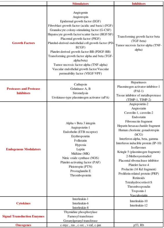
After birth, angiogenesis contributes to organ growth but during adulthood most of the blood vessels remain quiescent. However, ECs keep their remarkable ability of dividing rapidly and under some conditions, such as the cycling ovary, the pregnancy, the physical exercise, the wound healing, or in response to a specific stimulus (for example hypoxia); therefore angiogenesis can be reactivated. The normal and healthy body controls angiogenesis through a series of “on” switches, known as angiogenesis factors (cytokines) and “off” switches, known as endogenous angiogenesis inhibitors (table 1).
Introduction
6
Tab. 1. Endogenous positive and negative regulators of angiogenesis.
In many disorders, the perfect balance between angiogenesis modulators is compromised; angiogenesis has been implicated in more than 70 disorders so
Stimulators Inhibitors
Growth Factors
Angiogenin Angiotropin Epidermal growth factor (EGF) Fibroblast growth factor (acidic and basic) (FGF)
Granulocyte colony-stimulating factor (G-CSF) Hepatocyte growth factor/scatter factor (HGF/SF)
Placental growth factor (PlGF) Plateled-derived endothelial cell growth factor
(PD-ECGF)
Platelet-derived growth factor-BB (PDGF-BB) Transforming growth factor alpha and beta
(TGF-alpha/beta)
Tumor necrosis factor-alpha (TNF-alpha) Vascular endothelial growth factor/Vascular
permeability factor (VEGF/VPF)
Transforming growth factor beta (TGF-beta)
Tumor necrosis factor-alpha (TNF-alpha)
Proteases and Protease Inhibitors
Cathepsin Gelatinase A, B
Stromelysin
Urokinase-type plasminogen activator (uPA)
Heparinases Plasminogen activator-inhibitor-1
(PAI-1)
Tissue inhibitor of metalloprotease (TIMP-1, TIMP-2)
Endogenous Modulators
Alpha v Beta 3 integrin Angiopoietin-1 Endothelin (ETB receptor)
Erythropoietin Follistatin
Hypoxia Leptin Midkine (MK) Nitric oxide synthase (NOS) Platelet-activating factor (PAF)
Pleiotropin (PTN) Prostaglandin E Thrombopoietin Angiopoietin-2 Angiostatin Caveolin-1, caveolin-2 Endostatin Fibronectin fragment Heparin hexasaccharide fragment
Human chorionic gonadotropin (hCG)
Interferon-alpha, beta, gamma Interferon inducible protein (IP-10)
Isoflavones Kringle 5 (plasminogen fragment)
2-Methoxyestradiol Placental ribonuclease inhibitor
Platelet factor-4 Prolactin (16 Kd fragment) Proliferin-related protein (PRP) Retinoids Tetrahydrocortisol-S Thrombospondin Troponin-1 Vasculostatin Cytokines Interleukin-1 Interleukin-6 Interleukin-8 Interleukin-10 Interleukin-12
Signal Transduction Enzymes
Thymidine phosphorylase Farnesyl transferase Geranylgeranyl transferase
Introduction
7
far and the list is ever growing. In particular, when angiogenic growth factors are produced in excess over angiogenesis inhibitors, the balance is moved in favor of blood vessels growth. Vice versa, when inhibitors exceed stimulators, angiogenesis is stopped. Persistent and up-regulated angiogenesis is often a diagnostic factor of severe pathologies such as cancer, atherosclerosis, and diabetic retinopathy. Instead, insufficient angiogenesis is a characteristic of coronary artery disease (CAD), cardiac failure, tissue injury, etc. Angiogenesis has been implicated in more than 70 disorders so far and the list is ever growing. (Carmeliet 2003; Carmeliet, 2005).
1.4 VEGF family
New vessels growth and maturation require the sequential activation of a series of receptors by means of numerous ligands. However, among these VEGF signaling represents the key rate-limiting step (Ferrara et al., 2003). In mammalians, five VEGF ligands (occurring in several different splice variants and processed forms) have been identified:
VEGF-A VEGF-B
PlGF (placental growth factor) VEGF-C
VEGF-D
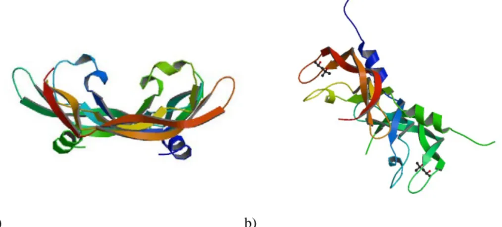
All these factors are secreted as dimeric and glycosilated proteins of approximately 40 kDa. Structurally (figure 2) they are characterized by a common motif represented by eight cysteine residues specifically spaced in a conserved domain. This is called “cysteine –knot motif”. The crystal structure of VEGF-A shows that the dimer is formed by two monomers ordered in an anti-parallel fashion, with the receptor-binding site located at each pole of the
Introduction
8
dimer. The VEGFs form also heterodimers if co-expressed in the same cell. Proteins that are structurally related to the VEGFs exist in parapoxvirus (VEGF-E) and snake venom (a group of proteins known as VEGF-Fs).
a) b)
Fig. 2. a) Structure of human Vascular Endothelial Growth Factor (PDB code: 1VPF); b) The crystal structure of human Placenta Growth Factor-1 (PLGF-1), an angiogenic protein at 2.0a
resolution (PDB code: 1FZV)
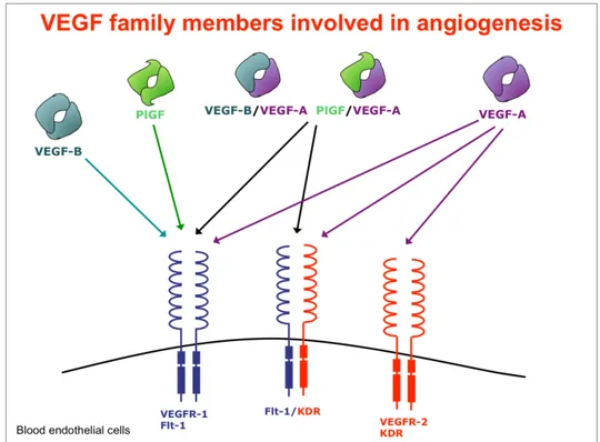
The VEGF family presents different biological and physical properties. VEGF-A, VEGF-B and PlGF are mainly involved in the angiogenesis, while VEGF-C and VEGF-D in the lymphangiogenesis. Their biological activities are due to the interactions with the extracellular domains of receptor tyrosine kinases (RTKs) (figure 3). These receptors are known as:
VEGFR-1 (also known as Flt-1, fms-like tyrosine kinase)
VEGFR-2 (also named KDR, kinase domain receptor, in human and Flk-1, Fetal liver kinase-1, in mouse)
VEGFR-3 (also known as Flt-4)
Some ligand isoforms are able to bind also co-receptors such as neuropilins (NP-1 and NP-2), other than heparin sulphate proteoglycans (HSPGs). The binding induces the RTKs dimerization. Each protein kinase monomer phosphorylates a distinct set of tyrosine residues in the cytosolic domain of its dimer partner (a process termed autophosphorylation) and finally a cascade of
Introduction
9
downstream proteins is activated. Through this fine mechanism, diverse angiogenic signals, including cell migration, survival and proliferation, are propagated. Thus, VEGRs play a key role in the signal transduction aimed at the formation of the new vessels and the regulation of vascular permeability (Rahimi , 2006; Roy et al., 2006; Otrock et al., 2007; Koch et al., 2011).
Fig. 3. Schematic representation of the Vascular Endothelial Growth Factors family members involved in angiogenesis.
A detailed description of some members of VEGF family is here after reported.
1.5 VEGF-A
VEGF-A (also known as VEGF) is the the most studied member of VEGF family. It plays a key role in the onset of angiogenesis, vasculogenesis and
Introduction
10
lymphangiogenesis. The vegf gene is located in the short arm of chromosome 6, formed by approximately 14 kb and containing 8 exons and 7 introns.
VEGF-A transcription is up-regulated in hypoxia condition through a fine mechanism. In particular, hypoxia-inducible factor–1α (HIF–1α) binds to the hypoxia responsive element (HRE) in the VEGF-A gene promoter region, which in turn increases the transcription of VEGF-A. Through the same mechanism, the transcription of other genes implicated in glucose transport, glycolysis and angiogenesis is also activated. Recent studies report the role of von Hippel-Lindau (VHL) tumor suppressor gene in HIF-1-dependent hypoxic responses. Also other pathways involving different growth factors can increase the expression of VEGF-A (Semenza, 2002; Mole at al. 2001). For instance, epidermal growth factor (EGF), transforming growth factors (TGF-α, TGF-β), keratinocyte growth factor, insulin-like growth factor-1, fibroblast growth factors (FGF) and platelet-derived growth factor, are able to up-regulate VEGF mRNA expression. This suggests that paracrine or autocrine systems deal with local hypoxia by regulating VEGF release. Some inflammatory cytokines (IL-1α, IL-6α) also induce VEGF-A gene expression supporting the hypothesis that VEGF-A is a mediator of permeability in inflammatory disorders. Finally, it has been also demonstrated that VEGF-A is up-regulated by mutations or amplification of Ras (Rat sarcoma) (Neufeld et al., 1999; Ferrara et al., 2003; Otrock at al., 2007).
VEGF exists in four different isoforms (comprising 121, 165, 189 and 206 amino acids in humans), which are generated by alternative splicing of a single pre-mRNA species. Among these, VEGF165 is the predominant isoform. However, isoforms that are expressed at lower extent were also identified (VEGF145 and VEGF183). Isoforms differ in their ability to bind to heparin sulfate and extracellular matrix (ECM) (Park et al., 1993; Ferrara et al., 2003). VEGF-A exerts its biologic effects through interaction with the receptors Flt-1 and KDR/Flk-1 and with the co-receptors NP-1 and NP-2. It is the most potent
Introduction
11
pro-angiogenic protein. VEGF-A induces proliferation, sprouting and tube formation of endothelial cells (ECs) and exerts many effects on a broad range of not-ECs types. It is also a potent survival factor, both in vitro and in vivo. In ECs it induces the expression of antiapoptotic proteins (Bcl-2 and A-1). VEGF-A was originally described as vascular permeability factor (VPF) but it is also involved in vascular leakage (a key process in inflammation and other pathological conditions) through the formation of intercellular gaps, vescico-vascular organelles, vacuoles and fenestration in some vescico-vascular beds. Furthermore, it increases the hydraulic conductivity of isolated microvessels, through an accentuation of calcium influx. This growth factor causes vasodilatation through the release of nitric oxide by inducing the endothelial nitric oxide synthase (eNOS). VEGF-A promotes the mobilization of hematopoietic stem cells (HSCs) from the bone marrow, monocyte chemotaxis, osteoblast-mediated bone formation and neuronal protection. Furthermore, it stimulates inflammatory cell recruitment and the expression of proteases implicated in pericellular matrix degradation in angiogenesis.
VEGF-A is essential mainly during embryonic and early postnatal development. The loss of a single vegf allele is lethal in the mouse embryo between days 11 and 12. These embryos present some developmental anomalies, not well-vascularized organs and a low number of nucleated red blood cells. Inhibition of VEGF during early postnatal life, increases mortality, stunts the body growth and impairs organ development and skeletal growth. In juvenile primates it results in abnormalities in physiological angiogenesis. On the other hand, transgenic mice with over-expression of VEGF-A in the skin, develops a psoriasis-like skin condition and accelerates experimental tumor growth. In humans, VEGF-A is expressed in most of all solid tumors as well as in some hematological malignancies. VEGF is also implicated in the pathogenesis of diabetes mellitus, having a major role in the onset of vitreous hemorrhages, retinal detachment, neovascular glaucoma and blindness
Introduction
12
correlated with this pathology. Recent studies showed the presence of VEGF in choroidal neovascular membranes of age-related macular degeneration (AMD) patients. It has been reported that this presence is correlated to neovascularization and vascular leakage that cause AMD. Finally, VEGF is involved in many inflammatory disorders and it cooperates in induction of angiogenesis associated to polycystic ovary syndrome, endometriosis and preeclampsia. (Ferrara et al., 2003; Tammela et al., 2005; Otrock et al., 2007; Roy et al. 2006).
1.6 PlGF
The human plgf gene is located on chromosome 14q24 and is formed by 7 exons spanning on 12 qD. Using alternative splicing processe, PlGF can be expressed in four isoforms, named PlGF-1, -2, -3 and -4 composed by 131, 152, 203 and 224 amino acids, respectively. They differ in the ability to bind heparan sulfate proteoglycans (Maglione et al., 1993a; DiPalma et al., 1996). Although some reports indicated an upregulation of PlGF in cells exposed to hypoxia, the analysis of promoter/enhancer region of PlGF did not show active HRE sequence as observed also for VEGF-A (Green et al., 2001; Oura et al., 2003; Selvaraj et al., 2003). However, PlGF gene promoter includes many recognition sequences for metal transcription factor 1 (MTF-1) and for NF-κB. These sequences are typically involved in the modulation of PlGF expression in hypoxic condition (Green et al., 2001). Moreover, overexpression of HIF-1α in endothelial cells or in primary cardiac and vascular cells up-regulates the expression of PlGF. Moreover, PlGF expression is modulated also by the forkhead/winged helix transcription factor FoxD1 (BF-2) in the developing kidney stroma. This is possible given the presence of a conserved HNF3b binding site on PlGF promoter region (Zhang et al., 2003). Finally, PlGF
Introduction
13
expression is also post-transcriptionally regulated by other growth factors and oncogenes (Maglione et al., 1993b).
The pro-angiogenic activity of PlGF is exerted through the binding and the activation of VEGFR-1. This growth factor shows the highest affinity if compared to those of the other members of the same family.
PlGF may also activate VEGFR-2 indirectly, using one of the following mechanisms:
VEGFR-1, once activated by PlGF, can transphosphorylate VEGFR-2 PlGF, upon binding to VEGFR-1, makes VEGF-A available for the
binding and activation of VEGFR-2
PlGF and VEGF-A, if co-expressed from the same cells, can form heterodimers able to activate 1 or to induce VEGFR-1/VEGFR-2 heterodimerization.
Furthermore PlGF-2 is able to bind the two coreceptors NP1 and NP2 (Autiero et al., 2003).
PlGF was originally discovered in placenta. It is also expressed in throphoblastic giant cells associated with the parietal yolk sac, during early embryonic development. At cellular level, the expression of PlGF was demonstrated in endothelial cells, thyroid transformed mouse embryonic fibroblast, NIH 3T3 cells and a limited number of tumor-derived cell lines. PlGF knockout mice do not have an evident phenotype. They born at medelian frequency, are healthy and fertile but present an impaired angiogenesis when pathological conditions are induced such as tumor growth, heart or limb ischemia, choroid neovascularization. This strongly indicates that PlGF is involved only in pathological angiogenesis and not in physiological angiogenic process. Overexpression of PlGF in the skin of transgenic mice results in a significant increase in the number and size of skin blood vessels, in number of mature smooth muscle-coated vessels and in enhanced vascular permeability. Accordingly, adenovirus-mediated PlGF transfer in the ischemic
Introduction
14
heart and limb was able to induce a strong angiogenic response, forming numerous larger vessels, with an efficacy almost comparable to that of VEGF-A (Luttun et al., 2002). The same approach used in xenograft tumors did not show an increase of tumor volume and vessel density, however it generated an increment of the vessel lumen, of the inflammatory infiltrate and of the vessel maturation (Tarallo et al., 2010). Recombinant PlGF homodimer or PlGF/VEGF heterodimer significantly promoted angiogenesis in ischemic conditions (Luttun et al., 2002; Autiero et al., 2003).
Futher studies demonstrated that PlGF promoted pathological angiogenesis by stimulating vessel growth and maturation. In particular, it acts on the growth, migration and survival of endothelial cells, increases the proliferation and recruitment of smooth-muscle cells and supports the proliferation of fibroblasts. Finally, PlGF is crucial for the recruitment and maturation of bone marrow derived progenitors and the differentiation and activation of monocyte-macrophage (De Falco, 2012).
1.7 VEGFR-1 and VEGFR-2
Human VEGFR-1 and VEGFR-2 are transmembrane glycoproteins of 180 and 200 kDa, respectively. They are structurally related to the platelet-derived growth factor (PDGF) receptor family and contain (figure 4):
an extracellular domain of 7 extracellular immunoglobulin (Ig) like domains
a single transmembrane region a regulatory juxtamembrane domain
an intracellular tyrosine kinase domain, interrupted by a kinase insert domain.
Introduction
15
Figure 4. Representative structure of vascular endothelial growth factor (VEGF) tyrosine kinase receptors. The VEGF receptor family is represented by seven immunoglobulin-like
loops in the extracellular domain, which binds VEGF. Ligand induces the formation of receptor dimer to activate autophosphorylation of tyrosine residues on the cytoplasmic
domain. Ig = immunoglobulin; VEGF = vascular endothelial growth factor; Y- = phosphorylated tyrosine residues.
Both the receptors bind VEGF with high affinity. The ligand-binding region is localized within the second and third Ig domains, while the fourth Ig domain is essential for dimerization of VEGF receptors. In addition, Flt-1 also acts as a receptor for VEGF-B and PlGF, whilst KDR also binds to VEGF-C and VEGF-D and the viral homolog VEGF-E.
Upon ligand binding, the tyrosine kinase activity of VEGFR1 is ten-fold weaker than that of VEGFR-2. This information prompts further investigations to understand at molecular level the different biological roles of these receptors.
VEGFRs undergo alternative splicing to generate soluble forms of the receptors. Soluble VEGFR-1 (sFlt-1) is composed by the first six domains of extracellular portion of the receptor. This form binds VEGF-A and PlGF with the same affinity of the full length receptor, it functions as an endogenous VEGF inhibitor (Tugues et al., 2011; Shibuya, 2013). As recently reported, it plays a pivotal role to maintain cornea avascularity. However, when an injury
Introduction
16
of cornea occurs with an increase of VEGF-A level, it titles sFlt-1 and binds to Flk-1 inducing angiogenesis (Ambati et al., 2006; Shibuya, 2013). Similarly, soluble VEGFR-2 (sKDR) is important in ocular lymphoangiogenesis context (Pavlakovic et al., 2010).
KDR and Flt-1 are both expressed in endothelial cells. VEGFR1 is also expressed in monocyte/macrophages, dendritic cells, osteoclasts, pericytes, trophoblasts, in mesangial cells, smooth muscle cells and also in bone marrow stem/progenitors derived cells. Its transcription is up-regulated by hypoxia, via a HIF-1-dependent mechanism and upon activation of macrophages. Non endothelial expression of VEGFR-2 has been observed in vascular endothelial progenitors, retinal progenitor cells, hematopoietic stem cells, neuronal cells, osteoblasts, pancreatic duct cells, and megakatyocytes.
VEGFR-1 knock out mices die at embryonic day 8.5-9.0, due to the over-growth and disorganization of blood vessels. This suggested that VEGFR-1, trapping VEGF-A and preventing activation of VEGFR-2, plays a negative regulatory role during the development of primitive vascular network.
Several tyrosine residues in VEGFR-1 intracellular domain have been identified as autophosphorylation sites using various experimental approaches. For instance, phosphorylation of Y1169 allows the binding and in turn the activation of phospholipase C (PLC)γ1 regulating endothelial cell proliferation via the mitogen activated protein kinase (MAPK) pathway. It is worth noting that VEGF-A and PlGF don’t induce the same autophosphorylations in VEGFR-1. This suggests that the two ligands stabilize two distinct conformations of the receptor intracellular domain in the activated VEGFR1 dimer or induce distinct modes of association with accessory molecules, such as heparin sulfated proteoglycans or neuropilins,. As a result, the availability of VEGFR-1 tyrosine residues as substrates for the kinase is different in response to the diverse ligand bound (Shibuya 2006). Finally, it has been
Introduction
17
demonstrated synergy between VEGFR-1 and VEGFR-2, by a “cross-talking mechanism” (Tjwa et al., 2003).
VEGFR-2 null mices die at embryonic day 8.5-9.0, due a lack of vasculogenesis and very poor hematopoietic development. This means that VEGFR-2 plays an essential role in survival, growth and differentiation of endothelial cell progenitors.
Thus, during early embryogenesis, the two VEGFRs have opposite roles in angiogenesis: VEGFR-2 is a positive signal transducer, whereas VEGFR-1 is a suppressor. It is necessary a coordinated signaling between the two receptors to obtain a balanced expansion and differentiation of the endothelial precursor pool.
VEGFR-2 activity is also important for migration of ECs during adulthood. Several studies have been carried out in order to clarify the signal transduction by VEGFR-2. Therefore some autophosphorilation sites have been identified. Y951 mediates the binding to T cell specific adapter (TSAd), which in turn binds to Src, thus regulating actin cytoskeleton and cell migration. Phosphorilation of Y1175 induces the activation of PLCγ that in turn stimulates the protein kinase C(PKC) pathway leading to inositol phosphate formation and calcium mobilization. Furthermore, Raf-MEK-MAP kinase pathway and subsequent DNA synthesis are regulated through the phosphorylation at this site. Y1175 also binds to the adapter molecule Shb, that is also implicated in the activation of PI3-kinase and its downstream effector Akt and MAPK p38 via VEGFR-2. This site is also involved in the differentiation of hemangioblasts. (Shibuya 2006, Shibuya 2013)
Recent studies give the first indications about the role of heterodimerization between VEGFR-1 and VEGFR-2 in the regulation of endothelial cell homeostasis. According to this research VEGFR1 − 2 activation mediates VEGFR phosphorylation, endothelial cell migration, sustained in vitro tube formation and vasorelaxation via the nitric oxide pathway, but not
Introduction
18
proliferation or endothelial tissue factor production, confirming that these functions are controlled by VEGFR-2 homodimers. Moreover VEGFR1 − 2 inhibits VEGF-A-induced prostacyclin release, phosphorylation of ERK1/2 MAP kinase and mobilization of intracellular calcium from primary endothelial cells. These findings indicate that VEGFR-1 subunits modulate VEGF activity predominantly by forming heterodimer receptors with VEGFR-2 subunits and such heterodimers regulate endothelial cell homeostasis (Cudmore et al., 2012).
1.8 Anti-angiogenic therapy in cancer
Expanding tumor tissues rapidly exhaust the available oxygen supply and become hypoxic. As described partially above, the activation of hypoxia-inducible factor (HIF) signaling triggers VEGF expression, not only in tumor cells but also in tumor-associated stromal cells. VEGF, meets the tumor’s oxygen requirements and promotes therefore tumor growth and metastasis, by stimulating vascular growth. For this reason tumor tissue is often characterized by a superficial dense vascular network (Kubota, 2011).
With the aim of suppressing tumor progression and metastatic spread, in the last years angiogenic therapy has been developed. Most current anti-cancer chemotherapeutic drugs, used in the clinical setting, indiscriminately target all rapidly dividing cells (e.g., at the level of DNA replication and protein synthesis) and therefore cause severe adverse effects, such as immunosuppression, intestinal problems and hair loss. Also if anti-angiogenic agents theoretically may have fewer side effects, the clinical practice has evidenced the appearance of important side effects such as bleeding, thrombotic events, hypertension, proteinuria, leucopenia, lymphopenia, hypothyroidism. They are due to physiological angiogenesis blockage.
Introduction
19
Blocking VEGF appeared immediately a reasonable anti-angiogenic modality. Several studies of angiogenesis inhibition by administration of VEGF blockers have demonstrated significant tumor-suppression effects in various types of cancers. Treatment of mice carrying human tumors with an anti-VEGF neutralizing Mab (monoclonal antibody) significantly inhibited xenograft tumor growth. In 2003, the Food & Drug Administration (FDA) approved Bevacizumab (Avastin; Genentech Inc.), a humanized variant of a VEGF neutralizing Mab, as the first anti-angiogenic agent for combination treatment with chemotherapeutic agents in metastatic CRC (colorectal cancer) and subsequently for treatment of NSCLC (non-small-cell lung cancer) or MBC (metastatic breast cancer). VEGF-TrapR1R2 (Aflibercept; Regeneron Inc.), a chimeric soluble receptor that neutralizes circulating VEGFs, is currently in clinical trials. Additionally, blockade of VEGF receptors inhibits tumor growth. Receptor tyrosine kinases inhibitors (RTKIs), such as sunitinib (SU11248; Sugen), pazopanib (Votrient; GSK), sorafenib (Bay 43-9006; Nexavar) vendatanib (Caprelsa; AstraZeneca), cabozantinib (XL184; Exelixis), axitinib, tivozatinib and linifanib have recently been developed, for the treatment of many types of cancer. A number of studies have reported their significant therapeutic efficacy (Shojaei, 2012).
Recently also PlGF becomes an interesting target. An antibody against placental growth factor (PlGF) inhibits growth of VEGF-resistant tumors without affecting healthy vessels; interestingly, targeting PlGF not only inhibits vessel growth but also the recruitment of angiogenic macrophages (Fischer, 2007).
Despite the encouraging conditions, it is well to report that anti-angiogenesis therapy is facing three major challenges nowadays (figure 5):
inherent/acquired resistance
Introduction
20
lack of validated predictive biomarkers to select patient population and to monitor tumors responses to the therapy
Fig. 5. Three major challenges of antiangiogenic therapy
Many patients show a lack of response to inhibitors of angiogenesis . Certainly, this is in part due to the very modest doses that are given to patients (5 mg/kg body weight, once every 2 weeks), compared to mice in preclinical study (10 mg/kg body weight, twice a week), to reduce possible off-target toxicities, such as hemorrhagic and thrombotic events . However, the extent of refractoriness is highly variable from one cancer to another. Mechanisms underlying resistance and/or enhanced invasiveness during treatment with AIs have been extensively studied and can be resumed in the following main points:
both tumor and non-tumor compartments contribute to resistance to anti-angiogenic agents
resistance to AIs may occur independent of class of agents i.e. antibodies or small molecule inhibitors
Introduction
21
resistance occurs independently of affinity of the anti-VEGF antibody limited bioavailability of therapeutic agent in tumor mass might
account for lack of response to the therapy
in absence of VEGF signaling, other mechanism (mediated for example by FGFs, angiopoietins, NRP1) can offset with their proangiogenic effects
in tumors, some blood vessels are covered with a dense pericyte coat (whose recruitment is mainly regulated by platelet-derived growth factor (PDGF)-B/PDGF receptor-β, transforming growth factor-β (TGF-β) and angiopoietin/Tie signaling) that could represent a reason of a minor responsiveness to VEGF blockers by these vessels ]
hypoxia-tolerant cancer cells, called cancer stem cells, survive in poorly oxygenated niches and elicit tumor adaptation to anti-angiogenesis; some reports suggest that the resultant selection of tumor cells renders tumors even more invasive and metastatic
chronic exposure of endothelial cells to anti-VEGF drugs may lead epigenetic modulation of expression of anti-apopotic genes such as
bcl-2 and survivin. Absence of the proapoptotic Bcl-bcl-2 homology 3
(BH3)-only Bcl-2 family member Bim in endothelial cells completely abolishes the effect of VEGF blockade macrophages are also involved in vascular development, independently of VEGF signaling; histological test of various cancer tissues reveals a vast accumulation of macrophages, that coordinate various aspects of tumor angiogenesis Given and modest responses observed to antiangiogenesis therapy in the clinic, it is essential to identify a set of biomarkers to select populations of likely responders and/or to monitor disease progression and response over the course of treatment in the clinic. From some studies, CD31 expression has been proposed as biomarker of response in breast cancer patients treated with bevacizumab and chemotherapy. Quantification of circulating endothelial
Introduction
22
progenitor cells, cytokines and/or growth factor in serum could also be another strategy to monitor response to angiogenesis inhibitors. Because of VEGF inhibition results in a reduction in NO (nitric oxide) synthesis, leading to vasoconstriction and increase in blood pressure, hypertension is another biomarker of response to these drugs.
In conclusion, in order to surmount several challenges associated with anti-angiogenesis therapy, advances in understanding the molecular and cellular bases of tumor angiogenesis are required, as well as new drugs are required to pave the path towards a more efficient and successful clinical application (Tonra and Hicklin, 2007; Hsu and Wakelee, 2009; Shojaei, 2012).
Project aims and approaches
23
CHAPTER 2:
Project aims and approaches
Project aims and approaches
25
2.1 Project aims
The work presented in this thesis was aimed to support a rational design of new antiangiogenic drugs or treatment strategies. To this end, the whole work was focused on the following objectives:
Performing an expression proteomic study of cell cultures treated with some dimers of VEGF family. In particular, following the incubation of Human Umbilical Vein Endothelial Cells (HUVECs) and Human Embryonic Kidney 293 cells, over-expressing stably human Flt-1, (HEK-293hFlt-1) with VEGF, PlGF, VEGF/PlGF, we meant to identify the proteins and differentially modulated following each treatment. Information relating to functional genomics can represent a valuable contribution to the further understanding of the molecular bases of angiogenesis and for the development of therapeutic strategies more effective and safer than those currently approved.
Identifying novel putative inhibitors of angiogenesis, performing a small library of natural compounds screening. On the basis of the experimental evidence reported in the literature on their involvement in pathological angiogenesis, we chose the axis VEGF/Flt-1 and PlGF/Flt-1,as target for this study.
2.2 Proteomic approach
“Proteomic” (term coined in 1994) is the science that describes the entire set of proteins expressed by the entire genome of a cell in the lifetime or, in a less universal sense, at any one time, under specific conditions. Providing these informations, proteomic promises to bridge the gap between our understanding
Project aims and approaches
26
of genome sequence and cellular behavior (Wilkins et al., 1996; Patterson et al., 2003).
The completion of human genome sequencing represented undoubtedly one of the most exciting biological achievement. One of the interesting aspects of this effort is the finding that, though they’re very different organisms from the phenotypic point of view, the number of protein-encoding genes found for humans (~30000) is not extremely different from those calculated for phylogenetically remote organism, such as a yeast cell (~6000), a fly (~13000), a worm (~18000), a plant (~26000). It’s clear that the physiological complexity of an organism is not a consequence of a mere genes number. Indeed, the existence of an open reading frame (ORF) doesn’t imply perforce that the gene is functional (Pandey and Mann, 2000). Many studies document the disparity between the relative expression levels of mRNA (transcriptome) and those of their corresponding proteins; in facts, through gene splicing mechanism, a single gene can code for multiple proteins and each protein can undergo numerous post-translational modifications, including phosphorylation, acetylation, ubiquitylation, SUMOylation, palmitoylation, transglutamination and proteolytic cleavage (Mann and Jensen, 2003). These aspects, not predictable from genome, increase the functional diversity of a cell system. Moreover, the complexity of an organism is also due to the ability of intracellular signaling pathways to interact with each other forming complex networks. This complexity is due to the overlapping functions of some proteins, to their connections, and to their spatio-temporal relationship in the cell. Finally, many cellular processes are very often performed not by individual proteins, but by protein macromolecular complexes (Pieroni et al., 2008; Preisinger, et al., 2008).
Proteomic analysis is certainly a valid first step in functional annotating the genome. In particular, differential proteomics, that consists in the comparison of distinct proteomes (for example: normal versus diseased cells, diseased
Project aims and approaches
27
versus treated cells and so on) is of paramount importance, in all areas of biological research (cancer research, toxicology, pharmacology and so on) (Patterson et al., 2003).
Several approaches can be used to achieve this type of study. They typically involve electrophoresis and/or chromatography combined with chemical or metabolic labeling and mass spectrometry (Gevaert and Vandekerckhove, 2000; Fuchs et al. 2005).
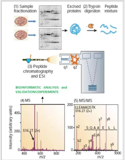
In this work, the proteomic profiles of cultured cells stimulated with VEGF dimers have been studied by a gel-based approach (Rabilloud, 2002; Wulfkuhle et al. 2003; Monteoliva and Albar, 2004). Classically this strategy involves the achievement of then following steps (figure 6):
1. Separation of complex protein mixtures, usually by two-dimensional gel electrophoresis, 2-DE
2. Protein visualization and image analysis 3. Excising the spots for following analyses
4. In gel digestion of proteins and pooling of the released peptides 5. Analysis of the peptide mixtures by mass spectrometry
6. Matching peptide masses against protein databases to obtain proteins identification
2-DE is a relatively simple but powerful method for high-resolution analysis of complex protein mixtures.
All proteins in an electric field migrate at a speed that is dependent on their conformation, size and electric charge. 2DE uses the latter two characteristics to allow high-resolution separation of proteins. As first described by O’Farrel and J. Klose in 1975, isoelectric focusing (IEF) separates proteins on the basis of their charge (separation in first dimension), while Sodium Dodecyl Sulphate - PolyAcrylamide Gel Electrophoresis (SDS-PAGE) on the basis of their molecular weights (separation in second dimension). The result is an array of
Project aims and approaches
28
protein spots, characterized by precise coordinates x and y: isoelectric point (pI) and apparent molecular weight (Mw), respectively.
Separated proteins can be visualized by staining or autoradiography. Subsequently computer-based analysis is needed to detect differentially expressed proteins. The most known commercial software are: Progenesis, Nonlinear Dynamics; Image Master 2D Platinum and Melanie Software, Amersham Biosciences; PDquest, Bio-Rad.
Fig. 6. Proteomic approach workflow.
A reliable protein separation by 2 DE depends on some parameters. First the choice of sample preparation protocol: it is the critical influential factor for
Project aims and approaches
29
isoelectric focusing which in turn affects the two-dimensional gel result in terms of quality and protein species distribution (Shaw and Riederer, 2003; Monteoliva and Albar, 2004). Since immobilized ph gradient strips (IPG-strip) were developed variability in experimental conditions has greatly decreased; this is now undoubtedly the most widespread strategy for comparing distinct states of more proteomes (Weiss and Görg 2009; Görg et al., 2009).
Protein identification is based on mass spectrometry data. The starting point is a protein spot, which may be a single protein or a complex mixture of proteins. An enzyme, often trypsin, digests the proteins to peptides. Chromatography is used to regulate the flow of peptides into the mass spectrometer. Peptides selected are then induced to fragment, possibly by collision, to capture an MS/MS spectrum. For each MS/MS spectrum, software is used to determine which peptide sequence in a database of protein or nucleic acid sequences gives the best match. Each entry in the database is digested, in silico, using the known specificity of the enzyme, and the masses of the intact peptides calculated. If the calculated mass of a peptide matches that of an observed peptide, the masses of the expected fragment ions are calculated and compared with the experimental values. The result is a list of candidate proteins with different confidence levels (Fenn et al. 1989; Henzel et al. 1993; Aebersold and Mann, 2003; Roepstorff, 2012)
2.3 Natural product for drug discovery
Natural compounds have been the source of inspiration for chemists and physicians for millennia, representing the richest font of novel compound classes and an essential wellspring of drugs and drug leads (figure 7). According to a recent survey by David J. Newman, Gordon M. Cragg, and Kenneth M. Snader of the national cancer institute, 61% of the 877
small-Project aims and approaches
30
molecule new chemical entities introduced as drugs worldwide during 1981– 2002 can be traced to or were inspired by natural products (Newman et al. 2003). These include natural products (6%), natural product derivatives (27%), synthetic compounds with natural-product-derived pharmacophores (5%), and synthetic compounds designed on the basis of knowledge gained from a natural product (that is, a natural product mimic; 23%). In certain therapeutic areas, the output is higher: 78% of antibacterials and 74% of anticancer compounds are natural products or have been derived from, or inspired by, a natural product. These numbers are not surprising since it is know that natural products evolved for self-defence. Despite that record of productivity, the use of natural products as a starting point for drug discovery was de-emphasized in many big pharmaceutical companies in 1990s, when combinatorial chemistry had place and the reasons were primarily practical. However, after several years it was clear that database of natural products have a major number of unused scaffolds, and couldn’t be discarded as starting points for new drugs discovery. The differences between synthetic compounds and natural products are remarkable, especially in their structural properties (Dobson, 2004; Rosén et al., 2009). On average, natural products have higher molecular weights; incorporate fewer nitrogen, halogen, or sulphur atoms, but more oxygen atoms and are sterically more complex, with more bridgehead tetrahedral carbon atoms, rings, and chiral centres. This gives them a high “sterical complexity” due to the fact that the enzymes used for biosynthesis, as well as their molecular targets, are inherently three-dimensional and chiral. Furthermore, nature has a limited palette of building blocks at its disposal, and thus has to generate novelty by branching out common intermediates into different scaffolds.
Moreover natural compounds are a really good starting point for the set up of libraries to be tested for drug discovery (figure 7), not only for their complex and diversified chemical space but also for their ability to interact with
Project aims and approaches
31
biomolecules. Lynn H. Caporale in her book "Darwin in the genome" (Caporale, 2002), writes: “even though natural products may not have coevolved with human proteins, they have emerged in nature to interact with biomolecules….varied genomes, based on similar chemistry, have spread across the earth. Indeed we share our gene families with other organisms. Whether inside bacteria or inside us, there is a limited number of ways that the structural components of proteins, such as -helices and -sheets, can arrange themselves in space and interact with each other and with other molecular structures”. Herbert Waldmann, from Max Planck institute for molecular physiology, offers a similar analysis, based on the premise that natural products evolved to perform a function that is achieved by binding to proteins. Therefore, natural products should be able to penetrate biological barriers and make their way to certain cells or organs in which they will exert the effect. Thus, natural products are already biologically validated to reach and bind specific proteins. Looking at all proteins and analyzing them for structural features, elements of conservatism and diversity may be found. The conservative elements are the domains: the parts that fold to compact secondary structures. Among the hundreds of thousands of human proteins, the number of distinct domains is only about 600 to 8,000. Consequently, proteins that may seem altogether different are quite similar when viewed structurally, but diversity lies in the precise details since similar domains may have very different amino acid sequences (Kirkpatrick, 2003).
Project aims and approaches
32
A number of bioactive plant compounds have been recently tested for their antiangiogenic potential. Among the known angiogenesis inhibitors compounds derived from natural sources, like flavonoids, sulphated carbohydrates, or triterpenoids are playing a prominent role. The underlying mechanisms are complex and in part unknown. In this regard, we should recall the recent work by Tarallo et al which proved an interesting antiangiogenic activity of amentoflavone. This biflavonoid can bind to VEGFs preventing the interaction and phosphorylation of VEGF receptor 1 and 2 (VEGFR-1,VEGFR-2) and to inhibit endothelial cell migration and capillary-like tube formation induced by VEGF-A or placental growth factor 1 (PlGF-1) at low μm concentration. In vivo, amentoflavone is able to inhibit VEGF-A-induced chorioallantoic membrane neovascularization as well as tumor growth and associated neovascularization, as assessed in orthotropic melanoma and xenograft colon carcinoma models (Tarallo et al., 2011).
Finally, ASA404, a flavonoid compound, is a “vascular disrupting agent” capable of induction of apoptosis in tumor associated endothelial cells, resulting in the inhibition blood flow in tumor mass. ASA404 is currently in advance stage of clinical development in combination with chemotherapic drugs in NSCLC patients.
For all these reasons natural products are considered: a valid source for investigational new antiangiogenic agents to treat many disorders.
Results and discussion
33
CHAPTER 3:
Results and discussion
Results and discussion
35
3.1 Proteomic study
In order to identify differentially expressed proteins both in endothelial cells and tumor cells stimulated by VEGF, PlGF and VEGF/PlGF, separation and quantification of relative protein extracts have been performed by a gel-based proteomic approach.
At the outset of this work, no comparative proteomic analysis of the selected cells expressing one or both VEGF receptors and treated with PlGF and VEGF/PlGF had been produced, while few works can be found on VEGF-treated cells (such as HUVECs). Data produced by others, as discussed below, report some findings corroborating our data.
3.1.1 Electrophoretic separation of proteins
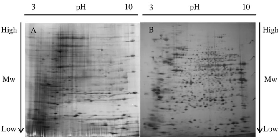
In the present study, alterations in the cellular proteome induced by VEGF, PlGF, and heterodimer treatments have been investigated using HUVECs and HEK-293 cells stably overexpressing hFlt-1 (HEK-293-Flt-1). Both cell lines underwent serum-starvation, followed by incubation with the selected growth factors for 24 hours. Total protein extracts were separated by 2-D electrophoresis (2DE) in order to obtain consistently well-separated protein profiles. For HEK-293Flt-1 protein extracts, it has been necessary to optimize the classical protocol of sample preparation, a crucial step for a successful 2 DE. Replacing sulphydryl reductants dithiothreitol DTT, the most common reductant agent, with tris (2-carboxyethyl) phosphine TCEP, we were able to solubilize the samples more effectively to get 2D maps better resolved and more easily analyzable (figure 8).
Silver staining compatible with mass spectrometry have been used for a sensitive detection of separated proteins (figures 9, 10).
Results and discussion
36
Fig. 8. 2D gel of HEK-293Flt-1 protein extracts; a) Gel obtained with utilization of DTT; b) Gel obtained with utilization of TCEP.
Fig. 9. 2D gel of protein extracts of HEK-293Flt-1, following the treatment with: b) VEGF, c) PlGF, d) heterodimer; a) represents the control (not induced). Each gel has been produced in triplicate. 3 pH 10 3 pH 10 Mw Mw High Low Low High A B 3 pH 10 3 pH 10 Mw Mw High Low Low High A B 3 pH 10 3 pH 10 Mw Mw High Low Low High C D
Results and discussion
37
Fig. 10. 2D gel of protein extracts of HUVECs, following the treatment with: b) VEGF, c) PlGF, d) heterodimer; a) represents the control (not induced). Each gel has been produced in triplicate.
3.1.2 Image analysis and statistics
In order to perform image analysis, 2D gel images must be converted into digital data. This has been addressed acquiring images by charge-coupled device (CCD) camera systems and then analyzing them using commercial image processing software.
Schematically, workflow approach to 2D image analysis was:
Step 1. 2D gel images, once loaded, had automatically examined from "quality control" application of the software. In general, the output of
3 pH 10 3 pH 10 Mw Mw High Low Low High A B 3 pH 10 3 pH 10 Mw Mw High Low Low High C D
Results and discussion
38
this operation is a description of the properties of the image and of the problems (for example: saturation of the image, due to a wrong staining of the gel) associated with it. Thanks to the high quality of resolution of our gels, no problems were revealed.
Step 2. The most appropriate reference image to which align all the other images was selected.
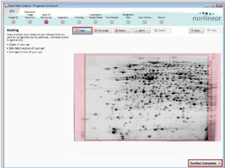
Step 3. In this phase a mask of disinterest was placed on the image to exclude specific areas from the analysis (figure 11). In our work only the extremely lateral margins of the gels had been masked.
Fig. 11.Page of Progenesis SameSpots. Masked area are evidenced in pink
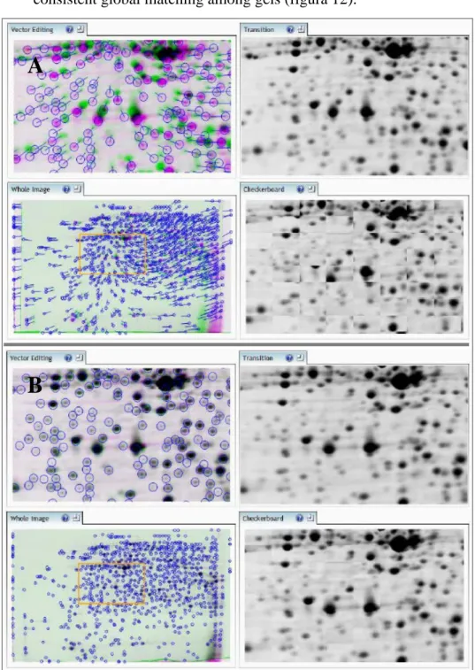
Step 4. Image alignment was the process of matching gel images with the reference image. In order to achieve the best possible results, it was crucial that the alignment was highly accurate. Even if the gels showed very good resolution in the separation of proteins, as a starting point we needed to align a few spots manually. These spots, acting as "guide
Results and discussion
39
vectors" for the subsequent automatic alignment, contributed to a very consistent global matching among gels (figura 12).
Fig 12 Alignment with Progenesis SameSpots. Vectors are colored in blu. A) Gels are not aligned; B) Gels are perfectly aligned.
A
Results and discussion
40
Step 5. After reviewing the alignment quality, all spots in every gel were detected and quantified. Among these, some were removed based on position, area, normalized volume and combinations of these spot properties. Anyway, the most important process of this step, essential to determine the volume of the protein in the spot, was the background subtraction and normalization. Background subtraction was performed by software, subtracting the lowest intensity value of the image pixels outside the spot's outline from the intensity value of every pixel inside the spot outline. Normalization was performed using the “total spot volume” method, which results in all spot volumes being expressed as a fraction of the total spot volume within a gel (figura 13). This allowed to correct for protein loading differences between gels and to identify any differences in expression due to only biological change.
Fig. 13. Background subtraction performed by
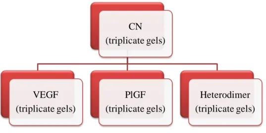
Step 6. At this phase one or more groups for analyzed images were built up. For this study, we grouped the gel images to reflect the biological groupings (figure 14); thus we assembled the group of three gels, representing a specific treatment (VEGF, PlGF, Heterodimer),
Results and discussion
41
and that relating the control (CN). After selecting two groups (control vs treatment), software calculated the data of differential protein expression (Fold-change and relative p-value).
Step 7. At this stage results had been revised, changing detection zones, using a set of tools, such as editing, splitting, merging of spots. This software permits to process a single image, but the effects are applied across all images in the experiment.
Fig. 14. Scheme: every block represents a group of three gels relateve to a specific biological condition. We compared each specific treatment block to control. This strategy have been
applied to both cell cultures.
Step 8. Finally it was possible to perform multivariate statistics on selected spots. The statistical analysis of the differentially expressed proteins (only statistically significant spots, p<0.05) was presented in a form of interactive graphical representations. In particular, for “Principal Component Analysis” (PCA), the software used spot expression levels across gels to determine the principle axes of expression variation. Transforming and plotting the most significant
CN (triplicate gels) VEGF (triplicate gels) PlGF (triplicate gels) Heterodimer (triplicate gels)
Results and discussion
42
expression data in principle component space, gels were separated according to expression variation. PCA plot allowed us to observe if gels group according to the experimental conditions and to identify gel outliers.
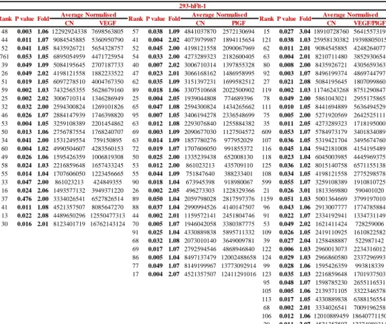
In this study, protein spots showing a fold change higher than 1.3 and statistically significant (p value < 0.05), were considered as differentially expressed proteins. Achieved data (tables 2, 3) showed that following the VEGF treatment, HUVECs exhibit significantly altered expression, compared to the control, of a total of 23 protein spots (figure 15); in particular, 2 protein spots are down-regulated and 21 up-regulated. Instead, in HEK-293Flt-1, after the same treatment, 24 protein spots show a significant variation of their abundance (figure 16) and specifically 19 proteins were down-regulated and 5 proteins showed up-regulated expression. PlGF modifies the expression of 61 protein spots (17 down-regulations and 44 up-regulation) in HUVECs (figure 17) and of 30 protein spots (18 down-regulations and 12 up-regulation) in HEK-293Flt-1 (figure 18). Finally, due to VEGF/PlGF treatment, 43 protein spots were found to show markedly altered expression in HUVECs (figure 19); among these regulated protein spots, 8 were down-regulated and 35 up-regulated; in HEK-293Flt-1 instead, 36 protein spots modify their abundance (figure 20), with 30 spots showing a significative down-regulation and 6 spots a significative down-regulation.
Examples of some spot patterns are shown in figures 21, 22:
in HUVECs spot 5, spot 20 and spot 33 decrease their abundance after the treatment with VEGF, PlGF or the heterodimer, respectively, while spot 29, spot 56 and spot 46 show a significantly higher abundance. in HEK-293Flt-1 spot 40, spot 63 and spot 75 decrease their abundance
after the treatment with VEGF, PlGF or the heterodimer, respectively, while spot 37, spot 17 and spot 68 show a significantly higher abundance.