Dipatimento di Scienze Chimiche Biologiche Farmaceutiche ed Ambientali
Dottorato
di
Ricetca
in
o'Biologia
Applicata
eMedicina
Sperimentale"
XXXI
ciclo
Curriculum:
Sciettze
del
farmaco
SSD:
CFIIM/0S
IonNUFICATION
OT
NBw
TynoSINASn
INHTBIToRS
vrA CoprpurATroNAL
Sruorns,
SvNTHESIS
AND
Srnu
cruRAL
CHenecrERrzATr
oN
Ph.I).
thesis of: LauraIELO
fuW
Supenrisor:Prof. Laura
DE LUCA
ff
'
D-WEts
Coordinator
of thePh.D.
colrrse: Prof.Maria
AssuntaLO GULLO
2
INDEX
Abbreviations
9Preface
13SECTION I
15Aim of the work
15CHAPTER 1 TYROSINASE ENZYME
161.1. Enzymatic function 16
1.2. Structure of the enzyme 17
1.3. Ty catalytic mechanism 19
1.3.1. Ortho-quinones formation by oxidation of phenols: from oxy- to deoxy- Ty 20
1.3.2. Ortho-quinones formation by oxidation of catechols: from oxy- to met- Ty 21
1.3.3. Auto-activation and the lag period: from met- to deoxy-Ty 22
1.3.4. Ty inactivation by catechols and resorcinols: from oxy- to deact-Ty 23
1.4. Sources of TyS 24
1.4.1. Tys in Mushrooms 25
1.4.1.1. Ty from Agaricus bisporus 25
1.4.2. Tys in Bacteria 27
1.4.2.1. Ty from Bacillus megaterium 27
1.4.3. Tys in Plants, Vegetables and Fruits 28
1.4.4. Tys in Mammals 28
1.4.4.1. Human Ty 30
1.4.4.2. TYRP1 31
1.4.4.3. TYRP2 32
1.5. Melanin 33
CHAPTER 2 TYROSINASE INHIBITION
36Introduction 36
2.1 Different approaches for hyperpigmentation treatment 36
2.1.1 Inhibition of Ty mRNA transcription 36
3
2.1.3 Increase of Ty degradation rate 37
2.1.4 Indirect regulation of Ty activity 38
2.1.5 Inhibition of Ty catalytic activity 38
2.2 Ty “true inhibitors” 39
2.2.1 Polyphenols 42
2.2.1.1 Flavonols 43
2.2.1.2 Flavones, flavanones and flavanols 43
2.2.1.3 Isoflavonoids 45
2.2.1.4 Chalcones 47
2.2.1.5 Stilbene 50
2.2.1.6 Coumarines 52
2.2.2 Peptides and Peptidomimetics 52
2.2.3 Biphenyl derivatives 53
2.2.4 Indole derivatives 55
2.2.5 Thiourea and Thiosemicarbazone derivatives 56
2.2.6 Hydroxycinnamic acid derivatives 57
2.2.7 Kojic acid derivatives 58
2.2.8 Human Ty inhibitors 59
CHAPTER 3 PHARMACEUTICAL INTEREST OF Ty
623.1 Ty related disorders 62
3.1.1 Vitiligo 62
3.1.2 Oculocutaneous albinism type 1 (OCA1) 62
3.1.3 Melasma 63
3.1.4 Melanoma 63
3.2 Applications of TyIs 64
3.2.1 Agriculture and Food Industry 64
3.2.2 Cosmetic Industry 65
CHAPTER 4 MOLECULAR MODELING
67Introduction 67
4.1 Molecular docking 67
4.1.1 Algorithms in molecular docking 69
4
4.1.1.2 Stochastic algorithms 69
4.1.1.3 Deterministic algorithms 70
4.1.2 Scoring functions 70
4.1.2.1 Empirical scoring functions 71
4.1.2.2 Knowledge-based scoring functions 71
4.1.2.3 Molecular mechanics force fields (FF) 71
4.1.3 GOLD software 72
4.1.3.1 Genetic algorithm in GOLD program 72
4.1.3.2 Scoring function in GOLD program 73
4.2 Pharmacophore modeling 73
4.2.1 Ligand Scout 73
4.3 Virtual screening database 75
CHAPTER 5 RESULTS AND DISCUSSION
775.1 Starting point: Discovery of a promising Ty inhibitor and structural modifications 77
5.1.1 Structural modifications of derivative 18 indolic scaffold 78
5.1.2 Inhibitory activity of derivatives 36, 39a-c, 45 on TyM 80
5.1.3 Docking pose of derivative 18 in the active site of TyM 81
5.1.4 Evaluation of the activity of the main molecular fragments of compound 18 82
5.1.5 Synthesis of derivatives 46-48 83
5.1.6 X-ray studies of derivative 18 in complex with TyBm 84
5.2 Design and synthesis of a series of 4-(4-Fluorobenzyl)piperidine and 1-(4-Fluorobenzyl)piperazine derivatives 85
5.2.1 Docking studies of derivative 48 85
5.2.2 Synthesis of 4-(4-Fluorobenzyl)piperidine and 1-(4-Fluorobenzyl) piperazine analogs 86
5.2.3 Biological activity of derivatives 51-58 87
5.2.4 X-ray studies of derivative 51 in complex with TyBm 88
5.2.5 Docking studies of compound 51 89
5.2.6 Design of derivatives 59-70 90
5
5.2.8 Biological activity of derivatives 59-70 91
5.2.9 Docking studies of derivative 59 93
5.2.10 Crystal structure of derivative 65 in comparison with docking studies 94
5.2.11 Insight into the fundamental interactions between derivative 65 and TyM 96
5.2.12 Synthesis of derivatives 71-94 97
5.2.13 Biological activity of derivatives 71-94 98
5.2.14 Docking studies of derivatives 77, 81, 84 and 87 100
5.2.15 Cytotoxicity effect in B16F10 melanoma cells of derivative 87 102
5.2.16 Modification of the benzoyl ring of derivative 57 with other aromaticor heteroaromatic rings 103
5.2.17 Biological activity of derivatives 95-103 103
5.2.18 Docking studies of derivatives 98-100 105
5.3 Generation of pharmacophore model for the development of new TyM inhibitors 106
5.3.1 Docking studies 111
5.3.2 Synthetic pathways used to obtain compounds 104 and 105 112
5.3.3 Inhibitory activities of compounds 104 and 105 on TyM 113
5.4 Concerning Human Tyrosinase 114
5.5 Conclusion 115
CHAPTER 6 EXPERIMENTAL SECTION
1186.1 Chemistry 118
6.1.1 General procedure for the synthesis of 1-(1-H-Benzimidazol-1-yl)- 3-chloropropan-1-one (35), 3-Chloro-N-(3-methyl-1,2-oxazol- 5-yl) propanamide (38a), 3-Chloro-N-phenyl-propanamide (38b), Tert-butyl-3-(3-chloropropanoylamino)pyrazole-1-carboxylate (41) 118 6.1.2 General procedure for the synthesis of 1-(1-H-Benzimidazol-1-yl)-
3-(4-(4-fluorobenzyl) piperidin-1-yl)propan-1-one (36), 3-[4-(4- Fluorobenzyl)piperidin-1-yl]-N-(3-methyl-1,2-oxazol-5-yl)propanamide (39a), 3-{4-[(4-Fluorophenyl)methyl]piperidin-1-yl}-N-phenyl propanamide (39b) and 3-[4-(4-Fluorobenzyl)piperidin-1-yl]-N-
6
(1-H-pyrazol-3-yl) propanamide (39c) 119 6.1.3 Amino group Boc-protection of 3-aminopirazole (37c) 120 6.1.4 Amino group Boc-deprotection of tert-butyl-3-(3-chloropropanoyl
amino)pyrazole-1-carboxylate (42) 121 6.1.5 Synthesis of 3-{4-[(4-Fluorophenyl)methyl]piperidin-1-yl}propane nitrile (44) 121 6.1.6 Synthesis of 3-[4-(4-Fluorobenzyl)piperidin-1-yl]-1-phenyl-4-butan-2- one (45) 121 6.1.7 Synthesis of 4-[(4-Fluorophenyl)methyl]-1-methylpiperidine (47) 122 6.1.8 Synthesis of 1-Ethyl-4-(4-fluorobenzyl)piperidine (48) 122 6.1.9 Synthesis of 1-(5,6-dimethoxy-1H-indol-3-il)ethanone (46) 123 6.1.10 General procedure for the synthesis of 1-[4-(4-Fluorobenzyl)
piperidin-1-yl]ethanone (51), 1-[4-(4-Fluorobenzyl)piperidin-1-yl] propan-1-one (52), 1-[4-(4-Fluorobenzyl)piperidin-1-yl]-2-methyl propan-1-one (53), [4-(4-Fluorobenzyl) piperidin-1-yl](phenyl) methanone (54), 1-{4-[(4-Fluorophenyl)methyl]piperazin-1-yl} propan-1-one (55), 1-{4-[(4-Fluorophenyl)methyl]piperazin-1-yl}-2-methylpropan-1-one (56), {4-[(4-Fluorophenyl)methyl]piperazin-1- yl}-phenyl-methanone (57) 123 6.1.11 Synthesis of derivative 1-[4-(4-Fluorobenzyl)piperazin-1-yl]
ethanone (58) 125 6.1.12 General procedure for the synthesis of [4-(4-Fluorobenzyl)piperazin-
1-yl]methanone derivatives (59-61) 125 6.1.13 General procedure for the synthesis of [4-(4-Fluorobenzyl)piperazin-
1-yl]methanone derivatives (62-67, 71-83, 95-100) 126 6.1.14 General procedure to synthesize [4-(4-Fluorobenzyl)piperazin-1-
yl]methanone derivatives (84-89, 101-103) 132 6.1.15 General procedure to synthetize 4-(4-Fluorobenzyl)piperazin-1-yl]
(hydroxyphenyl)methanone derivatives (68-70, 90) and 4-[2-(4- hydroxyphenyl) ethyl]-1,2-benzenediol (104) 134 6.1.16 General procedure to synthetize 4-(4-Fluorobenzyl)piperazin-1-yl]
7
6.1.17 Synthesis of derivative 4-(bromomethyl)-1,2-dimethoxybenzene
(107) 137
6.1.18 Synthesis of 1,2-dimethoxy-4-[2-(4-methoxyphenyl)ethyl]benzene (108) 137
6.1.19 Synthesis of derivative 1,1’-(methylenedisulfanediyl)bis(4-fluoro benzene) (105) 138
6.2 Docking analysis 138
6.3 Mushroom tyrosinase inhibition assay 139
6.4 Kinetic analysis of the tyrosinase inhibition 140
APPENDIX
141Aim of the work
142CHAPTER 1 CARBENOIDS
1431.1. Introduction 143
1.2. Classical methods for preparing carbenoids species 145
1.2.1. Metal-halide exchange 146
1.2.2. Metal-proton exchange 147
1.2.3. Metal-sulfinyl exchange 147
1.2.4. Metal-tin exchange 148
1.3. Lithium carbenoids homologation reactions: electrophilic partners 148
1.3.1. Weinreb amides 148
1.3.2. α,β-Unsatured ketones 150
1.3.3. Heterocumulenes 152
1.3.4. Imines 153
1.3.5. Disulfides and Diselenides 154
1.4. Fluorocarbenoids 154
1.5. Carbenoids and microfluidic techniques 155
1.6. Aziridines 155
CHAPTER 2 RESULTS AND DISCUSSION
1572.1. C1 or C2 homologations of imines to aziridines through a single synthetic operation 157 2.1.1. α-Halomethyl-trifluoromethyl aziridines as useful synthons in
8
preparative chemistry 165
2.1.2. X-ray analisys of compounds 43 and 67 167
2.2. Conclusion 167
CHAPTER 3 EXPERIMENTAL SECTION
1683.1. General methods 168
3.1.1. General Procedure 1 and spectral data of Chlorotrifluoroimidates 1-32 168
3.1.2. General Procedure 2 and spectral data of Chloroaziridines 33-53 187
3.1.3. General Procedure 3 and spectral data of Chloromethylaziridines 54-84 200
3.1.4. General Procedure 4 and spectral data of Fluoromethylaziridines 85-91 221
3.1.5. General procedure and spectral data of 2-(chloromethyl)-1-[4- (methylsulfinyl) phenyl]-2-(trifluoromethyl)aziridine (92) 226
3.1.6. General procedure and spectral data of 1-{3-[2-(chloromethyl)-2- (trifluoromethyl) -1-azidirinyl]phenyl}ethanone (93) 227
3.1.7. General procedure and spectral data of N2-allyl-N1-butyl-2-(chloro methyl) -3,3,3- trifluoro -N2- {4- [(E)- phenyldiazenyl]phenyl} -1,2 propane diamine (94) 228
9
Abbreviations
Abs Absorbance
ADMET Absorption, distribution, metabolism, excretion and toxicity
APT Attached proton test
AUC Area under the curve
CA Caffeic acid
CicloDOPA Indole leukodopachrome
COSY Correlation spectroscopy
CPME Cyclopentyl methyl ether
CyHex Cyclohexane
DCE Dichloroethane
DCM Dichloromethane
DHI 5,6-Dihydroxyindole
DHICA 5,6-Dihydroxyindole-2-carboxilic acid
DMF N,N-Dimethylformammide
DMSO Dimethyl sulfoxide
DOPA 3,4-Dihydroxyphenylalanine
DTBB 4,4’-Di-tert-butylbiphenyl
EDG Electron-donating group
EDIPA N,N-Diisopropilethylamine
EF Enrichment factor
EGF Epidermal growth factor
ER Endoplasmic reticulum
EtOAc Ethyl acetate
EtOH Ethanol
EWG Electron-withdrawing group
10
FF Force fields
Gas Genetic algorithms
GB/SA Generalized-born/surface area
HBA H-bond acceptor
HBD H-bond donor
HBTU N,N,N,N-Tetramethyl-O-(1H-benzotriazol-1-yl)uronium hexafluorophosphate
HEM Human epidermal melanocytes
HEMn-MP Human epidermal melanocytes neonatal moderately pigmented donor
HMBC Heteronuclear multiple bond correlation
HMPA Hexamethylphosporamide
HNB 4-(6-Hydroxy-2-naphthyl)1,3-benzendiol
HOPNO 2-Hydroxypyridine-N-oxide
HRMS High resolution mass spectrometry HSQC Heteronuclear single quantum coherence
IL-1α Interleukin 1 alpha
i-PrMgCl Isopropylmagnesium chloride
LBDD Ligand-based drug design
LBVS Ligand-based virtual screening
LDA Lithium diisopropylamide
LiHMDS Lithium bis(trimethylsilyl)amide
LNCy2 Lithium dicyclohexylamide
LTMP Lithium 2,2,6,6-tetramethylpiperidide
MA Matching algorithms
MC Monte Carlo
MCSS Multiple copy simultaneous search
11
MeCN Acetonitrile
MeLi Methyllithium
MeLi LiBr Methyllithium lithium bromide
MeOH Methanol
MITF Microphthalmia-associated transcription factor
MPA Methyl ester of p-coumaric acid
MW Microwave
n-BuLi n-Butyllithium
n-BuNH2 n-Butylamine
NI Negatively ionizable areas
NMR Nuclear magnetic resonance
NOESY Nuclear overhauser effect spectroscopy
NP Natural product
OCA Oculocutaneous albinism
OPT Optional
OTMS Trimethoxyoctadecylsilane
PB/SA Poisson Boltzman/surface area
PBA Phenyl benzoic acid
PCA p-Coumaric acid
PDB Protein Data Bank
PI Positively ionizable areas
PTU Phenylthiourea
RMSD Root-mean-square distance
ROC Receiver operating characteristic SARs Structure-activity relationships
SBDD Structure-based drug design
12
TAT Twin-arginine traslocation
t-Boc tert-Butyloxycarbonyl
TEA Triethylamine
TFA Trifluoroacetic acid
TGF-β1 Trasforming growth factor β1
THF Tetrahydrofuran TLC Thin-layer-chromatography TMBC 2,4,2’,4’-Tetrahydroxy-3-(3-methyl-2-butenyl)chalcone TMEDA N,N,N’,N’-Tetramethylethane-1,2-diamine TMSCH2Li Trimethylsilylmethyllithium TMSCl Trimethylsilyl chloride TNF-α Necrosis factor α TP Positive hit
TyBm Tyrosinase from Bacillus megaterium
TyH Human tyrosinase
TyIs Tyrosinase inhibitors
TyM Tyrosinase from Agaricus bisporus
TyRPs Tyrosinase related proteins
Tys Tyrosinases
UV Ultraviolet
VS Virtual screening
WA Weinreb amide
13
PREFACE
The dissertation of this PhD thesis is divided in two parts: the first section and an appendix concerning my research works performed at University of Messina and University of Vienna respectively. The first section is focused on the development of new synthetic Tyrosinase inhibitors (TyIs) useful for the treatment of skin disorders. In particular, in the first three chapters I examined the target - in general - reporting its structure, function and catalytic mechanism (Chapter 1); an overview of the known inhibitors previously developed (Chapter 2); the disorders related to the dysfunction of Tyrosinases (Tys) activity and the applications of TyIs (Chapter 3).
For the design of new TyIs a rational approach was employed, thus in Chapter 4 are illustrated all the computational methods used for this purpose. Chapter 5 describes the results obtained concerning synthetic procedures, biological activity, structure-activity relationships (SARs).
In Chapter 6 full experimental procedures employed for the synthesis are presented, as well as, docking studies, biochemical and pharmacological assays of the designed TyIs. The in vitro biochemical assays have been performed at University of Messina in collaboration with Prof. Maria Paola Germanò. The cytotoxicity effect in B16F10 melanoma cells has been evaluated in collaboration with Prof. Francesca Fais at the Department of Life Science and Environment, University of Cagliari. The crystal structures of the synthesized compounds in complex with the Tyrosinase extracted from Bacillus megaterium (TyBm) have been obtained in the laboratories of Prof. Fishman atthe Department of Biotechnology and Food Engineering, Technion Israel Institute of Technology, Israel.
In the Appendix of this dissertation, the research work performed during the ten-months of external experience at University of Vienna - Department of Pharmaceutical Chemistry under the supervision of Prof. Vittorio Pace – is documented. It deals with the development of novel synthetic methods based on the use of homologating carbenoidic-like reagents. In particular, we established an unprecedented protocol enabling the telescoped C1 or C2 homologation of imine-type surrogates (i.e. chlorotrifluoroimidates) to the corresponding halo- or halomethylaziridines through a single synthetic operation. Such an Appendix is divided in 3 chapters structured as
14
follows: in Chapter 1 the general features and reactivity of carbenoids and a brief overview on aziridines chemistry are provided; in Chapter 2 I reported the results on the telescoped homologation of chlorotrifluoroimidates through lithium carbenoids and representative X ray conducted in collaboration with Dr. A. Roller, Insitute of Inorganic Chemistry, University of Vienna; in Chapter 3 the experimental procedures employed, characterization data conducted in collaboration with Prof. W. Holzer (Department of Pharmaceutical Chemistry, University of Vienna), are provided.
Key words:
Tyrosinase; arylpiperidine-piperazine; docking studies; organic synthesis; lithium halocarbenoids; aziridines.15
SECTION I
AIM OF THE WORK
Tys (EC 1.14.18.1) are metalloenzymes, existing in all life domains, involved in the mammal biosynthesis of melanin. An excessive production of melanin can cause serious skin diseases. Thus, Tys inhibition is an established strategy to avoid these side effects and the development of TyIs gained high interest in the therapy of skin pathologies, as well as, in dermocosmetic treatments. Over the past 30 years several TyIs such as hydroquinone and kojic acid were developed, but unfortunately they showed relevant human toxicity. For this reason, there is still an urgent needing for new derivatives with better pharmacological characteristics. A web-research performed using the most popular database, PubMed, highlighted how the interest of this target, related to the treatment of skin diseases, is increased during the years considering the growing of the number of scientific publications from 1987 to date, as reported in graphic 1.
Graphic 1: Correlation between years and number of publications related to Ty inhibition
through a PubMed research.
Thus, the purpose of my PhD project was the development of new synthetic TyIs with better pharmacological profile. Starting from a “lead compound” previously identified by my research group, a rational approach was employed to design new derivatives with various structural modifications clarifying the structure-activity relationships (SARs). In particular, a combination of crystallographic and docking studies were used and the so planned compounds were then synthetized and their biological activity was evaluated.
16
CHAPTER 1 TYROSINASE ENZYME
1.1 Enzymatic function
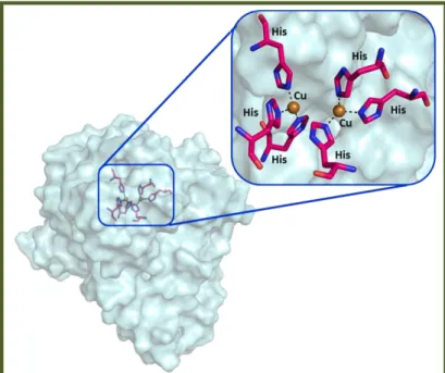
Tys are ubiquitous metalloenzymes exhibited across the most diversified life domains. They belong to the “type-3 copper” protein family as well as catechol oxidases, hemocyanins and laccases. In the active site of this protein family there is a conserved region characterized by six histidine residues coordinating two copper ions (CuA and CuB) located in a four helical bundle (figure 1).[1]
Figure 1: Tys active site. The six histidine residues are represented by pink stick and the two
copper ions by brown spheres. The picture was generated using PyMol.[2]
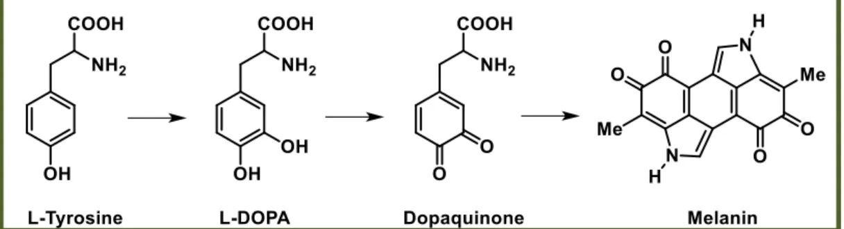
Tys catalyze the first two steps of the Raper-Mason pathway, the hydroxylation of L-tyrosine to L-DOPA and the subsequent oxidation of L-DOPA to L-dopaquinone. The first activity is called tyrosine hydroxylase (monophenolase activity) and the second one o-diphenol oxidase, catechol oxidase or DOPA oxidase (diphenolase activity). Once dopaquinone is formed by the oxidative action of these enzymes, the pathway progresses through a series of spontaneous reactions leading to the final melanin pigments (figure 2). In animal melanocytes, all steps after L-dopaquinone formation were also thought to proceed spontaneously to form melanin pigment, but around 1980, a number of growing pieces of evidence indicated a lack of correlation among Tys activity, melanin formation in animal skin and hair and blood levels of melanocortin, the animal hormone that controls the melanogenic capacity of
17
melanocytes. Thus, it was highlighted the possibility that the hormonal control of melanogenesis acted on the other proteins involved in mammalian melanogenesis. Soon, data strongly suggested the existence of a post-Ty regulation in this process beyond L-dopaquinone or L-dopachrome formation.[3]
Figure 2: Raper-Mason pathway for the formation of melanin pigments.
1.2 Structure of the enzyme
The structure of the enzyme can be divided in three parts: N-terminal domain, central domain and C-terminal domain (figure 3).
N-terminal domain. It is a transit peptide determining the final location of the enzyme. In plant, it directs the enzyme to the chloroplast; in human and mouse, it seems to be involved in melanosome transfer;[4] in mushrooms, since the Ty
enzyme is cytoplasmatic, it does not contain a transit peptide although in some cases it is associated with the cell wall;[5] in bacteria the N-terminal domain is
characterized by a TAT signal peptide responsible for proteins secretion.[1, 6]
Central domain. It is characterized by six conserved histidine residues, coordinating by the CuA and CuB oxidizing ions.[1] This copper pair is the site of
interaction of Ty with both molecular oxygen and its phenolic substrates. It is interesting also to note the presence of thioether bridge between one histidine residue of the active site and one cysteine residue observed in the central domain of Ty of N. crassa.[7] It seems that this bond can regulate the activity of the enzyme.
In mushroom and Aspergillus Tys, it is also possible the formation of a thiother bridge. This is not the case of known prokaryotic, plants and mammalian Tys.[8]
C-terminal domain. It is a transmembrane domain. In Agaricus bisporus and
Neurospora crassa, Ty is a soluble cytosolic enzyme and does not contain a
18
transmembrane domain, however the presence of a transmembrane helix was suggested in this domain;[9, 10] mammalian Tys are melanosomal membrane
proteins possessing a carboxyl tail oriented to the cytoplasm and a single membrane-spanning helix in the C-terminal portion of the proteins.[11]
Figure 3: General structure of TyS. The picture was generated using PyMol.[2]
Tys active sites, as well as, most type-3 copper proteins, are provided by an additional protein domain defined as “placeholder”.[8, 12] Usually, it is a bulky aromatic residue
such as Phe or Tyr but, also a Leu residue can be present therein. It is located above the active site and is oriented parallel to the second coordinating CuB histidine residue. It was supposed that its role is to control the enzymatic activity and to prevent an undesirable oxidation of phenolic compounds, since in most cases the enzymatic activity is possible only after the removal of placeholder. An additional position which covers the active site is located above the CuA, according to literature, acting as blocker residue that participates in substrate orientation.[1, 13] However, while in some
enzyme its role is extremely important, in others, it is neglectable.[1]
Tys from different sources possess both common and diverse features. The common features are related to the overall folding and the active site of the enzyme, in which the central copper-binding domain is conserved, containing strictly conserved amino acids residues.[14] The differences are relative to the signal sequence, carboxyl tail,
19
important for the formation of disulfide bridges.[15] Indeed, there are 17 cysteine
residues in humans and mouse, 11 in plants, 0 or 1 in prokaryotes. They seem to have an important role in the correct folding of protein and in the acquisition process of copper ions.[8]
1.3 Ty catalytic mechanism
Ty is characterized by four discrete oxidation states (deoxy, oxy, met and deact form) relying on the oxidation state of the two copper ions in the active site (figure 4).
Figure 4: The four discrete oxidation states of Ty. Picture modified from reference [16].
Native Ty is present mainly as met-Ty, in which a hydroxyl ion is bound to the two copper ions [Cu(II)]. Phenols bind to met-Ty but are not oxidized by this form of the enzyme unlike catechols. In catechols oxidation process, met-Ty is reduced to deoxy state in which both coppers are now in the Cu(I) oxidation state. Deoxy-Ty rapidly binds dioxygen to give oxy-Ty in which the two oxygen atoms are held between the copper ions in the active site. The first oxidizing form of the enzyme is oxy-Ty which
20
oxidizes phenols and catechols by a monooxygenase and oxidase mechanism, respectively (figure 5).
Figure 5: Oxidation process of phenols and catechols by a monooxygenase and oxidase
mechanism respectively. Picture modified from reference [16].
Therefore, both phenols and catechols are oxidized by oxy-Ty to ortho-quinones in presence of dioxygen. During the catecholic cycle, a catechol is occasionally treated as a phenol and it is oxidized by oxy-Ty through a monooxygenase mechanism leading to the irreversible formation of deact-Ty in which one of the copper atoms has been reduced to the Cu(0) state and may diffuse out of the active center. This minor pathway eventually leads to total inactivation of the enzyme by catechols.[1, 16]
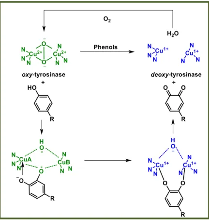
1.3.1 Ortho-quinones formation by oxidation of phenols: from oxy- to deoxy-Ty. The phenol with its oxygen atom binds to CuA leading to a complex in which the substrate is bound to both copper ions by the electrophilic monoxygenation of the ring. This complex forms the ortho-quinone and deoxy-Ty by homolytic dissociation. The
deoxy-Ty binding oxygen restores oxy-Ty and the phenol-oxidation cycle continues
21
Figure 6: Ortho-quinones formation by oxidation of phenols: from oxy- to deoxy-Ty.
Picture modified from reference [16].
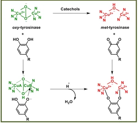
1.3.2 Ortho-quinones formation by oxidation of catechols: from oxy- to met-Ty. Catechols oxidation was proposed to proceed through CuB active site binding, unlike phenols that bind to CuA.[16] However, recent studies on Ty from Bacillus megaterium
showed that both tyrosine and L-DOPA substrates were similarly oriented toward CuA trough π-π interactions with the second coordinating CuB histidine residue.[1] The
oxidation cycle of catechols involves two steps characterized by deprotonation of the hydroxyl groups in which the oxygen atoms are coordinated with both copper atoms. In the first step, in which oxy-Ty is converted to met-Ty, the catechol/enzyme complex dissociates with the release of one of the oxygen atoms leading to the corresponding
ortho-quinone and water. The resultant met form of the enzyme retains the oxidation
state of the active site copper ions [Cu(II)] to which the remaining oxygen atom is coordinated, probably in a protonated form. In the second step of the process, another molecule of catechol reduces the active site copper ions to Cu(I) yielding deoxy-Ty and a second molecule of ortho-quinone. The oxidation states of the active site copper ions [Cu(II)] are then restored by binding dioxygen (figure 7).[16]
22
Figure 7: Ortho-quinones formation by oxidation of catechols: from oxy- to met-Ty.
Picture modified from reference [16].
1.3.3 Auto-activation and the lag period: from met- to deoxy-Ty.
The initial in vitro monoxidation of phenolic substrate is extremely slow and oxidation slowly accelerates to its maximum velocity during an initial induction period defined as “lag period”. In order to allow the binding of oxygen for monooxygenase activity, the copper atoms have to be in the Cu(I) state. The redox potential of copper favors a “resting state” of the active center atoms in the oxidized form Cu(II)2. The activation of
the monooxygenase function requires reduction of the active site copper atoms to Cu(I)2. There are four ways which can be followed:
. direct reduction by hydrogen peroxide;
. reduction by a reducing agent such as ascorbate; . redox exchange with other metals;
. reduction by a catecholic substrate.
23
Figure 8: Auto-activation and the lag period: from met- to deoxy-Ty.
Picture modified from reference [16].
The length of the “lag period” depends on several factors: the source of the enzyme; monophenol concentration (it is longer when the concentration of monophenol is growed); the concentration of the enzyme (it decreases with the growing of enzyme concentration but never totally disappearing) and after all, the presence of catalytic amount of o-diphenol or transition metal ions that completely abolish the lag period.[14]
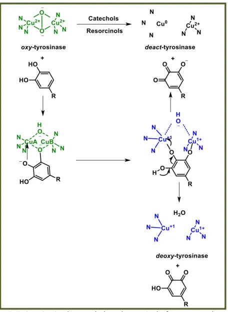
1.3.4 Ty inactivation by catechols and resorcinols: from oxy- to deact-Ty.
Ty inactivation mechanism was proposed to proceed in different ways but not satisfactory explanation was available until 2007 when Land and coworkers proposed that catecholic substrates may sometimes be processed as phenols and oxidized by the monooxygenase pathway.[16, 17] Normally, the monooxygenation of a catechol conducts
to the formation of an intermediate, which then gives 3-hydroxyquinone and deoxy-Ty. In the presence of an additional hydroxyl substituent, it supplies an alternative method of intermediate fragmentation. The reductive elimination of one of the copper atoms can lead to the deprotonation and quinone formation. This mechanism supports the idea that the inactivation process is related to the initial Ty concentration and, explains how the oxidative activity of Ty diminishes with the loss of half the active-site copper atoms, according to Dietler and Lerch.[18] (figure 9).
24
Figure 9: Ty inactivation by catechols and resorcinols: from oxy- to deact-Ty.
Picture modified from reference [16].
1.4 Sources of Tys
As above mentioned, Tys are widespread in nature. They are present in various prokaryotes, as well as, in fungi, mammals, arthropods and plants: in mammals they are responsible for the melanin formation in skin and hair color; in fruits and vegetables for browning, following by cell damage; in plants, sponges and many invertebrates are important for wound healing and primary immune responses; in arthropods they play a crucial role in sclerotization; in bacteria they protect DNA from UV damage.[1]
25
1.4.1 Tys in Mushrooms
Tys were isolated from different fungus. In particular, from Agaricus bisporus,[19]
Neurospora crassa,[20] Amanita muscaria,[21] Lentinula edodes,[22] Aspergillus oryzae,[23]
Portabella mushrooms, Pycnoporus sanguineus[24] and Lentinula boryana.[25, 26]
1.4.1.1 Ty from Agaricus bisporus
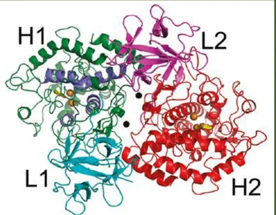
Ty from Agaricus bisporus (TyM) is characterized by a H2L2 tetrameric structure with
molecular weight of 120 kDa. In figure 10, the tetrameric structure of TyM co-crystalized with the natural inhibitor tropolone (PDB code 2Y9X) is reported.[27]
Figure 10: H2L2 tetrameric structure of TyM: H-1 (green), L-1 (cyan), H-2 (red) and L-2 (magenta). The holmium stabilizing ions and the copper ions are represented by black and
brown spheres respectively; tropolone inhibitor is reported like yellow stick. Picture modified from reference[27].
The L subunit (14 kDa) possesses a β-trefoil fold, consisting of 12 antiparallel β-strands assembled in a cylindrical barrel of six 2-stranded sheets. The biological role of this subunit is still unknown so, further research is needed. The H subunit (43 kDa) contains 13 α-helices, 8 mostly short β-strands and many loops. Its core structure is similar to the Bacillus megaterium Ty. It is larger if compared to the other type 3 copper proteins, possessing 100-120 more residues present in loops connecting the secondary structure elements of the core domain. The H subunit includes the Ty core region,[28] starting
with the conserved Arg20 until Tyr365, containing the binuclear copper site, in which each copper ion is coordinated by three histidine residues. This site is located at the
26
heart of two pairs of antiparallel α-helices, which make an angle of nearly 90°, at the bottom of a spacious cavity in the surface of the H subunit. The first copper ion, CuA, is coordinated from His61, His85 and His94; the second one, CuB from His259, His263 and His296. Four of these His residues (61, 94, 259, 263) make hydrogen bonds with a carbonyl oxygen atom of a peptide, thus restricting their side chain rotational freedom and may contribute to the affinity for the metal. Whereas, His85 makes a thioether bond with Cys83 fixing its side chain orientation. His296 does not possess direct interactions with other protein residues but, its side chain is held in position by a hydrogen bond with an internal water molecule (figure 11).[27]
Figure 11: Active site of TyM (PDB code 2Y9X). CuA and CuB copper ions are represented by
brown spheres and the main aminoacids of the pocket as green stick. The covalent thioether bond between Cys83 and His85 is shown as yellow stick.
The picture was generated using PyMol.[2]
Some Phe residues are important such as Phe90, which is positioned between His94, His259 and His296 while, Phe292 is placed between His61, His263 and His296, restricting the histidine side-chain conformations to maintain the integrity of the copper binding site.[29] The distance between CuA and CuB is 4.5 ± 0.2 Å, adopting a
planar trigonal geometry. A water molecule or hydroxyl bridge binds the two copper ions, completing the four-coordinate trigonal pyramidal coordination sphere for both copper ions. Other important residues present in the active site are His244, Glu256, Asn260 and Ala 286.[27]
27
1.4.2 Tys in Bacteria
Tys have been reported in several species such as Streptomyces,[30] Rhizobium,
Symbiobacterium thermophilum, Pseudomonas maltophilia, Sinorhizobium meliloti, Marinomonas mediterranea, Thermomicrobium roseum, Bacillus thuringiensis, Pseudomonas putida,[31] Streptomyces castaneoglobisporus, Ralstonia solanacearum,
Verrucomicrobium spinosum[32] and Bacillus megaterium.[26, 33]
1.4.2.1 Ty from Bacillus megaterium
Ty from Bacillus megaterium (TyBm) is a homodimer consisting of two ellipsoid asymmetric monomers. The main secondary structure elements are α-helices, with the two copper ions placed at the core of a four-helical bundle. At the interface of the dimer are present some interaction between residues Trp41-Tyr267 and Arg37-Asn270. In particular, a hydrophobic center is formed by the interaction between Trp41-Tyr267 and Trp269-Phe48, including van der Waals and π-π interactions (figure 12).[33]
Figure 12: Homodimeric structure of TyBm. The stabilizing residues interactions are
represented by black stick; copper ions like brown spheres and the 4 α-helices as a cartoon. Picture modified from reference [33].
The active site of TyBm is relatively exposed and no “placeholder” was identified. However, the Val208 residue, situated on a loop adjacent to CuA, was proposed as a modified gatekeeper controlling the entrance to the active site of TyBm. Movement of Val208 could direct the substrate correctly into the active site. Another important residue seems to be Arg209, positioning in proximity to the entrance of the active site, adjacent to His208. The movement in the position of Arg209 can cause pKa changes in
28
the area, which could modulate the catalytic activity in the presence of a substrate. Each copper ion is coordinated by three histidine residues: CuA by His42, His69 and His60; CuB by His204, His208 and His231 (figure 13).[33]
Figure 13: Active site of TyBm (PDB code 3NM8). The His residues are represented as pink stick
and copper ions as brown spheres. The picture was generated using PyMol.[2]
1.4.3 Tys in Plants, Vegetables and Fruits
In fruits and vegetables Tys have been extracted from Monastrell grape, apple,[34]
sunflower seed,[35] and Solanum melongena.[36] In plants, Tys are localized in the
chloroplasts of healthy plant tissues, whereas its substrates are contained in the vacuole. Portulaca grandiflora (Portulacaceae) is a potent source of Tys.[37] It generally
causes undesired enzymatic browning of farm products which subsequently leads to a significant decrease in the nutritional and market values. Since in plants the enzyme cannot catalyze the hydroxylation of monophenols, it only shows the diphenolase activity and, for this reason, it is called catechol oxidase (instead of Ty).[1]
1.4.4 Tys in Mammals
In mammals the Tys proteins family is composed of three members: the authentic Ty and two related proteins (TYRPs) named TYRP1 and TYRP2, respectively. All the three members of this family are metal-containing glycoproteins with a single transmembrane α-helix. They possess a common architecture of the tridimensional structure and are anchored to the melanosomal membrane through a C-terminal fragment sharing approximately 40% aminoacid identity and 70% similarity (figure 14).[38]
29
Figure 14: Alignment of human Ty and Ty-related proteins: amino acidic sequences.
Picture modified from reference [38].
The Ty and TYRPs structures are composed by four conserved regions: N-terminal signal peptide, a large intra-melanosomal domain, a single transmembrane α-helix and a small, flexible C-terminal cytoplasmic domain. The intra-melanosomal domain contains a cysteine (Cys)-rich subdomain and a catalytic tyrosinase-like subdomain with two metal ion-binding sites. The Cys-rich domain is only found in mammalian Ty and TYRPs and is composed by an “EGF-like” region (epidermal growth factor) and another Cys-rich fragment at the central part of the sequence, between the two metal binding sites. Its core structure is formed by two pairs of short antiparallel β-strands from which a long loops emerge. It interacts with the tyrosinase-like subdomain via its
30 N-terminus and a long loop emerging from the EGF-like core and it is located far from
the active site, at the opposite side of the molecule, suggesting that it unlikely affects the catalytic activity of TYRP1. The bulk of the protein is a globular intramelanosomal domain followed by a single transmembrane fragment and a small C-terminal tail oriented to the cytosol of melanocytes. The intramelanosomal domains of the three proteins are similar in length and contain a binuclear metal ion-binding motif with six conserved His residues. However, the C-terminal tails show very low homology and they were used to generate specific antibodies, PEP1, PEP7 and PEP8 for TYRP1, Ty and TYRP2, respectively. The specific reactivity of these antibodies against each C-tail allowed the differentiation, characterization and quantitation of the three proteins. The human melanogenic enzymes contain six or seven putative N-glycosylation sites, which are important for the proteins maturation. Ty contains seven N-glycosylation motifs (at Asn 86, 111, 161, 230, 290, 337, 371). TYRP1 has six sites (at Asn 96, 104, 181, 304, 350, 385), all glycosylated with various lengths of carbohydrate chains in the crystal structure. TYRP2 also contains six N-glycosylation motifs (at Asn 170, 178, 237, 300, 342, 377), four equivalent to the TYRP1 sites.[3, 38]
1.4.4.1 Human Ty
Unfortunately, no crystal structure of human Ty (TyH) is available and its precise catalytic mechanism is still under debate.[39] Nevertheless, a good model of TyH was
generated on the basis of TYRP1 crystal structure co-crystallized with tyrosine substrate.[40] Assuming that tyrosine binds equally to TyH and TYRP1, the model
suggests that the p-hydroxyl group interacts with the water molecule present between the two copper ions and with a serine residue (Ser394 in TYRP1 and Ser380 in TyH). Moreover, this serine residue seems to have an important role in substrate activation (figure 15).[38]
31
Figure 15: TyH model generated on the basis of TYRP1 crystal structure.
Picture modified from reference [38].
1.4.4.2 TYRP1
TYRP1 is a redox enzyme although it is unclear the reaction that it catalyzes. However, the most accepted function attributed to TYRP1 was the DHICA (5,6-dihydroxyindole-2-carboxylic acid) oxidase activity needed after TYRP2 action in the distal phase of melanogenesis.[41] It has a globular compact shape with strict interactions between the
Cys-rich and Ty subdomains. The Ty-like subdomain possesses a four-helix bundle connected by long loops; it is similar to TyBm sharing 32% sequence identity. Two disulfide bonds stabilize the Ty subdomain. The core of Cys-rich subdomain has an epidermal growth factor (EGF)-like fold similar to the structure of the human epidermal growth factor, formed by two pairs of short antiparallel β-strands from which long loops emerge. They are stabilized by disulfide bonds. The subdomain associated with Ty-like subdomain is located far from the active site, at the opposite side of the molecule, making a direct effect on the activity of TYRP1. The active site contains two Zn ions instead of Cu (figure 16).[40]
32
Figure 16: TYRP1 crystal structure (PDB code 5M8L). The four chains are represented as
cartoon. The picture was generated using PyMol.[2]
Interestingly, TYRP1 binds typical Ty substrates and inhibitors (tyrosine, mimosine, kojic acid and tropolone). The binding of these compounds occurs via aromatic stacking interactions with His381, ligation of their keto- and hydroxyl groups to the zinc ions and hydrogen-bonding interactions with Ser394.[3, 38, 40]
1.4.4.3 TYRP2
TYRP2 contains two zinc ions in the active site. The TYRP2 gene was attributed to the enzyme dopachrome tautomerase which catalyzes the conversion of dopachrome to DHICA.[42-44] Tautomerization is a type of isomerization rearranging dopachrome to
DHICA, rather than to DHI (5,6-dihydroxyindole) which is generated spontaneously by decarboxylation.[3] Dopachrome with its two hydroxyl groups was proposed to bind the
zinc ions in a bidentate mode, each displacing a water molecule (figure 17).[40, 45] DHI is
less stable and more toxic than DHICA:[46] in the eumelanin biosynthetic pathway it has
an important role as regulatory control, preventing cytotoxicity and premature cell death.[47] The cytotoxicity of DHI has been exploited as a targeting concept in
33
Figure 17: Homology model of intramelanosomal domain of TYRP2 based on crystal structure
of TYRP1. Picture modified from reference [38].
1.5 Melanin
Ty is the key enzyme of melanin biosynthesis, performing a crucial role in skin, hair, eyes pigmentation and in skin protection from UV radiations.[48]
Melanogenesis can be regulated at three different levels:
1. Gene: Melanocytes migrate to the epidermis and hair follicles during embryo development.
2. Cellular: Melanosomes regulate melanogenesis according to their size, number and densities.
3. Subcellular: Gene expression defined by Ty, TYRP1, and TYRP2 enzymes controls melanogenesis at subcellular level.[49]
Despite the complexity of melanin biosynthetic pathway, the only limiting step is the conversion of L-tyrosine to L-DOPA since, in presence of oxygen, the subsequent reactions occur spontaneously at physiological pH value.[50] Melanocytes can produce
different kind of melanin (figure 18):
Eumelanin: It is a black or brown pigment and it is produced by the intramolecular cyclization of L-dopaquinone amine group, leading to the formation of the indole leukodopachrome (cicloDOPA). The redox reaction between leukodopachrome and L-dopaquinone leads to dopachrome and L-DOPA. Dopachrome decomposes gradually to give DHI or, can undergo enzymatic transformation by
dopacromo-34
tautomerase, thus forming DHICA. The dihydroxyindole DHI and DHICA are subsequently oxidized to eumelanin.[51]
Feomelanin: It is a yellow or red pigment and it is produced from L-dopaquinone by addition of thiolic compound (generally cysteine or glutathione) producing 5-S-cysteinyl-dopa or glutationildopa. Subsequent oxidations give the benzothiazine intermediate 1,4-benzothiazinylalanine and then feomelanin (figure 18).[51]
Allomelanin: It is a dark pigment and it is the less studied and most heterogeneous class of melanin pigments. It derives from phenolic monomers unlike tyrosine and does not contain dopaquinone-derived motifs in its structure based on other chinoid building blocks. Generally, it does not present nitrogen atoms and may have different characteristics depending on the organisms that we take into account. It can originate from catechols (especially in plants), 4-hydroxyphenylacetate (in some bacteria) or dihydroxynaphthalene (especially in microorganisms). In rare cases, allomelanin contains units of meta-diphenol unlike the usual ortho and para positions occupied in eumelanine.[14]
35
In addition, another type of melanin is the neuromelanin, a dark pigment present in the brain in the substantia nigra-pars compacta, the locus ceruleus, the dorsal motor nucleus of the vagus nerve (cranial nerve X) and medial nuclei of the raphe. Ty constitutes also the key enzyme in the biosynthetic pathway of neuromelanin, thus it seems involved in Parkinson’s diseases.[52-54]
36
CHAPTER 2 TYROSINASE INHIBITION
Introduction
Although a significant number of TyIs were identified so far, developing new agents featuring drug-like properties is an urgently demanded task. Unfortunately, only few of the known Tyls could reach clinical applications as skin-whitening agents, due to safety concerns and weak whitening effects.[49, 55]
2.1 Different approaches for hyperpigmentation treatment
Suppression of melanin production by melanocytes represents an approach for the hyperpigmentation treatment. Ty is the enzyme that directly modulates the amount of melanin production. Thus, an interesting approach to treat these disorders is regulating the Ty activity through: the transcription of its mRNA, its maturation via asparagine-linked oligosaccharide processing, the modulation of its catalytic activity and/or its degradation.[56]
2.1.1 Inhibition of Ty mRNA transcription
One of the possible approaches that can be used to decrease Ty activity is the regulation of the transcription of its encoding gene. Decreases of Ty mRNA levels in cultured melanoma cells can be induced by incubation with the thymidine analogs such as: 5-bromodeoxyuridine,[57] tumor promoter
12-O-tetradecanoylphorbol-13-acetate,[58-60] transforming growth factor β1 (TGF-β1)[61] necrosis factor α (TNF-α).[62]
To reduce the production of melanin level it is possible also to use factors enabling the decrease of levels of mRNAs encoding Ty and/or microphthalmia associated transcription factor in cultured melanoma cells, melanocytes or melanoblasts.[63]
Examples of these factors are: hydrogen peroxide,[64] ceramide,[65] and
lysophosphatidic acid.[56, 66]
2.1.2 Aberrant Ty maturation
Human and murine Tys are glycoproteins with six highly conserved N-glycosylation sites. An abnormal glycosylation process in the endoplasmic reticulum (ER) or in the Golgi apparatus inhibits the correct protein folding and maturation, leading to hypopigmentation. Therefore, glycosylation inhibitors, such as glucosamine and
37
tunicamycin, inhibit the melanin synthesis in cultured melanoma cells with no apparent decrease in Ty levels.[67] Glutathione,[68] ferritin,[56] feldamycin[69] and calcium
D-pantetheine-S-sulfonate[70] showed the same effect. N-Butyldeoxinojirimicin, acting
as inhibitor of the ER-processing enzymes α-glucosidases I and II, blocks the Ty activity in B16 melanoma cells with little appreciable change in Ty level.[56, 71-73]
2.1.3 Increase of Ty degradation rate
The Ty synthesis and degradation are related processes influencing enzyme activity. When a TyI decreases the Ty level, acting also on its mRNA levels, it accelerates the degradation rate of Ty. Previously it was reported that the rate of enzymatic degradation increases as a consequence of the acidification of the melanoma cell culture medium, indicating that the degradation of the enzyme may depend on the environmental conditions surrounding the melanocytic cells.[74] Several intrinsic factors
in the epidermis and other factors can regulate Ty degradation, such as[56]:
TGF-β1: Keratinocytes synthesize and secrete numerous cytokines such as IL-1α, TNF-α[75] and TGF-β1.[76] Among them, TGF-β1 increases in a dose-dependent
manner Ty inhibition and TYRP1 activity in B16 melanoma cells following treatment with cycloheximide.[56, 61]
TNF-α: TNF-α is a cytokine present at the epidermal and dermal level, secreted during inflammatory processes or even in response to UV exposure. Although its role is not yet clear, recent studies suggested that this cytokine decreases the stability of Ty and TYRP1 reducing the levels of the corresponding mRNA.[56, 62]
Linoleic acid: It is an unsaturated fatty acid (C18:2) representing the major component of biological cell membrane. Its topical application causes a decrease in UV-induced skin hyperpigmentation. Linoleic acid increases the degradation of Ty with little change in mRNA Ty levels.[56, 60]
2,2’-Dihydroxy-5-5’-dipropyl-biphenyl: It is a phenolic compound decreasing melanin synthesis by degradation of Ty in cultured melanoma cells.[56, 77]
acetate/phospholipase D2: Tetradecanoylphorbol-13-acetate activates phospholipase D2 that is an enzyme which hydrolyzes phosphatidylcholine to generate phosphatidic acid. The over expression of phospholipase D2 decreases Ty levels in cultured melanoma cells.[56, 78]
38
25-Hydroxycholesterol: It is an oxysterol regulating cholesterol homeostasis which decreases melanin synthesis in mouse melanocytes enhancing Ty degradation via a proteasome-independent mechanism.[56, 79]
Phenylthiourea (PTU): PTU is a potent Ty inhibitor decreasing the stability of the enzyme in a similar way of linoleic acid.[56, 80]
2.1.4 Indirect regulation of Ty activity
Skin pigmentation depends on environmental factors external to melanocytes, apart from the intracellular regulation of Ty activity. Thus, melanin synthesis can be also modulated by the indirect regulation of Ty activity. An example is given by the inhibition of cell-to-cell communication between keratinocytes and melanocytes. This process, that normally activates melanogenesis through paracrine cytokines, such as endothelin-1, can be inhibited with: chamomilla extract;[81] inflammation inhibitors
such as glabridin;[82] trans-4-aminomethylcyclohexanecarboxylic acid, known as
tranexamic acid.[56, 83]
2.1.5 Inhibition of Ty catalytic activity
In the last twenty years, numerous TyIs have been identified, from both natural and synthetic sources. Often the definition of "tyrosinase inhibitor" can be too general and non specific, since it is sometimes used in reference to inhibitors of melanogenesis, whose action mainly resides in some interference in melanin formation, independently of any direct inhibition of the enzyme. The inhibition of Ty activity, observed experimentally, may be the result of different mechanisms of action. Thus, it is possible to classify the inhibitors in six different categories[14]:
1. Reducing agents: They reduce dopaquinone to its L-dopa precursor avoiding the subsequent formation of dopachrome and therefore of melanin. An inhibitor acting with this mechanism is, for example, ascorbic acid.
2. o-Dopaquinone scavengers: They are for example thio-containing compounds, which react with o-dopaquinone forming colorless products. As a result, the melanogenic process is slowed until the inhibitors are completely consumed. 3. Alternative enzyme substrates: Among them there are some phenolic compounds,
39
When these phenolic compounds show a good affinity towards the enzyme, the formation of the dopachrome is prevented and, for this reason, they are often erroneously classified as inhibitors.
4. Non-specific enzymatic inactivators: They non-specifically denature the enzyme inhibiting its activity. They are for examples bases or acids.
5. Specific Ty inactivators: E.g. suicidal inactivators (or mechanism-based inactivators). They act as substrates of Ty and form covalent bond with the enzyme causing an irreversible inhibition of activity.
6. Specific Ty inhibitors: They reversibly bind the enzyme reducing its catalytic activity.
Among these six different classes of compounds only the specific Ty inactivators (5) and the specific Ty inhibitors (6) are considered "true inhibitors", since they are able to bind the enzyme and inhibit its activity.[14]
2.2 Ty “true inhibitors”
The “true” TyIs are compounds able to bind the enzyme and inhibit its activity. Among them are reported derivatives from both natural and synthetic sources. Commonly, TyIs activity is assayed trough in vitro studies employing TyM commercially available and using kojic acid as reference compound (IC50 = 17.76 µM ).[84] Kojic acid
(5-hydroxy-2-(hydroxymethyl)-gamma-pyrone) is a fungal metabolite isolated from various species of Aspergillus niger and penicillum.[85] It acts as a good chelator for transition metal
ions and a good "scavenger" of free radicals. It is employed as a whitening cosmetic agent for the skin and as a food additive to prevent enzymatic browning, although its usage in cosmetic is limited due to its instability and side effects.[86]
Considering the mechanisms of action, TyIs can be classified into four classes (figure 19):
40
Competitive inhibitor: It is a substance that binds the active site of the enzyme in a manner that prevent substrate engaging. It might be a copper chelator, non-metabolizable analog or derivative of the true substrate.
Uncompetitive inhibitor: It is a compound that can bind only to the enzyme-substrate complex.
Non-competitive inhibitor: It is a substance that binds the enzyme not in the catalytic site but in a different place compared to the substrates.
Mixed inhibitor (competitive and uncompetitive): It is a compound that can bind not only the free enzyme but also the enzyme-substrate complex. For most mixed-type inhibitors, the equilibrium binding constants for the free enzyme and the enzyme-substrate complex, respectively, are different.[14]
Figure 19: Mechanisms of action of reversible inhibitors. E, S, I and P represent the enzyme,
substrate, inhibitor and product respectively; ES is the enzyme-substrate complex; EI e ESI are the inhibitor-enzyme complex and enzyme-substrate-inhibitor.
Picture modified from reference [14].
Another kind of inhibitors are the irreversible ones. They are also called specific inactivators, forming a reversible non-covalent complex with the enzyme (EI or ESI), that then reacts to produce the covalently modified “dead-end complex” Ei. The rate at
which Ei is formed is called the inactivation rate or kinact (figure 20). Irreversible
inhibitors display time-dependent inhibition, and their potency cannot be characterized by an IC50 value. This is because the amount of active enzyme in a given
concentration of irreversible inhibitor will be different depending on how long the inhibitor is pre-incubated with the enzyme. In contrast to the huge number of reversible inhibitors that have been identified, rarely irreversible inhibitors of Ty were found until now.[14]
41
Figure 20: Mechanism of action of irreversible inhibitors. E and Ei represent the enzyme and the inactivate enzyme respectively; S, I and P are the substrate, inhibitor and product; ES, EI
and ESI are the intermediates. Picture modified from reference [14].
Suicide substrates belong to the family of irreversible inhibitors. The mechanism of action of the suicide substrate has been extensively studied by Waley,[87] who
proposed a simple branched reaction pathway as reported in figure 21, in which an intermediate Y may give either an active enzyme and product, or an inactive enzyme. The intermediate Y has a choice of reaction, governed by the partition ratio r, where r = (k+3)/(k+4). The r value is referred as the molar proportion for inactivation, i.e., the number of molecules of inhibitors required to completely inactivate one molecule of the enzyme. It may be determined by plotting the fractional activity remaining against the ratio of the initial concentration of inhibitor to that of the enzyme. The intercept on the abscissa is 1 + r in the plot, when r > 1.[88] As in general irreversible inhibitors,
the inhibitory strength of a suicide substrate is also not determined by an IC50 value
but expressed by its r value, where a smaller r value of a suicide substrate means fewer inhibitor molecules are needed to inactivate all the enzyme activity and being more powerful inhibition.[14]
42
Figure 21: Mechanism of action of suicide substrates. E and Ei represent the enzyme and the inactivate enzyme respectively; P is the product; X is the first intermediate and Y is another
intermediate. Picture modified from reference [14].
The primary canon to take into account to evaluate an inhibitor, in addition to the mechanism of action, is its inhibitory strength. It is usually expressed as the inhibitory IC50 value, which is the concentration of an inhibitor needed to inhibit half of the
enzyme activity in the tested condition.[14] The K
i value represents the ligand-binding
affinity to the enzyme. The lower Ki value means higher binding affinity, whereas
higher Ki values means lower binding affinity. The Ki value for non-competitive
inhibitors is essentially the same numerical value as the IC50 of the inhibitors, whereas
for competitive inhibitors, the Ki is about one-half that of the numerical values of
IC50.[55]
2.2.1 Polyphenols
Polyphenols are a group of compounds containing multiple phenolic moieties. Flavonoids are the most numerous and best-studied polyphenols: they are present in the leaves, seeds, barks and flowers of plants and are responsible for the characteristic red and blue colors of berries, wines and certain vegetables. Flavonoids can be divided into seven groups: flavones, flavonols, flavanones, flavanols, isoflavonoids, chalcones and chatechin.[14] The general structure of flavonoids is reported in figure 22: they
differ in the conjugation of A and B rings and in the arrangement of the substituents, such as hydroxyl, methoxy, glycosides etc.[86]
43
Some flavonoids such as kaempferol,[89] quercetin[90] and morin[91] possess inhibitory
activity against Ty, while other such as catechin and rhamnetin act as substrates suppressing Ty activity like a cofactor[92] or acting as a free radical scavenger such as
rhamnetin.[86, 93]
2.2.1.1 Flavonols
Usually they are competitive inhibitors and their 3-hydroxy-4-keto moiety has a key role in copper chelation.[89, 94] Important flavonols are quercetin, myricetin,
kaempferol.[91, 95] A synthetic derivative of kaempferol, 6-hydroxykaempferol resulted
two times more active than kaempferol.[96] Although many flavonols have been
identified as TyIs, the most active of these, quercetin, showed only 20% of the inhibitory strength of kojic acid. Thus, they have little potential in applications of skin whitening or food antibrowning.
2.2.1.2 Flavones, flavanones and flavanols
Citrus species extracts contain a huge number of flavonoids such as nobiletin (5,6,7,8,3',4'-hexamethoxyflavone), naringin (5,7,4'-trihydroxyflavone) and the neohesperidin (5,7,3'-trihydroxy-4'-methoxyflavone), but they possess low inhibitory activity compared to kojic acid.[97, 98]
44
Also Morus species extracts contain flavonoids: norartocarpetin (5,7,2',4'-tetrahydroxyflavone), isolated from the steam barks, is 10.4-fold more active than kojic acid;[99] streppogenin (5,7,2',4'-tetrahydroxyflavanone) is a flavanone extracted from
the roots of the plant possessing a very similar chemical structure than norartocarpetin (flavone) and thus similar activity against TyM.[100]
Other two potent inhibitors are dihydromorin (5,7,2',4'-tetrahydroxyflavanol) and artocarpetin (5,2',4'-trihydroxy-7-methoxyflavone) isolated from Artocarpus heterophyllus wood.[101, 102]
45
Taxifolin (5,7,3',4'-tetrahydroxyflavanol) instead, was isolated from the sprout of
Polygonum hydropiper showing the same inhibitory activity of kojic acid.[103]
In addition to the monomers, a flavone-flavanone dimer was isolated from marine plants Garcinia subelliptica, being 3.6-fold more active than kojic acid.[104]
2.2.1.3 Isoflavonoids
A general structure of isoflavonoids is illustrated in figure 23. Chang et al.[14] reported
that the number and position of hydroxyl groups in the A ring can affect both the mode and inhibitory strength of different compounds. An isoflavone presenting hydroxyl groups in both C6 and C7 of A ring increases more than ten times both the inhibitory activity and the affinity in comparison with isoflavones that have not
46
hydroxyl groups in A ring or only on the C7. Switching hydroxyl groups on C7 and C8 the mode of action completely changes, passing from a reversible competitive to an irreversible suicide form.
Figure 23: Isoflavonoids general structure.
From the roots of Glycyrrhiza species different isoflavonoids with Ty inhibitory activity were isolated. Among them, glabridine, a non-competitive inhibitor, resulted 15-fold more active than kojic acid.[82] Glyasperin C was isolated from the same part of the
plant presenting an inhibitory activity 2-fold more than glabridine.[105]
Lee et al. reported three TyIs extracted from Lespedeza cyrtobotrya: haginin A (2',3'-dimethoxy-7,4'-dihydroxyisoflav-3-ene) is a non-competitive inhibitor 10-fold more active than kojic acid; dalbergioidin (5,7,2’,4’-tetrahyroxyisoflavan) is a non-competitive inhibitor; calycosin (4'-methoxy-7,4'-dihydroxyisoflavone) possesses inhibitory activity comparable to that of kojic acid showing two different mechanisms of action: inhibition of the enzyme activity and reduction of its expression.[106, 107]
47
2.2.1.4 Chalcones
They are characterized by two aromatic rings in trans configuration separated by three carbon atoms, two of them linked through a double bond and the third is a carbonyl group (figure 24).
Figure 24: General structure of chalcones.
From the roots of various species of Glycyrrhiza were isolated three chalcones derivatives including: licuraside, isoliquiritin and licochalcone A, the last one resulted 5.4-fold more active than kojic acid.[108]
48
Another potent inhibitor isolated from Sophora flavescens is kuraridin, which is 34-fold more active than kojic acid.[108]
Recently, 2,4,2',4'-tetrahydroxy-3-(3-methyl-2-butenyl)-chalcone (TMBC) extracted from the stems of Morus nigra, has proved to be a potent inhibitor being 26-fold more potent than kojic acid.[109] The 4-resorcinol moiety (2,4-dihydroxyl group) in the
aromatic ring detains a crucial role in chalcones inhibitory activity.[110, 111] Moreover,
the simultaneous presence of a lipophilic moiety can contribute to increase the activity.[14]
49
Some natural chalcones isolated from Morus australis resulted good TyIs. In particular, compound 1 resulted more potent than arbutin (IC50= 164 µM) with an IC50 value of
0.21 µM.[49, 112]
Radhakrishnan et al. reported some azachalcones as TyIs. Compounds 2 and 3 resulted more potent than kojic acid with an IC50 value of 1.70 and 2.30 µM respectively,
revealing that the presence of pyridine ring was important for the activity.[113] They
also identified some chalcones with oxime functionality such as compounds 4 and 5 with an IC50 value of 4.77 and 7.89 µM, respectively.[49, 114]

![Figure 3: General structure of TyS. The picture was generated using PyMol. [2]](https://thumb-eu.123doks.com/thumbv2/123dokorg/4565513.38022/18.892.281.700.269.615/figure-general-structure-tys-picture-generated-using-pymol.webp)
![Figure 4: The four discrete oxidation states of Ty. Picture modified from reference [16]](https://thumb-eu.123doks.com/thumbv2/123dokorg/4565513.38022/19.892.240.738.399.872/figure-discrete-oxidation-states-ty-picture-modified-reference.webp)