*
CHAPTER 5
Sources and procurement
of HSC
H.E. Johnsen, F. Lanza, S. Fruehauf, J. Petriz, T. Carr, J. Walewski, A. Wahlin, H. Schrezenmeier, L.M. Knudsen
1. Introduction
Mobilised PBSC have become the main source for auto- or allo-HSCT following mye-loablative therapy in patients with lympho-haematopoietic malignancies or solid tumours. Classical strategies for PBSC mobilisation include administration of growth factors, mainly G-CSF alone or in combination with other cytokines or marrow suppressive CT. HSC mobilisation and collection have been optimised in numerous clinical trials (1), but a significant proportion of patients mobilise an insufficient number of HSC, resulting in an inadequate graft.
In recent years, the biology of haematopoiesis has been elucidated, leading to the development of new experimental mobilisation strategies including administration of new growth factors, chemokine ligands, adherence molecules or chemotherapy (2). Improved understanding of these new developments may enable sufficient PBSC to be obtained for transplantation even in patients with impaired haematopoiesis, thus enabling them to receive potentially curative high dose treatment.
This chapter will focus on what the EBMT Committee on Graft Evaluation consider as the “Gold Standard” for priming regimens, mobilisation, harvest and transplantation of autologous HSC, including a proposal for assessment of graft quality and finally strategies for handling of “poor mobilisation”.
2. Definition and identification of HSC
It is a widely accepted assumption that there are adult SC located in various organs of the body, dedicated to the replenishment of specific tissues such as blood. The results of HSCT have proven that stem cells exist in the haematopoietic system and have stimulated methods for identification and isolation of stem and progenitor cells. This has created a new discipline in medicine: “Stem Cell Research”, which focuses on stem cell research and development including cell substitution in the management of diseases. Stem cell laboratories have a great need for protocols and procedures describing the exact handling of techniques used for identification, isolation and characterisation of haematopoietic stem cells. The actual status of practical standardised techniques has recently been published in a comprehensive form by a series of active SC researchers (2).
Before such standards are applied to clinical practice one has to recognise the uncer-tainty that exists in the identification of HSC. This was pointed out by Potten and Loeffler (3), who realised that the characteristics of a stem cell relate to its future potential and can only be studied effectively by allowing the cell to express that potential. Therefore, to characterise a cell as a SC, allowing it to differentiate, the original cell is lost; and at the same time one may only see a limited range of responses. They continued by pointing out the analogy with Heisenberg’s Uncertainty
80
THE EBMT HANDBOOK2004 REVISED EDITION
Principle. Originally formulated in the theory of quantum physics, it says that “the very act of measuring the properties of a certain body inevitably alters the characteristics of that body, thus giving rise to uncertainty in the evaluation of its properties”. Stem cell assays all observe the response after a perturbation to the system. So far no scientist has identified the HSC and it is still an open question how it should be defined. The answer is awaited and in the meantime we have to accept well studied and described standard protocols for identification and enumeration by differentiation markers (4) as surrogate markers for estimating SC potential.
3. Priming and mobilisation/engraftment and recovery
Many studies have documented faster engraftment after transplantation with PBSC compared to BM SC. Despite the success of PBSCT, the exact mechanisms involved in PBSC mobilisation and homing are not completely understood (1, 5). In adults, small quantities of PBSC are present in the PB during steady state haematopoiesis, suggesting a continuous migration and exchange of HSC between the BM and other organs. Most studies investigating mechanisms of mobilisation of progenitors and SC have focused on changes in the properties of these cells; little work has been done to investigate the potential contributions of cell traffic and marrow stroma to this phenomenon. Damage to the stroma from the disease or previous exposure to CT/RT may, in part, explain the quantitatively poor mobilisation frequently seen in some patients and understanding the mechanism of mobilisation may contribute to the development of more predictable and efficacious mobilisation protocols. However, engraftment failure or prolonged time to blood cell recovery following reinfusion may also be a consequence of impaired trafficking or stromal homing function and consequently mirror mobilisation capacity. In other words, the SC quality is not solely a quantitative matter but needs to be evaluated by taking other aspects into consideration including demographic, disease and therapy related variables (vide
infra)(see Table 3.1.). Future research should be focused on the immediate molecular and biochemical events (priming) leading to release and migration (mobilisation) as well as homing (engraftment) and expansion by differentiation (blood cell recovery), which ultimately may allow us to release and home progenitor cells more effectively.
4. Priming and harvest strategies: “The gold standard”
Mobilisation of a sufficient number of HSC into the PB to ensure a rapid and sustained engraftment can be achieved by administration of growth factors such as rhG-CSF alone or by administration of myelosuppressive CT such as high-dose Cy (1–4 g/m2) (see Table 4.1.). The combination of myelosuppressive CT and myeloid growth factor mediated expansion has been shown to be synergistic. It is, however, still uncertain which regimen is optimal regarding the maximum yield of progenitor cells and tumour cell contamination, but data support the experience that patients who fail to mobilise SC with growth factors alone may benefit from CT plus growth factor administration.
In daily practice the priming regimen is often one of the final CT cycles administrated as induction or salvage. Ideally, however, the tumour specific therapeutic approach and the priming strategy should be considered as a two step process: The first aim
CHAPTER 5 •Sources and procurement of HSC
Table 3.1: Variables (risk factors) for harvest and engraftment of PBSC Harvest outcome (CD34+ cells)
Age Sex
Disease type (diagnosis) Disease stage
Haematopoiesis impaired (y/n) Therapy response (y/n) Type of therapy
Priming regimen w. CT (y/n) Blood CD34+ cell level Apheresis strategy
Engraftment time (Tree lineage recovery)
Age Sex
Disease type (diagnosis) Disease stage
Growth factor levels Graft CD34+ cell number
CD34 subsets (growth factor, chemokine receptors and adherence molecules)
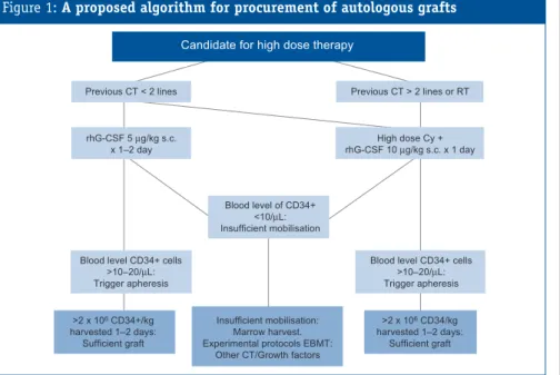
Day 0:
Growth factor administration 1–2 times per day s.c. or
*Cy dose 1–4 g/m2+ rhG-CSF 1 time per day s.c.
Day 3 or *10 / 4 or *11:
Measure blood CD34 level and proceed to apheresis when CD34+ cells > 10–20/µL
Day 4 or *11–7 or *14:
Harvest a minimum of CD34+ cells/kg pt weight depending on centre experience Table 4.1: The “Gold Standard” priming procedure and timing of PBSC harvest
being to obtain optimal disease control before growth factor (or chemotherapy-based) priming and then focusing on SC harvest and graft quality. The reason for such a strategy is to ensure an optimal disease response before high dose therapy, known by several studies to be one of the major prognostic variables.
Handling of the procedure needs close cooperation between the “Leukapheresis Unit” and “The Stem Cell Laboratory” to fulfil the regulations given in the directive of the European parliament and of the Council in 2003 (6) setting standards of quality and safety for the donation, procurement, testing, processing, storage, and distribution of human tissues and cells – including HSC. On the professional rather than the regulatory level, the JACIE recommendations on “Standards for haematopoietic progenitor cell collection, processing & transplantation from the joint accreditation committee of ISCT-Europe and EBMT” (7) are designed to provide minimum guidelines for facilities and individuals performing HSCT and therapy or providing support services for such procedures. These Standards have no intention to include all procedures and practices that a facility or individual should implement, if the standard of practice in the community or governmental laws or regulations establish additional requirements (7). Each facility and individual should analyse their practices and procedures to determine whether additional standards apply, including standard operating procedures (SOP) for stem cell enumeration, apheresis, freezing, thawing, registration and transplantation as well as the outcome of the procedures.
One major problem is the occurrence of poor mobilisers defined as patients who do not obtain a CD34+ cell level sufficient to proceed to harvest before Day 14 of priming. The algorithm given in Figure 1is a proposal for a common strategy in Europe, which will allow EBMT members to collaborate in clinical trials focussed on “poor mobilisation”.
5. Standard for CD34+ enumeration
The success of SC laboratories during the last decade is documented by the elimination of the risk of engraftment failure - not by the introduction of PBSC grafting, but very likely due to improved quality assessment procedures. This has been achieved by hard work on a very simple technology (Figures 2 and 3) allowing us to identify patients at risk of engraftment failure, due to either lack of mobilisation or obtaining an insufficient harvest (8).
The current situation regarding CD34+ cell enumeration has been analysed and published as an European Survey on flow cytometry determination of CD34-expressing cells (8). The existing problems in quality assessment were discussed during the first committee session held at the annual 1999 EBMT meeting. One of the major 82
concerns was paragraph D4.000 on quality management in the 1999 JACIE Standards document stating: “D4.130: A nucleated cell count shall be performed for any component after collection and after any subsequent processing (if applicable). D4.131: CD34+ cell count shall be performed. D4.132: The target should be to transfuse a minimum of 2 x 10⁶CD34+ cells per kg body weight, but lower numbers
may be acceptable in specific cases. (This does not apply to BM or CB)”.
The 1999 subcommittee meeting concluded that the numbers given in paragraph D4.132 were not appropriate, as no convincing data exist from single- or multicentre studies to document either a common protocol for CD34+ cell enumeration or a strategy for clinical validation of numbers. This has now been accepted and the recent version of “Standards for HSC collection, processing & transplantation” (7) do not give exact numbers for a sufficient graft. This reflects the problem of preparing guidelines for quality assessments and is the background for a recent proposal of an optional European Reference Protocol on CD34+ cell enumeration by flow cytometry and a strategy for its validation by clinical end-points (8-10).
We do not yet know the minimum safe number of CD34+ cells needed for clinical engraftment of all lineages, as this may vary depending on the stem and progenitor cell subset composition in a given patient or autograft. However, we do know that
CHAPTER 5 •Sources and procurement of HSC
84
Figure 2: Standard for flow cytometry enumeration of CD34+ cells
“State of the art” single platform method to enumerate CD34+ progenitor cells from all sources of stem-progenitor cells currently used in either clinical or research laboratory settings (i.e., bone marrow, mobilised peripheral blood and cord blood). This protocol allows the determination of the absolute CD34+ cell count directly from the flowcytometer (so called “single platform analysis”). Briefly, a single platform approach facilitates the absolute count determination of leukocytes subsets directly from the cytometer, by either adding a known number of fluorescent microbeads to the sample or by calculating the number of cells in a known volume (“volumetric assay”). Assessment of the ratio between the number of fluorescent beads and CD34+ cells counted, together with a lyse-no-wash sample preparation regimen allows a precise and reliable enumeration of CD34+ cells. A more detailed description of the sequential Boolean gating strategy used for list mode data analysis has been published in Keeney et al (J Biol Regul Homeost Agent, 2003: 17, 247-253). Briefly, the multiparameter definition of haemopoietic stem/progenitor cells (HPC) is based on their light scatter properties, dim expression of CD45 and dim to bright expression of CD34, and utilizes fluorescent counting beads. A sample from a 2 hours old apheresis product is diluted 4x and stained with CD34 PE and CD45 FITC. After 20 min, the sample is lysed with 2 ml NH4Cl containing 1 µg 7-AAD. After 10 min, counting beads are added and the sample is analysed immediately thereafter. Plot 1: CD45+ events [region R1] (CD45-FITC vs. SS (side scatter); plot 2: HPC are selected as CD34+ and low side scatter (region R2). Plot 3 shows CD34+ cells in the CD45 vs. SS plot. In plot 4, HPC are further selected by selecting the cluster of CD45dimevents (region R3), and in plot 5, HPC are finally identified
by placing a light scatter region on lymphocytes (lymhs) and blasts (region R4). Plot 6 shows light scatter characteristics of lymphocytes selected in region R5 (plot 1) to allow optimal placement of the lymphocytes-blast region R4. The lower limit of CD45+ expression by the CD34+ events is verified by quadrant analysis in Plot 7. Plot 8 shows counting beads, selected by their bright fluorescence (region R7); region R7 is placed on single beads
a graft content of more than 5–10 x 106CD34+ cells per kg of body weight is safe,
resulting in fast recovery of ANC and platelets before day 14 and 21, respectively, in a major fraction of patients and, most important, only has a minor risk of engraftment failure. In a committee survey of 1600 patients from nine published papers (8), including a minimum of 50 patients each, it was concluded that the overall median time to ANC and platelet recovery is 11 days (range 2–93) and 11 days (range 0–1000+), respectively. From 15 studies with information about patients receiving low numbers of reinfused cells (seeTable 5.1), it is concluded that no definite lower level exists to document groups of patients at high risk for prolonged cytopenia and late recovery, based on CD34 numbers below 1 x 10⁶/kg, 2 x 10⁶/kg, 2.5 x 10⁶/kg,
3 x 10⁶/kg or 5 x 10⁶/kg (8). From such data, it is obvious that we will never obtain
a cut-off number of CD34+ cells delineating an “insufficient” or “safe” graft. We need CHAPTER 5 •Sources and procurement of HSC
Figure 3: Standard for flow cytometry enumeration of CD34+ cells
Standard method to exclude “non-viable” CD34+ progenitor cells. The addition of the (not a viability dye) dye 7-AAD (7-aminoactinomycin D) to the single platform method permits the determination of the absolute numbers of viable and non-viable CD34+ cell numbers from all samples examined. This method is recommended for the analysis of old (older than 6–8 hours) and manipulated sample preparations (purged, submitted to cell selection techniques, to ex vivo expansion or to gene therapies protocols, or potentially damaged by shipping to another site for analysis). It is also the standard method for the analysis of cryopreserved and thawed cell preparations, which consistently reflect those cells actually reinfused to the patient, and thus to be considered the most important variable determining graft success or failure
86
THE EBMT HANDBOOK2004 REVISED EDITION
to reconsider these terms and translate CD34 counts into probabilities of obtaining clinical efficacy, avoiding toxicity and retaining safety, evaluated by proper end-points (vide infra).
6. Handling of “poor mobilisation”
“Poor mobilisation”can be defined as procedures involving patients who do not obtain a CD34+ cell level (>10–20/µL) triggering a sufficient harvest before Day 14 of priming. Such patients, if primed with CT, should continue growth factor administration and CD34 enumeration until the blood platelet level has risen to >50–100 x109/L - before
secondary strategies should be considered. Many different secondary approaches exist, but none has been documented superior to others; such approaches include: Double dose rhG-CSF, high dose VP-16 administration or ultimately BM harvest. Unfortunately, the impact of new biological active molecules has not been evaluated in the "poor mobiliser" population. It is our hope that the collaborative effort of the EBMT committee on graft evaluation may give birth to well-designed and focused protocols, which include sufficient numbers of patients in a short time.
At present the best recommendation for poor mobilisers is to perform a BM harvest targeting a level of 1 x 10⁶CD34+ cells per kg of body weight during one or two
procedures. One major concern, however, is that such patients are at risk for partial graft failure or delayed blood cell recovery, which needs to be taken into consideration when the individual optimal supportive therapy is designed.
7. Clinical validation of SC protocols: Prospective registration studies
In general, new protocols, moving from the laboratory bench to the clinic, have to pass different stages before they are validated and can be implemented in routine practice. In parallel with the well-established guidelines for the development of therapeutic trials through clinical phases I–IV, there are four different phases believed to be necessary and informative for the clinical validation of a laboratory technique (e.g. CD34 enumeration).
Table 5.1: Blood cell recovery following HSCT of low number of CD34+ cells (8)
CD34+ cells ≤1 x 10⁶/kg ≤2 x 10⁶/kg ≤2.5 x 10⁶/kg # patients 37 159 116
Overall median days to ANC > 0.5 x 10⁹/L 15 (11–21) 15 (13–18) 13 (11–15)
Overall median days to platelet > 20 x 109/L 30 (22–43)
17 (14–20) 17 (14–21)
In the first phase, a technique such as CD34 enumeration is established in the laboratory and analysed for specificity, sensitivity, reproducibility and accuracy. The second phase documents the likely clinical impact by single centres analysing retrospective material/data. The third phase involves a single centre in prospective evaluation; evolving into the most important phase, phase IV, which is a multicentre prospective evaluation based upon important clinical end-points. Ideally, phases II–III should document the usefulness of the technique in question, convincing one or more centres to participate in a phase IV validation trial, which however is only rarely performed.
Previously, the impact of graft related factors together with disease and therapy related variables have been described by critical end points such as delayed engraftment, graft failure, regimen related death etc. which, however, are rare observations in actual clinical practice. To day, haematological toxicity is considered one of the secondary end points important for graft evaluation - which in practice focuses on the primary impact of health economic end points. A recent proposed strategy for analysing such pretransplant variables has defined clinically relevant primary, secondary and tertiary objectives by clinically relevant end points such as supportive transfusions, antibiotic administration, time in hospital, organ toxicities and safety, related both to safe delivery of conditioning and to the risk of tumour cell reinfusion.
Introduction of such end points, graded in binary fashion as an acceptable or unacceptable post-transplant outcome has allowed us to estimate prognostic models and illustrate individual quality assessment based on probability evaluation. The protocol established in a co-operative group of subcommittee members from EBMT is breaking new waves in quality assessment of autografting by help of demographic, disease and graft related variables. Such prognostic models will ultimately allow us to calculate an individual patient specific predictor of outcome, which will have a major impact on clinical practice.
Recently, the regulatory authorities have begun to show interest in haematopoietic SC products and transplantation. On a professional rather than a regulatory basis the objectives must be to promote quality of medical and laboratory practice to ensure that appropriate standards of work and product quality are established and maintained as proposed in the following.
8. Quality assessment of HSC autografting by probability estimation Today haematological toxicity is considered as one of the secondary end points important for graft evaluation - which in practice focuses more on the primary impact of health economic end points, including supportive care. Clinical end points for
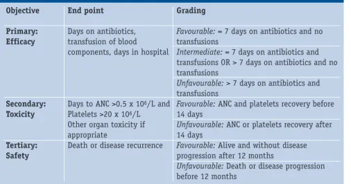
such evaluation have recently been proposed (9) (see Table 8.1). In accordance with clinical evaluation of other therapies in medicine (e.g. CT) the primary objective is to analyse efficacy, which in context of post-transplant supportive care may be defined by e.g. days on antibiotics and transfusions of blood products. Secondary objectives may be to analyse toxicity, defined by e.g. associated organ toxicity including time to blood cell recovery. Tertiary objectives may be to analyse safety defined by risk for early relapse or death.
By such analysis it is possible to identify prognostic variables defined by clinical end points and illustrate that individual pretransplant probability evaluation, including CD34 enumeration and targeting may be an important intervention step in quality assessment of autografting (Figure 4).
88
THE EBMT HANDBOOK2004 REVISED EDITION
0.26 0.24 0.22 0.20 0.18 0.16 0.14 0.12 0.10 0.08 0.06 Probability of favo u rable Males - Females CD34/days -2 0 2 4 6 8 10 12 14 16
Figure 4: The estimated probability for primary outcome depending on a varying CD34+ cell number
Estimate for an identified female (blue)/male (grey) patient who was candidate for high dose therapy, suffering from stage III Multiple Myeloma, and means values for all other identified significant variables (Hgb, beta-2 microglobulin, S-Creatinine, length of priming, CD34+ cell number harvested per day) and in partial remission (PR) (unpublished). The estimated model for a favourable outcome has been given by the following equation Logit (Favourable) = -2.497+0.953+0.002*S-Creatinine+0.021*Haemoglobin-0.22*Length of priming + 0.085*CD34/days. At mean values of S-Creatinine, Haemoglobin and Length of priming the function is reduced to Logit (Favourable) = (-1.48 +0.085*CD34/days) used to illustrate that the probability of a favourable outcome increases with the average CD34+ cell number
References
1. Fruehauf S, Seggewiss R. It's moving day: Factors affecting peripheral blood stem mobiliza-tion and strategies for improvement. Br J Haematol 2003; 122: 360-375. 2. Klug CA, Jordan CT, Editors. Hematopoietic Stem Cell Protocols. 2002.
3. Potten CS, Editor. Stem Cells. 1997.
4. Flow cytometric methods. J Biol Regul Homeost Agents 2003; 17: 211-280.
5. Lapidot T, Petit I. Current understanding of stem cell mobilization: the roles of chemokines, proteolytic enzymes, adhesion molecules, cytokines, and stromal cells. Exp Hematol 2002; 30: 973-981.
6. EU Directive. Directive of the european parliament and of the council on setting standards of quality and safety for the donation, procurement, testing, processing, storage, and distribution of human tissues and cells. 2002/0128(COD). EU 2003.
7. The Joint Accreditation Committee of ISCT-Europe and EBMT. Standards for hematopoietic progenitor cell collection, processing & transplantation. Second Edition - Europe June 2003.
8. Serke S, Johnsen HE. A European reference protocol for quality assessment and clinical validation of autologous haematopoietic blood progenitor and stem cell grafts. Bone Marrow Transplant 2001; 27: 463-470.
9. Johnsen HE, Lanza F. Quality assessment of autologous hematopoietic blood progenitor and stem cell grafting: A prospective registration study. J Biol Regul Homeost Agents 2002; 16: 272-288.
10. Homepage for the European Group for Blood and Marrow Transplantation. http://www.ebmt.org. 2003.
CHAPTER 5 •Sources and procurement of HSC
Table 8.1: Proposed graded clinical end points in quality assessment Objective Primary: Efficacy Secondary: Toxicity Tertiary: Safety End point Days on antibiotics, transfusion of blood components, days in hospital
Days to ANC >0.5 x 10⁶/L and
Platelets >20 x 106/L
Other organ toxicity if appropriate
Death or disease recurrence
Grading
Favourable: = 7 days on antibiotics and no
transfusions
Intermediate: = 7 days on antibiotics and
transfusions OR > 7 days on antibiotics and no transfusions
Unfavourable: > 7 days on antibiotics and
transfusions
Favourable: ANC and platelets recovery before
14 days
Unfavourable: ANC or platelets recovery after
14 days
Favourable: Alive and without disease
progression after 12 months
Unfavourable: Death or disease progression