29
Journal of Cardiovascular Echography / Jan-Mar 2015 / Vol 25 | Issue 1 CASE REPORT
In December 2013, a 45-year-old Caucasian male with no history of cardiovascular disease, heavy-smoker, was referred to the Cardiology Unit at University Hospital of Messina (Messina, Italy) for recurrent supraventricular tachycardia of undetermined origin. There were no significant cardiac anomalies in previously performed electrocardiogram (ECG) and transthoracic echocardiogram.
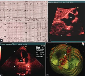
The patient complained with unjustified palpitation since 1 week, emphasized over the last 12 hours. Physical examination revealed arrhythmic heart beats (135 bpm) and 2/6 Levine systolic murmur with predominant tricuspid component. Blood pressure was 120/70 mmHg. Twelve-lead ECG showed chaotic atrial tachycardia [Figure 1a] with intermittent fibrillation, but no significant anomalies of the QRS complex and ST-T segment.
Access this article online Quick Response Code:
Website: www.jcecho.org DOI:
10.4103/2211-4122.158424
Address for correspondence
Prof. Cesare de Gregorio, Dipartimento di Medicina Clinica e Sperimentale, Azienda Ospedaliera Universitaria di Messina, Messina, Via Consolare Valeria, 98125 Messina, Italy. E-mail: [email protected]
ABSTRACT
We report the case of a 45-year-old Caucasian male with negative cardiovascular history, heavy-smoker, who was referred to our Cardiology Unit for recurrent inexplicable tachycardia. Chaotic atrial tachycardia with intermittent fibrillation was observed at ECG, whereas a smoothed mass (approximately sized 8 × 8 cm) was unexpectedly found at echocardiography likely infiltrating the right atrial wall. Multi-detector computed tomography confirmed the mediastinal mass and the digital post-processing clearly identified its anatomic characteristics and invasivity. This study demonstrates that recurrent and refractory atrial arrhythmias can be early signs of cardiac infiltrating mediastinal masses. The combined approach by echocardiography and computed tomography was confirmed to provide precise anatomical and functional characteristics of the arrhythmogenic disease in this patient.
Key Words: Arrhythmias, echocardiography, extracardiac masses, mediastinal masses, right atrium
Recurrent Supraventricular Arrhythmias as
the First Clinical Warning of a Right Atrium
Infiltrating Pulmonary Carcinoma
Cesare de Gregorio, Giampiero Speranza, Pietro Pugliatti, Ines Paola Monte
1,
Giuseppe Andò
Department of Clinical and Experimental Medicine, University Hospital of Messina, Messina, 1Department of Medical and Pediatric Sciences, University of Catania, Catania, Italy
Opportunely, the patient was already on warfarin by his generalist and the INR was 2.8. He was admitted to Cardiology Unit and given Amiodaron infusion. Sinus rhythm restored in 4-hour time and symptoms relieved. The day after, while leaving the hospital, he had a new paroxysm. Before deciding on treatment, a trans-thoracic echocardiogram was performed. Unexpectedly, a smoothed mass (approximately sized 8 × 8 cm) was disclosed next to (likely infiltrating) the free wall of the right atrium [Figure 1b and c]. The oncology team was then contacted and a multi-detector computed tomography of the chest was carried out. A mediastinal mass was confirmed (measured 9.8 × 8.5 cm) to infiltrate the inferior cava vein and the right atrial wall [Figure 1d]. Shortly, the patient underwent bronchoscopy which allowed to make diagnosis of primary bronchial squamous cell carcinoma, requiring urgent chemotherapy and possible subsequent surgery.
CASE REPORT
Journal of Cardiovascular Echography / Jan-Mar 2015 / Vol 25 | Issue 1
30
de Gregorio, et al.: Arhythmogenic extracardiac mass
DISCUSSION
This report deals with a rare, but possible, recurrence of supraventricular arrhythmias as the first clinical
presentation of pulmonary malignancy, as reported by previous studies.[1,2] Transthoracic echocardiography was
confirmed to be a powerful diagnostic tool for extracardiac masses, but the combined approach with multidetector computed tomography merely provided a realistic anatomic picture[3], also revealing the pathophysiology of refractory
atrial arrhythmias.
Routine ultrasound examination of the heart should be inclusive of extracardiac structures in high-risk patients with unexplained clinical features. Fast-track protocols during routine ultrasound examination are often empiric, whereas inherent algorithms should be established by either National or International Specialty Societies.[4]
REFERENCES
1. Korecki J, Kamiński KA, Lisowska A, Musiał WJ. Supraventricular tachycardia and pulmonary hypertension at the presentation of Hodgkin’s disease. Acta Cardiol 2005;60:655-7.
2. Sick P, Baldauf G, Schuler G. Mediastinal space-occupying lesion as a rare cause of atrial arrhythmia. Z Kardiol 2000;89:781-7.
3. Juanpere S, Cañete N, Ortuño P, Martínez S, Sanchez G, Bernado L. A diagnostic approach to the mediastinal masses. Insights Imaging 2013;4:29-52.
4. Alkhouli M, Sandhu P, Wiegers SE, Patil P, Panidis J, Pursnani A. Extracardiac findings on routine echocardiographic examinations. J Am Soc Echocardiogr 2014;27:540-6.
Figure 1: Panel a: ECG showing chaotic atrial arrhythmias with
alternance of either sinus rhythm, premature atrial beats and atrial fibrillation. Panel b: Subcostal echo view showing the contact edge of the pulmonary mass (arrows) with the right atrium. Panel c: Apical 4-chamber view showing a large extracardiac mass next to the right atrium (RA). Panel d: Digital post-processing of MDCT scan (OsiriX Open Medical Software, Release 3.9.4t, Foundation OsiriX, Genève, Switzerland) showing the mediastinal mass (arrows) infiltrating the pericardium, the right atrial wall and the inferior cava vein
d c
b a
How to cite this Article: Gregorio Cd, Speranza G, Pugliatti P, Monte IP,
Andò G. Recurrent supraventricular arrhythmias as the first clinical warning of a right atrium infiltrating pulmonary carcinoma. J Cardiovasc Echography 2015;25:29-30.
Source of Support: Nil, Conflict of Interest: None declared. [Downloaded free from http://www.jcecho.org on Monday, June 1, 2020, IP: 95.249.185.155]