Downloaded from www.microbiologyresearch.org by IP: 192.167.111.12
On: Wed, 27 Jan 2016 08:23:04
Case Report
A case of abscess after BCG vaccine
in an immunocompetent child without other
clinical signs
Francesco d’Aleo, Roberta Bonanno, Antonella Lo Presti Costantino,
Salvatore Arena, Angelina Midiri, Giuseppe Mancuso, Carmelo Biondo
and Concetta Beninati
Correspondence Concetta Beninati [email protected]
Department of Pediatric, Gynecological, Microbiological and Biomedical Sciences, University of Messina, Messina, Italy
Introduction: Bacille Calmette–Gue´rin (BCG), an attenuated strain ofMycobacterium bovis, is a rare cause of infection, with few published cases in immunocompetent individuals.
Case presentation: We present the case of a cutaneous abscess in an immunocompetent infant returning from Morocco, where he received a BCG vaccination. The abscess developed at the site of inoculation in the forearm (a non-recommended site) in the absence of
lymphadenopathy or systemic signs. The lesion did not recur after aspiration of the abscess and further treatment was not required.
Conclusion: Infections caused byM. bovis BCG may be difficult to diagnose without systemic signs or lymphadenopathy but should be suspected in children returning from regions where BCG vaccination is widely applied. The present report suggests that abscess formation after BCG vaccination is a continuing problem, particularly in tuberculosis-endemic areas and when recommendations concerning dosage or injection techniques are not followed. Moreover, we highlight here the importance of combining phenotypic and genotypic methods for quick identification ofMycobacterium bovis BCG in abscess drainage fluids.
Keywords: Abscess; BCG vaccine; erythematous skin lesion; mycobacteriosis; Mycobacterium bovis BCG abscess; needle aspiration; swelling.
Received 22 June 2015 Accepted 14 September 2015
Introduction
Bacille Calmette–Gue´rin (BCG), an attenuated strain of Mycobacterium bovis, is the main component of the tuber-culosis vaccine, which was first used in humans in 1921 (Gue´rin, 1957) and is administered worldwide to prevent tuberculosis (Gyldenløve et al., 2012; Lotte et al., 1984). Local or systemic side effects due to primary BCG vacci-nation are relatively uncommon, and are mostly seen in cases of overdose, fortuitous revaccination or poor injec-tion techniques. Only a few cases have been reported in the literature (Cuchet et al., 2004; Diniz et al., 2014; Lussier et al., 1999; Murphy et al., 1989; Okazaki et al., 2005; Zaı¨em et al., 2014). In Italy, because the general population is at low risk for acquiring tuberculosis infection, BCG vac-cination for the entire population is not presently rec-ommended. However, since 1970, BCG has been administered to children aged 6–9 years in some provinces, and to tuberculin-skin-test-negative high-risk groups such as army soldiers, children infected with hepatitis C virus,
and children in contact with tuberculosis patients or living in high prevalence areas. We report the case of a localized cutaneous abscess in an infant who had pre-viously received a BCG vaccination in Morocco. A strain of M. bovis BCG was identified using multiplex PCR of the regions of difference (RD) (Nakajima et al., 2010). This case highlights the importance of considering BCG-induced infection in the differential diagnosis of cutaneous lesions occurring in non-immunocompromised patients returning from areas where BCG vaccination is widely administered.
Case report
A female infant (1 year old) was brought by her parents to the Unit of Pediatric Surgery of the University of Messina Polyclinic ‘G. Martino’ for evaluation of an inflammatory lesion on her right forearm. She was born and raised in Italy but had travelled to Morocco with her parents 2 months before the consultation. In Morocco, she received a BCG vaccination in the internal aspect of her right fore-arm, which is not a recommended site. She had no past
Abbreviations: BCG, bacille Calmette – Guerin; RD, regions of difference.
JMM Case Reports (2015) DOI 10.1099/jmmcr.0.000103
G2015 The Authors. Published by Microbiology Society
Downloaded from www.microbiologyresearch.org by IP: 192.167.111.12
On: Wed, 27 Jan 2016 08:23:04
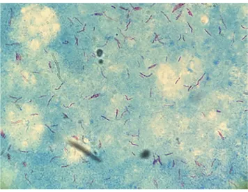
medical history and no known contact with tuberculosis. Information on the vaccine manufacturer and technique of inoculation was not available. Six weeks later, she devel-oped a painful swelling, and an erythematous and infil-trated skin lesion at the injection site. There were no associated systemic signs or symptoms and no lymphade-nopathy. At presentation, there was a circular erythema-tous swollen area of 2 cm diameter with indurated margins at the site of the inoculation (Fig. 1). A needle aspiration of the lesion was performed and 2 ml pus was collected. As a cause of the lesion, the parents suggested vaccination, and BCG-induced abscess was immediately suspected. A Gram stain and routine bacterial culture were not performed. A Ziehl–Neelsen stain showed acid-fast bacilli (Fig. 2), and mycobacteria were grown after 14 and 18 days in an MGIT system (Becton Dickinson) and on Lo¨wenstein–Jensen medium, respectively. The micro-organism was identified by multiplex PCR of the RD (Behr et al., 1999; Parsons et al., 2002) as M. bovis BCG. The strain was negative in a pyrazinamidase test, an assay that is often used for the presumptive differen-tiation between M. tuberculosis and Mycobacterium bovis. As the latter species does not produce pyrazinamidase and is resistant to pyrazinamide, the result of this test was compatible with M. bovis/ BCG. The abscess was disin-fected and treated with gentamicin sulfate ointment daily for 2 weeks to prevent secondary bacterial infection. No treatment with isoniazid or other antituberculosis drugs was considered to be required. The lesion improved considerably within 1 month and was completely cleared
within 3 months. No relapse has been observed after 4 months of follow-up.
Discussion
Tuberculosis remains a global public health problem affect-ing both humans and animals. The estimated worldwide incidence rate was 127 cases per 100 000 of the population in 2013 (WHO, 2014). Tuberculosis was second only to human immunodeficiency virus/AIDS as the greatest killer worldwide in 2013. Indeed, 9 million people fell ill and 1.5 million died from the disease (WHO, 2014). BCG vacci-nation can significantly reduce the overall risk of tuberculo-sis and is particularly effective against disseminated forms of the disease. Abscess formation at the site of BCG vaccination is uncommon if injection is performed intradermally at the recommended sites (in the deltoid region or in the upper external part of the thigh) using the indicated dosage and hygienic techniques. Thus, in the case presented here, it is possible that abscess formation was the result of injection at a non-recommended site and/or subcutaneous, instead of intradermal, inoculation. Differential diagnosis of a cutaneous lesion in a patient arriving from endemic areas includes a number of different conditions ranging from a variety of infections to tumours. For example, differential diagnosis should include staphylococcal abscess, spirotri-chosis, nocardiosis, chromomycosis, leishmaniasis, tertiary syphilis, hidradenitis and all forms of panniculitis, to name only a few. Differential diagnosis with cutaneous tuberculosis is fundamental in patients from underdeve-loped countries, particularly in high-risk populations such as immigrants from endemic region. The BCG vaccine is rarely administered in developed countries, so it is not easy for clinicians to diagnose any complication related to the administration of this vaccine. Therefore, health Fig. 1. Erythematous and infiltrated skin lesion at the injection
site on the right upper arm at the vaccination site.
Fig. 2. Acid-fast bacilli in a Ziehl–Neelsen stain of needle aspira-tion material from the lesion.
F. d’Aleo and others
Downloaded from www.microbiologyresearch.org by IP: 192.167.111.12
On: Wed, 27 Jan 2016 08:23:04
professionals should consider BCG as possible cause of cutaneous lesions developing in patients returning from areas where BCG vaccine is widely administered.
The clinical diagnosis must always be confirmed by histo-pathology and microbiological cultures.
In our case, with the exception of local swelling and erythema, there were no general or local signs of infection. A diagnosis of abscess caused by BCG was suspected, as the site of lesion corresponded to that of the BCG inoculation. For quick identification of the species, we ran a PCR assay in which three primers were used to detect the RD1, RD9 and RD10 regions. The strain we isolated lacked all three regions and was therefore identified as a BCG strain (Par-sons et al., 2002). Non-BCG strains of M. bovis may display one or all three of these regions, while M. tuberculosis strains display all three (Behr et al., 1999). BCG vaccines consist of live attenuated strains of M. bovis. They were first used for immunization against tuberculosis in 1921 and are considered safe. Ninety per cent of the vaccines are produced from four different strains of M. bovis: Pas-teur 1173P2, Danish 1331, Glaxo 1077 and Tokyo 1729. These BCG strains differ in their immunogenicity, efficacy and side effects (Starke & Connelly, 1994). Transitory adverse reactions arising from the use of BCG vaccine occur in 0.1–1.7 % of cases in immunocompetent infants (Lotte et al., 1984). In addition to ulceration and abscess formation, rare adverse reactions to BCG vaccination include keloid formation and regional lymphadenitis. Localized abscesses without regional lymphadenopathy in immunocompetent hosts, such as the one reported here, are relatively uncommon (Fitzgerald & Duclos, 1991). The pathogenesis is not clear, and causes might include technical errors in vaccine administration including over-dosing. There are variable recommendations for manage-ment of post BCG abscess and suppurative lymphadenitis. These range from no treatment to needle aspiration, drug treatment, surgical drainage, surgical excision or a combi-nation of these. Some authors suggest that needle aspiration is the management of choice, although this is not always effective and additional therapy may be required (Lussier et al., 1999; Okazaki et al., 2005). A similar case to one described here was reported by Lussier et al. (1999) in a young woman. In their case, in contrast to ours, the lesion did not resolve after needle aspiration and treatment with isoniazid was required. Similarly, a 3-month isoniazid course was necessary in the case reported by Okazaki et al. (2005) in which a BCG-derived abscess developed in a region some distance from the vaccination site. The man-agement of BCG-induced abscess with erythromycin is highly controversial (Murphy et al., 1989). In conclusion, healthcare personnel should be aware that extensive utiliz-ation of the BCG vaccine in many countries has increased the occurrence of BCG-derived complications and that these should therefore be considered in the differential diag-nosis of patients arriving from tuberculosis-endemic areas.
Acknowledgements
Part of this work was performed using funds from the Ministero dell’Universita` e della Ricerca of Italy (Grant MIUR n.4/13 ex art 11 D.M.503 to Scylla Biotech Srl).
References
Behr, M. A., Wilson, M. A., Gill, W. P., Salamon, H., Schoolnik, G. K., Rane, S. & Small, P. M. (1999). Comparative genomics of BCG vaccines by whole-genome DNA microarray. Science284, 1520–1523.
Cuchet, E., Templier, I., Brion, J. P. & Be´ani, J. C. (2004).[A persistent ulceration after a fortuitous BCG revaccination]. Ann Dermatol Venereol131, 1077–1079, (in French).
Diniz, L. M. O., Guimara˜es, T., Oliveira, M., Pinto, J. A. & de Miranda, S. S. (2014).Lymphadenitis caused by infection with an isoniazid-and rifampin-resistant strain of Mycobacterium bovis BCG in an infant with IFN-c/IL-12 pathway defect. J Bras Pneumol 40, 188–192.
Fitzgerald, M. J. & Duclos, P. (1991).The reporting and management of adverse reactions to bacillus Calmette–Gue´rin (BCG) vaccination. Can Dis Wkly Rep17, 98–100.
Gue´rin, C. (1957).The history of BCG. In BCG Vaccination against Tuberculosis, pp. 48–53. Edited by S. R. Rosenthal. Boston: Little, Brown & Co.
Gyldenløve, M., Andersen, A˚ . B. & Halkjær, L. B. (2012).Cutaneous necrotic ulceration due to BCG re-vaccination. Hum Vaccin Immunother8, 423–424.
Lotte, A., Wasz-Ho¨ckert, O., Poisson, N., Dumitrescu, N., Verron, M. & Couvet, E. (1984). BCG complications. Estimates of the risks among vaccinated subjects and statistical analysis of their main characteristics. Adv Tuberc Res21, 107–193.
Lussier, N., Bourgault, A. M., Gaudreau, C. & Turgeon, P. (1999).
A complication of BCG vaccine: a case of localized cutaneous abscess due to Mycobacterium bovis. Can J Infect Dis10, 257–259.
Murphy, P. M., Mayers, D. L., Brock, N. F. & Wagner, K. F. (1989).
Cure of bacille Calmette-Gue´rin vaccination abscesses with erythromycin. Rev Infect Dis11, 335–337.
Nakajima, C., Rahim, Z., Fukushima, Y., Sugawara, I., van der Zanden, A. G., Tamaru, A. & Suzuki, Y. (2010). Identification of Mycobacterium tuberculosis clinical isolates in Bangladesh by a species distinguishable multiplex PCR. BMC Infect Dis10, 118.
Okazaki, T., Ebihara, S., Takahashi, H., Asada, M., Sato, A., Seki, M., Ohto, H. & Sasaki, H. (2005).Multiplex PCR-identified cutaneous tuberculosis evoked by Mycobacterium bovis BCG vaccination in a healthy baby. J Clin Microbiol43, 523–525.
Parsons, L. M., Brosch, R., Cole, S. T., Somosko¨vi, A., Loder, A., Bretzel, G., Van Soolingen, D., Hale, Y. M. & Salfinger, M. (2002).
Rapid and simple approach for identification of Mycobacterium tuberculosis complex isolates by PCR-based genomic deletion analysis. J Clin Microbiol40, 2339–2345.
Starke, J. R. & Connelly, K. K. (1994). Bacille Calmette–Gue´rin vaccine. In Vaccines, pp. 439–473. Edited by S. A. Plotkin & E. A. Mortimer, Jr. Philadelphia, PA: W.B. Saunders.
WHO (2014).Global Tuberculosis Report 2014. Geneva: World Health Organization.
Zaı¨em, A., El Ferjani, S., Lakhoua, G., Sahnoun, R., Badri, T., Kastalli, S., Daghfous, R., Lakhal, M. & El Aidli, S. (2014). Probable disseminated BCG infection in a 10-month-old child after BCG vaccination. Hum Vaccin Immunother10, 2081–2082.
Abscess after BCG vaccine in an immunocompetent child