Università degli Studi di Ferrara
DOTTORATO DI RICERCA IN
SCIENZE DELL’INGEGNERIA
CICLO XXII
COORDINATORE Prof. STEFANO TRILLO
DUROTAXIS MODELLING FOR TISSUE
ENGINEERING APPLICATIONS
Settore Scientifico Disciplinare ING/IND 22
Dottorando Tutore
Dott. STEFANONI FILIPPO Prof. MOLLICA FRANCESCO
“I know that I know nothing” (Socrates)
PREFACE
This thesis comes from my three years PhD period at the University of Ferrara. I began to be a PhD student on January 2007 and I finished on December 2009. During these years a lot of events took place but now I remember when I was in Aachen for my abroad stage and Dragos, a Romanian PhD student working with me, told me: “When you start your PhD you are like a student, when you finished it, you are like a worker”. I think Dragos was right or at least, this happen to me during my PhD. Three years ago I was still a student trying to study something useful and not only to take an exam. Now I have changed my mind and I see things in a different way.
During these years I studied many subjects and problems on polymeric materials, biomaterials and not only, supervised by Professor (or Engineer as he prefers) Francesco Mollica.
I participated as a student at the following summer schools and lectures:
“Nonlinear Computational Solid and Structural Mechanics. Theoretical
formulation, FEM technology and computations”. IMATI-CNR, Università
degli studi di Pavia. Pavia, 14-18 Maggio 2007.
“14th CIRMIB Biomaterials School”. Ischia (NA). 9-13 Luglio 2007
I collaborated with the Dental Clinic Section and with the Orthodontic School of the University of Ferrara through Dr. Luca Lombardo and Dr. Nicola Mobilio supervising students in their thesis:
Francesco Zampini (2007), Facoltà di Medicina e Chirurgia, Corso di Laurea in Odontoiatria e Protesi Dentaria, Tesi di laurea in Ortognatodonzia.
“Valutazione al FEM delle tensioni che si generano attorno ad una minivite ad ancoraggio osseo mono e bicorticale”. Supervisor: Prof.
Giuseppe Siciliani.
Paolo Contiero (2008), Facoltà di Medicina e Chirurgia, Corso di Laurea in Odontoiatria e Protesi Dentaria, Tesi di laurea. “Valutazione comparativa
della distribuzione dei carichi masticatori su impianti in Titanio e Zirconia mediante analisi agli elementi finiti”. Supervisor: Prof. Santo
Catapano.
Laura Attorresi (2009), Facoltà di Medicina e Chirurgia, Scuola di Specializzazione in Ortognatodonzia, Tesi di Specializzazione in Ortognatodonzia. “Distribuzione dello stress sulla superficie radicolare in
seguito all’applicazione di forze sul lato vestibolare e linguale”.
Supervisor: Prof. Giuseppe Siciliani.
From this collaboration a paper was published and others are in progress:
“Optimal Palatal Configuration for Miniscrew Applications”. Lombardo L, Gracco
A, Zampini F, Stefanoni F, Mollica F. Angle Orthodontist. 2010;80(1):145-152.
I also made some research activity in collaboration with Vortex Hydra S.r.l. on mathematical modelling of plants for concrete roof tiles:
Francesco Mollica, Filippo Stefanoni (2009), Relazione tecnica finale. “Sviluppo di
un modello matematico del processo di estrusione utilizzato da Vortex Hydra per realizzare manufatti in malta cementizia”.
But I decide to write my thesis only on the main topic I studied, i.e. Tissue Engineering and in particular cell migration. This research was made in collaboration with CRIB (Interdisciplinary Research Centre on Biomaterials, University of Naples “Federico II”) and in particular with Dr. Maurizio Ventre. I do not know why this happened and how cell migration came to me and I also think that it is very strange and new for an Engineer to study such a subject, but this is the so called “interdisciplinary” of Tissue Engineering and in this research field you have to be prepared to everything as my supervisor learn me.
Thus I structured my thesis following my personal path into this new subject. The first chapter is introductory to the general concepts of Tissue Engineering. It is very general, because the field is very broad but it is necessary to outline the rationale behind the subsequent studies. The second chapter is about the physical
phenomenon I dealt with, i.e. cell migration. Here I put notions I studied from papers and books useful for me as a base and to develop new ideas. Then, the last three chapters are on my original work. Also if they are different in methods, they have in common the phenomenon of Durotaxis that is the real subject of this thesis. As better explained inside, this particular condition of cell migration is studied from a mathematical and an experimental point of view in chapters 3 and 4 and a possible applications of the phenomenon was developed in chapter 5.
Some of the material contained here was used for oral presentations and scientific talks in scientific congresses and Universities:
“Un Modello Numerico per la Durotassi”. F Stefanoni, M Ventre, F Mollica, PA Netti. Congresso Nazionale Biomateriali SIB, Follonica (GR),17-19 Settembre 2008.
“Durotaxis: Modeling and Experimental Validation”. F Stefanoni, M Ventre, M Diez, VA Schulte, MC Lensen, F Mollica, PA Netti. 22nd European Conference on Biomaterials, European Society for Biomaterials. Lausanne (CH), 7-11 Settembre 2009.
“A Numerical Model for Durotaxis”. Stefanoni F, Ventre M, Diez M, Schulte VA, Lensen MC , Mollica F, Netti PA. XIX Congresso AIMETA. Ancona, 14-17 Settembre 2009.
“Cellular behavior on micro- and nanopatterned hydrogels”. Diez M, Chen J, Mela P, Schulte VA, Cesa CM, Stefanoni F, Ventre M, Mollica F, Netti PA, Möller M, Lensen MC. ESF-EMBO Symposium: Biological Surfaces and Interfaces. Sant Feliu de Guixols (ESP),27 June-2 July 2009.
“Guiding cell migration: a key for tissue engineering”. Ventre M, Netti PA, Mollica F, Stefanoni F. Scientific Talk at DWI-RWTH, Aachen (D), 24 October 2008.
“Studio dei modelli numerici per la determinazione delle proprietà dei
tessuti in crescita”. F Stefanoni, M Ventre. Scientific Talk at CRIB (VIV),
Napoli 15 Giugno 2007.
I do not know what will happen to me in the next years and I do not know if I will continue to study these subjects. Anyway, this is the work I have done.
AKNOWLEDGMENTS
First of all I want to thank my supervisor Francesco Mollica. I know I have been his first PhD student so this was a new experience also for him. He was very careful with me and always ready to help me and to discuss concepts and ideas. We have had good and bad experiences working together and I hope that both have learn something from them. Surely I did.
Then thanks to Maurizio Ventre. He introduced me in the world of migrating cells and he was the first “Biological Engineer” I met. He also introduced me in Naples and in the fantastic world of the “Scamorza Affumicata”: I never forgot this experience! I want also to thank him and his mother, for giving me hospitality every time I went to Naples.
I also want to thank Dr. Marga C. Lensen for giving me the possibility to pass a two month period (9 February-7 April 2009) at the DWI-RWTH in Aachen (Germany) to study substrata for my experiments and all the people I met there.
Some words also for people I met at CRIB and during scientific congresses, schools and lectures: Ilaria De Santo, Maria Iannone, Silvia Orsi, Vincenzo Guarino, Edmondo Battista, Maria Grazia Raucci, Antonio Gloria, Ciccio Urciuolo, Tiziana Punzi, Tiziano Serra and Giampiero Pampolini. I really enjoyed those times!
I cannot forget people from the University of Ferrara daily with me: Mirko Morini, Alberto Minotti, Cristian Ferrari, Anna Vaccari, Michele Pinelli and all the people here around.
In the end, I want to thanks my friends Carla, Giuseppe and Nicola, my parents Daniele and Cristina, my brother Simone, my grandmothers Ilde and Bruna, Ilaria, Nicola, Ester and Ruben, my aunt Laura, Paolo and Andrea. Every one of them is important for me and for my well-being, so they also contribute to this thesis!
TABLE OF CONTENTS PREFACE ……….……….……….……….5 AKNOWLEDGMENTS………9 TABLE OF CONTENTS………10 ABSTRACT……….………...13 SOMMARIO………..3
CHAPTER 1 – Tissue Engineering 1. Introduction………...………18 2. Methods……….………21 3. Cell Culture………...……23 4. Scaffolds………29 5. Bioreactors………33 6. Applications………...………34 7. References………36
CHAPTER 2 – Cell Migration, Durotaxis and Collagen Deposition 1. Introduction………..………….39
2. Cell and Cell Functions………..………….………39
3. Cell Cytoskeleton and Movement...………44
4. The Process of Cell Movement……….………49
5. Influencing Factors of Cell Motion……….………52
6. Durotaxis………...……54
7. Cell Migration and Collagen Deposition....………..……56
8. References………58
CHAPTER 3 – A Computational Model for Durotaxis 1. Introduction………...…62
2. The Langevin Equation in the Modelling of Cell Migration………63
3. A Discrete Model for Durotaxis………..……66
4. Two Particular Cases for Preliminary Validation……….…70
6. References………77
CHAPTER 4 – Experimental Validation of the Model 1. Introduction………...81
2. Preparation of the Samples………81
3. Cell Migration Experiments over Biphasic Substrata……….84
4. Experimental Results...………...85
5. Comparison with the Model..……….87
6. Results and Conclusions………89
7. References………89
CHAPTER 5 - Study and Design of a Durotaxis-based Substratum 1. Introduction……….………..92
2. Polyethylene glycol (PEG)……..………92
3. Preparation of the Materials………...…93
4. Cell Migration Experiments………95
5. Results and Remarks………..96
6. The “Pattern of Elasticity” Substrata………...102
7. Preliminary Cell Adhesion Experiments…………..………..107
8. Conclusions………109
9. References……….109
ABSTRACT
Tissue Engineering is a very promising research field for the development of natural biological substitutes that restore damaged tissue functions. Cells play a crucial role in tissue regeneration and repair due to their characteristics of proliferation and differentiation, cell-to-cell interaction, biomolecular production and extracellular matrix formation. In particular cell migration is a phenomenon that is involved in different physiological processes such as morphogenesis, wound healing and new tissue deposition. In the absence of external guiding factors it is essentially a phenomenon that shares quite a few analogies with Brownian motion. The presence of biochemical or biophysical cues, on the other hand, can influence cell migration in terms of speed, direction and persistence, transforming it in a biased random movement. Recent studies have shown that cells, in particular fibroblasts, are able to recognize the mechanical properties of a substratum over which they move and that these properties direct the motion through a phenomenon called durotaxis. The aim of this thesis is to study this phenomenon for a better understanding of cell behaviour in durotaxis conditions and for Tissue Engineering applications. In order to do that, in the first part of the work a mathematical model for the description of durotaxis is presented. The model is based on a stochastic differential equation for the cell velocity which is derived from the Langevin equation: cell movement is affected by two forces, namely a deterministic one representing the dissipative effects of the system, and a stochastic one which is due to all the probabilistic processes that might affect cell motility (random fluctuations in motile sensing, response mechanisms, etc.). The original contribution of this work concerns the stochastic force, which has been modified to account for the directions of highest perceived local stiffness through a finite element scheme that reminds the cellular probing mechanism. Numerical simulations of the model provide individual cell tracks that can be qualitatively compared with experimental observations. The present model is solved for two important cases that are reported in literature and a comparison with experimental data obtained on PDMS substrata is presented. The degree of agreement is satisfactory thus the model could be utilized to quantify relevant parameters of cell migration as a function of substratum mechanical properties.
The second part of the work is concerned on the study and development of a durotaxis-based substratum, able to guide cells in their migration and in particular, able to guide cells along straight path. It was proved, in fact, that a relation exist between the alignment of collagen produced by fibroblasts or others tissue cells and their migration. Thus, the idea is to obtain an aligned tissue made of new collagen, giving to the cells the conditions to move along straight-lines through the mechanical properties of the substratum. To realize this substratum Polyethylenglycole (PEG) was used. First, smooth PEG was synthesized and cell migration experiments was performed over it to better understand its response. Then a specific technique was developed to produce durotaxis-based PEG substrata, and preliminary experiments of cell adhesion over it were performed showing aligned adhesion of cells over them.
SOMMARIO
L’Ingegneria dei Tessuti è un campo di ricerca molto promettente che si occupa dello sviluppo di sostituti biologici naturali che possono riparare tessuti danneggiati o non più in grado di svolgere le loro funzioni. Le cellule svolgono un ruolo cruciale in questo campo, per la loro capacità di proliferare e differenziare, per la loro interazione reciproca e per la loro capacità di produrre biomolecole e matrice extracellulare. In particolare, la migrazione delle cellule è un fenomeno importante in diversi processi fisiologici tra cui la morfogenesi, la cicatrizzazione e la deposizione di nuovo tessuto. In assenza di fattori esterni, si tratta di un fenomeno simile al moto Browniano di particelle. D’altro canto, la presenza di fattori biochimici o biofisici può avere un’influenza sul moto cellulare in termini di velocità, direzione e persistenza, rendendolo meno casuale. Studi recenti hanno dimostrato che le cellule e in particolare i fibroblasti, sono in grado di riconoscere le proprietà meccaniche di un substrato sopra il quale si muovono: queste sono quindi in grado di modificare il moto cellulare secondo un fenomeno chiamato durotassi.
Lo scopo di questa tesi è di studiare tale fenomeno, per meglio capire il comportamento delle cellule in queste condizioni e per possibili applicazioni in Ingegneria dei Tessuti. Per far ciò, nella prima parte del lavoro è stato sviluppato un modello matematico per la descrizione della durotassi. Il modello è basato su un’equazione differenziale stocastica per la velocità, derivante dall’equazione di Langevin: il movimento cellulare è influenzato da due forze, una deterministica che rappresenta gli effetti dissipativi del sistema, l’altra stocastica, dovuta a tutti I fattori probabilistici che possono avere un effetto sul moto (fluttuazioni casuali nel meccanismo di percezione dei segnali, nel meccanismo di risposta, etc.). Il contributo originale del lavoro riguarda il termine stocastico, che è stato modificato in modo da considerare la direzione di maggior rigidezza percepita dalla cellula tramite un algoritmo agli elementi finiti. Le simulazioni numeriche del modello forniscono le singole traiettorie cellulari che possono quindi essere comparate direttamente con le osservazioni sperimentali. Il modello viene risolto in due casi noti da letteratura e viene riportato un confronto con dati sperimentali ottenuti su substrati di PDMS. Il grado di accordo risulta essere buono e quindi il modello puo essere usato per
quantificare alcuni parametri del moto cellulare in base alla proprietà meccaniche del substrato.
La seconda parte del lavoro riguarda invece lo studio e lo sviluppo di un substrato che, grazie al fenomeno della durotassi, sia in grado di guidare le cellule lungo traiettorie rettilinee. E’ stato infatti provato che esiste una relazione tra l’allineamento del collagene prodotto da fibroblasti e altre cellule del tessuto connettivo, e il loro moto. L’idea è quindi quella di ottenere, grazie ai substrati, nuovo tessuto collagineo allineato. Per realizzare ciò è stato utilizzato Polietilenglicole (PEG). Per prima cosa sono stati fatti esperimenti preliminari di migrazione su PEG liscio, per testarne la risposta. Dopodichè è stata sviluppata una nuova tecnica per produrre substrati di PEG in grado di guidare le cellule tramite durotassi. Esperimenti preliminari di adesione cellulare sono stati eseguiti mostrando un buon grado di allineamento delle cellule.
CHAPTER 1
Tissue Engineering
1. Introduction
One of the most famous definitions of Tissue Engineering (TE in the remainder) was given by Professor Robert Langer and Professor Joseph P. Vacanti in their article published on “Science” in 1993:
“Tissue Engineering is an interdisciplinary field that applies the principles of engineering and the life sciences toward the development of biological substitutes that restore, maintain, or improve tissue function.”
As an engineer-PhD student trying to study phenomena related to TE, I can say that the most important word in this statement is “interdisciplinary”. When you heard about this term just by someone else or during your lectures at University, you can not imagine its real meaning. But when you start your practical research in TE and when you keep contact with other researchers studying analogous phenomena, you feel the effect of this apparently abstract term in creating curiosity inside you, opening your mind to other points of view and expanding your knowledge. Nevertheless the other side of the coin is that you need time to understand concepts far from your background and you need to be psychologically prepared to study problems in which you can not deeply understand all the aspects.
So if you survive and if you are able to appreciate the positive aspects, TE is a very exciting research field that includes a great variety of phenomena that have as the main subject biological tissues.
TE represents the confluence of different lines of work from three quite different fields: clinical medicine, engineering and life science. The most obvious precursors to TE lie in the clinical domain. In fact, thinking of a surgeon, one of his frequent problems is about the removal of organs or of body structures. Sometimes this removal can be life-saving but the patient must cope with the functional effects of
tissue loss and in some cases, with the psychological impacts of disfigurements. And for those vital organs whose complete removal is incompatible with life he needs some way of replacing or reconstituting essential functions.
To solve these problems surgeons have adopted different strategies. They have sought to reconstruct anatomic structures using the patient’s own tissues as raw material; they have pressed artificial materials into service as prostheses; and, most spectacularly, they have brought patients back from the brink of death by transplanting an ever-wider range of vital organs, primarily living organs, but in a few cases, with only very limited success to date, prototype artificial organs as well.
However, with experience, surgeons have come to understand in detail not only the benefits of such measures, but their limitations as well. Anatomic reconstruction using the patient’s own tissue can cause substantial morbidity at the donor site; the improvised structures are usually functionally inferior to the natural organs they replace and less durable as well. Poor compatibility between artificial materials and mechanical systems and the internal environment and physiologic requirements of the human body can lead to dysfunctional interactions and new failure modes. Transplantation of living organs brings with it profound immunologic complications and the number of patient who can be treated in this way will always be severely constrained by the limited supply of organ suitable for use.
For a surgeon, then, the development of engineered tissues is a logical next step in the ongoing effort to improve the match between its various reparative and reconstructive contrivances and the requirements of human anatomy and physiology. So the clinical perspective on TE is strongly applications-oriented but viewed the other way around, in terms of enabling knowledge and technologies, TE is remarkable for the breadth of its footprint in fundamental and applied biomedical research.
In Table 1 is possible to see some fields and subfields involved in TE, just to have an idea of its interdisciplinary body and of the range, depth and character of the inputs to the field.
Cell and Developmental Biology
Cell differentiation, morphogenesis and tissue assembly Cell-cell and cell-matrix interactions
Cell isolation and selection Cell culture
Angiogenesis Stem cells
Basic Medical and Veterinary Sciences
Anatomy Cytology
Physiology and patho-physiology
Transplantation Science
Applied immunology, immunosuppresion immunomodulation and immunoisolation Organ preservation
Biomaterials
Natural and synthetic, biodegradable and non-biodegradable polymers Polymer chemistry
Ceramics
Cell interactions with biomaterials
Controlled release of bioactive molecules Microencapsulation
Microfabrication techniques 3D fabrication techniques Surface chemistry
Biophysics and Biomechanics
Molecular and cell transport
Micro- and macrocirculatory dynamics Cells and tissue mechanics
Biomedical Engineering
Bioreactors
Membranes and filtration
Musculoskeletal joint engineering Biomedical sensors
Biomedical signal processing, feedback and control
Electrical and mechanical engineering of biohybrid systems Engineering design and system analysis
Quantitative tissue characterization Biosensors and bioelectronics
Table 1: Research field and subfields that have contributed to TE (National Science Foundation, 2003)
It is unclear who first used the term “Tissue Engineering” to mean what it does today. Realistically it was invented several times independently before it became of usage, but its origin can be clearly traced to a specific individual. In 1985, Y.C. Fung, a pioneer in the field of biomechanics and of bioengineering more broadly, submitted a proposal to National Science Foundation, for an Engineering Research Centre to be entitled “Centre for the Engineering of Living Tissues”. Fung’s concept drew on the traditional definition of tissue as a fundamental level of analysis of living organism,
between cell and organs. The proposal was not accepted, but the concept was born, so in the following years it took its shape, reaching the definition reported above of Langer and Vacanti.
2. Methods
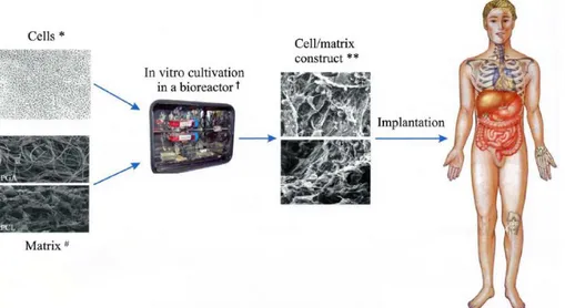
Tissue Engineering is a broad term describing a set of tools at the interface of the biomedical and engineering sciences that uses living cells or attract endogenous cells to aid tissue formation or regeneration, and thereby produce therapeutic or diagnostic benefit. More practically, the most frequent procedure lies in seeding cells on a retaining structure composed of synthetic polymers or natural materials; then a tissue is matured in vitro in a bioreactor and the construct is thus implanted in the appropriate anatomic location as a prosthesis.
The retaining structure on which cells are seeded is called “scaffold”, a generic term indicating an artificial structure made, for example, of a bioresorbable polymer in a porous configuration or, of natural material such as collagen or chemically treated tissue: a scaffold is a sort of house for cells before the implantation, that furnishes them all the necessary conditions for spreading, proliferating and then for the generation of new tissue. A bioreactor is a device or a system that supports a biologically active environment, in which cells can proliferate and elaborate extracellular matrix (ECM). After the in vitro growth, the construct is implanted in the appropriate anatomic location, where in vivo remodelling is intended to recapitulate the normal functional architecture of an organ or tissue (Fig. 1).
Fig. 1: Tissue engineering process: Cells, Matrix, Bioreactor and Cell/matrix construct (Shieh and Vacanti, 2005).
The key processes occurring during the in vitro and in vivo phases of tissue formation and maturation are:
Cell proliferation, sorting and differentiation; Extracellular matrix production and organization; Degradation of the scaffold;
Remodelling and potentially growth of the tissue.
The illustrated procedure is comprehensive of all the feasible operations, but sometimes incomplete procedures are adopted for example implanting directly the scaffold in the host without using of the bioreactor or using a scaffold that recruits endogenous cells directly inside the patient.
In any case, the three principal components of TE are cells, scaffold and bioreactor and all the parameters related to them have an impact upon the ultimate result. In Table 2 a series of these parameters are reported as factors that can be taken into account in a TE process.
Cells Biodegradable matrix/Scaffold
Source Architecture/Porosity/Chemistry
Allogenic Composition/Charge
Xenogenic Homogeneity/Isotropy Autologous Stability/Resorption rate
Type/Phenotype Bioactive molecules/Ligands Single versus multiple types Soluble Factors Differentiated cells from primary or other
tissue
Mechanical Properties
Adult bone marrow stem cells Strength
Pluripotent embryonic stem cells Compliance
Density Ease of manufacture
Viability Bioreactor Conditions Gene expression Nutrients/Oxygen
Genetic manipulation Growth Factors
Perfusion and flow conditions Mechanical Factors
Pulsatile Hemodynamic shear stresses Tension/Compression
Table 2: Parameters involved in TE (Biomaterials Science, 2004)
3. Cell Culture
Cells play a crucial role to tissue regeneration and repair due to their characteristics of proliferation and differentiation, cell-to-cell interaction, biomolecular production, and extracellular matrix formation (details about cell functions are reported in chapter 2 of this thesis). As shown in Table 2, the sources of cells used in TE can be autologous, i.e. from the host, allogeneic, i.e. from another individual of the same species, or xenogeneic, i.e. from another individual of another species. Ideal donor cells for TE would be those that are easily accessible, that can easily expand without permanently altering the phenotype (i.e. all the observable properties of an organism, that are produced by the interaction of its genetic constitution, the genotype, and the environment) and function and without transmitting species-specific pathogens (agents producing a disease, e.g., virus or bacterium) that are multipotent to differentiate or transdifferentiate into a variety of tissue- or organ-specific cells with specialized function, and that have the least immunologic response.
Some cells, such as keratinocytes, fibroblasts, chondrocytes, endothelial cells, smooth muscle cells or skeletal muscle satellite cells, proliferate rapidly. They are good tissue-specific cell sources for TE. Two Food and Drug Administration (FDA)-approved living skin products engineered in the laboratory have been applied to a patient with diabetic or venous skin ulcers, and a FDA-approved autologous cell
product also has been used to repair an articular cartilage. However, other cells, such as hepatocytes or adult cardiomyocytes, proliferate slowly or not at all. Therefore, alternative sources of cells are needed.
Recent advances in stem cell biology have had a marked impact on the progress of TE. Stem cells, which are capable of self-renewal and differentiation into various cell lineages, hold great promise for treating affected tissue in which the source of cells for repair is limited or not readily accessible. Cells derived from human embryonic blastocysts (a structure formed in the early embryogenesis of mammals), after undifferentiated proliferation in vitro for 4-5 months, still maintain the developmental potential to form trophoblast (cells forming the outer layer of the blastocyst) and derivatives of all three embryonic germ layers including gut epithelium (endoderm), cartilage, bone, smooth muscle, and striated muscle (mesoderm), and neural epithelium, embryonic ganglia, and stratified squamous epithelium (ectoderm). Although these cell lines should be useful in human regenerative medicine, the ethical and legal issues are still under debate.
Adult bone marrow stem cells can replicate as undifferentiated cells that have the potential to differentiate into lineages of mesenchymal tissue, including bone, cartilage, fat, tendon, muscle and marrow stroma. They display a stable phenotype, remain as a monolayer in vitro, and could be induced to differentiate exclusively into adipocytic, chondrocytic, or osteocytic lineages. To date, the isolation of various autologous adult stem cells, including mesenchymal, hematopoietic, neural, muscle, and hepatic stem cells, are being investigated actively, because they are immunocompatible and have no ethical concerns. Nevertheless, there are a number of technical obstacles, such as how to isolate stem cell preparations without contamination by other cells, how to control the permanent differentiation to the desired cell types, and how to increase the production of the large number of cell needed to create tissue.
Other strategies aim at optimizing cells for TE and are focused on the host-immune response to allogenic or xenogenic cell. Starting from this point, researchers are trying to create “universal donor cells” by masking histocompatibility proteins on the cell surface to reduce the cell’s antigeniticy (Shieh and Vacanti, 2005).]
The more recent research in this field is about nuclear transfer, or “therapeutic cloning”: it is a process wherein the nucleus of a somatic cell is injected into an
unfertilized enucleated oocyte. This transformation probably involves deletion of the existing epigenetic state (the actual expression of the genes of an organism) and expression. Through this nuclear manipulation any differentiated somatic cell can potentially be reprogrammed back to totipotency, which results in redifferentiation to the full repertoire of adult cells for any individual tissue repair. Although the goal of therapeutic cloning is to generate replacement cells and tissue that are genetically identical to those of the donor, non-self-mitochondria proteins derived from the recipient oocytes could render cloned tissue immunogenetic. All these findings bring closer the promise of therapeutic cloning and TE. The combination of nuclear transfer, gene therapy and cell transplantation as a possible applicable paradigm for genetic and phenotypic correction is a challenge to many active scientist worldwide. Regardless of the types of strategy, cells for TE practically come from cell culture. In fact, animal or plants cells, removed from tissues, will continue to grow if supplied with the appropriate nutrients and conditions and when carried out in laboratory, the process is called cell culture. This allows single cells to act as independent units, much like a microorganism such as a bacterium or fungus. Cells are capable of dividing, increase in size and, in a batch culture, they can continue to grow until limited by some culture variable such as nutrient depletion.
Cells can be isolated from tissue for ex vivo culture in several ways. They can be easily purified from blood, however only the white cells are capable of growth in culture. Mononuclear cells can be released from soft tissue by enzymatic digestion with enzymes such as collagenase, trypsin or pronase, which break down the ECM. Alternatively pieces of tissue can be placed in growth media and cells that grow out are available for culture.
Cultures normally contain cells of one type although mixed cultures, especially of bacteria, are common in food sciences and wastewater treatment studies. The cells in culture may be genetically identical (homogeneous population) or may show some genetic variations (heterogeneous population). A homogeneous population of cells derived from a single parental cell is called a “clone”. Therefore all cells within a clonal population are genetically identical.
Cells that are cultured directly from a subject are known as primary cells. With the exception of some derived from tumours, most primary cell cultures have limited lifespan. After a certain number of population doublings cells undergo the process of
senescence and stop dividing, while generally retaining viability. After several sub-cultures onto fresh media, the cell line will either die out or transform to become a continuous cell line Such cell lines show many alterations from the primary cultures including change in morphology, chromosomal variation and increase in capacity to give rise to tumours in host with weak immune systems. An established or immortalised cell line has acquired the ability to proliferate indefinitely either through random mutation or deliberate modification. There are numerous well-established cell lines representative of particular cell types and the major repositories are the American Type Culture Collection (ATCC) and the European Collection of Cell Cultures (ECACC). Some examples of immortalised lines are reported in Table 3. Cells are grown and maintained at an appropriate temperature and gas mixture, typically 37°C and 5% CO2 for mammalian cells, in a cell incubator (Fig. 2).
Fig. 2: Cell incubator at CRIB (Interdisciplinary Research Centre on Biomaterials, University of Naples “Federico II”)
Culture conditions vary widely for each cell type and variation of conditions for a particular cell type can result in different phenotypes being expressed. Aside from temperature and gas mixture, the most commonly varied factor in culture system is the growth medium. Recipes for growth media can vary in pH, glucose concentration, growth factors and the presence of other nutrients. The growth factors used to supplement media are often derived from animal blood such as calf serum.
surface, e.g. the cells of the bloodstream. There are also cell lines that have been modified to be able to survive in suspension cultures so that they can be grown to a higher density than adherent conditions would allow. Adherent cells require a surface such as tissue culture plastic, which may be coated with ECM components to increase adhesion properties and provide other signals needed for growth and differentiation. Most cells derived from solid tissue are adherent. Another type of adherent culture is organotypic culture which involves growing cells in a three dimensional environment as opposed to two dimensional culture dishes. This three dimensional culture system is biochemically and physiologically more similar to in vivo tissue, but is technically challenging to maintain because of many factors such as diffusion. As cells generally continue to divide in culture, they generally grow to fill the available area or volume. This can generate nutrient depletion in the growth media and accumulation of apoptotic or necrotic cells. Further cell-cell contact can stimulate cell life-cycle arrest, causing cell to stop dividing (contact inhibition or senescence) or cellular differentiation.
Among the common manipulations carried out on culture cells are media changes (directly by aspiration in adherent cultures), passaging cells (i.e. transferring a small number of cells into a new vessel to allow the culture for a longer time, using trypsin-ethylenediaminetetraacetic acid to detached the adherent cells) and transfecting cells (i.e. the introduction of foreign DNA by transfection). These are generally performed using tissue culture methods that rely on sterile technique. This technique aims to avoid contamination with bacteria, yeast or other cell lines. Manipulations are typically carried out in a biosafety hood or laminar flow cabinet to exclude contaminating micro organism (Fig. 3)
Fig. 3: Laminar flow cabinet at CRIB (Interdisciplinary Research Centre on Biomaterials, University of Naples “Federico II”)
There are a number of applications for animal cell cultures besides TE. They are utilized to investigate the normal physiology or biochemistry of cells (studies of cell metabolism), to test the effect of various chemical compounds or drugs on specific cell types (normal or cancerous cell type) or to synthesize valuable biologicals (specific proteins or viruses) from large scale cell cultures. The advantage of using cell culture for any of these applications is the consistency and reproducibility of results that can be obtained from using a batch of clonal cells. The main disadvantage is that, after a period of continuous growth, cell characteristics can change and may become quite different from those found in the starting population. Cells can also adapt to different culture environments (e.g. different nutrients, temperatures, salt concentrations, etc.) by varying the activities of their enzymes.
Cell Line Name Meaning Species Tissue Morphology
293-T Human Embryonic Kidney NIH 3T3 3-Day Transfer, Inoculum-3 x 10⁵ cells
Mouse Embryo Fibroblasts
NIH L929 Mouse Fibroblasts
ALC Murine Bone Marrow Stroma cells
HCA2 Human Fibroblast
HEK-293 Human
Embryonic Human
Embryonic
Kidney
HeLa Henrietta Lacks Human Cervical Cancer Epithelial Cells HL-60 Human
Leukaemia Human Myeloblast Blood Cells HMEC Human Mammary
Epithelial Cells Human Epithelial Cells HUVEC
Human Umbilical Vein Endothelial Cells
Human Umbilical Cord
Vein Endothelial Cells MCF-7 Michigan Cancer Foundation-7 Human Mammary Gland Invasive Breast Ductal Carcinoma
MC3T3-E1 Mouse Osteoblast
MDCK II Madin Darby
Canine Kidney Dog Kidney Epithelial Cells MyEnd Myocardial
Endothelial Mouse Endothelial Cells
RenCa Renal Carcinoma Mouse Renal Carcinoma
Cells
T2 Human
T Cell
Leukaemia/B Cell Line Hybridoma
U373 Human
Glioblastoma-Astrocytoma Epithelial Cells Vero Cells
African Green Monkey
Kidney Epithelial Cells
WM39 Human Skin Primary Melanoma
Cells
DU-145 Human Prostate Cancer Prostate Cancer Cells
A2780ADR Human Ovary Epithelial Cells
Hepa1c1c7
Clone 7 of Clone 1 Hepatoma Line 1
Mouse Hepatoma Epithelial Cells
NCI-H69/CPR Human Lung
Lung Carcinoma Cells
Table 3: Some examples of immortalised cell lines
4. Scaffolds
In the first phase of the production of an engineered tissue, the cultured, stem or cloned cells are seeded onto a scaffold. The rationale behind the use of such a system is based on empirical observations: dissociated cells tend to reform their original structures when given the appropriate environmental conditions in cell
culture. For example, capillary endothelial cells form tubular structures and mammary epithelial cells form acini that secrete milk on the proper substrata in vitro (Folkman and Haudenschild, 1980). Although isolated cells have the capacity to reform their respective tissue structure, they do so only to a limited degree since they have no intrinsic tissue organization and are hindered by the lack of template to guide restructuring. Then, most organ cell types are anchorage-dependent and require the presence of a suitable substratum in order to survive and retain their ability to proliferate, migrate and differentiate Moreover, tissue cannot be transplanted in large volumes because diffusion limitations restrict the interaction with the host environment for nutrients, gas exchange, and elimination of waste products. Therefore, the implanted cells will survive poorly more than a few hundred m from the nearest capillary or other source of nourishment. From these observations comes the approach to regenerate tissue by attaching isolated cells to biomaterials that serve as a guiding structure for initial tissue development.
Cell morphology on scaffold correlates with cellular activities and functions: strong cell adhesion and spreading often favours proliferation while rounded cell shape is required for cell-specific function. For example it has been demonstrated that the use of substrata with patterned surfaces morphologies or varied ECM surface coatings can modulate cell shape and function (Chen et al., 1998; Mooney et al., 1992, Singhvi et al., 1994). Also gene expression in cells is regulated differently by bi-dimensional versus three-bi-dimensional scaffolds (Aulthouse et al., 1989).
Early works in TE demonstrated that bovine chondrocytes seeded onto a synthetic biodegradable scaffolding could produce neo-cartilage after transplantation into athymic mice. Cartilage can be created in predetermined shapes and dimensions by using cell transplantation on appropriate polymer templates even in a complex three dimensional architecture like a human ear (Shieh et al., 2004). The delicate three dimensional polymer scaffolds of high porosity and surface are crucial to structural TE such as bone and cartilage (Shieh and Vacanti, 2005).
Thus scaffolds are designed to guide cell organization and growth allowing diffusion of nutrients to them. In general, the ideal scaffold should be three dimensional, highly porous with an interconnected pore network, biocompatible and bioresorbable with a controlled degradation rate; it should have an appropriate surface for cell adhesion, proliferation and differentiation and it should maintain proper mechanical properties.
It can be produced from natural material (collagen, fibrin, alginate, hydroxyapatite) or synthetic polymers (see Table 4). Natural materials may closely mimic the native cellular environment, whereas synthetic polymers have the advantages of being able to better control material properties. Synthetic bioresorbable polymers that are fully degradable into the body’s natural metabolites by simple hydrolysis under physiological conditions are the most attractive scaffold materials. These synthetic polymers must possess unique properties specific to the tissue of interest as well as satisfy some basic requirements in order to serve as an appropriate scaffold.
Development of biomaterials poses significant challenge for TE scaffolds. The goal of early or first-generation biomedical materials, during the 1960s and 1970s, was to attain suitable physical properties to match the replaced tissue with a common feature of biological inertness. Second generation biomaterials were designed to produce bioactive responses that could elicit a controlled reaction in the physiologic environment. Such bioactive (ceramic, hydroxyapatite) or resorbable (polyglycolide, polyactide) materials have been applied in the medical needs of many fields successfully. Third-generation biomaterials are combining these two properties and are being designed to stimulate specific cellular responses at the molecular level. In fact several synthetic bioresorbable polymers are activated by either cells or genes and are designed to improve the complicated biological event of tissue repair. Incorporation of a signal peptide such as RGD (a small sequence of amino-acids, Arg-Gly-Asp) into the biomaterial has attempted to mimic the ECM, modulate cell adhesion and induce cell migration. An intermediate density of adhesive ligands is crucial for optimal cell migration. With recent advances in nanotechnology, nanoscale clustering of RGD peptides at surfaces using comb polymer is more effective for inducing cell adhesion and migration.
Materials Applications
Poly(-hydroxy esters)
Poly(L-lactic acid), PLLA Bone, cartilage, nerve
Poly(glycolic acid), PGA Cartilage, tendon, urothelium, intestine, liver, bone
Poly(D,L-lactic-co-glycolic acid), PLGA Bone, cartilage, urothelium, nerve,
RPE
PLLA-bonded PLGA fibres Smooth muscle PLLA coated with collagen or poly(vinyl
alcohol), PVA
PLLA and poly(ethylene glycol), PEG, block copolymer
Bone
PLGA and PEG blends Soft tissue and tubular tissue Poly(L-lactic acid-co--caprolactone),
PLLACL
Meniscal tissue, nerve Poly(D,L-lactic acid-co--caprolactone),
PDLLACL
Vascular graft
Polyurethane/poly(L-lactic acid) Small-calibre arteries
Poly(lysine-co-lactic acid) Bone, cartilage, nerve Poly(propylene fumarate), PPF Bone
Poly(propylene fumarate-co-ethylene glycol), P(PF-co-EG)
Cardiovascular, bone PPF/-tricalcium phosphate (PPF/-TCP) Bone
Poly(-caprolactone) Drug delivery Polyhydroxyalkalonate (PHA) Cardiovascular
Polydioxanone Bone
Polyphosphates and polyphosphazenes Skeletal tissue, nerve Pseudo-poly(amino-acids) Bone
Tyrosine-derived polyiminocarbonates Tyrosine-derived polycarbonate
Tyrosine-derived polyacrilates
Table 4: Scaffold materials and their applications (Biomaterials Science, 2004)
The techniques used to manufacture synthetic bioresorbable polymers into suitable scaffold depends on the properties of the polymer and its intended application as it is possible to see in Table 5. Scaffold processing usually involves heating the polymers above their glass transition or melting temperature, dissolving them in organic solvents and incorporating and leaching of porogens (gelatine microsphere, salt crystal, etc.) in water. The processes usually result in a decrease in molecular weight and have profound effects on biocompatibility, mechanical properties and other characteristics of the formed scaffold. Incorporation of large bioactive molecules such as proteins into the scaffolds and retention of their activity have been a major challenge.
Processing technique Examples
Fibre bonding PGA fibres, PLA-reinforced PGA fibres Solvent casting and particulate leaching PLA, PLGA, PPF foams
Superstructure engineering PLA, PLGA membranes Compression molding PLA, PLGA foams
Extrusion PLA, PLGA conduits
Freeze-drying PLGA foams
Solid freeform fabrication Complex 3D PLA, PLGA structures
Table 5: Examples of Scaffolds Processed by Various Techniques (Biomaterials Science)
In any case, the design requirements of a tissue engineering scaffold are specific to the structure and function of the tissue to be regenerated.
5. Bioreactors
The third component of TE is the bioreactor. The in vitro cultivation of 3D-constructs in the bioreactor that supports efficient nutrition of cells, possibly combined with the application of mechanical stimulation to direct cellular activity, differentiation and function, is an important step towards the development of functional grafts. Furthermore, the bioreactor provides a more well-defined culture condition than in vivo tissue regeneration, thus it is useful for systematic, controlled studies of cellular differentiation and tissue development in response to biochemical and mechanical cues. Today, a wide variety of bioreactor types, such as spinner flasks, perfusion systems, rotating wall vessel (RWV) or pulsatile flow reactor (Chen and Hu, 2006), have been developed for TE of tissues such vocal fold (Titze et al., 2005), retina (Dutt et al., 2003) and several others that include skin, muscle, ligament, tendon, bone, cartilage and liver.
Ideally, a TE bioreactor should enable robust control of environmental factors (e.g. pH, O2, temperature, nutrient transfer and waste removal) at defined levels and also
allow for aseptic operation (e.g. sampling and feeding) and automated processing steps. These attributes are pivotal not only for controlled, reproducible investigations but also for routine manufacturing of tissues for clinical applications.
Among these parameters, diffusion limitations of mass transport have severely curtailed efforts to engineer tissues that normally have high vascularity and cellularity. In particular, the O2 level is critical in the production of ECM components
in the context of cartilage engineering despite controversy concerning whether high or low oxygen concentration is more beneficial. It is well-established that mechanical forces improve or accelerate tissue regeneration in vitro. Fluid dynamics originated stress, induced by the fluid flowing across the construct surface and into the porous space, is believed to be the most important mechanical stimulus in activating the
mechanotransduction signalling. Consequently, fluid flow-induced shear stress is frequently used as a mechanical stimulus. Additionally, specific criteria for different tissues must be met. For example, pulsatile radial stress of tubular scaffolds seeded with smooth muscle cells improves structural organization of the engineered blood vessels, and enables the vessels to remain open for four weeks following in vivo grafting (Niklason et al., 1999). The engineered artificial arteries require cyclic stretching/distension of constructs which enhances the proliferation and matrix organization by human heart cells. The cyclic stretch also increases tissue organization and expression of elastin by smooth muscle cells and improves the mechanical properties of tissues generated by skeletal muscle cells (Powell et al. 2002). Dynamic deformational loading or shear of chondrocytes embedded in a three-dimensional environment stimulates glycosaminoglycan (GAG) synthesis and enhances the mechanical properties of the resultant engineered cartilage. Translational and rotational strain of mesenchymal progenitor cells embedded in a collagen gel induces cell alignment, formation of orientated collagen fibres, and upregulation of ligament specific genes. Mechanical compression and cyclic hydrostatic fluid pressure are important regulators of cell physiology (e.g. alters gene expression and ECM synthesis) and can facilitate tissue formation, particularly in the context of musculoskeletal TE. Thus, specific mechanical loading conferred by the bioreactor might not only enhance the development of an engineered tissue but also direct the differentiation of multi-potent cells along specific lineages.
6. Applications
The technology of TE has been shown to be feasible; some products are already on the market and there is potential for the development of new products with significant clinical implant. Translation of research from the laboratory to the clinic requires animal studies and many questions remain about the suitable animal models for human conditions. Long-term rather than short-term investment money, business plans geared to realistic cost/benefit trade-offs, less hype, more sophisticated personnel skilled at product development and manufacturing scale-up are needed to
move the field toward the clinic. Along with these, continued progress on the fundamental side is needed to provide support for the translational advancements. In the last two decades over 30 tissues of the body, with many showing sophisticated structure and function have been studied in animal replacement models. Five engineered tissues have been approved by FDA; several academic institutes as well as companies are making efforts to develop new products for regenerative medicine. One skin product, composed of human neonatal dermal fibroblasts grown on biodegradable scaffold and cryopreserved, has been used to treat diabetes releted foot ulcers. Another product contains multi-layered skin, including both dermal and epidermal components. Several types of cartilage replacement therapy, as well as replacement therapies for corneas, blood vessels and bone, have been successfully used in clinical trials. Injection of autologous chondrocytes to correct vesicoureteral reflux in children and patients with urinary incontinence appears to be effective and safe.
Earlier work in TE of the muskoskeletal system addressing muscle, cartilage and bone was focused on using cell in conjunction with synthetic biocompatible scaffolds. Autologous fetal myoblast TE can be a viable alternative for diaphragmatic replacement in a lamb model. The engineered cartilage in the shape of a human ear was first reported. Further in vitro and in vivo studies in auricular TE bordered on actual clinical application. The significant accumulation of knowledge of optimal conditions for cartilage TE allows for the ability to engineer other types of cartilage tissue, such as those for nasoseptum, temporo-mandibular joint disc, composite tracheal tissue, meniscus and joint resurfacing.
For an osteochondral joint defect, in vitro generation of osteochondral tissue composites based on biodegradable polymer scaffolds with chondrogenic and osteogenic cells may provide better osteochondral repair with the development of a well-defined tissue-to-tissue interface. The formation of small phalanges and whole joints from bovine-cell source transplanted onto biodegradable polymer matrices in athymic mice was further described. Moreover the successful replacement of an avulsed phalanx with engineered bone suggests that the use of tissue-engineered bone may be an effective approach to the treatment of bone loss to trauma or disease.
In cardiovascular TE, the goal is to develop artificial blood vessels and heart valves. For blood vessel, the large diameter (major to 5 mm) grafts were commercialized by using Dacron and expanded polytetrafluoroetylhene (Gore Tex ®). These materials lack growth potential; however they have a limited use in pediatric cardiovascular surgery. “Living” vascular graft engineered from autologous cells and biodegradable polymers functioned well in the pulmonary circulations as demonstrated in lambs. This work has evolved into the clinical applications of transplantation of a tissue-engineered pulmonary artery in a child with a complex congenital heart disease and pulmonary atresia. But, the TE of small-calibre blood vessel has been difficult and further investigation is ongoing.
For TE of heart valves, it has been demonstrated that a tissue-engineered valve leaflet constructed from its cellular components can function in the pulmonary valve position in lambs. A whole tri-leaflet tissue-engineered heart valve was then developed and implanted in the pulmonary position with appropriate function for 120 days in a lamb model.
For nerve TE, researchers have created a tubular nerve guidance conduit with a biodegradable scaffold and cultured Schwann cells, which posses the macro-architecture of a poly-fascicular peripheral nerve; works on this model have demonstrated the feasibility of in vivo regeneration through the conduit. Furthermore a biodegradable nerve guidance conduit loaded with growth factors was developed by using materials originally designed for drug delivery applications. Different designs of conduits seeded with Schwann cells are under investigation to promote guided peripheral nerve regeneration.
7. References
[1] Aulthouse AL, Beck M, Griffey E, Sanford J, Arden K, Machado MA, Horton WA. Expression of the human chondrocyte phenotype in vitro. In Vitro Cellular & Developmental Biology. 1989;25(7):659-668.
[2] Chaudry A. Cell Culture. The Science Creative Querterly. 2004. (http://www.scq.ubc.ca/).
[3] Chen CS, Mrksich M, Huang S, Whitesides GM, Ingber DE. Micropatterned surfaces for control of cell shape, position, and function. Biotechnology Progress. 1998;14(3):356-363.
[4] Chen HC; Hu YC. Bioreactors for tissue engineering. Biotechnology Letters. 2006;28:1415-1423.
[5] Dutt K, Harris-Hooker S, Ellerson D, Layne D, Kumar R, Hunt R. Generation of 3D retina-like structures from a human retinal cell line in a NASA bioreactor. Cell Transplantation. 2003;12(7):717-731.
[6] Folkman J, Haudenschild C. Angiogenesis by capillary endothelial cells in culture. Transactions of the Ophthalmological Societies of the United Kingdom. 1980;100:346-353.
[7] Folkman J; Haudenschild C. Angiogenesis in vitro. Nature. 1980;288(5791):551-556.
[8] Hench LL; Polak JM. Third-generation biomedical materials. Science. 2002;295(5557):1014-1017.
[9] Langer R, Vacanti JP. Tissue Engineering. Science. 1993;260(5110):920-926. [10] Masters JR. HeLa cells 50 years on: the good, the bad and the ugly. Nature Reviews Cancer. 2002;2:315-319.
[11] Mooney D; Hansen L; Vacanti J; Langer R; Farmer S; Ingber D. Switching from differentiation to growth in hepatocytes control by extracellular matrix. Journal of Cellular Physiology. 1992;151(3):497-505.
[12] National Science Foundation (U.S.A.). The Emergence of Tissue Engineering as a Research Field. 2004. (http://www.nsf.gov/pubs/2004/nsf0450/start.htm).
[13] Niklason LE, Gao J, Abbott WM, Hirschi KK, Houser S, Marini R, Langer R. Functional arteries grown in vitro. Science. 1999;284(5413):489-493.
[14] Powell CA, Smiley BL, Mills J, Vandenburgh HH. Mechanical stimulation improves tissue-engineered human skeletal muscle. American Journal of Physiology-Cell Physiology. 2002;283(5):C1557-C1565.
[15] Ratner BD, Hoffman AS, Schoen FJ, Lemons JE. Biomaterials Science, Second Edition: An Introduction to Materials in Medicine (pp. 709-749). Elsevier
AcademicPress, 2004.
[16] Shieh SJ; Terada S; Vacanti JP. Tissue engineering auricular reconstruction: in vitro and in vivo studies. Biomaterials. 2004;25(9):1545-1557.
[17] Shieh SJ; Vacanti JP. State-of-the-art tissue engineering: From tissue engineering to organ building. Surgery. 2005;137(1):1-7.
[18] Singhvi R, Kumar A, Lopez GP, Stephanopoulos GN, Wang DIC, Whitesides GM, Ingber DE. Engineering cell-shape and function. Science. 1994;264(5159):696-698.
[19] Tabata Y. Tissue regeneration based on tissue engineering technology. Congenital Anomalies. 2004;44(3):111-124.
[20] Titze IR, Broadhead K, Tresco P, Gray S. Strain distribution in an elastic substrate vibrated in a bioreactor for vocal fold tissue engineering. Journal of Biomechanics. 2005;38(12):2406-2414.
CHAPTER 2
Cell Migration, Durotaxis and Collagen Deposition
1. Introduction
As seen in chapter 1, cells and their functions play an important role in TE applications. Understanding functions and mechanisms of cells is strictly related to scaffold design and materials selection for this kind of applications.
In this chapter information about cell structure and normal cell function are exposed, giving particular attention to the process of cell movement. This process is explained and external influencing factors are introduced. In particular cell movement in conditions of durotaxis is described from the physical point of view as it is the main topic of this thesis. Finally a recent finding on the relation between cell movement and collagen deposition of fibroblasts are presented.
2. Cell and Cell Functions
The Latin term “cellula”, meaning small room, is due to Robert Hooke, one of the first users of the microscope, who in 1665 was able to obtain thin slice of cork and observing them with his ancient instrument he noted a lot of small cells in the structure, like a hive (Fig. 1). Obviously Hooke was not observing cells, but their walls, nevertheless he opened the way to the study of these unknown structures of living matter.
Fig. 1: Cork cell structure as seen by Robert Hooke (Encyclopedia Britannica)
After this discover it took almost two centuries before the microscope was able to put its lights inside the cellular spaces, and before Mathias Schleinden, for vegetables, and Theodor Schwann, for animals, described the characteristics of the various cell types and tissues, and recognized the common structure of all cells. Then with the refining of microscope techniques and chemical analysis and with the help of electronic microscopy, the image of the cell becomes clearer and clearer and it confirmed its structural uniformity.
Composed of nucleic acids, proteins, and other large and small molecules, cells constitute the basic structural building blocks of all living matter. They are held together by cell-to-cell junctions to form tissues comprising four general types: epithelium, connective tissue, muscle, and nerve. Organs are assembled from these basic tissues, glued together by a largely proteinaceous extracellular matrix (ECM) synthesized by the individual cells. The organs, in turn, perform the various functions required by the intact living organism, including circulation, respiration, digestion, excretion, movement, and reproduction.
Conceptually, cells may be viewed as independent collections of self-replicating enzymes and structural proteins that carry out certain general functions. The most essential cell attributes are:
• Self-replication
• Acquisition of nutrients • Movement
• Communication
• Catabolism of extrinsic molecules
• Production of chemicals (especially proteins)
• Degradation and renewal of senescent intrinsic molecules • Energy generation
Intracellular constituents exist in an environment made of water, ions, sugars, and small-molecular-weight molecules called the cytosol or cytoplasm. Within the cytosol there is also a source of energy, typically adenosine triphosphate (ATP). Although long conceptualized as a randomly diffusing bag of soluble molecules, the cell is, in fact, a structurally highly ordered and functionally integrated assembly of organelles, cytoskeletal elements, and enzymes.
The cytosol is delimited and protected from the environment by a phospholipids bilayer, the plasma membrane, which permits the cell to maintain cytosolic constituents at concentrations different from those in the surrounding environment. Because of its hydrophobic inner core, the plasma membrane is impermeable to charged and large polar molecules; however, it permits specific passage to incoming or outgoing material (ions, amino acids, etc.) by channel or transport proteins inserted through it. Most nutrient acquisition is thereby accomplished by the movement of substances either through pores or by energy-driven transport. Cells also have the capacity to internalize material from the outside environment by capturing bits of the extracellular environment in invaginated folds of the plasma membrane called vesicles. Depending on the volume and size of the ingested material, the process may be called phagocytosis ("cell eating") or pinocytosis ("cell drinking"). Transcytosis is the movement of vesicles from one side of a cell to another, and it may play an important role in mediating the increased vascular permeability that occurs around tumours or at sites of inflammation. The plasma membrane may also express a variety of specific surface molecules that facilitate interactions with other cells, soluble ligands (e.g., insulin), and with the extracellular matrix.
Many of a cell's normal housekeeping functions are compartmentalized within membrane-bounded intracellular organelles (Fig. 2) thus permitting adjacent regions of the cell to have vastly different chemistries. By isolating certain cellular functions within distinct compartments, potentially injurious degradative enzymes or toxic metabolites can be kept at usefully high concentrations locally without causing damage to more delicate intracellular constituents. Moreover, compartmentalization also allows the creation of unique intracellular environments (e.g., low pH, high calcium, or high concentration of a potent enzyme) that permit more efficient functioning of certain chemical processes, enzymes, or metabolic pathways.
Fig. 2: General Schematic of a typical mammalian cell, demonstrating the general organization and major organelles (Biomaterials Science, 2004)
The enzymes and structural proteins of the cell are constantly being renewed by ongoing synthesis tightly balanced with intracellular degradation. Oversight for the new synthesis of macromolecules, including deoxyribonucleic acid (DNA) and ribonucleic acid (RNA), is provided by the nucleus. New proteins destined for the plasma membrane or for secretion into the extracellular environment are synthesized and packaged in the rough endoplasmic reticulum (RER) and Golgi apparatus; proteins intended for remaining in the cytosol are synthesized on free ribosomes. Smooth endoplasmic reticulum (SER) may be abundant in certain cell types where it is used for steroid hormone and lipoprotein synthesis, as well as for the modification of hydrophobic compounds into water-soluble molecules for export. Degradation of internalized molecules or senescent self-molecules into their constituent amino acids,