ERRA T U M
Open Access
Erratum to: Switching of mesodermal and
endodermal properties in hTERT-modified and
expanded fetal human pancreatic progenitor cells
Kang Cheng
1, Antonia Follenzi
2, Manju Surana
3, Norman Fleischer
3and Sanjeev Gupta
4*Erratum
After publication of our article [1], errors were noticed
in the composition of data in Figures threeC, fiveD and
sixA (Figs. 1c, 2d and 3a here respectively). The data
from original gels were incorrectly compiled or modified
by the first author, Dr. K. Cheng, which was not noticed
by the other authors.
In figure threeC (Fig. 1c here) the original gels for the
following genes were incorrectly represented
– GATA-2,
GATA-6, ISL-1, Pdx1, CGA, GK, TGF-α, TGF-β1, TGF-β2,
TGF-β2R, and GAPDH. Expression of these genes in
mature islets was verified by additional studies.
In figure fiveD (Fig. 2d here) some of the lanes were cut
out of the composition, and others were mislabeled.
In figure sixA (Fig. 3a here) the published figure was
erroneously composed with incorrect or distorted images.
The correct figures are provided here. These errors do
not affect the results or conclusions of our study.
Please note the change in corresponding author email
address since the publication of our original article.
* Correspondence:[email protected]
4
Hepatology Division, Department of Medicine, Cancer Research Center, Diabetes Research Center, Center for Human Embryonic Stem Cell Research, Marion Bessin Liver Research Center, Ruth L. and David S. Gottesman Institute for Stem Cell and Regenerative Medicine Research, and Institute for Clinical and Translational Research, Albert Einstein College of Medicine, Ullmann Bldg., Rm 625, 1300 Morris Park Avenue, Bronx, NY 10461, USA Full list of author information is available at the end of the article
© 2015 Cheng et al. Open Access This article is distributed under the terms of the Creative Commons Attribution 4.0 International License (http://creativecommons.org/licenses/by/4.0/), which permits unrestricted use, distribution, and reproduction in any medium, provided you give appropriate credit to the original author(s) and the source, provide a link to the Creative Commons license, and indicate if changes were made. The Creative Commons Public Domain Dedication waiver (http://creativecommons.org/publicdomain/zero/1.0/) applies to the data made available in this article, unless otherwise stated.
Chenget al. Stem Cell Research & Therapy (2015) 6:189 DOI 10.1186/s13287-015-0176-0
Fig. 1 Initial characterization of fetal pancreatic cells. a and b show morphology of cells in culture after 2 d and 7 d. Note epithelial morphology of EpCAM-positive cells. c shows RT-PCR for genes as indicated. Lanes 1 to 6 show results from mature human pancreatic islets, intact fetal pancreas, cells after early term culture (1 to 2 d) or longer culture (10 to 14 d). For comparisons,β-actin and glyceraldehyde phosphate dehydrogenase (GAPDH) genes were included
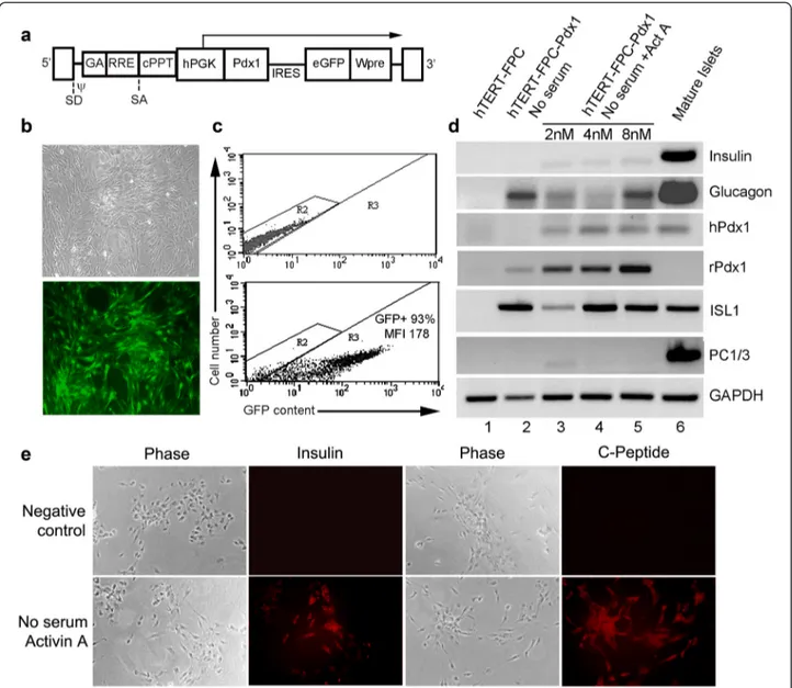
Fig. 2 Induction of insulin-expression in hTERT-FPC by Pdx1-LV. a shows schematic of LV with rat Pdx1 and GFP genes driven by hPGK promoter - IRES, intervening internal ribosomal entry site, cPPT, central polypurine tract, Wpre, posttranscriptional regulatory element of the woodchuck hepadnavirus. b shows Pdx1-LV-transduced hTERT-FPC under phase contrast (top) and under epifluorescence for GFP. c shows flow cytometric quantitation of GFP in nontransduced cells (top panel) and Pdx1-LV-transduced hTERT-FPC. MFI = mean fluorescence intensity. d shows RT-PCR for gene expression in control hTERT-FPC (lane 1), Pdx1-LV-transduced hTERT-FPC cultured without serum (lane 2) and without serum plus activin A (lane 3), and mature pancreatic islets (lane 4). e shows insulin and c-peptide expression in negative control hTERT-FPC-Pdx1 cells, where primary antibodies were omitted, and cells with expression of both insulin and c-peptide. Orig. Mag., × 200
Author details
1
Hepatology Division, Department of Medicine, Albert Einstein College of Medicine, Ullmann Bldg., Rm 625, 1300 Morris Park Avenue, Bronx, NY 10461, USA.2Department of Pathology, Albert Einstein College of Medicine, Ullmann Bldg., Rm 625, 1300 Morris Park Avenue, Bronx, NY 10461, USA.
3
Endocrinology Division, Department of Medicine, Diabetes Research Center, Albert Einstein College of Medicine, Forchheimer Bldg., Rm 505, 1300 Morris Park Avenue, Bronx, NY 10461, USA.4Hepatology Division, Department of Medicine, Cancer Research Center, Diabetes Research Center, Center for Human Embryonic Stem Cell Research, Marion Bessin Liver Research Center, Ruth L. and David S. Gottesman Institute for Stem Cell and Regenerative Medicine Research, and Institute for Clinical and Translational Research, Albert Einstein College of Medicine, Ullmann Bldg., Rm 625, 1300 Morris Park Avenue, Bronx, NY 10461, USA.
Received: 1 September 2015 Revised: 1 September 2015 Accepted: 1 September 2015
Reference
1. Cheng K, Follenzi A, Surana M, Fleischer N, Gupta S. Switching of mesodermal and endodermal properties in hTERT-modified and expanded fetal human pancreatic progenitor cells. Stem Cell Res Ther. 2010;1:6.
Submit your next manuscript to BioMed Central
and take full advantage of:
• Convenient online submission
• Thorough peer review
• No space constraints or color figure charges
• Immediate publication on acceptance
• Inclusion in PubMed, CAS, Scopus and Google Scholar
• Research which is freely available for redistribution
Submit your manuscript at www.biomedcentral.com/submit
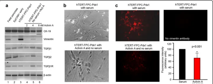
Fig. 3 Phenotype alterations in fetal pancreatic cells. a shows RT-PCR for epithelial marker, CK-19, and mesenchymal marker, vimentin, along with TGF-β1, TGFβ2 and their receptors under various conditions indicated. b shows morphological changes in LV-Pdx1-transduced hTERT-FPC during culture with serum and in the absence of serum plus addition of Activin A (bottom panel). These data indicated that cells became more rounded and less flattened in the absence of serum and presence of Activin A. c shows changes in vimentin expression by immunostaining in LV-Pdx1-transduced hTERT-FPC cultured with serum (top left), and with Activin A and no serum (bottom left). No immunostaining was detected when vimentin antibody was omitted (top right). The panel at bottom right in c shows quantitation of vimentin immunofluorescence signals by image analysis to indicate that culture without serum and with activin A perturbed cell phenotype, which was in agreement with morphological changes in LV-Pdx1-transduced hTERT-FPC