1
http://journals.tubitak.gov.tr/zoology/
© TÜBİTAK
doi:10.3906/zoo-1911-32
Nonpharmacological treatment options for Alzheimer’s disease: from animal testing to
clinical studies
Hasan TÜRKEZ1, Mehmet Enes ARSLAN1,*, Antonio Di STEFANO2, Ivana CACCIATORE2, Adil MARDİNOĞLU3,4
1Department of Molecular Biology and Genetics, Faculty of Science, Erzurum Technical University, Erzurum, Turkey 2Department of Pharmacy, University “G. d’Annunzio” of Chieti-Pescara, Chieti, Italy
3Centre for Host-Microbiome Interactions, Faculty of Dentistry, Oral & Craniofacial Sciences, King’s College London, London, UK 4Science for Life Laboratory, KTH - Royal Institute of Technology, Stockholm, Sweden
* Correspondence: [email protected]
1. Introduction
In 2014, the World Council for Dementia (WDC)
asked the Alzheimer’s Association (AA) to assess the
state of evidence of cognitive decline and dementia for
modifiable risk factors. Interestingly, in contrast to some
preclinical results, the AA declared that there was not
sufficient evidence supporting the links between various
modifiable risk factors including regular physical activity,
healthy diet, smoking, obesity, and lifelong learning/
cognitive training or stimulation and cognitive decline at
the 2014 WDC meeting in London
(Baumgart et al., 2015).
Although there is no definitive relationship between
individual demographic information and Alzheimer’s
disease (AD) treatments, better patient investigations can
be achieved by integrating a comprehensive assortment
of patient information based on sex, age, education,
lifestyle, medical history, and environmental exposures.
Also, this demographic analysis can be used to ameliorate
AD symptoms by diversification of the patient’s lifestyle
in nonpharmacological directions. Nonpharmacological
treatment options involve prevention strategies as the best
medicine since there are limited numbers of approaches
available for AD therapy. Therefore, it is common to try
to treat AD in its early stages, with or without medication.
Nonpharmacological strategies may be based on physical
activity, brain stimulation, social communication, and
diet-chemical substances (Peng et al., 2016).
2. Nonpharmacological studies on animal models
Most information comes from animal studies for the
positive effects of a physically and cognitively positive
lifestyle on AD. Studies on the effects of environmental
enrichment (EE), including residences with increased
opportunities for physical activity and cognitive status,
were conducted in various transgenic mouse models of
AD, partially inconsistent, but predominantly yielding
positive results on behavior and nerve pathology
(Nithianantharajah and Hannan, 2006). EE appears to
affect AD-like pathology through multiple mechanical
pathways, including hippocampal neurogenesis, amyloid
plaques, glial pathology, formation neurotrophic factors,
and AD-related factors (Ambrée et al., 2006; Wolf et al.,
2006; Herring et al., 2009; Beauquis et al., 2013). Recent
findings on animal models suggest that early exposure to
Abstract: Despite extensive pharmacological approaches, there is no curative therapy for Alzheimer’s disease (AD) or other types of
dementias. While current pharmacological options alleviate some symptoms of AD, they can lead to various adverse effects. Hence, nonpharmacological treatment options for AD are often considered with the assumption that they are safe, effective, and economic in managing patients. Furthermore, studies on animal models have suggested that environmental exposures like diet, music, or reward-related actions can stimulate neuronal regeneration and differentiation without using any pharmacological factors. The aim of this review is to provide a summary of nonpharmacological treatment options for the management of cognitive, emotional, and behavioral symptoms of AD. In addition, this review provides an overview of the challenging and encouraging experiences and recent studies and problems in cognitive training related to animal models. Nonpharmacological studies of AD are discussed in this literature review in terms of animal models, physical activity, brain stimulation, and the role of social communication.
Key words: Alzheimer’s disease, nonpharmacological, animal models, therapy, treatment options
Received: 20.11.2019 Accepted/Published Online: 30.01.2020 Final Version: 00.00.2020
EE is more effective in reducing AD-related psychological
deficiencies than late onset of amyloidogenesis and
indicates long-term protective effects (Verret et al., 2013).
The effects of EE also depend on the severity and the
direction of the disease. In a rapid degeneration of the APP/
PS1KI mouse model, EE did not enhance most behavioral
and physiological markers of pathology (Cotel et al., 2012).
According to these investigations, animal models are very
useful and varied to study nonpharmacological aspects
of AD, while other studies have shown that this diversity
can make studies more complex and irrelevant when an
inappropriate model organism is selected.
3. Physical activity
Clinical studies showed that physical activities (PAs) such
as 40 min of ergo-cycling, running in place, and
stair-climbing for 12 consecutive weeks improved neurogenesis
by enhancing cerebral blood flow in brain areas related to
the pathogenesis of AD (Chen et al., 2016). Recent findings
have revealed that PA leads to improvement in cognitive
function via several Aβ-dependent and independent
mechanisms, such as reducing the levels of beta-amyloid
(especially Aβ
1-42), amyloid precursor protein (APP),
beta-site APP-cleaving enzyme 1 (BACE1), presenilin (PS) 1,
and apolipoprotein E (APOE), or increasing the activities
of neprilysin (NEP) and insulin-degrading enzyme
(IDE) (Ebrahimi et al., 2017). Moreover, experimental
analysis put forth that the maintenance of PA, mainly
aerobic exercise and descending a ladder, modulated the
symptomatic progression of AD (Soni et al., 2019). The
frequency, intensity, or duration of PA was reported to be
responsible for raising brain-derived neurotrophic factor
(BDNF) signaling and altering small noncoding RNAs
(Nigam et al., 2017; Silva et al., 2017; Stephen et al., 2017).
PA improved brain blood flow, enlarged hippocampal
volume, and improved neurogenesis. Therefore, PA
has been considered to exhibit fewer side effects and
better adherence in comparison to medication-based
applications (Cass, 2017).
The suggested molecular mechanisms underlying the
prevention of AD development are based on different
pathways. The first is related to the interaction between PA
and the APOE ε4 allele. The APOE ε4 allele is considered
as the most significant genetic risk factor that may lead to
vascular damage and impaired cholesterol transport. A
recent study revealed that APOE ε4 allele-carrying patients
benefitted more from PA intervention than noncarriers in
terms of cognitive and neuropsychiatric functions as well
as physical measures (Jensen et al., 2019b). The second is
associated with the modulation of neuroinflammation in
AD. With that, the main mediators of neuroinflammation
such as interleukin-6 (IL-6) and soluble trigger receptor
expressed from myeloid cells 2 (sTREM2) were modulated
in AD patients after 16 weeks of PA (Jensen et al., 2019a).
Third, PA provided protection of cognitive functions
against Aβ-induced memory deficits associated with the
formation of oxidative stress and hippocampal cellular
disorganization (Rossi Dare et al., 2019). Fourth, PA
consisting of aerobic and resistance exercises decreases
acetylcholinesterase (AChE), thus improving cognition
and memory functions of patients with AD (Farzi et al.,
2019).
4. Brain stimulation
A number of noninvasive brain stimulation practices
have been suggested for AD patients and healthy older
adults to improve cognitive impairments associated
with physiological and pathological aging. The most
effective approaches for stimulating the brain are
repetitive transcranial magnetic stimulation (rTMS) and
transcranial direct current stimulation (tDCS) techniques.
While rTMS delivers strong magnetic pulses to the cortex
via the scalp, tDCS delivers weak electrical currents to the
scalp for modulating neuronal transmembrane potential
towards hyperpolarization or depolarization. However,
due to adverse effects and failure to provide measurable
differences between patients and placebo conditions 3
months after stimulation, the results of brain stimulation
have been ambiguous (Cotelli et al., 2014; Eliasova et al.,
2014; Khedr et al., 2014; Hsu et al., 2015).
5. Social communication
Hallucinatory experiences, social isolation, and loneliness
are more common in AD patients than in healthy
individuals (El Haj et al., 2016). Autopsy studies of 89
brains of AD patients showed that larger social networks
were associated with decreased negative effects on
cognition (Jedrziewski et al., 2014). Music and theater
therapies exert positive impacts on cognition in patients
with AD (Riello and Frisoni, 2001; Van Dijk et al., 2012a,
2012b). Listening to preferred music led to the activation
of the supplementary motor area, which is concerned
with memory. Moreover, listening to preferred music
stimulated corticocortical and corticocerebellar networks
that affect brain function (King et al., 2019). Likewise,
AD patients who joined a living-room theater activity
offered by professional actors recalled more memories and
exhibited less socially isolated behavior (van Dijk et al.,
2012a, 2012b).
6. Diet and disease relationships
Recent findings of epidemiological investigations
established that diet and nutrition are the main modifiable
risk factors for AD development. Different research
showed that diet can even change animal behaviors
and population habitat in nature. In consequence,
supplementations of antioxidants, certain vitamins,
polyphenols, and polyunsaturated fatty acids in the daily
diet along with eating fish, fruits, vegetables, and coffee
reduce the risk of AD. Adhering to a healthy diet, such
as Japanese, Argentinian, and Mediterranean diets, is
closely related to a reduced risk of AD (Hu et al., 2013).
Therefore, the combination of Mediterranean and
Dietary Approaches to Stop Hypertension (DASH) diets,
called MIND (Mediterranean-DASH Intervention for
Neurodegenerative Delay), has become prominent in
reducing AD risk. A recent cohort study revealed that the
MIND diet might radically alleviate the cognitive decline
due to age-related neurodegenerative diseases such as AD
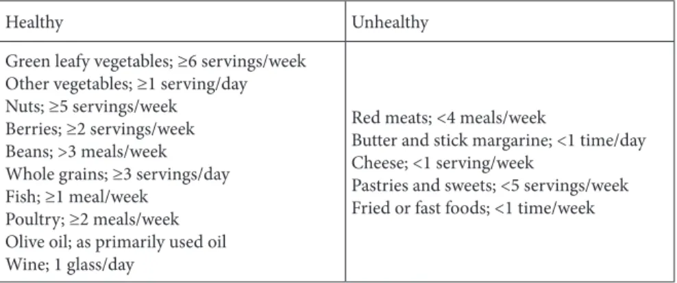
or other dementia types. This combined diet encourages
the consumption of brain-healthy foods like green leafy
vegetables, berries, beans, whole grains, olive oil, fish,
and poultry. The MIND diet also limits the consumption
of animal-based foods and foods high in saturated fat
(Table 1) (Morris et al., 2015a, 2015b). Also, heavy metal
accumulation negatively affects not only human brain
functions but also animal physiology.
A growing number of experimental studies have
revealed that oxidative stress plays a key role in both
initiation and progression of AD via lipid oxidation,
DNA oxidation, and glycoxidation, eventually leading
to mitochondrial defects. Therefore, antioxidant-based
therapeutics are assessed as promising tools for the
treatment of AD (Feng and Wang, 2012; Moneim, 2015).
Ascorbic acid, also known as vitamin C, has shown
therapeutic benefits against AD-related pathological
conditions in experimental animal studies. The
mechanisms behind the beneficial effects include
scavenging free radicals, inhibiting membrane lipid
peroxidation, modulating neuronal bioenergetics, and
antiproteolytic properties (Olajide et al., 2017). Moreover,
it was reported that vitamin D supplementation improved
the Mini-Mental State Exam (MMSE) score for cognitive
impairments in AD (Annweiler, 2014). On the other hand,
a high intake of vitamin E via diets is associated with the
incidence of AD. In contrast to this, randomized controlled
trials determined that treatment with vitamin E suspended
functional decline in patients with mild to moderate AD,
but vitamin E did not exhibit cognitive benefits in patients
or in generally healthy older individuals (Shinohara and
Yamada, 2015). The intake of probiotics was also suggested
as another type of nutrition-based nonpharmacological
treatment option due to their efficacy of modulating
proinflammatory cytokines related to gut microbiota
alterations with aging (Mendiola-Precoma et al., 2016).
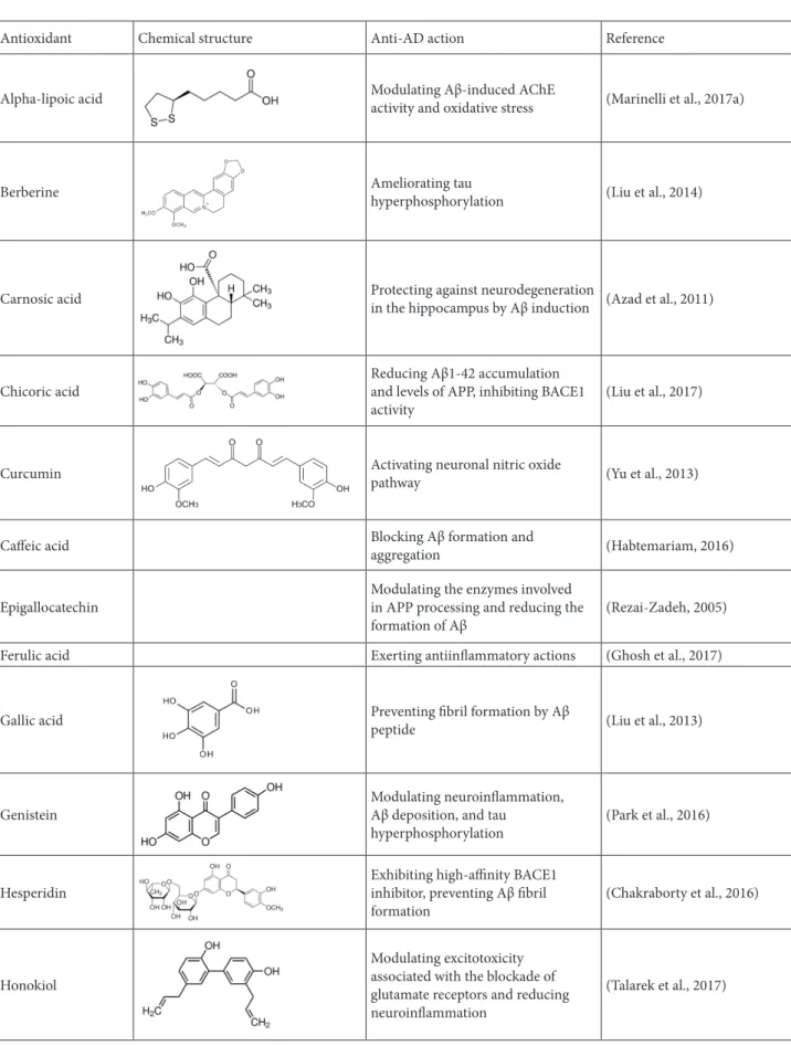
Several polyphenolic compounds obtained from fruits,
vegetables, herbs, and nuts also have neuroprotective
properties against AD and these naturally occurring
phytocompounds support memory and cognitive
function. The possible molecular mechanisms behind
their therapeutic potentials are generally associated with
alleviation of oxidative stress-mediated damage, protein
folding, and neuroinflammation (Essa et al., 2012; Shal et
al., 2018). The common phytocompounds and their specific
functions in AD therapy are summarized in Table 2.
It is still unclear how phytocompounds reach the brain.
Also, neither the quantity nor the biologically active form
that is required for exerting therapeutic actions is known
(Albarracin et al., 2012). About 500 years ago, the Swiss
scientist Paracelsus stated that “Poison is in everything, and
nothing is without poison. The dosage determines it either
a poison or a remedy”. Accordingly, at relatively higher
doses or under certain conditions antioxidant-containing
functional food ingredients including carotenoids,
vitamins C and E, and polyphenols, like flavonoids, show
prooxidant activities (Terada et al., 1999; Rietjens et al.,
2002). Lipoic acids (LAs) and their derivatives, known
as anti-Alzheimer molecules that prevent beta amyloid
accumulation, can be found in different plants such as
spinach, broccoli, and potatoes. In addition,
phenol-lipoyl hybrids are synthesized from LA derivatives and
these molecules are shown to be effective against AD
Table 1. Dietary components of the MIND diet.
Healthy Unhealthy
Green leafy vegetables; ≥6 servings/week Other vegetables; ≥1 serving/day Nuts; ≥5 servings/week
Berries; ≥2 servings/week Beans; >3 meals/week Whole grains; ≥3 servings/day Fish; ≥1 meal/week
Poultry; ≥2 meals/week Olive oil; as primarily used oil Wine; 1 glass/day
Red meats; <4 meals/week
Butter and stick margarine; <1 time/day Cheese; <1 serving/week
Pastries and sweets; <5 servings/week Fried or fast foods; <1 time/week
Table 2. Protection mechanisms by certain phytocompounds against various neurotoxic insults in AD.
Antioxidant Chemical structure Anti-AD action Reference
Alpha-lipoic acid Modulating Aβ-induced AChE activity and oxidative stress (Marinelli et al., 2017a)
Berberine Ameliorating tau hyperphosphorylation (Liu et al., 2014)
Carnosic acid Protecting against neurodegeneration in the hippocampus by Aβ induction (Azad et al., 2011)
Chicoric acid Reducing Aβ1-42 accumulation and levels of APP, inhibiting BACE1
activity (Liu et al., 2017)
Curcumin Activating neuronal nitric oxide pathway (Yu et al., 2013)
Caffeic acid Blocking Aβ formation and aggregation (Habtemariam, 2016)
Epigallocatechin Modulating the enzymes involved in APP processing and reducing the
formation of Aβ (Rezai-Zadeh, 2005)
Ferulic acid Exerting antiinflammatory actions (Ghosh et al., 2017)
Gallic acid Preventing fibril formation by Aβ peptide (Liu et al., 2013)
Genistein Modulating neuroinflammation, Aβ deposition, and tau
hyperphosphorylation (Park et al., 2016)
Hesperidin Exhibiting high-affinity BACE1 inhibitor, preventing Aβ fibril
formation (Chakraborty et al., 2016)
Honokiol
Modulating excitotoxicity associated with the blockade of glutamate receptors and reducing neuroinflammation
Icariin Preventing Aβ1-42 aggregation (Liu et al., 2015)
Leucomicin Preventing Aβ1-42 induced oxidative stress (Türkez, 2018)
Lycopene Inhibiting NF-κB activity and regulating neuroinflammatory
cytokines (Sachdeva and Chopra, 2015)
Naringenin Alleviating Aβ-induced impairments, lipid peroxidation, and apoptosis via
the estrogenic pathway. (Ghofrani et al., 2015)
Nobiletin Reducing Aβ plaques, NFTs, and cognitive impairments (Nakajima et al., 2015)
Oleuropein Counteracting amyloid-β peptide and tau aggregation (Martorell et al., 2016)
Quercetin Minimizing Aβ1-40 and Aβ1-42 amounts, decreasing
BACE1-mediated cleavage of APP
(Sabogal-Guáqueta et al., 2015)
Rosmarinic acid Preventing Aβ-sheet assembly (Cornejo et al., 2017)
Resveratrol
Promoting the removal of Aβ peptides
and antiinflammatory content (Braidy et al., 2016)
Rutin
Inhibiting Aβ aggregation, supporting antioxidant SOD, CAT, and GSH-Px
enzyme activities (Yu et al., 2015)
Sulforaphane Reducing cholinergic neuron loss (Zhang et al., 2014)
Tangeretin
Alleviating cholinergic deficits, reducing Aβ accumulation, inhibiting tau hyperphosphorylation and increasing NEP levels
(Braidy et al., 2017a)
Tannic acid Inhibiting BACE1 activity and aggregation of tau, disrupting Aβ
symptoms with synergistic effects due to their antioxidant
and antiamyloid properties (Cacciatore et al., 2016; Pagoni
et al., 2020). Another synthesized derivative of LA is
LA-GPE (R-α-lipoyl-Gly-l-Pro-l-Glu dimethyl ester), which
has antioxidant and enzyme inhibitory features. LA-GPE
molecules effectively ameliorate side effects of AD by
inhibiting AChE enzyme activity, increasing antioxidant
status, and preventing necrotic cell death (Marinelli et al.,
2017).
Therefore, the uptake of one kind of antioxidant
substance in overdose leads to detrimental conditions.
Due to this, the application of only one
antioxidative-containing compound against AD is not rational or
advantageous. The effectiveness of antioxidant therapy is
also allied with the optimum starting period of treatment.
As oxidative stress comes into existence very early in the
progression of AD, it is obvious that antioxidant therapy
will be helpful if started early before other harmful and
irreversible conditions arise (Persson et al., 2014).
7. Conclusion
About 110 years after the first description of AD,
neuroscientists have still not determined an exact
therapeutic approach against AD progression. Meanwhile,
the number of elderly people with AD is increasing all
over the world. In addition, the costs for fighting AD
are stressing the economies of even developed countries.
Unfortunately, many global drug companies are reluctant
to invest in the development of novel anti-AD formulations
and the current pharmacological or etiological treatment
options provide limited and insufficient results
without the exact cure of AD. Moreover, these existing
pharmacological applications exhibit side effects in
patients. At this point, nonpharmacological options
involving regular physical activity, brain stimulation,
improving social communication abilities, and altering
nutrition style present effective, safe, and economic
prevention and treatment opportunities to moderate AD.
However, the above-mentioned options seem to be not
exclusive and may be considered as complementary. The
uses of nonpharmacological options for patients with AD
have revealed clear potential benefits for quality of life.
Therefore, the efficacy of nonpharmacological treatment
approaches needs to be further investigated. For their
translation to clinical application, more standardized trials
with larger patient sizes are urgently required. In addition,
the integration of animal models in these studies can
ensure that results be obtained quickly and safely. Also,
using model organisms can reduce expenses and eliminate
many ethical issues associated with nonpharmacological
studies. This review has summarized 20 years of literature
studies that investigate nonpharmacological treatment of
AD related to nutritional habits, living conditions, and
social status.
Conflict of interest
The authors declare no potential conflicts of interest with
respect to research, authorship, and/or publication of this
paper.
References
Albarracin, SL, Stab B, Casas Z, Sutachan JJ, Samudio I et al. (2012). Effects of natural antioxidants in neurodegenerative disease. Nutritional Neuroscience 15 (1): 1-9. doi: 10.1179/1476830511Y.0000000028
Ambrée O, Leimer U, Herring A, Görtz N, Sachser N et al. (2006). Reduction of amyloid angiopathy and Aβ plaque burden after enriched housing in TgCRND8 mice. American Journal of Pathology 169 (2): 544-552. doi: 10.2353/ajpath.2006.051107 Annweiler C (2014). Vitamine D et maladie d’Alzheimer : d’une
curieuse idée à une possibilité de traitement. Biologie Aujourd’hui 208 (1): 89-95 (in French). doi: 10.1051/ jbio/2014005
Azad N, Rasoolijazi H, Joghataie MT, Soleimani S (2011). Neuroprotective effects of carnosic acid in an experimental model of Alzheimer’s disease in rats. Cell Journal 13 (1): 39-44. Baumgart M, Snyder HM, Carrillo MC, Fazio S, Kim H et al.
(2015). Summary of the evidence on modifiable risk factors for cognitive decline and dementia: a population-based perspective. Alzheimer’s and Dementia 11 (6): 718-726. doi: 10.1016/j.jalz.2015.05.016
Beauquis J, Pavía P, Pomilio C, Vinuesa A, Podlutskaya N et al. (2013). Environmental enrichment prevents astroglial pathological changes in the hippocampus of APP transgenic mice, model of Alzheimer’s disease. Experimental Neurology 239: 28-37. doi: 10.1016/j.expneurol.2012.09.009
Braidy N, Behzad S, Habtemariam S, Ahmed T, Daglia M et al. (2017a). Neuroprotective effects of citrus fruit-derived flavonoids, nobiletin and tangeretin in Alzheimer’s and Parkinson’s disease. CNS & Neurological Disorders - Drug Targets 16 (4): 387-397. doi: 10.2174/18715273166661703281 13309
Braidy N, Jugder BE, Poljak A, Jayasena T, Mansour H et al. (2016). Resveratrol as a potential therapeutic candidate for the treatment and management of Alzheimer’s disease. Current Topics in Medicinal Chemistry 16 (17): 1951-1960. doi: 10.217 4/1568026616666160204121431
Braidy N, Jugder BE, Poljak A, Jayasena T, Nabavi SM et al. (2017b). Molecular targets of tannic acid in Alzheimer’s disease. Current Alzheimer Research 14 (8): 861-869. doi: 10.2174/1567205014 666170206163158
Cacciatore I, Marinelli L, Fornasari E (2016). Novel NSAID-derived drugs for the potential treatment of Alzheimer’s disease. International Journal of Molecular Sciences 17: 1035. doi: 10.3390/ijms17071035
Cass SP (2017). Alzheimer’s disease and exercise. Current Sports Medicine Reports 16 (1): 19-22. doi: 10.1249/ JSR.0000000000000332
Chakraborty S, Bandyopadhyay J, Chakraborty S, Basu S (2016). Multi-target screening mines hesperidin as a multi-potent inhibitor: implication in Alzheimer’s disease therapeutics. European Journal of Medicinal Chemistry 121: 810-822. doi: 10.1016/j.ejmech.2016.03.057
Chen WW, Zhang X, Huang WJ (2016). Role of physical exercise in Alzheimer’s disease. Biomedical Reports 4 (4): 403-407. doi: 10.3892/br.2016.607
Cornejo A, Aguilar Sandoval F, Caballero L, Machuca L, Muñoz P et al. (2017). Rosmarinic acid prevents fibrillization and diminishes vibrational modes associated to β sheet in tau protein linked to Alzheimer’s disease. Journal of Enzyme Inhibition and Medicinal Chemistry 32 (1): 945-953. doi: 10.1080/14756366.2017.1347783
Cotel MC, Jawhar S, Christensen DZ, Bayer TA, Wirths O (2012). Environmental enrichment fails to rescue working memory deficits, neuron loss, and neurogenesis in APP/PS1KI mice. Neurobiology of Aging 33 (1): 96-107. doi: 10.1016/j. neurobiolaging.2010.02.012
Cotelli M, Manenti R, Petesi M, Brambilla M, Rosini S et al. (2014). Anodal tDCS during face-name associations memory training in Alzheimer’s patients. Frontiers in Aging Neuroscience 6: 38. doi: 10.3389/fnagi.2014.00038
Ebrahimi K, Majdi A, Baghaiee B, Hosseini SH, Sadigh-Eteghad S (2017). Physical activity and beta-amyloid pathology in Alzheimer’s disease: a sound mind in a sound body. EXCLI Journal 16: 959-972. doi: 10.17179/excli2017-475
El Haj M, Jardri R, Larøi F, Antoine P (2016). Hallucinations, loneliness, and social isolation in Alzheimer’s disease. Cognitive Neuropsychiatry 21 (1): 1-13. doi: 10.1080/13546805.2015.1121139
Eliasova I, Anderkova L, Marecek R, Rektorova I (2014). Non-invasive brain stimulation of the right inferior frontal gyrus may improve attention in early Alzheimer’s disease: a pilot study. Journal of the Neurological Sciences 346: 318-322. doi: 10.1016/j.jns.2014.08.036
Essa MM, Vijayan RK, Castellano-Gonzalez G, Memon MA, Braidy N et al. (2012). Neuroprotective effect of natural products against Alzheimer’s disease. Neurochemical Research 37: 1829-1842. doi: 10.1007/s11064-012-0799-9
Farzi MA, Sadigh-Eteghad S, Ebrahimi K, Talebi M (2019). Exercise improves recognition memory and acetylcholinesterase activity in the beta amyloid-induced rat model of Alzheimer’s disease. Annals of Neurosciences 25: 121-125. doi: 10.1159/000488580 Feng Y, Wang X (2012). Antioxidant therapies for Alzheimer’s
disease. Oxidative Medicine and Cellular Longevity 2012: 472932. doi: 10.1155/2012/472932
Ghofrani S, Joghataei MT, Mohseni S, Baluchnejadmojarad T, Bagheri M et al. (2015). Naringenin improves learning and memory in an Alzheimer’s disease rat model: Insights into the underlying mechanisms. European Journal of Pharmacology 764: 195-201. doi: 10.1016/j.ejphar.2015.07.001
Ghosh S, Basak P, Dutta S, Chowdhury S, Sil PC (2017). New insights into the ameliorative effects of ferulic acid in pathophysiological conditions. Food and Chemical Toxicology 103: 41-55. doi: 10.1016/j.fct.2017.02.028
Habtemariam S (2016). Protective effects of caffeic acid and the Alzheimer’s brain: an update. Mini-Reviews in Medicinal Chemistry 17 (8): 667-674. doi: 10.2174/13895575166661611 30100947
Herring A, Ambrée O, Tomm M, Habermann H, Sachser N et al. (2009). Environmental enrichment enhances cellular plasticity in transgenic mice with Alzheimer-like pathology. Experimental Neurology 216: 184-192. doi: 10.1016/j. expneurol.2008.11.027
Hsu WY, Ku Y, Zanto TP, Gazzaley A (2015). Effects of noninvasive brain stimulation on cognitive function in healthy aging and Alzheimer’s disease: a systematic review and meta-analysis. Neurobiology of Aging 36 (8): 2348-2359. doi: 10.1016/j. neurobiolaging.2015.04.016
Hu N, Yu JT, Tan L, Wang YL, Sun L et al. (2013). Nutrition and the risk of Alzheimer’s disease. BioMed Research International 2013: 524820. doi: 10.1155/2013/524820
Jedrziewski MK, Ewbank DC, Wang H, Trojanowski JQ (2014). The impact of exercise, cognitive activities, and socialization on cognitive function. American Journal of Alzheimer’s Disease & Other Dementias 29 (4): 372-378. doi: 10.1177/1533317513518646
Jensen CS, Bahl JM, Østergaard LB, Høgh P, Wermuth L et al. (2019a). Exercise as a potential modulator of inflammation in patients with Alzheimer’s disease measured in cerebrospinal fluid and plasma. Experimental Gerontology 121: 91-98. doi: 10.1016/j.exger.2019.04.003
Jensen CS, Simonsen AH, Siersm V, Beyer N, Frederiksen KS et al. (2019b). Patients with Alzheimer’s disease who carry the APOE ε4 allele benefit more from physical exercise. Alzheimer’s and Dementia: Translational Research and Clinical Interventions 5: 99-106. doi: 10.1016/j.trci.2019.02.007
Khedr EM, El Gamal NF, El-Fetoh NA, Khalifa H, Ahmed EM et al. (2014). A double-blind randomized clinical trial on the efficacy of cortical direct current stimulation for the treatment of Alzheimer’s disease. Frontiers in Aging Neuroscience 6: 275. doi: 10.3389/fnagi.2014.00275
King JB, Jones KG, Goldberg E, Rollins M, MacNamee K et al. (2019). Increased functional connectivity after listening to favored music in adults with Alzheimer dementia. Journal of Prevention of Alzheimer’s Disease 6 (1): 56-62. doi: 10.14283/ jpad.2018.19
Liu J, Liu Z, Zhang Y, Yin F (2015). A novel antagonistic role of natural compound icariin on neurotoxicity of amyloid β peptide. Indian Journal of Medical Research 142 (2): 190-195. doi: 10.4103/0971-5916.164254
Liu Q, Chen Y, Shen C, Xiao Y, Wang Y et al. (2017). Chicoric acid supplementation prevents systemic inflammation-induced memory impairment and amyloidogenesis via inhibition of NF-κB. FASEB Journal 31 (4): 1494-1507. doi: 10.1096/ fj.201601071R
Liu X, Zhou J, Abid MDN, Yan H, Huang H et al. (2014). Berberine attenuates axonal transport impairment and axonopathy induced by calyculin a in N2a cells. PLoS One 9 (4): e93974. doi: 10.1371/journal.pone.0093974
Liu Y, Pukala TL, Musgrave IF, Williams DM, Dehle FC et al. (2013). Gallic acid is the major component of grape seed extract that inhibits amyloid fibril formation. Bioorganic and Medicinal Chemistry Letters 23 (23): 6336-6340. doi: 10.1016/j. bmcl.2013.09.071
Marinelli L, Fornasari E, Di Stefano A, Turkez H, Arslan ME et al. (2017). (R)-α-Lipoyl-Gly-l-Pro-l-Glu dimethyl ester as dual acting agent for the treatment of Alzheimer’s disease. Neuropeptides 66: 52-58. doi: 10.1016/j.npep.2017.09.001 Martorell M, Forman K, Castro N, Capó X, Tejada S et al. (2016).
Potential therapeutic effects of oleuropein aglycone in Alzheimer’s disease. Current Pharmaceutical Biotechnology 17: 994-1001. doi: 10.2174/1389201017666160725120656 Mendiola-Precoma J, Berumen LC, Padilla K, Garcia-Alcocer G
(2016). Therapies for prevention and treatment of Alzheimer’s disease. BioMed Research International 2016: 1-17. doi: 10.1155/2016/2589276
Moneim A (2015). Oxidant/antioxidant imbalance and the risk of Alzheimer’s disease. Current Alzheimer Research 12 (4): 335-349. doi: 10.2174/1567205012666150325182702
Morris MC, Tangney CC, Wang Y, Sacks FM, Barnes LL et al. (2015a). MIND diet slows cognitive decline with aging. Alzheimer’s and Dementia 11 (9): 1015-1122. doi: 10.1016/j.jalz.2015.04.011 Morris MC, Tangney CC, Wang Y, Sacks FM, Bennett DA et al.
(2015b). MIND diet associated with reduced incidence of Alzheimer’s disease. Alzheimer’s and Dementia 11 (9): 1007-1014. doi: 10.1016/j.jalz.2014.11.009
Nakajima A, Aoyama Y, Shin EJ, Nam Y, Kim HC et al. (2015). Nobiletin, a citrus flavonoid, improves cognitive impairment and reduces soluble Aβ levels in a triple transgenic mouse model of Alzheimer’s disease (3XTg-AD). Behavioural Brain Research 289: 69-77. doi: 10.1016/j.bbr.2015.04.028
Nigam SM, Xu S, Kritikou JS, Marosi K, Brodin L et al. (2017). Exercise and BDNF reduce Aβ production by enhancing α-secretase processing of APP. Journal of Neurochemistry 142: 286-296. doi: 10.1111/jnc.14034
Nithianantharajah J, Hannan AJ (2006). Enriched environments, experience-dependent plasticity and disorders of the nervous system. Nature Reviews Neuroscience 7: 697-709. doi: 10.1038/ nrn1970
Olajide OJ, Yawson EO, Gbadamosi IT, Arogundade TT, Lambe E et al. (2017). Ascorbic acid ameliorates behavioural deficits and neuropathological alterations in rat model of Alzheimer’s disease. Environmental Toxicology and Pharmacology 50: 200-211. doi: 10.1016/j.etap.2017.02.010
Pagoni A, Marinelli L, Di Stefano A (2020) Novel anti-Alzheimer phenol-lipoyl hybrids: synthesis, physico-chemical characterization, and biological evaluation. European Journal of Medicinal Chemistry 186: 111880. doi: 10.1016/j. ejmech.2019.111880
Park YJ, Ko J, Jeon S, Kwon YH (2016). Protective effect of genistein against neuronal degeneration in ApoE-/- mice fed a high-fat diet. Nutrients 8 (11): 692. doi: 10.3390/nu8110692
Peng X, Xing P, Li X, Qian Y, Song F et al. (2016). Towards personalized intervention for Alzheimer’s disease. Genomics, Proteomics & Bioinformatics 14 (5): 289-297. doi: 10.1016/j. gpb.2016.01.006
Persson T, Popescu BO, Cedazo-Minguez A (2014). Oxidative stress in Alzheimer’s disease: Why did antioxidant therapy fail? Oxidative Medicine and Cellular Longevity 2014: 427318. doi: 10.1155/2014/427318
Rezai-Zadeh K (2005). Green tea epigallocatechin-3-gallate (EGCG) modulates amyloid precursor protein cleavage and reduces cerebral amyloidosis in Alzheimer transgenic mice. Journal of Neuroscience 25 (38): 8807-8814. doi: 10.1523/ JNEUROSCI.1521-05.2005
Riello R, Frisoni GB (2001). Music therapy in Alzheimer’s disease: is an evidence-based approach possible? Recenti Progressi in Medicina 92 (5): 317-321 (in Italian with an abstract in English).
Rietjens IM, Boersma MG, Haan L, Spenkelink B, Awad HM et al. (2002). The pro-oxidant chemistry of the natural antioxidants vitamin C, vitamin E, carotenoids and flavonoids. Environmental Toxicology and Pharmacology 11: 321-333. doi: 10.1016/S1382-6689(02)00003-0
Rossi Dare L, Garcia A, Alves N, Ventura Dias D, de Souza MA et al. (2019). Physical and cognitive training are able to prevent recognition memory deficits related to amyloid beta neurotoxicity. Behavioural Brain Research 365: 190-197. doi: 10.1016/j.bbr.2019.03.007
Sabogal-Guáqueta AM, Muñoz-Manco JI, Ramírez-Pineda JR, Lamprea-Rodriguez M, Osorio E et al. (2015). The flavonoid quercetin ameliorates Alzheimer’s disease pathology and protects cognitive and emotional function in aged triple transgenic Alzheimer’s disease model mice. Neuropharmacology 93: 134-145. doi: 10.1016/j. neuropharm.2015.01.027
Sachdeva AK, Chopra K (2015). Lycopene abrogates Aβ (1–42)-mediated neuroinflammatory cascade in an experimental model of Alzheimer’s disease. Journal of Nutritional Biochemistry 26 (7): 736-744. doi: 10.1016/j. jnutbio.2015.01.012
Shal B, Ding W, Ali H, Kim YS, Khan S (2018). Anti-neuroinflammatory potential of natural products in attenuation of Alzheimer’s disease. Frontiers in Pharmacology 9: 548. doi: 10.3389/fphar.2018.00548
Shinohara M, Yamada M (2015). Vitamin E and Alzheimer’s disease. Brain and Nerve 67 (12): 1509-1513 (in Japanese with an abstract in English).
Silva GJJ, Bye A, el Azzouzi H, Wisløff U (2017). MicroRNAs as important regulators of exercise adaptation. Progress in Cardiovascular Diseases 60 (1): 130-151. doi: 10.1016/j. pcad.2017.06.003
Soni M, Orrell M, Bandelow S, Steptoe A, Rafnsson S et al. (2019). Physical activity pre- and post-dementia: English Longitudinal Study of Ageing. Aging and Mental Health 23 (1): 15-21. doi: 10.1080/13607863.2017.1390731
Stephen R, Hongisto K, Solomon A, Lönnroos E (2017). Physical activity and Alzheimer’s disease: a systematic review. Journals of Gerontology Series A, Biological Sciences and Medical Sciences 72 (6): 733-739. doi: 10.1093/gerona/glw251
Talarek S, Listos J, Barreca D, Tellone E, Sureda A et al. (2017). Neuroprotective effects of honokiol: from chemistry to medicine. BioFactors 43 (6): 760-769. doi: 10.1002/biof.1385 Terada A, Yoshida M, Seko Y, Kobayashi T, Yoshida K et al. (1999).
Active oxygen species generation and cellular damage by additives of parenteral preparations: selenium and sulfhydryl compounds. Nutrition 15: 651-655. doi: 10.1016/S0899-9007(99)00119-7
Türkez H, Arslan ME (2018). Neuroprotective effects of leucomicine sesquiterpene on differentiated SH-SY5Y neuroblastoma cell line. Neuroendocrinology 107: 35-36.
Van Dijk A, van Weert JCM, Dröes RM (2012). Does theatre improve the quality of life of people with dementia? International Psychogeriatrics 12: 99-116. doi: 10.1017/S1041610211001992
Van Dijk AM, van Weert JCM, Dröes RM (2012). Theater als contactmethode in de psychogeriatrische zorg: effecten op gedrag, stemming en kwaliteit van leven van mensen met dementie. Tijdschrift Voor Gerontologie En Geriatrie 43 (6): 283-295. doi: 10.1007/s12439-012-0042-9
Verret L, Krezymon A, Halley H, Trouche S, Zerwas M et al. (2013). Transient enriched housing before amyloidosis onset sustains cognitive improvement in Tg2576 mice. Neurobiology of Aging 34: 211-225. doi: 10.1016/j.neurobiolaging.2012.05.013 Wolf SA, Kronenberg G, Lehmann K, Blankenship A, Overall R et
al. (2006). Cognitive and physical activity differently modulate disease progression in the amyloid precursor protein (APP)-23 model of Alzheimer’s disease. Biological Psychiatry 60 (12): 1314-1323. doi: 10.1016/j.biopsych.2006.04.004
Yu SY, Zhang M, Luo J, Zhang L, Shao Y et al. (2013). Curcumin ameliorates memory deficits via neuronal nitric oxide synthase in aged mice. Progress in Neuro-Psychopharmacology and Biological Psychiatry 45: 47-53. doi: 10.1016/j. pnpbp.2013.05.001
Yu XL, Li YN, Zhang H, Su YJ, Zhou WW et al. (2015). Rutin inhibits amylin-induced neurocytotoxicity and oxidative stress. Food & Function 6 (10): 3296-3306. doi: 10.1039/C5FO00500K Zhang R, Zhang J, Fang L, Li X, Zhao Y et al. (2014). Neuroprotective
effects of sulforaphane on cholinergic neurons in mice with Alzheimer’s disease-like lesions. International Journal of Molecular Sciences 15 (8): 14396-14410. doi: 10.3390/ ijms150814396