Abstract
Purpose: To investigate the diagnostic accuracy of MRI for Placenta Accreta Spectrum (PAS) placental invasion diagnosis and clinical outcome prediction in women with placenta previa, using a novel MRI-based predictive model.
Methods: Thirty-eight placental MRI exams performed on a 1.5T scanner were retrospectively reviewed by two radiologists in consensus. The presence of T2 dark bands, myometrial thinning, abnormal vascularity, uterine bulging, placental heterogeneity, placental protrusion sign, placental recess and percretism signs was scored using a 5-point scale. Pathology and clinical intrapartum findings were the standard of reference for PAS placental invasion while intrapartum/peripartum bleeding and emergency hysterectomy defined the clinical outcome. Receiver operating
characteristic (ROC) analysis and discriminant function analysis were performed to test the
predictive power of MRI findings for both PAS placental invasion and clinical outcome prediction. Results: Abnormal vascularity and percretism signs were the two most predictive MRI features of PASplacental invasion. The area under the curve (AUC) of the predictive function was 0.833 (cut-off 0.39, 67% sensitivity, 100% specificity, p=0.001). Percretism signs and myometrial thinning were the two most predictive MRI features of poor outcome. AUC of the predictive function was 0.971 (cut-off -0.55, 100% sensitivity, 77% specificity, p<0.001).
Conclusion: The diagnostic accuracy of MRI, especially considering the combination of the most predictive MRI findings, is higher when the target of the prediction is the clinical outcome rather than the PASplacental invasion.
Keywords
Magnetic resonance;
Placenta Accreta Spectrum (PAS); Placenta Previa;
Placental Invasion; Invasive placenta; Intrapartum bleeding; Clinical outcome.
Introduction
Placenta Accreta Spectrum (PAS) is the general term applied to abnormal adherence of the placental trophoblast to the uterine placenta. The spectrum includes the attachment of the placenta to myometrium without intervening decidua (placenta accreta), the invasion of the myometrium (placenta increta) and the infiltration of the surrounding organs through the uterine serosa (placenta percreta) [1]Invasive placenta is defined when the chorionic villi adhere to the myometrium (placenta accreta), invade the myometrium (placenta increta) or infiltrate the surrounding organs through the uterine serosa (placenta percreta). Previous caesarean section and placenta previa are the two most important risk factors [2]. Pregnancies with invasive placenta are more likely to have a preterm delivery and develop potentially fatal (7% cases) massive bleeding caused by the abnormal invasion of the uterus by the placenta [3, 4]. More in detail, placental implantation abnormalities, including placenta previa and placenta accreta, can have
catastrophic consequences for both mother and fetus, especially as pregnancy progresses to term
[5]. The damage of myometrial circulation by the placental invasion is responsible for maternal bleeding and potential fetal compromise [6]. The presence of placenta previa represents a risk factor for vasa previa and premature rupture of membranes (PROM) that can lead to vessel tearing and rapid fetal bleeding [5]. Moreover, placenta previa is associated with dysregulated interface function (cell-free human placental lactogen mRNA is elevated in maternal circulation of these patients) leading to an increased risk of fetomaternal hemorrhage [7]. Up to 40-60% of the peripartum hysterectomies are due to invasive placenta [8]. The International Federation of Gynecology and Obstetrics (FIGO) recently developed guidelines and recommendations to improve the diagnosis and the management (conservative versus nonconservative) of PAS
disorders, thus reducing the burden of maternal mortality and long-term sequelae that arise from this disease [9-13].
In this context, the prenatal diagnosis of invasive placenta plays an essential role in the delivery management. Although ultrasound (US) still represents the first line examination for the antenatal
care, a growing interest towards magnetic resonance (MR) imaging was developed in recent years [14, 15]. In fact, compared to US, MR has the advantage of high soft tissue contrast resolution and provides a panoramic view of the tissues and organs surrounding the uterus. In this way, it can be helpful to add topographic and morphologic information representing a complementary tool in cases with equivocal ultrasound findings or when additional information is needed [14, 16, 17].
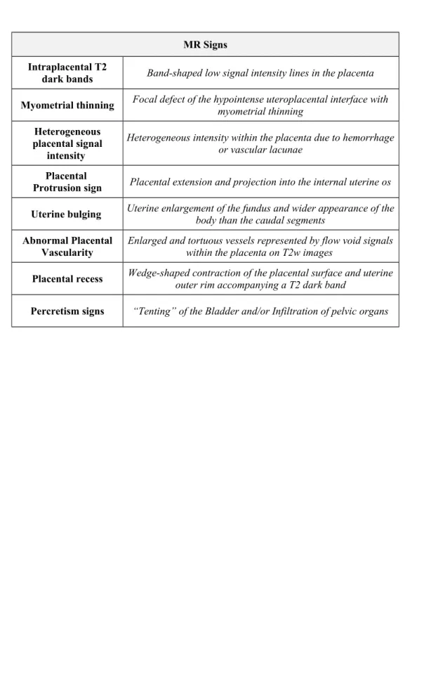
Moreover, several studies described some MR predictive "signs" of placental invasion, such as the presence of dark intraplacental bands in T2-weighted (T2w) images, myometrial thinning,
heterogeneous placental signal intensity, placental protrusion sign, abnormal uterine bulging sign, abnormal placental vascularity, placental recess, tenting of the bladder and/or infiltration of pelvic organs (Table 1) [18-27]. In this regard, a recently published study investigated the potential role of the MR to predict the clinical outcome in terms of treatment (conservative or not) and bleeding (massive or minor) of women with invasive placenta previa [26]. However, none of the above mentioned studies investigated the best combination of predictive MR signs correlating MR findings with both PAS and clinical outcome.
However, none of the above mentioned studies investigated the best combination of predictive MR signs shifting the focus from the placental invasion to the clinical outcome.
Therefore, the aim of our study was to investigate the diagnostic accuracy of MRI for PAS placental invasion diagnosis and clinical outcome prediction in women with placenta previa, using a novel MRI-based predictive model.
Methods Patients
All our procedures involving human subjects were in accordance with the ethical standards of the institutional and/or national research committee and with the 1964 Helsinki declaration and its later amendments or comparable ethical standards. All imaging data were retrospectively retrieved from PACS and informed consent was waived.
Fifty consecutive parturients with placenta previa and clinically and/or ultrasound suspect of PAS from June 2014 to May 2017 were retrospectively selected.
Fifty consecutive parturients who underwent a clinically indicated MRI of the placenta from June 2014 to May 2017 were retrospectively included.
Inclusion criteria were: (1) availability of MR imaging and (2) surgery outcome/pathology. Among the fifty potentially eligible patients, 12 were excluded. More specifically, nine were excluded due to patient transfer to another facility/hospital before deliver, one had vaginal delivery, one underwent to emergency delivery before MR examination and in one case the ultrasound findings of placenta previa were not confirmed by the MR exam.
Among them, 12 were excluded. More specifically, nine were excluded due to patient transfer to another facility/hospital before deliver, one case did not have surgery outcome nor pathology, one underwent to emergency delivery before MR examination and in one case the ultrasound findings of placenta previa were not confirmed by the MR exam.
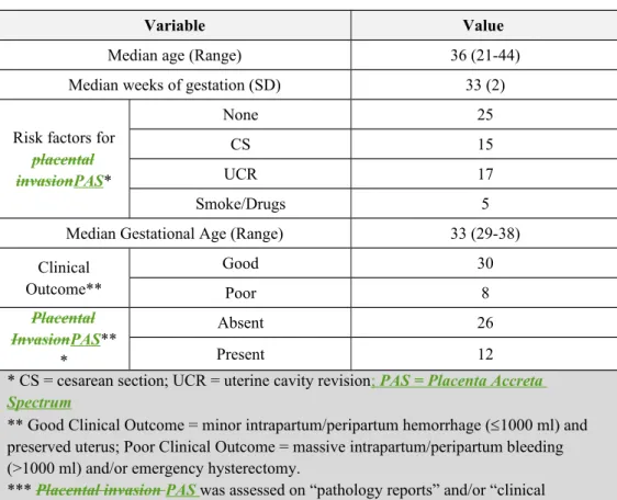
Finally, a total of 38 patients (median age 36 years; range 21-44) were included in the study. All the MR exams were performed within a week from the US examination. The baseline characteristics of all patients included in this study are shown in Fig. 1.
MRI technique
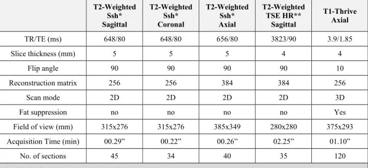
The MRI studies were performed on a 1.5T Scanner (Achieva, Philips Medical System, Best, the Netherlands).MR protocol comprised T2w images (including high resolution sequences) in sagittal, coronal, and axial orientations by using a fast spin-echo sequence; T1-Thrive were obtained in axial plane. MR protocols and sequence parameters are summarized in Table 2Table 1.
Imaging Analysis
Two radiologists, with at least five years of experience in abdominal imaging MR, in consensus reviewed the MR images assessing the following features (Fig. 2):
1. Intraplacental T2 dark bands: band-shaped low signal intensity lines in the placenta 2cm (Fig. 2a) [18].
2. Myometrial thinning: focal defect of the hypointense uteroplacental interface with myometrial thinning or indistinctness of myometrial delineation on T2w images (Fig. 2b) [18, 22].
3. Intraplacental abnormal vascularization: enlarged and tortuous vessels represented by flow void signals with a diameter > 6 mm within the placenta on T2w images (Fig. 2c) [20]. 4. Uterine bulging: loss of normal “pear shape” of the uterus, with the enlargement of the
fundus and the wider appearance of the body than the caudal segments (Fig. 2d) [18, 19]. 5. Heterogenous intraplacental sign: heterogeneous intensity within the placenta due to
hemorrhage or vascular lacunae (Fig. 2e) [18, 23].
6. Placental protrusion sign: the placenta extends and project into the internal uterine os (Fig. 2f) [21].
7. Placental recess: it is described as a placental deformity with the contraction of the placental surface and uterine outer rim. It shows a wedge-shaped contour and accompanies a T2 dark band (Fig. 2g) [24].
8. Percretism signs: direct invasion of adjacent organs (bladder or rectum) (Fig. 2h) [19]. The presence of each MR sign was qualitatively assessed according to a 5-points scale: 1=absent, 2=probably absent, 3=indeterminate, 4=probably present, 5=certainly present.
In case of disagreement the two readers reviewed MR images a second time after two weeks. If the disagreement persisted a third radiologist (with 15 years of experience in abdominal imaging MR) decided the final score.
Standard of reference
Both “pathology reports” and “clinical intrapartum findings” were used as standard of reference to confirm the presence of invasive placenta. The clinical outcome was assessed after a
multidisciplinary team (gynecologists and radiologists) consensus based on clinically records. consensus, consisted of gynecologists and radiologists. The poor outcome group was defined as parturient with massive intrapartum/peripartum bleeding (>1000 ml) and/or emergency
hysterectomy (non-conservative management). The good outcome group was defined as parturient with minor intrapartum/peripartum hemorrhage (1000 ml) and preserved uterus (conservative management).
Statistical analysis
The frequency distribution of qualitative MRI features regarding the presence/absence of invasive placenta and poor/good clinical outcome was calculated by using Fisher exact test. Qualitative scores were dichotomized considering 1, 2, 3 as negative scores and 4, 5 as positive rating. The diagnostic power of each MR feature was calculated by receiver operating characteristic (ROC) curve analysis. All the MR features were included in a stepwise discriminant function analysis. This analysis was conducted to determine whether a set of variables is effective in predicting category membership. Wilks’ lambda was the variable selection method and F value was the criterion for entry and removal from the equation. In this way, at each step the variable that minimizes the overall Wilks’ lambda is included in the model. Coefficients were also determined indicating the unique contribution of each variable to the predictive function. Finally, a ROC curve analysis of the predictive function (both for invasive placenta and for clinical outcome) was used to determine a cutoff with relative sensitivity and specificity. A p-value ≤ .05 was considered statistically
significant. All statistical analyses were performed by using IBM SPSS Statistics software, version 20 (IBM, Armonk, NY).
Out of 38 patients, 12 (31.6%) were diagnosed with PAS invasive placenta (6 with placenta accreta accretism and 6 with placenta percretapercretism) and 26 (68.4%) did not show
PASplacental invasion. When considering the clinical outcome, 8 (21.1%) parturient underwent emergency hysterectomy and/or blood transfusion due to massive bleeding (>1000 ml) and 30 (78.9%) showed minor intrapartum/peripartum hemorrhage (1000 ml) and preserved uterus (Figs. 3, 4). In detail, all the 6 patients that revealed PAS invasive placenta with placenta percreta
percretism at pathology underwent emergency isterectomy. Further patient characteristics are summarized in Table 3Table 2.
The readers showed disagreement in five patients. In three of them it concerned the myometrial thinning and in two it regarded the uterine bulging sign. In all the five cases, the third
radiologist decided the final score.
The results of frequency distribution and the diagnostic accuracy (by means AUC and ROC curve) of each MR sign regarding PAS placental invasion and clinical outcome are shown in Table 4Table 3. None of the 38 parturients showed the placental protrusion sign at MRI examinations. This MR sign was excluded from the further analysis.
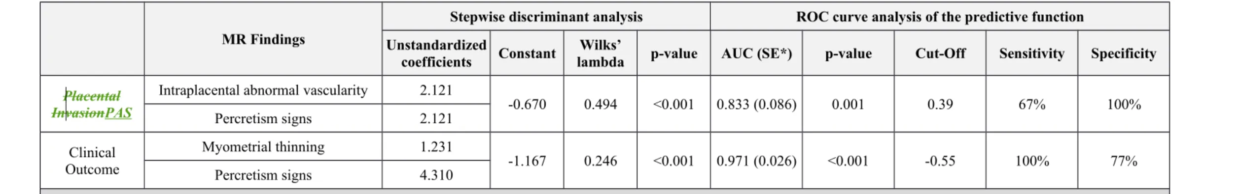
Diagnostic performance of the predictive models is summarized in Table 5 Table 4 and Figure 5. The Discriminant Analysis provided two specific predictive models, one for the PAS diagnosis (present in 12/38 patients) and one for the clinical outcome (delivery management) prediction (8/38 patients with non-conservative management). In detail, the predictive model selected two MR signs for the predictive function of PASplacental invasion: intraplacental abnormal vascularity and percretism signs. AUC of the predictive model for PAS placental invasion was 0.833 with a cut-off of 0.39 (67% sensitivity and 100% specificity, p=0.001). When considering the predictive function of clinical outcome, myometrial thinning and percretism signs were selected. AUC of the predictive model for clinical outcome was 0.971 with a cut-off of -0.55 (100% sensitivity and 77% specificity, p<0.001).
Discussion
The aim of our study was to investigate the role of MR for the PAS placental invasion diagnosis and the clinical outcome prediction in the prenatal planning of women with placenta previa. Our results demonstrated that the diagnostic performance of MR improves when the MR findings are
combined to predict the conservative versus non-conservative management. The rationale of this approach was that the delivery management prediction may represent an added value in the delivery management.The rationale of this approach was that the solely placental invasion
prediction may have limited influence on the delivery management. In fact, treatment strategies for PAS placental invasion depend not only on the presence or absence of invasive placenta but mostly on the type of PAS placental invasion (presence or not of adjacent organ invasion) and on the entity of bleeding. For instance, they range from conservative strategies (urotonics, intrauterine balloon tamponade and uterine artery embolization) to the hysterectomy in case of parturients with adjacent organ invasion or massive bleeding [28]. Additionally, if on one hand the presence of invasive placenta represents a risk factor for massive bleeding, on the other hand not all
intrapartum/peripartum bleeding are uniquely related to the presence of invasive placenta. In this regard, the delivery management may be influenced also by other causes of uterine hemorrhage including lack of uterine tone (not predictable with MR), lacerations, retained placental tissue, uterine inversion, infection and coagulation defects In fact, lack of uterine tone, lacerations, retained placental tissue, uterine inversion, infection and coagulation defects are related to the entity of uterine hemorrhage [29].
In this context, in our study the diagnostic performance improved considering the MR findings taken individually and combining the MR findings with the best diagnostic performances in a developed predictive model. In detail, percretism signs were found to be the most important predictive variable, both considering the PAS placental invasion and the clinical outcome
prediction. When the percretism signs were combined with myometrial thinning they differentiated parturients that underwent hysterectomy and/or intrapartum/peripartum massive bleeding with an
AUC of 0.971 (p<0.001). On the other hand, when percretism signs were combined to
intraplacental abnormal vascularity they predicted the presence of PAS invasive placenta with an AUC of 0.833 (p=0.001). Interestingly, the predictive power increased in terms of sensitivity (from 67% to 100%) if the subject moves from the PAS placental invasion to the clinical outcome, meaning that none of poor outcome patients was missed. These results may have a beneficial effect from a clinical point of view because they focus the attention on “critical” patients, thus
representing an alarm for clinicians, allowing to arrange in advance blood products and the most appropriate surgical team. In this context, the solely placental invasion prediction may result “incomplete” since not all the PAS invasive placentas develop massive bleeding and needs of hysterectomy. In fact, in our study population, the 33% (4/12) of PAS invasive placentas was treated conservatively by using uterotonics, intrauterine balloon tamponade and/or uterine artery embolization.
Looking at MR findings individually, intraplacental dark band, myometrial thinning, intraplacental abnormal vascularity, uterine bulging, placental recess and percretism signs showed significant differences between poor clinical outcome and good clinical outcome women. More in detail, myometrial thinning, percretism signs and placental recess showed the highest diagnostic accuracy (AUC of 0.883, 0.875 and 0.858 respectively). To the best of our knowledge the study by Chen et al., recently published, was the only one investigating the potential role of MRI findings for the prediction of clinical outcome [26]. The chosen criteria to define good and poor clinical outcome (massive/minor bleeding and conservative treatment/hysterectomy) were the same of our study. They demonstrated that dark band, percretism signs and placental protrusion sign were more frequently observed in patients with poor outcome and that dark band was the only significant predictor of poor outcome. Compared to their results, in our study percretism signs showed a 4-times greater predictive power (Table 5Table 4) compared to the second variable selected
First of all, the study population is different. In fact, Chen et al. included only women that
underwent uterine artery embolization assisted cesarean section. Secondly, some differences in the criteria for the image analysis existed. For example, they did not evaluate the placental recess and intraplacental abnormal vascularity. Furthermore, the definitions of MRI findings were slightly different compared to ours. For instance, we considered dark band as present only when they were equal or greater than 2 cm accordingly to Lax et al., while they assessed the presence of dark band regardless any dimensional limit [18].
When considering the PASplacental invasion, intraplacental abnormal vascularity and percretism signs showed the best diagnostic accuracy (AUC=0.750, p=0.014) followed by placental recess (AUC=0.731, p=0.024). These results are in line with those ofDerman et al. that in 2011 firstly identified the presence of enlarged tortuous flow voids on T2-weighted images as MR criterion of placental invasion [20]. Ueno et al. hypothesized that the intraplacental abnormal vascularity was related to the abnormal development of maternal arteries where the placenta adheres closely to the myometrium with subsequent infiltration of the placenta [21]. Placental recess was a more recently defined MR finding consisting of a wedge shaped placental deformity accompanied by a dark band. It showed promising results with high accuracy for the diagnosis of placental invasion [24]. The first MRI-based predictive model for the placental invasion was developed by Ueno et al. in 2016, and included abnormal vascularity, dark band, uterine bulging, heterogeneous placenta, placental protrusion sign and myometrial thinning [27]. Compared to their results, in our study we did not observe significant differences between the two groups in terms of dark band, myometrial thinning, heterogeneous placenta, and placental protrusion sign. In this regard, no parturients with placental protrusion sign were observed in our study, probably due to the small number of invasive placenta included in our population. In accordance with recent studies demonstrating that the prediction of placental invasions improved when at least two MRI signs are combined together, our stepwise discriminant analysis selected two MRI findings (percretism signs and intraplacental abnormal
vascularity) for the predictive function [25, 30]. The specificity of this function is however burdened with a relatively lower sensitivity meaning that 34% (4/12) of placental invasions PAS were missed. This percentage is nonetheless higher than that reported by Sato et al. (22% for abnormal vascularity) and comparable to that described by Valentini et al. (75% for intraplacental abnormal vascularity and of 50% for percretism signs) [24, 25].
We are aware that this study has some limitations. First of all, due to the fact that placental
invasion PAS and poor clinical outcome (non-conservative management) represent relatively rare conditions, our sample size is relatively small and this may have affected the results of statistical analysis. In this regard, we adopted restrictive selection criteria by including only patients with placenta previa to increase the homogeneity of our study population. Second, the MRI findings used in our study were established accordingly the recent literature. However, as discussed above, a certain grade of heterogeneity is undoubtedly still present and should be considered when
comparing our results with those of other studies. Third, due to the retrospective nature of the study, our results should be considered as preliminary report for future large perspective researches.
Conclusions
In conclusion, the diagnostic performance of MRI is higher when the target is the clinical outcome prediction rather than the PAS placental invasion diagnosis. In particular, the combination of two MR findings, namely percretism signs and myometrial thinning for clinical outcome, and percretism signs and intraplacental abnormal vascularity for PASplacental invasion, reached a higher
diagnostic accuracy compared to that of the individual findings. Further studies are warranted to confirm these results in larger sample size.
Author Contributions All authors were involved in patient management and wrote and/or reviewed the report. Written consent to publication was obtained.
Compliance with Ethical Standards
Conflict of Interest All authors declare that they have no conflict of interest.
Ethical Approval All procedures performed in this study involving human participant were in accordance with the ethical standards of the institutional and/or national research committee and with the 1964 Helsinki declaration and its later amendments or comparable ethical standards. Informed Consent Informed consent was obtained from the patients included in this study.
References
1. Silver, R.M. and D.W. Branch, Placenta Accreta Spectrum. N Engl J Med, 2018. 378(16): p. 1529-1536.
2. Higgins, M.F., et al., Real increasing incidence of hysterectomy for placenta accreta following previous caesarean section. Eur J Obstet Gynecol Reprod Biol, 2013. 171(1): p. 54-6.
3. Committee on Obstetric, P., Committee opinion no. 529: placenta accreta. Obstet Gynecol, 2012. 120(1): p. 207-11.
4. Hudon, L., M.A. Belfort, and D.R. Broome, Diagnosis and management of placenta percreta: a review. Obstet Gynecol Surv, 1998. 53(8): p. 509-17.
5. Vintzileos, A.M., C.V. Ananth, and J.C. Smulian, Using ultrasound in the clinical management of placental implantation abnormalities. Am J Obstet Gynecol, 2015. 213(4 Suppl): p. S70-7.
6. Jauniaux, E., S. Collins, and G.J. Burton, Placenta accreta spectrum: pathophysiology and evidence-based anatomy for prenatal ultrasound imaging. Am J Obstet Gynecol, 2018. 218(1): p. 75-87.
7. Umazume, T., et al., Occult fetomaternal hemorrhage in women with pathological placenta with respect to permeability. J Obstet Gynaecol Res, 2016. 42(6): p. 632-9. 8. D'Arpe, S., et al., Emergency peripartum hysterectomy in a tertiary teaching hospital: a
14-year review. Arch Gynecol Obstet, 2015. 291(4): p. 841-7.
9. Allen, L., et al., FIGO consensus guidelines on placenta accreta spectrum disorders: Nonconservative surgical management. Int J Gynaecol Obstet, 2018. 140(3): p. 281-290. 10. Jauniaux, E., et al., FIGO consensus guidelines on placenta accreta spectrum disorders:
Introduction. Int J Gynaecol Obstet, 2018. 140(3): p. 261-264.
11. Jauniaux, E., et al., FIGO consensus guidelines on placenta accreta spectrum disorders: Prenatal diagnosis and screening. Int J Gynaecol Obstet, 2018. 140(3): p. 274-280. 12. Jauniaux, E., et al., FIGO consensus guidelines on placenta accreta spectrum disorders:
Epidemiology. Int J Gynaecol Obstet, 2018. 140(3): p. 265-273.
13. Sentilhes, L., et al., FIGO consensus guidelines on placenta accreta spectrum disorders: Conservative management. Int J Gynaecol Obstet, 2018. 140(3): p. 291-298.
14. Fadl, S., et al., Placental Imaging: Normal Appearance with Review of Pathologic Findings. Radiographics, 2017. 37(3): p. 979-998.
15. Rahaim, N.S. and E.H. Whitby, The MRI features of placental adhesion disorder and their diagnostic significance: systematic review. Clin Radiol, 2015. 70(9): p. 917-25. 16. Zaidi, S.F., et al., Comprehensive Imaging Review of Abnormalities of the Placenta.
Ultrasound Q, 2016. 32(1): p. 25-42.
17. Blaicher, W., et al., Magnetic resonance imaging of the normal placenta. Eur J Radiol, 2006. 57(2): p. 256-60.
18. Lax, A., et al., The value of specific MRI features in the evaluation of suspected placental invasion. Magn Reson Imaging, 2007. 25(1): p. 87-93.
19. Baughman, W.C., J.E. Corteville, and R.R. Shah, Placenta accreta: spectrum of US and MR imaging findings. Radiographics, 2008. 28(7): p. 1905-16.
20. Derman, A.Y., et al., MRI of placenta accreta: a new imaging perspective. AJR Am J Roentgenol, 2011. 197(6): p. 1514-21.
21. Ueno, Y., et al., Novel MRI finding for diagnosis of invasive placenta praevia: evaluation of findings for 65 patients using clinical and histopathological correlations. Eur Radiol, 2014. 24(4): p. 881-8.
22. Bour, L., et al., Suspected invasive placenta: evaluation with magnetic resonance imaging. Eur Radiol, 2014. 24(12): p. 3150-60.
23. Masselli, G. and G. Gualdi, MR imaging of the placenta: what a radiologist should know. Abdom Imaging, 2013. 38(3): p. 573-87.
24. Sato, T., et al., Placental recess accompanied by a T2 dark band: a new finding for diagnosing placental invasion. Abdom Radiol (NY), 2017. 42(8): p. 2146-2153. 25. Valentini, A.L., et al., The morbidly adherent placenta: when and what association of
signs can improve MRI diagnosis? Our experience. Diagn Interv Radiol, 2017. 23(3): p. 180-186.
26. Chen, T., et al., Conventional MRI features for predicting the clinical outcome of patients with invasive placenta. Diagn Interv Radiol, 2017. 23(3): p. 173-179.
27. Ueno, Y., et al., Evaluation of interobserver variability and diagnostic performance of developed MRI-based radiological scoring system for invasive placenta previa. J Magn Reson Imaging, 2016. 44(3): p. 573-83.
28. Bailit, J.L., et al., Morbidly adherent placenta treatments and outcomes. Obstet Gynecol, 2015. 125(3): p. 683-9.
29. Committee on Practice, B.-O., Practice Bulletin No. 183: Postpartum Hemorrhage. Obstet Gynecol, 2017. 130(4): p. e168-e186.
30. Noda, Y., et al., Prenatal MR imaging diagnosis of placental invasion. Abdom Imaging, 2015. 40(5): p. 1273-8.
Tables
Table 1Table 2. MR sequences and parameters used for the study. MR Signs
Intraplacental T2
dark bands Band-shaped low signal intensity lines in the placenta Myometrial thinning Focal defect of the hypointense uteroplacental interface withmyometrial thinning
Heterogeneous placental signal
intensity
Heterogeneous intensity within the placenta due to hemorrhage or vascular lacunae
Placental
Protrusion sign Placental extension and projection into the internal uterine os Uterine bulging Uterine enlargement of the fundus and wider appearance of thebody than the caudal segments Abnormal Placental
Vascularity Enlarged and tortuous vessels represented by flow void signalswithin the placenta on T2w images
Placental recess Wedge-shaped contraction of the placental surface and uterineouter rim accompanying a T2 dark band Percretism signs “Tenting” of the Bladder and/or Infiltration of pelvic organs
T2-Weighted Ssh* Sagittal T2-Weighted Ssh* Coronal T2-Weighted Ssh* Axial T2-Weighted TSE HR** Sagittal T1-Thrive Axial TR/TE (ms) 648/80 648/80 656/80 3823/90 3.9/1.85 Slice thickness (mm) 5 5 5 4 4 Flip angle 90 90 90 90 10 Reconstruction matrix 256 256 384 384 256 Scan mode 2D 2D 2D 2D 3D
Fat suppression no no no no Yes
Field of view (mm) 315x276 315x276 385x349 280x280 375x293
Acquisition Time (min) 00.29” 00.22” 00.26” 02.25” 01.10”
No. of sections 45 34 40 35 120
*= Single shot; **=Turbo spin echo high resolution
Variable Value
Median age (Range) 36 (21-44)
Median weeks of gestation (SD) 33 (2) Risk factors for
placental invasionPAS* None 25 CS 15 UCR 17 Smoke/Drugs 5
Median Gestational Age (Range) 33 (29-38) Clinical Outcome** Good 30 Poor 8 Placental InvasionPAS** * Absent 26 Present 12
* CS = cesarean section; UCR = uterine cavity revision; PAS = Placenta Accreta Spectrum
** Good Clinical Outcome = minor intrapartum/peripartum hemorrhage (1000 ml) and preserved uterus; Poor Clinical Outcome = massive intrapartum/peripartum bleeding (>1000 ml) and/or emergency hysterectomy.
*** Placental invasion PAS was assessed on “pathology reports” and/or “clinical intrapartum findings”
Table 3Table 4. Diagnostic accuracy of MR findings for “Placental InvasionPAS” and “Clinical Outcome”.
MR Findings p-value* ROC curve analysis AUC (SE**) p-value Intraplacental T2 dark bands PASPlacental Invasion 0.157 0.638 (0.099) 0.177 Clinical Outcome 0.003 0.804 (0.085) 0.009 Myometrial thinning PASPlacental Invasion 0.481 0.577 (0.102) 0.451 Clinical Outcome <0.001 0.883 (0.053) 0.001 Intraplacental abnormal vascularization PASPlacental Invasion <0.001 0.750 (0.098) 0.014 Clinical Outcome <0.001 0.717 (0.118) 0.063 Uterine bulging PASPlacental Invasion 0.045 0.673 (0.100) 0.090 Clinical Outcome 0.002 0.808 (0.097) 0.008 Heterogeneous intraplacental sign PASPlacental Invasion 0.164 0.641 (0.097) 0.167 Clinical Outcome 0.117 0.675 (0.105) 0.133 Placental recess PASPlacental Invasion 0.002 0.731 (0.099) 0.024 Clinical Outcome <0.001 0.858 (0.095) 0.002 Percretism signs PAS Placental Invasion <0.001 0.750 (0.098) 0.014 Clinical Outcome <0.001 0.875 (0.094) 0.001 *= Fisher’s exact test
**= Standard error
ROC= Receiver Operating Characteristic curve AUC= Area Under the ROC Curve
Table Table 5.4. Stepwise discriminant analysis and ROC curve analysis of the predictive functions.
MR Findings
Stepwise discriminant analysis ROC curve analysis of the predictive function Unstandardized
coefficients Constant lambdaWilks’ p-value AUC (SE*) p-value Cut-Off Sensitivity Specificity
Placental InvasionPAS
Intraplacental abnormal vascularity 2.121
-0.670 0.494 <0.001 0.833 (0.086) 0.001 0.39 67% 100% Percretism signs 2.121 Clinical Outcome Myometrial thinning 1.231 -1.167 0.246 <0.001 0.971 (0.026) <0.001 -0.55 100% 77% Percretism signs 4.310
Figure Legends
Figure 1. Flowchart of the study population.
Figure 2. Sagittal T2-weighted (a-d, f-h) and axial T1-weighted (e) images showing MRI criteria. a Intraplacental dark band (white arrow) with the major diameter longest than 2 cm. b Myometrial thinning (white arrow): focal defect of the hypointense uteroplacental interface with myometrial thinning or indistinctness of myometrial delineation. c Intraplacental abnormal vascularity: enlarged and tortuous vessels (white arrow) with a diameter > 6 mm. d Uterine bulging (white arrows): loss of normal “pear shape” of the uterus, with the wider appearance of the body than the caudal segments. e Heterogenous intraplacental sign: heterogeneous intensity within the placenta due to hemorrhage or vascular lacunae (white asterisks). f Placental protrusion sign: the placenta extends and projects (white arrow) into the internal uterine os. g Placental recess: the contraction of the placental (white arrow) surface accompanying a dark band (white asterisk). h Percretism signs: direct invasion (white arrow) of bladder (B).
Figure 3. A case of good outcome in a 33-year old pregnant with invasive placenta. Sagittal (a, c) and coronal (b) T2-weighted images and axial T1-weighted image (d) showing abnormal flow voids (white arrow in a and b), uterine bulging (black arrow in c) and heterogeneous signal intensity due to focal hemorrhage (white asterisks in d). The patient had minor bleeding and underwent conservative treatment (bakri balloon and b-lynch suture). Invasive placenta with accretism was demonstrated at the delivery.
Figure 4. A case of poor outcome in a 36-year old pregnant with invasive placenta. Sagittal (a, b, c, d) T2-weighted images showing intraplacental dark bands (white asterisks in a), placental recess (black arrow in b), intraplacental abdnormal vascularity (white arrow in c), myometrial thinning with focal indistinctness of its delineation (black asterisk in c) and signs of percretism with bladder invasion (white P” in d). The patient underwent emergency hysterectomy and PAS placental invasion with percretism was histologically confirmed.
Figure 5. ROC curve analysis of MR findings and predictive functions for PAS placental invasion and clinical outcome.