Molecular Sciences
Article
Polymorphisms of Dopamine Receptor Genes and
Risk of L-Dopa–Induced Dyskinesia in
Parkinson’s Disease
Cristoforo Comi1,*,†, Marco Ferrari2,†, Franca Marino2, Luca Magistrelli1, Roberto Cantello1, Giulio Riboldazzi3, Maria Laura Ester Bianchi3, Giorgio Bono3and Marco Cosentino2
1 Movement Disorders Centre, Neurology Unit, Department of Translational Medicine,
University of Piemonte Orientale, 28100 Novara, Italy; [email protected] (L.M.); [email protected] (R.C.)
2 Center of Research in Medical Pharmacology, University of Insubria, 21100 Varese, Italy;
[email protected] (M.F.); [email protected] (F.M.); [email protected] (M.C.)
3 Departments of Biotechnology and Life Science, University of Insubria, 21100 Varese, Italy;
[email protected] (G.R.); [email protected] (M.L.E.B.); [email protected] (G.B.)
* Correspondence: [email protected]
† These authors contributed equally to this work. Academic Editor: Emil Alexov
Received: 31 October 2016; Accepted: 16 January 2017; Published: 24 January 2017
Abstract:L-dopa–induced dyskinesia (LID) is a frequent motor complication of Parkinson’s disease (PD), associated with a negative prognosis. Previous studies showed an association between dopamine receptor (DR) gene (DR) variants and LID, the results of which have not been confirmed. The present study is aimed to determine whether genetic differences of DR are associated with LID in a small but well-characterized cohort of PD patients. To this end we enrolled 100 PD subjects, 50 with and 50 without LID, matched for age, gender, disease duration and dopaminergic medication in a case-control study. We conducted polymerase chain reaction for single nucleotide polymorphisms (SNP) in both D1-like (DRD1A48G; DRD1C62T and DRD5T798C) and D2-like DR (DRD2G2137A, DRD2C957T, DRD3G25A, DRD3G712C, DRD4C616G and DRD4nR VNTR 48bp) analyzed genomic DNA. Our results showed that PD patients carrying allele A at DRD3G3127A had an increased risk of LID (OR 4.9; 95% CI 1.7–13.9; p = 0.004). The present findings may provide valuable information for personalizing pharmacological therapy in PD patients.
Keywords:Parkinson’s disease; SNPs; personalized medicine; disease progression; motor complications
1. Introduction
Levodopa-induced dyskinesia (LID) is a disabling motor complication of long-term levodopa therapy in Parkinson’s disease (PD) [1,2]. The risk factors of LID include young age at PD onset, severe degeneration of nigrostriatal neurons, longer exposure and higher total daily dose of levodopa [2]. The pathophysiology of LID is quite complex and not fully understood. Nonetheless, strong evidence supports the contribution of dopamine as a major player in LID development [3–5].
The existence of profound inter-individual heterogeneity suggests that genetic predisposition may be a relevant determinant of LID [2,6]. Several variants of DR genes have been detected, and their role has been characterized in Alzheimer’s disease, schizophrenia, bipolar disorder, and addiction [7–9]. Work from our group showed that the TT genotype at DRD1 rs686 may predispose PD patients to developing visual hallucinations (VHs) while subjects with GG at DRD1 rs4532 display a shorter time to VHs [10]. A few studies explored the possible influence of DR variants on LID development in
PD, but the reported results were not subsequently confirmed. One study showed that PD patients carrying the rs6280 single nucleotide polymorphism (SNP) at DRD3 have earlier onset of peak dose dyskinesia [11]. An additional, more recent report suggested a higher risk of LID in patients carrying the TTCTA haplotype at the DRD2/ANKK1 region [12]. Finally, Kaplan and colleagues did not find a significant correlation between SNPs at DRD2 and LID [13].
Due to the lack of confirmed results on the role of polymorphic DR variants in LID development, the present pilot study was designed to further investigate DR genetics in a small but accurately characterized cohort of Italian PD patients. To this end, we selected a panel of DR variants, giving priority to the most frequent and functionally characterized ones, and compared the frequency of all variants in two matched subgroups of PD patients with and without LID.
2. Results
Dopamine Receptor (DR) Genotypes
There were no significant differences in demographic and clinical characteristics between patients with and without LID (Table1). All DR alleles were in Hardy-Weinberg equilibrium (data not shown), and we did not find any linkage disequilibrium between considered SNPs. The frequencies of DR genotypes in patients with and without LID are shown in Table2. Using Fisher’s exact test (recessive model), we found that the risk of LID was, on average, 4.9 (95% CI 1.7–13.9) times higher in subjects carrying the A allele in the rs6280 (25G>A) of the DRD3, p = 0.004. The same results were obtained using dominant and codominant models (data not shown). This significant association was confirmed by a two-way ANOVA test (p = 0.002). Moreover, we found a trend for an association between the A allele in rs1800497 (2137G>A) and LID, p = 0.010; however, this association was not statistically significant after the Bonferroni correction. No other SNP studied was associated with LID (Table3).
Table 1.Demographic and clinical features of study population.
Feature No Dyskinesia Dyskinesia p
Number of subjects 50 50
Gender, male/female 28/22 28/22 na
Age at onset, mean ± SD 65.1 ± 5.6 63.3 ± 9.8 ns Disease duration (years) mean ± SD 10.8 ± 4.2 12.1 ± 5.2 ns Dyskinesia onset (years) mean ± SD na 7.6 ± 4.2 na
UPDRS III, mean ± SD *
ON 24 ± 10 23 ± 9 ns
OFF 29 ± 13 28 ± 12 ns
Hoehn and Yahr, median (range) * 3 (1–4) 3 (1–4) na L-dopa treatment duration (years) mean ± SD 8.9 ± 3.4 9.6 ± 3.3 ns Medication dose LED (mg/day), mean ± SD * 612.6 ± 242.6 741 ± 279.6 ns
* These variables were collected at time of event in patients with dyskinesia and at an equal time point from onset in each paired patient without dyskinesia; ns: not significant; na: not applicable; SD: standard deviation.
Table 2. Dopamine Receptor (DR) Frequency in Parkinson’s disease (PD) patients with and without dyskinesia.
Gene SNP Genotype
No Dyskinesia Dyskinesia Dyskinesia
Dyskinesia P (a) P (b) OR (95% CI)
DRD1 rs4532 A/A 11 (22%) 11 (22%) ns ns ns A/G 21 (42%) 20 (40%) G/G 18 (36%) 19 (38%) rs686 C/C 17 (34%) 15 (30%) ns ns ns C/T 25 (50%) 25 (50%) T/T 8 (16%) 10 (20%) DRD5 rs6283 T/T 33 (66%) 35 (70%) ns ns ns T/C 13 (26%) 15 (30%) C/C 4 (8%) 0 (0%)
Table 2. Cont.
Gene SNP Genotype
No Dyskinesia Dyskinesia Dyskinesia
Dyskinesia P (a) P (b) OR (95% CI)
DRD2 rs1800497 G/G 36 (72%) 22 (44%) ns ns ns G/A 11 (22%) 25 (50%) A/A 3 (6%) 3 (6%) rs6277 C/C 21 (42%) 11 (22%) ns ns ns C/T 20 (40%) 28 (56%) T/T 9 (18%) 11 (22%) DRD3 rs6280 G/G 26 (52%) 9 (18%) 0.0001 0.0001 4.9 (2.0–12.2) G/A 18 (36%) 21 (42%) A/A 6 (12%) 20 (40%) rs1800828 G/G 38 (76%) 39 (78%) ns ns ns G/C 8 (16%) 11 (224%) C/C 4 (8%) 0 (0%) DRD4 nR VNTR 48 bp repetition 4/4 33 (66%) 31 (62%) ns ns ns 4/7 16 (32%) 18 (36%) 7/7 1 (2%) 1 (2%) rs747302 C/C 39 (78%) 42 (84%) ns ns ns C/G 11 (22%) 8 (16%) G/G 0 (26.4%) 0 (0%)
Notes: (a), by χ2-test for trend; (b), by Fisher Exact Test. ns: not significant.
Table 3.Dopamine receptor (DR) gene variants analyzed in the study.
Receptor Gene Variant Change Frequency Effects Score
D1-like
D1 DRD1 rs4532 −48A>G 60 (%)
Association with nicotine dependence [14], tobacco smoking in schizophrenia [15], and alcohol dependence [16] and resistance to
schizophrenia treatment [17].
+1
rs686 62C>T 55 (%)
Higher DRD1 gene expression and association with nicotine dependence [14], alcohol dependence [17], and tobacco smoking in
schizophrenia [15].
+1
D5 DRD5 rs6283 978T>C 30 (%) na na
D2-like
D2 DRD2 rs1800497 2137G>A(Taq1A) 15 (%) Lower striatal DR D2density in healthy [18]. +1
rs6277 957C>T 50 (%)
Decreased DR D2mRNA stability and
translation, and reduced dopamine-induced up-regulation of DR D2membrane expression
in vitro [19], and lower DR D2expression in
cortex and thalamus of healthy subjects [20]. +1
D3 DRD3 rs6280
25G>A
(Ser9Gly) 60 (%)
Higher dopamine binding affinity in vitro [21], association with alcohol dependence [22] and
heroin dependence [23].
−1
rs1800828 −712G>C 20 (%) na na
D4 DRD4 rs747302 −616C>G 10 (%)
No effect on DR D4mRNA expression in
human post-mortem brain tissue samples [23], and no association with heroin dependence [24].
na
7 48-base
pair VNTR 20 (%)
Trend toward reduced DR D4mRNA expression
in human post-mortem brain tissue samples [25], lower response to stimulants and
requirements of higher doses of methylphenidate [26].
+1
Note: na: not applicable.
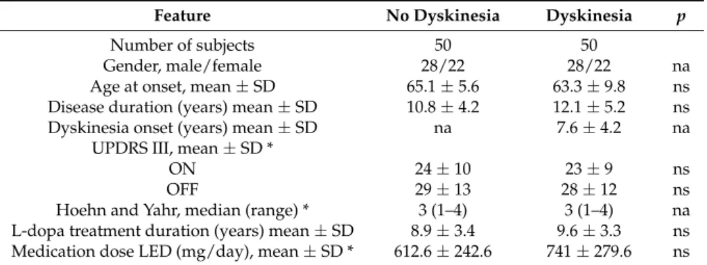
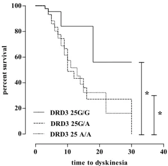
In Kaplan–Meier analysis, patients with DRD3 rs6280 AA and AG had significantly shorter times to LID when compared with patients with the GG genotype (median 10 and 13, respectively, vs. 18 years; log rank p = 0.005, see Figure1). No other SNP studied was associated with the timing of LID onset.
Int. J. Mol. Sci. 2017, 18, 242 4 of 8 In Kaplan–Meier analysis, patients with DRD3 rs6280 AA and AG had significantly shorter times to LID when compared with patients with the GG genotype (median 10 and 13, respectively, vs. 18 years; log rank p = 0.005, see Figure 1). No other SNP studied was associated with the timing of LID onset.
Figure 1. Correlations between rs6280 (DRD G25A) and time to dyskinesia. * = p < 0.005.
3. Discussion
This study provides the first evidence in an Italian cohort of PD patients that DR variability may predispose a person to LID. In fact, we found that the DRD3 G25A variant at rs6280 was independently associated with LID development after adjusting for gender, age at PD onset, H&Y stage, and duration of levodopa treatment. Furthermore, this variant was also associated with an earlier development of LID. Our results confirm a previous observation showing an association between the G25A allele at rs6280 DRD3 and an earlier onset of peak dose dyskinesia in Korean PD patients [11]. In addition, we also replicated the negative findings of Kaplan and colleagues regarding the possible correlations between SNPs at DRD2 and LID [13]. As regards the other DR SNPs analyzed in our study, there are no previously published data on PD patients. Our findings are negative, but we cannot exclude that one or more of such DR variations might show some relevance in other PD populations.
The precise mechanism through which the D3 receptor predisposes one to LID is open to discussion. Data on monkey models of PD showed that D3 expression was more abundant in animals with LID than in those without LID [27]. Furthermore, the DRD3 SNP rs6280 was shown to provide a higher binding affinity to dopamine [21] but also a higher susceptibility to tardive dyskinesia in patients with psychosis [28,29]. Dopamine receptor hypersensitivity may indeed be a possible mechanism involved in LID development [30], and the higher frequency of the DRD3 G25A genotype in PD patients with LID may be explained by a role of this variant in the sensitization process of the basal ganglia circuitry.
Along with dopamine, a number of reports indicate that other signaling pathways may be of relevance in the context of LID predisposition. Genetics of adenosine, serotonin, glutamic acid and endocannabinoid receptors have been investigated [31], and the exclusive focus of our study on dopamine receptor genetics may be seen as a limitation. Nonetheless, our purpose was to shed light onto a series of previous hypotheses implying the genetics of dopamine pathways in LID predisposition. Such hypotheses were intriguing, but had never been confirmed before.
The possibility that clinical differences in the two patient groups may have influenced the findings of our study was carefully evaluated. Therefore, we performed a strict sample matching: for each patient with LID we selected a paired patient with the same features, i.e., gender, age at onset,
0 10 20 30 40 0 20 40 60 80 100 DRD3 25G/A DRD3 25G/G DRD3 25 A/A
*
*
time to dyskinesia p erc en t s u rv iv alFigure 1.Correlations between rs6280 (DRD G25A) and time to dyskinesia. * = p < 0.005.
3. Discussion
This study provides the first evidence in an Italian cohort of PD patients that DR variability may predispose a person to LID. In fact, we found that the DRD3 G25A variant at rs6280 was independently associated with LID development after adjusting for gender, age at PD onset, H&Y stage, and duration of levodopa treatment. Furthermore, this variant was also associated with an earlier development of LID. Our results confirm a previous observation showing an association between the G25A allele at rs6280 DRD3 and an earlier onset of peak dose dyskinesia in Korean PD patients [11]. In addition, we also replicated the negative findings of Kaplan and colleagues regarding the possible correlations between SNPs at DRD2 and LID [13]. As regards the other DR SNPs analyzed in our study, there are no previously published data on PD patients. Our findings are negative, but we cannot exclude that one or more of such DR variations might show some relevance in other PD populations.
The precise mechanism through which the D3 receptor predisposes one to LID is open to discussion. Data on monkey models of PD showed that D3 expression was more abundant in animals with LID than in those without LID [27]. Furthermore, the DRD3 SNP rs6280 was shown to provide a higher binding affinity to dopamine [21] but also a higher susceptibility to tardive dyskinesia in patients with psychosis [28,29]. Dopamine receptor hypersensitivity may indeed be a possible mechanism involved in LID development [30], and the higher frequency of the DRD3 G25A genotype in PD patients with LID may be explained by a role of this variant in the sensitization process of the basal ganglia circuitry.
Along with dopamine, a number of reports indicate that other signaling pathways may be of relevance in the context of LID predisposition. Genetics of adenosine, serotonin, glutamic acid and endocannabinoid receptors have been investigated [31], and the exclusive focus of our study on dopamine receptor genetics may be seen as a limitation. Nonetheless, our purpose was to shed light onto a series of previous hypotheses implying the genetics of dopamine pathways in LID predisposition. Such hypotheses were intriguing, but had never been confirmed before.
The possibility that clinical differences in the two patient groups may have influenced the findings of our study was carefully evaluated. Therefore, we performed a strict sample matching: for each patient with LID we selected a paired patient with the same features, i.e., gender, age at onset, disease duration, and therapy. Such a rigorous design had the downside of markedly restricting the sample size. Indeed, the main limitation of our study lies in the relatively small number of enrolled patients, which nonetheless reached the minimum estimated size to assess [10,32]. A further limitation is the
candidate gene approach. Indeed, a more extensive analysis with direct sequencing of the five DRs might add relevant information to uninvestigated variations possibly playing a role in the genetics of LID.
4. Patients and Methods 4.1. Patients
We enrolled 100 consecutive patients with idiopathic PD: 50 patients who had experienced LID during their disease course, and 50 who had never complained of LID. Patients were all Caucasian Italian and were matched for age, gender, disease duration and treatment. The main features of the study population are shown in Table1. Patients were enrolled at the Movement Disorders Centers of the University of Piemonte Orientale, Novara, and University of Insubria, Varese, Italy according to the following inclusion criteria: (1) clinical diagnosis of PD according to the UK Parkinson’s Disease Society Brain Bank criteria [33]; (2) age at onset >40 years; (3) active and longitudinal follow-up >4 years; (4) treatment with levodopa; (5) reliable data concerning time of levodopa treatment initiation and time of LID presentation. The study was approved by the local Ethics Committee (Novara, protocol number 9606) and patients were enrolled after having read and signed an informed consent form [10].
Outcome measures were obtained retrospectively from clinical records, in the context of a larger collaborative initiative aimed at identifying the genetic determinants of PD progression [10]. A review of available data in routine clinical records of each center was performed and agreement was found on the following evaluations: detailed collection of patients’ history, complete neurological examination, Unified Parkinson’s Disease Rating Scale (UPDRS) score, Hoehn and Yahr (H&Y) stage, presence and time to development of LID [10].
All patients had undergone a longitudinal follow-up with assessments every three to six months performed by a neurologist expert in movement disorders. UPDRS score, H&Y stage, total L-dopa daily dose equivalent (LED), were recorded at the time of the event in patients with LID and at an equal time point from onset of PD in each paired patient without LID. LED was calculated according to Tomlinson et al. [34].
4.2. Genotyping
Samples of 3 mL venous blood were collected from each patient and genomic DNA was obtained using a standard DNA extraction protocol (Qiagen Inc., Hilden, Germany). The following DR variants: rs4532 (−48A>G and rs686 (62C>T) in DRD1; rs1800497 (2137G>A) and rs6277 (957C>T) in DRD2; rs6280 (25G>A) and rs1800828 (−712G>C) in DRD3; rs747302 (−616C>G), and 7 48-base pair VNTR in DRD4; and rs6283 (978T>C) in DRD5 were analyzed by real time PCR using a GeneAmp 9700 PCR System (ABI, Foster City, CA, USA) and pre-designed genotyping assays (ABI). The DRD4 7 48-base pair variable number tandem repeat (VNTR) was examined using a previously published method [35]. An example of PCR curve for each SNP is included in Figure S1.
4.3. Statistics
Genotype frequencies were analysed by the two-way ANOVA test, χ2-test for trend or by the Fisher’s exact test, as appropriate, and the odds ratio (OR) with 95% confidence interval (CI) was calculated using dominant, codominant and recessive model. Kaplan–Meier (KM) plot was used for correlations between patient genotype and time to LID. Curves were compared using Log-rank (Mantel–Cox) test [10]. Bonferroni correction was applied when multiple comparisons were performed. A p-value≤0.005 was considered statistically significant [10]. Presence of linkage disequilibrium was investigated using Haploview software [10].
5. Conclusions
In conclusion, our data provide a solid base towards the personalization of PD treatment since they may help in identifying fragile PD patients who would benefit from a less aggressive dopaminergic treatment.
Supplementary Materials:Supplementary materials can be found at www.mdpi.com/1422-0067/18/2/242/s1.
Acknowledgments:This work was supported by Institutional funding of the University of Piemonte Orientale (Fondi Ateneo-DIMET 2015).
Author Contributions: Study conception and design: Cristoforo Comi; Marco Ferrari; Franca Marino; Giorgio Bono; Marco Cosentino. Acquisition of data: Cristoforo Comi; Marco Ferrari; Luca Magistrelli, Roberto Cantello, Giulio Riboldazzi, Maria Laura Ester Bianchi. Analysis and interpretation of data: Cristoforo Comi; Marco Ferrari; Franca Marino; Giorgio Bono; Marco Cosentino. All authors were involved in drafting the article and/or revising it critically for important intellectual content, and all authors approved the final version to be published. All authors agree to be accountable for all aspects of the work in ensuring that questions related to the accuracy or integrity of any part of the work are appropriately investigated and resolved, and declare to have confidence in the integrity of the contributions of their co-authors.
Conflicts of Interest:The authors declare no conflicts of interest. References
1. Fahn, S. The spectrum of levodopa-induced dyskinesias. Ann. Neurol. 2000, 47, S2–S11. [PubMed]
2. Jankovic, J. Motor fluctuations and dyskinesias in Parkinson’s disease: Clinical manifestations. Mov. Disord.
2005, 20, S11–S16. [CrossRef] [PubMed]
3. Brotchie, J.M. Nondopaminergic mechanisms in levodopa-induced dyskinesia. Mov. Disord. 2005, 20, 919–931. [CrossRef] [PubMed]
4. Fabbrini, G.; Brotchie, J.M.; Grandas, F.; Nomoto, M.; Goetz, G.C. Levodopa-induced dyskinesias. Mov. Disord.
2007, 22, 1379–1389. [CrossRef] [PubMed]
5. Calabresi, P.; Di Filippo, M.; Ghiglieri, V.; Picconi, B. Molecular mechanisms underlying levodopa-induced dyskinesia. Mov. Disord. 2008, 23, S570–S579. [CrossRef] [PubMed]
6. Thanvi, B.; Lo, N.; Robinson, T. Levodopa-induced dyskinesia in Parkinson’s disease: Clinical features, pathogenesis, prevention and treatment. Postgrad. Med. J. 2007, 83, 384–388. [CrossRef] [PubMed]
7. Wong, A.H.; Buckle, C.E.; van Tol, H.H. Polymorphisms in dopamine receptors: What do they tell us? Eur. J. Pharmacol. 2000, 410, 183–203. [CrossRef]
8. McAllister, T.W.; Summerall, L. Genetic polymorphisms in the expression and treatment of neuropsychiatric disorders. Curr. Psychiatry Rep. 2003, 5, 400–409. [CrossRef] [PubMed]
9. Le Foll, B.; Gallo, A.; Le Strat, Y.; Lu, L.; Gorwood, P. Genetics of dopamine receptors and drug addiction: A comprehensive review. Behav. Pharmacol. 2009, 20, 1–17. [CrossRef] [PubMed]
10. Ferrari, M.; Comi, C.; Marino, F.; Magistrelli, L.; de Marchi, F.; Cantello, R.; Riboldazzi, G.; Bono, G.; Cosentino, M. Polymorphisms of dopamine receptor genes and risk of visual hallucinations in Parkinson’s patients. Eur. J. Clin. Pharmacol. 2016, 72, 1335–1341. [CrossRef] [PubMed]
11. Lee, J.Y.; Cho, J.; Lee, E.K.; Park, S.S.; Jeon, B.S. Differential genetic susceptibility in diphasic and peak-dose dyskinesias in Parkinson’s disease. Mov. Disord. 2011, 26, 73–79. [CrossRef] [PubMed]
12. Rieck, M.; Schumacher-Schuh, A.F.; Altmann, V.; Francisconi, C.L.; Fagundes, P.T.; Monte, T.L.; Callegari-Jacques, S.M.; Rieder, C.R.M.; Hutz, M.H. DRD2 haplotype is associated with dyskinesia induced by levodopa therapy in Parkinson’s disease patients. Pharmacogenomics 2012, 13, 1701–1710. [CrossRef] [PubMed]
13. Kaplan, N.; Vituri, A.; Korczyn, A.D.; Cohen, O.S.; Inzelberg, R.; Yahalom, G.; Kozlova, E.; Milgrom, R.; Laitman, Y.; Friedman, E.; et al. Sequence variants in SLC6A3, DRD2, and BDNF genes and time to levodopa-induced dyskinesias in Parkinson’s disease. J. Mol. Neurosci. 2014, 53, 183–188. [CrossRef] [PubMed]
14. Huang, W.; Ma, J.Z.; Payne, T.J.; Beuten, J.; Dupont, R.T.; Li, M.D. Significant association of DRD1 with nicotine dependence. Hum. Genet. 2008, 123, 133–140. [CrossRef] [PubMed]
15. Novak, G.; LeBlanc, M.; Zai, C.; Shaikh, S.; Renou, J.; DeLuca, V.; Bulgin, N.; Kennedy, J.L.; le Foll, B. Association of polymorphisms in the BDNF, DRD1 and DRD3 genes with tobacco smoking in schizophrenia. Ann. Hum. Genet. 2010, 74, 291–298. [CrossRef] [PubMed]
16. Batel, P.; Houchi, H.; Daoust, M.; Ramoz, N.; Naassila, M.; Gorwood, P. A haplotype of the DRD1 gene is associated with alcohol dependence. Alcohol Clin. Exp. Res. 2008, 32, 567–572. [CrossRef] [PubMed] 17. Ota, V.K.; Spíndola, L.N.; Gadelha, A.; dos Santos Filho, A.F.; Santoro, M.L.; Christofolini, D.M.;
Melaragno, M.I. DRD1 rs4532 polymorphism: A potential pharmacogenomic marker for treatment response to antipsychotic drugs. Schizophr. Res. 2012, 142, 206–208. [CrossRef] [PubMed]
18. Johnson, A.D.; Zhang, Y.; Papp, A.C.; Pinsonneault, J.K.; Lim, J.E.; Saffen, D.; Dai, Z.; Wang, D.; Sadée, W. Polymorphisms affecting gene transcription and mRNA processing in pharmacogenetic candidate genes: Detection through allelic expression imbalance in human target tissues. Pharm. Genom. 2008, 18, 781–791. [CrossRef] [PubMed]
19. Duan, J.; Wainwright, M.S.; Comeron, J.M.; Saitou, N.; Sanders, A.R.; Gelertner, J.; Gejman, P.V. Synonymous mutations in the human dopamine receptor D2 (DRD2) affect mRNA stability and synthesis of the receptor. Hum. Mol. Genet. 2003, 12, 205–216. [CrossRef] [PubMed]
20. Hirvonen, M.M.; Laakso, A.; Någren, K.; Rinne, J.O.; Pohjalainen, T.; Hietala, J. C957T polymorphism of dopamine D2 receptor gene affects striatal DRD2 in vivo availability by changing the receptor affinity. Synapse 2009, 63, 907–912. [CrossRef] [PubMed]
21. Lundstrom, K.; Turpin, M.P. Proposed schizophrenia-related gene polymorphism: Expression of the Ser9Gly mutant human dopamine D3 receptor with the Semliki Forest virus system. Biochem. Biophys. Res. Commun.
1996, 225, 1068–1072. [CrossRef] [PubMed]
22. Kang, S.G.; Lee, B.H.; Lee, J.S.; Chai, Y.G.; Ko, K.P.; Lee, H.J.; Han, D.M.; Ji, H.; Jang, G.H.; Shin, H.E. DRD3 gene rs6280 polymorphism may be associated with alcohol dependence overall and with Lesch type I alcohol dependence in Koreans. Neuropsychobiology 2014, 69, 140–146. [CrossRef] [PubMed]
23. Kuo, S.C.; Yeh, Y.W.; Chen, C.Y.; Huang, C.C.; Chang, H.A.; Yen, C.H.; Ho, P.S.; Liang, C.; Chou, H.W.; Lu, R.B.; et al. DRD3 variation associates with early-onset heroin dependence, but not specific personality traits. Prog. Neuro-Psychopharmacol. Biol. Psychiatry 2014, 51, 1–8. [CrossRef] [PubMed]
24. Vereczkei, A.; Demetrovics, Z.; Szekely, A.; Sarkozy, P.; Antal, P.; Szilagyi, A.; Sasvari-Szekely, M.; Barta, C. Multivariate analysis of dopaminergic gene variants as risk factors of heroin dependence. PLoS ONE 2013, 8, e66592. [CrossRef] [PubMed]
25. Simpson, J.; Vetuz, G.; Wilson, M.; Brookes, K.J.; Kent, L. The DRD4 receptor Exon 3 VNTR and 5’ SNP variants and mRNA expression in human post-mortem brain tissue. Am. J. Med. Genet. B Neuropsychiatr. Genet. 2010, 153, 1228–1233. [CrossRef] [PubMed]
26. Ptácek, R.; Kuzelová, H.; Stefano, G.B. Dopamine D4 receptor gene DRD4 and its association with psychiatric disorders. Med. Sci. Monit. 2011, 17, RA215–RA220. [CrossRef] [PubMed]
27. Bezard, E.; Ferry, S.; Mach, U.; Stark, H.; Leriche, L.; Boraud, T.; Gross, C.; Sokoloff, P. Attenuation of levodopa-induced dyskinesia by normalizing dopamine D3 receptor function. Nat. Med. 2003, 9, 762–767. [CrossRef] [PubMed]
28. Lerer, B.; Segman, R.H.; Fangerau, H.; Daly, A.K.; Basile, V.S.; Cavallaro, R.; Masellis, M. Pharmacogenetics of tardive dyskinesia: Combined analysis of 780 patients supports association with dopamine D3 receptor gene Ser9Gly polymorphism. Neuropsychopharmacology 2002, 27, 105–119. [CrossRef]
29. Steen, V.M.; Lovile, R.; MacEwan, T.; McCreadie, R.G. Dopamine D3-receptor gene variant and susceptibility to tardive dyskinesia in schizophrenic patients. Mol. Psychiatry 1997, 2, 139–145. [CrossRef] [PubMed] 30. Sethi, K.D.; Morgan, J.C. Drug-induced movement disorders. In Parkinson’s Disease & Movement Disorders,
5th ed.; Jankovic, J., Tolosa, E., Eds.; Lippincott Williams & Wilkins: Philadelphia, PA, USA, 2007; p. 401. 31. Sharma, S.; Singh, S.; Sharma, V.; Singh, V.P.; Deshmukh, R. Neurobiology ofL-DOPA induced dyskinesia
and the novel therapeutic strategies. Biomed. Pharmacother. 2015, 70, 283–293. [CrossRef] [PubMed] 32. Goetz, C.G.; Burke, P.F.; Leurgans, S.; Berry-Kravis, E.; Blasucci, L.M.; Raman, R.; Zhou, L. Genetic variation
analysis in Parkinson disease patients with and without hallucinations: Case-control study. Arch. Neurol.
2001, 58, 209–213. [CrossRef] [PubMed]
33. Hugher, A.J.; Daniel, S.E.; Kilford, L.; Lees, A.J. Accuracy of clinical diagnosis of idiopathic Parkinson’s disease: A clinical pathological study of 100 cases. J. Neurol. Neurosurg. Psychiatry 1992, 55, 181–184. [CrossRef]
34. Tomlinson, C.L.; Stowe, R.; Patel, S.; Rick, C.; Gray, R.; Clarke, C.E. Systematic review of levodopa dose equivalency reporting in Parkinson’s disease. Mov. Disord. 2010, 25, 2649–2653. [CrossRef] [PubMed] 35. George, S.R.; Cheng, R.; Nguyen, T.; Israel, Y.; O’Dowd, B.F. Polymorphisms of the D4 dopamine receptor
alleles in chronic alcoholism. Biochem. Biophys. Res. Commun. 1993, 196, 107–114. [CrossRef] [PubMed] © 2017 by the authors; licensee MDPI, Basel, Switzerland. This article is an open access
article distributed under the terms and conditions of the Creative Commons Attribution (CC BY) license (http://creativecommons.org/licenses/by/4.0/).