SCUOLA DOTTORALE IN BIOLOGIA Sezione BIOLOGIA MOLECOLARI E CELLULARI
(Ph.D. in Biology)
XXIII° CICLO
Oxidative stress in Huntington disease and protection by the hMTH1 hydrolase
Stress ossidativo nella corea di Huntington e protezione da parte della idrolasi hMTH1
Dottoranda Ilenia Ventura A.A. 2010/2011
Docente guida
Dr.ssa Margherita Bignami
Tutore Coordinatore Prof.ssa Renata Cozzi Prof.Paolo Mariottini
INDEX
ABBREVIATIONS……….…5
RIASSUNTO………...6
SUMMARY……….…8
I. INTRODUCTION………11
1. DNA damage by reactive oxygen species …...………...…..11
1.1 Oxidative DNA damage……...………...12
1.2 Oxidative DNA damage repair…………...……...13
1.3 Oxidation of purine nucleotides and role of MTH1………..17
2. Defective repair of oxidative damage and genomic instability………...20
3. Oxidative DNA damage and neurodegeneration ………...24
3.1 Parkinson’s disease...24
3.2 Huntington’s disease...26
3.3 Oxidative DNA damage and repeat instability in HD………..29
II. AIM………...………...…..…32
III. RESULTS………..…………..……….33
IV. DISCUSSION………..……….47
V. REFERENCES………51
ABBREVIATIONS
8-oxo-dG: 7,8-dihydro-8-hydroxyguanine, a very common lesion present in DNA after oxidative damage
8-oxo-dGTP: 8-oxo-2’-deoxyguanosine triphosphate 2-OH-dATP: 2-hydroxy-2’-deoxyadenosinetriphosphate ROS: reactive oxygen species
BER: Base Excision Repair, a DNA repair pathway dedicated to the removal of oxidative lesions
MMR: Mismatch repair, a DNA repair pathway dedicated to the removal of mispairs following DNA replication
NER: Nucleotide Excision Repair, a DNA repair pathway dedicated to the removal of helix-distorting lesions
PD: Parkinson’s disease HD: Huntington’s disease
ALS: Amyotrophic Lateral Sclerosis AD: Alzheimer’s disease
mtDNA: mitochondrialDNA Htt: huntingtin
MPTP: 1-methyl-4-phenyl-1,2,3,6-tetrahydropyridine 3-NP: 3-nitropropionic acid
OGG1: DNA glycosylase dedicated to the removal of 8-oxo-dG SSBs: Single Strand Breaks, substrate for the SSB repair pathway AP sites: Apurinic/Apyrimidinic sites are formed on the DNA structure following damage or as part of the repair process.
RIASSUNTO
Molte malattie neurodegenerative umane sono caratterizzate dall’accumulo di danno ossidativo nel DNA (8-oxo-7,8-dihydroguanine) dei neuroni colpiti. Ciò può avvenire sia attraverso l'ossidazione diretta della guanina nel DNA che tramite l'incorporazione di nucleotidi ossidati durante la replicazione. hMTH1 è una delle principali idrolasi umane che degrada i trifosfati ossidati (8-oxo-dGTP e 8-oxo-GTP) convertendoli nei rispettivi monofosfati e minimizzando così la loro incorporazione nel DNA/RNA. La Corea di Huntington (HD) è una malattia neurodegenerativa autosomica dominante in cui è stato riscontrata la presenza di un alto livello di DNA ossidato. Sebbene lo studio dei meccanismi attraverso i quali i difetti nel gene responsabile per HD portano a neurodegenerazione rimane incompleto, il coinvolgimento dello stress ossidativo è ormai chiaro. L'obiettivo di questo studio è quello di chiarire il rapporto tra stress ossidativo e neurodegenerazione che si verificano in HD. Per verificare se l'incorporazione di precursori ossidati degli acidi nucleici contribuisca alla neurodegenerazione è stato costruito un topo transgenico, in cui è stata espressa la proteina hMTH1 8-oxodGTPasi umana. In questo topo la neurodegenerazione striatale tipica di HD è stata indotta dal trattamento chimico con l’acido 3-nitropropionico (3-NP), un inibitore irreversibile della succinato deidrogenasi, che porta a disfunzione mitocondriale e alla formazione di lesioni striatali. I topi wild-type (wt) esposti al 3-NP sviluppano sintomi simili a quelli di HD. L’espressione di hMTH1 porta ad una forte protezione nei confronti di questi sintomi simil-HD, compresa la perdita di peso, la distonia e l’andatura anormale, la degenerazione striatale e la morte. Per caratterizzare ulteriormente il meccanismo alla base del ruolo neuroprotettivo di hMTH1, abbiamo esaminato l'effetto di hMTH1 anche in un modello genetico di HD. Come modello abbiamo usato due linee di progenitori neuronali striatali,
immortalizzate con un mutante temperatura sensibile di SV40 large T-antigen, derivate dal topo knock-in per il gene dell’huntintina (htt) e contenenti 111 ripetizioni della tripletta CAG (Hdh Q111), e da un
topo wild-type con 7 ripetizioni CAG (HdhQ7). Queste linee sono
state trasfettate con il cDNA di hMTH1 ed il ruolo protettivo di hMTH1 nei confronti dell’effetto citotossico associato ad uno stress ossidativo è stato studiato dopo esposizione delle cellule ad alcuni agenti ossidanti. L’effetto protettivo di hMTH1 è risultato molto evidente dopo trattamento sia con 3-NP, una tossina specifica mitocondriale che con un agente ossidante generico come l’H2O2.
Per verificare il meccanismo alla base di questa protezione abbiamo misurato i livelli di ossidazione, sia basali che dopo trattamento, nel DNA genomico e nel DNA mitocondriale. Simili esperimenti sono stati condotti anche in vivo con trattamento degli animali transgenici e wt con 3-NP ed analisi dei livelli di ossidazione in vari organi. L’effetto protettivo di hMTH1 osservato sia a livello dei nuclei che dei mitocondri suggerisce che i trifosfati ossidati giocano un ruolo cruciale nei meccanismi di morte indotti da agenti ossidanti in ambedue i comparti cellulari.
SUMMARY
Several human neurodegenerative disorders, including Huntington’s disease (HD), are characterized by the accumulation of 8−oxo−7,8−dihydroguanine (8−oxodG) in the DNA of affected neurons. This can occur either through direct oxidation of DNA guanine or via incorporation of the oxidized nucleotide during replication. Hydrolases that degrade oxidized purine nucleoside triphosphates normally minimize this incorporation. hMTH1 is the major human hydrolase, which degrades both 8−oxodGTP and 8−oxoGTP to the corresponding monophosphates. HD is an autosomal dominant neurodegenerative disease in which there is high level of oxidative DNA lesions. Although the mechanisms by which the gene defect responsible for HD leads to neuronal degeneration remains incompletely understood, the involvement of oxidative stress is well established. The aim of this study is to clarify the relationship between oxidative stress and neurodegeneration occurring in HD. To investigate whether the incorporation of oxidized nucleic acid precursors contributes to neurodegeneration occurring in HD, we constructed a transgenic mouse in which the human hMTH1 8−oxodGTPase is expressed. In this mouse an HD- like striatal neurodegeneration can be induced by treatment with 3-nitropropionic acid (3-NP). This is an irreversible inhibitor of succinate dehydrogenase that leads to mitochondrial dysfunction and formation of striatal lesions. Wild-type (wt) mice exposed to 3-NP acid develop neuropathological and behavioural symptoms that resemble those of HD. hMTH1 transgene expression conferred a dramatic protection against these HD-like symptoms, including weight loss, dystonia and gait abnormalities, striatal degeneration, and death. To further characterize the mechanism underlying the neuroprotective role of hMTH1, we examined the effect of hMTH1 in a genetic model of HD. This is based on SV40 large T antigen-immortalized progenitor striatal cells from mutant knock-in mice expressing the expanded CAG repeats in the huntingtin (htt) gene
(Hdh Q111
). The hMTH1 cDNA was transfected into Hdh Q111 cells as well as in their normal counterparts containing a wt CAG repeat (HdhQ7). The protective role of hMTH1 against oxidative stress was
investigated in these cell lines. In proliferating cell cultures hMTH1 provided a strong protection against the selective vulnerability of mutant htt-expressing cells following treatment with the mitochondrial toxin 3-NP or H2O2-induced oxidative DNA damage.
hMTH1 expression provided a safeguard effect also in this in vitro settings suggesting that oxidized triphosphates play a major role in both killing mechanisms. This indicates that the oxidative DNA damage modulated by hMTH1 can be causative for HD-like disease in the chemical model for HD (3-NP model) and may affect some phenotypic manifestations of the disease in the genetic model of HD.
I. INTRODUCTION
1. DNA damage by reactive oxygen species
DNA is intrinsically unstable and decays even in the absence of exogenous challenges from DNA-reactive chemicals and radiations. In fact, hundreds of DNA lesions occur in each mammalian cell every day from spontaneous decay, replication errors and cellular metabolism alone (Lindahl et al., 1993). Oxidative stress is a very important source of DNA damage resulting from cellular metabolism and from interaction with exogenous sources such as carcinogenic compounds, redox-cycling drugs and ionizing radiations. The excess of reactive oxygen species (ROS), beyond the antioxidant capacity of an organism, produces an oxidative stress causing continual low level DNA damage. Such a damage has been implicated in the aetiology of a variety of pathologies, including cancer. Damage to DNA by oxidative stress comprises oxidative damage to bases and sugar-phosphates, as well as single- or double-strand breaks (SSBs or DSBs) in DNA. DSBs are lethal unless repaired, while base damage may be mutagenic, cytotoxic or both. The most important ROS are the superoxide radical (O2•¯), he hydroxyl radical (OH•) and the hydrogen peroxide (H2O2). More than 20 different types of
base damage have been identified after exposure to oxidative stress. The prevalent damage to pyrimidines is the formation of thymine glycol, while the most common damage to purines is 7,8-dihydro-8-oxoguanine, commonly named 8-oxo-dG. Approximately, 180 guanines oxidize to 8-oxo-dG per mammalian cell per day (Lindahl et al., 1993). Because of the extreme ROS mutagenicity, cells have developed different systems to avoid this kind of dangerous lesions. The first line of defence against ROS is enzymatic inactivation of superoxide radical by superoxide dismutase and inactivation of the hydrogen peroxide by catalase. As a second level of protection, the incorporation of damaged bases into DNA can be prevented by the action of enzymes that hydrolyse oxidised dNTPs to the
corresponding dNMP. The third line of defence is repair of oxidative damage in DNA by specific repair mechanisms.
1.2 Oxidative DNA damage
An increasingly attention has been dedicated to the effects of oxidative damage in cells. In particular, endogenous oxidative damage has lately gained importance as it is seen as a plausible explanation for the emergence of pathological conditions. Nuclear DNA, like other molecules of living organisms, is continuously exposed to endogenously generated oxygen species. The bases and the 2-deoxyribose moieties may be oxidatively damaged. Up to now, more than 70 modified nucleosides generated after oxidative DNA damage have been identified. (Cadet et al, 2008).
Guanines are easily oxidized giving rise to two major products 2,6-diamino-4-hydroxy-5-formamidopyrimidine (FapyGua) and 8-oxo-dG. 8-oxo-dG is eventually further oxidized to produce guanidinohydantoin and spiroiminodihydantoin. The primary products formed from adenine are 7,8-dihydro-8-hydroxyadenine (8-OH-Ade) and 4,6-diamino-5-formamidopyrimidine (FapyAde). Damage to pyrimidines includes thymine glycol, 5-hydroxycytosine and dihydrouracil.
The 8-oxo-dG is the base that is more frequently encountered after oxidative damage, in such an extent to be considered a good marker for oxidative DNA damage. This high frequency is due to the fact that the guanine exhibits the lowest ionization potential among DNA components. Moreover, 8-oxo-dG has been extensively studied because of its mutagenic potential. This is due to its pairing ability with non-cognate adenine to form a stable Hoogsteen mispair containing two hydrogen bonds. Mutagenicity thus results in G > T transversions. It should be noted that also FapyGua mispairs with adenine thus representing another potential source of G > T transversions (reviewed in D’Errico et al., 2008).
ROS are also responsible for the generation of SSBs, discontinuities in one strand of DNA. SSBs can occur directly by disintegration of
the oxidized sugar or indirectly during DNA repair of oxidized bases and abasic sites (Caldecott, 2008). SSB can also arise as a result of abortive activity of Topoisomerase 1 (Topo1), which creates a DNA nick in order to relax the DNA helix during transcriptional and replicational stress.
Chromosomal SSBs can have an impact on cell fate in a number of ways if they are not repaired rapidly or appropriatedly. The most likely consequence of unrepaired SSBs in proliferating cells is the blockage or collapse of DNA replication forks during the S phase of the cell cycle, possibly leading to the formation of DSBs.
1.3 Oxidative DNA damage repair
Oxidative damage is one of the most common threats to genome stability since it can alter the genetic information contained in both nuclear and mitochondrial DNA (mtDNA). The oxidized purine 8-oxo-dG is a particularly frequent DNA lesion that, during replication, can form base pairs with adenine leading to the formation of GC→TA transversions (Shibutani et al, 1991). The extent of the threat posed by DNA 8-oxo-dG is emphasized by the existence of multiple and highly conserved repair systems that protects the genome against the mutagenic properties of 8-oxo-dG (Fig. 1). Base excision repair (BER) is the primary DNA repair pathway that corrects base lesions due to oxidative, alkylation, deamination, and depurination/depyrimidination damage. Two complementary arms of the BER pathway bring about the removal of 8-oxo-dG from DNA: short-patch and long-patch BER. The short patch BER pathway leads to a repair tract of a single nucleotide, while the long patch produce a repair tract of at least two nucleotides. In the first, the OGG1 DNA glycosylase initiates excision of the oxidized purine from resting DNA in which 8-oxo-dG is paired with C (A. Klungland et al,1999, O. Minowa et al, 2000). An additional BER pathway, initiated by the MUTYH DNA glycosylase, a homolog of the Escherichia coli MutY protein, removes adenine misincorporated opposite 8-oxo-dG during
replication (M.M. Slupska et al , 1999). During subsequent BER, a specialized DNA polymerase (polymerase ) will catalyze with high preference the accurate (incorporation of dCTP) bypass of 8-oxo-dG (B. van Loon et al, 2009). This will promote the eventual removal of the oxidized purine from DNA via OGG1-mediated processing of the 8-oxo-dG:C base pairs generated during repair.
In addition, the human apurinic/apyrimidinic endonuclease (APE1) and more recently Xeroderma Pigmentosum complementation group C (XPC) and human endonuclease VIII-like (NEIL) 1 proteins (Mokkapati SK et al, 2004) have been shown to enhance the activity of OGG1.
NEIL1, homologue of the bacterial Fpg/Nei DNAglycosylase, is a DNA glycosylase/AP lyase specific for many oxidized bases but with weak 8-oxo-dG excision activity. It carries out -elimination at the AP site and shares with OGG1 the ability to remove 8-oxoG from 8-oxoG:C bp. The mechanisms involved in OGG1 stimulation by APE1, XPC or NEIL1 seem to be an active displacement of the DNA glycosylase from its product, the abasic site.
Another repair system that acts on oxidized base-containing mismatches is mismatch repair (MMR). This versatile post- replicative repair system efficiently corrects single base mis- matches and loops of one to three extrahelical nucleotides that arise during the replication of repetitive DNA tracts. Error correction is initiated by the binding by one of the two mismatch recognition complexes (MutS, MutS) with overlapping specificities. Complete excision and replacement of the mismatched section of DNA involves heterodimeric complexes between theh MLH1 and hPMS2 (or hMLH3) proteins, PCNA, RPA, DNA polymerase , and hEXO1 (T.A. Kunkel et al, 2005; J. Jiricny, 2006). The human MutS factor recognizes DNA 8-oxo-dG as well as another oxidation product, 2-hydroxyadenine, in some contexts that resemble frameshift intermediates. This suggests that MMR might reduce the burden of these oxidized purines and prevents oxidative
damage-dependent frameshift formation (P. Macpherson et al, 2005; F. Barone et al, 2007).
Finally, the common feature of the injuries removed by nucleotide excision repair pathway (NER), seems to rely in the lesion’s ability to provoke a significant distortion of the double helix. The most relevant lesions removed by NER are cyclobutane pyrimidine dimers and [6-4] photoproducts, two major kinds of lesions produced by the shortwave length of UV light. Some factors of the NER, in particular CSA, CSB and XPC, are involved in the recognition of 8-oxo-dG:C pairs and might also contribute to protect the genome against oxidative DNA damage (D’Errico et al., 2008).
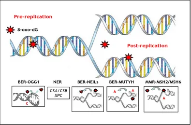
Figure 1: Repair pathways acting on 8-oxo-dG. Oxidation produces several
detrimental DNA alterations, including 8-oxo-dG (*), against which cells deploy multiple protective strategies. The OGG-1 DNA glycosylase initiates BER of 8-oxo-dG from 8-oxo-8-oxo-dG:C pairs. MUTYH–dependent removal of mismatched adenine incorporated opposite 8-oxo-dG during replication generates 8-oxo-dG:C pairs, a substrate for excision by OGG-1. CSA and CSB are also involved in the recognition of 8-oxo-dG:C pairs by a still undefined mechanism. MMR provides supplementary protection by excising incorporated 8-oxo-dG, escaped from hMTH1 cleansing of the oxidized dNTP pool Pre-replication Post-replication 8-oxo-dG BER-MUTYH MMR-MSH2/MSH6 NER CSA/CSB XPC BER-OGG1 C C A A A A A BER-NEILs Pre-replication Post-replication 8-oxo-dG BER-MUTYH MMR-MSH2/MSH6 NER CSA/CSB XPC BER-OGG1 C C A A A A A A A A BER-NEILs
1.4 Oxidation of purine nucleotides and role of MTH1
There are two models for the accumulation of oxidized bases in cellular DNA or RNA: one is a result of the direct oxidation of bases in DNA or RNA while the other is a result of the incorporation of oxidized nucleotides generated in nucleotide pools. Recent progress in studies of the sanitization of nucleotide pools, as well as DNA repair, has revealed that the impact of oxidation of free nucleotides is unexpectedly large, in comparison with the direct oxidation of DNA. Free nucleotides are thus more susceptible to oxidation by ROS than is DNA. An in vitro studies of Kamiya’s group indicated that dGTP is likely to be most susceptible to oxidation by the ROS known to be generated in vivo. However, there have been few reports measuring the in vivo concentration of 8-oxo-dGTP in the nucleotide pool. Recently, it has been reported that 8-oxo-dGTP is present in the 0.2–2 µM range in the mitochondrial dNTP pools of several rat tissues under normal conditions (Pursell et al, 2008).
It has been established that 8-oxo-dGTP and 2-OH-dATP are frequently misinserted opposite template adenine or guanine, respectively, in DNA by various DNA polymerases for bacterial genomes, and in the nuclear and mitochondrial DNA in mammals. It has been shown that these oxidized nucleotides indeed increased certain mutations when they were introduced into E. coli or mammalian cells.
8-oxo-dGTP misinserted opposite template adenine causes mainly A:T to C:G transversion mutation after two rounds of replication. 2-OH-dATP misinserted opposite guanine mostly, induces G:C to T:A transversion mutation (Nakabeppu Y et al, 2010).
E. coli mutT gene degrades 8-oxo-dGTP in the nucleotide pool to 8-oxo-dGMP and pyrophosphate thereby preventing incorporation of 8-oxo-dG into DNA (Maki and Sekiguchi, 1992). In the absence of mutT a strong mutator phenotype, with accumulation of AT→CG
transversions, is observed, which is mostly due to misinsertion of 8-oxo-dGTP opposite template adenine.
Several mutT homolog hydrolases (MTH1, MTH2, NUDT5 all sharing a Nudix motif) have been identified in mammalian cells (Ishibashi et al, 2003). The majority of the studies focused on the most abundant enzyme, the human MutT homolog-1, hMTH1 (K. Sakumi, 1993). In contrast to MutT, MTH1 efficiently hydrolyzes two forms of oxidized dATP, 2-OH-dATP and 8-oxo-dATP, as well as 8-oxo-dGTP. It also hydrolyzes the corresponding ribonucleotides, 2-OH-ATP, 8-oxo-GTP and 8-oxo-ATP.
The solution structure of MTH1 has been determined by multi-dimensional heteronuclear NMR spectroscopy (Mishima M et al, 2004). Despite the low sequence similarity outside the conserved nudix motif, the protein adopts a highly similar folding pattern to E. coli MutT. Among known proteins with the nudix motif, two other mammalian proteins, MTH2 (NUDT15) and NUDT5, were identified with the potential to hydrolyze either oxo-dGTP or 8-oxo-(d)GDP to 8-oxo-(d)GMP, respectively. NUDT5 also hydrolyzes 8-oxo-dADP and to a lesser extent 2-OH-dADP. The discovery of NUDT5 with 8-oxo-(d)GDPase activity, further revealed that MTH1 and MutT can both hydrolyze 8-oxo-dGDP as well as their triphosphate forms (Ishibashi et al, 2003). MTH1 also recognizes oxidized forms of dATP and ATP as mentioned above. Therefore, we expect that their diphosphate forms can be hydrolyzed by MTH1, suggesting that MTH1 is the most powerful enzyme for the sanitization of nucleotide pools. Gene knockdown experiments for MTH1, MTH2 and NUDT5 in cultured human cells revealed that MTH1 deficiency induced the highest occurrence of A:T to C:G transversion mutations when 8-oxo-dGTP was introduced into cells (Nakabeppu Y et al, 2010).
Although the protective role of sanitizing oxidized dNTPs in mammalian cells has received less attention than repair pathways acting on DNA, lately numerous lines of evidence indicate that oxidation of nucleic acid precursors affects important biological
processes including mutagenesis (Hori M. et al, 2010), senescence (Rai P. et al, 2009) and neurodegeneration.
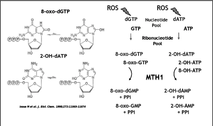
Figure 2: Chemical structure and repair of oxidized triphosphates. MTH1
efficiently hydrolyzes two forms of oxidized dATP, 2-OH-dATP and 8-oxo-dATP, as well as 8-oxo-dGTP. It also hydrolyzes the corresponding ribonucleotides, 2-OH-ATP, 8-oxo-GTP and 8-oxo-ATP.
Nucleotide Pool dGTP dATP ROS ROS 8 8--oxooxo--dGMPdGMP +
+ PPiPPi 22--OHOH+ + --PPiPPidAMPdAMP
MTH1
MTH1
8
8--oxooxo--GMPGMP +
+ PPiPPi 22--OHOH+ + PPiPPi--AMPAMP 8
8--oxooxo--dGTPdGTP 22--OHOH--dATPdATP ROS ROS Ribonucleotide Pool GTP ATP 8
8--oxooxo--GTPGTP 22--OHOH--ATPATP 8
8--OHOH--ATPATP
8-oxo-dGTP
Inoue M et al. J. Biol. Chem. 1998;273:11069-11074 2-OH-dATP Nucleotide Pool dGTP dATP ROS ROS 8 8--oxooxo--dGMPdGMP +
+ PPiPPi 22--OHOH+ + --PPiPPidAMPdAMP
MTH1
MTH1
8
8--oxooxo--GMPGMP +
+ PPiPPi 22--OHOH+ + PPiPPi--AMPAMP 8
8--oxooxo--GMPGMP +
+ PPiPPi 22--OHOH+ + PPiPPi--AMPAMP 8
8--oxooxo--dGTPdGTP 22--OHOH--dATPdATP ROS ROS Ribonucleotide Pool GTP ATP 8
8--oxooxo--GTPGTP 22--OHOH--ATPATP 8 8--OHOH--ATPATP Ribonucleotide Pool GTP ATP Ribonucleotide Pool GTP ATP 8
8--oxooxo--GTPGTP 22--OHOH--ATPATP 8
8--OHOH--ATPATP 8
8--oxooxo--GTPGTP 22--OHOH--ATPATP 8
8--OHOH--ATPATP
8-oxo-dGTP
Inoue M et al. J. Biol. Chem. 1998;273:11069-11074 2-OH-dATP
2. Defective repair of oxidative damage and genomic instability
In agreement with the role of DNA repair systems in maintaining genome stability, organisms in which repair of 8-oxo-dG has been impaired show accumulation of endogenously produced oxidized DNA bases, a mutator phenotype and increased susceptibility to tumor development (Nordstrand L.M. et al, 2007; Russo M.T. et al, 2007). The entity of these phenotypes varies however depending on the specific gene. Thus both Ogg1-/- and Mutyh-/- mice show increases in DNA 8-oxo-dG levels in an age- and tissue-specific fashion accompanied by moderate increases in mutation rates (Russo MT et al, 2004 and 2009). While initially a significant tumor-prone phenotype could not be recognized in these mice (Y. Xie et al 2004), more recent reports showed an increased cancer susceptibility of Ogg1-/- and Mutyh-/- mice affecting respectively the lung and the gastrointestinal tract and occurring late in life (19 months)(Sakumi K et al, 2003; Sakamoto K et al, 2007).
The protective role of these genes against neoplastic transformation associated with oxidative stress becomes however more obvious when mice are exposed to chemical or physical stresses. Thus an increased cancer incidence of the skin, lung, or intestinal tract has been reported in Ogg1-/- mice chronically exposed to UVB irradiation (Kunisada M et al, 2005), tobacco specific nitrosamines (Igarashi M et al, 2009), dimethylarsinic acid (Kinoshita A et al, 2007) or dextran sulfate (Liao J et al, 2008). Similarly an increased frequency of small intestinal tumors was observed in KBrO3-treated Mutyh-/- mice when compared to wild-type animals (Sakamoto K. et al 2007). When two independent functions affecting the removal of 8-oxo-dG, i.e. OGG1 and MUTYH, are impaired, a more penetrant phenotype was observed. Thus simultaneous deletions of both genes cause a considerable increase in DNA damage and tumor frequency (lung, ovarian, and gastrointestinal tract tumors) establishing a strong link between oxidative DNA damage and tumorigenesis.
The increased frequency of intestinal tumors that occurs when Mutyh deficiency is combined with heterozygous inactivation of the
APC gene, provides a second example of the possible importance of persistent DNA 8-oxodG and cancer. APCMin+/Mutyh-/-mice develop
many more intestinal adenomas than their single knockout counterparts. The overrepresentation of G:C→T:A transversions in the remaining APC allele are consistent with a role for oxidized DNA bases in carcinogenesis (Sieber O.M et al, 2004).
Thirdly, double inactivation of Ogg1/Mutyh or Ogg1/Csb has synergistic effects on DNA 8-oxodG accumulation in several organs. These two combinations have different effects in different organs. Because of its central role in replication error correction, cells or organisms in which inactivating mutations in hMSH2, hMLH1, hPMS2, or hMSH6 incapacitate MMR show a massive spontaneous mutator phenotype and MMR-defective mice succumb mostly to lymphomas very early in their life (Buermeyer A B et al,1999). Which is the contribution of oxidative DNA damage to the genetic instability of MMR-defective cells? It has been shown that the dNTP pool is an important source of DNA 8-oxo-dG and that MMR provides supplementary protection by excising incorporated oxo-dGMP (Colussi C et al, 2002). It has been also demonstrated that 8-oxo-dG derived from an oxidized pool of dNTPs contributes significantly to the mutator phenotype of MMR-deficient cells. The high spontaneous hprt mutation rate of MMR-defective Msh2-/- mouse embryonic fibroblasts was attenuated by overexpression of the hMTH1 protein. Molecular analysis of hprt mutants showed that the presence of hMTH1 reduced the incidence of mutations in all classes, including frameshifts, and also implicated incorporated 2-OH-dAMP in the mutator phenotype (Russo M.T et al, 2004). Double knockout Msh2 and Mutyh mice showed much larger increases of oxidative DNA damage than loss of single genes. This synergistic increase in 8-oxo-dG levels in several organs stresses an independent role for these repair proteins in controlling oxidative DNA damage in vivo. However simultaneous inactivation of Mutyh and Msh2 is associated with an apparent paradox: increased levels of oxidative DNA damage but retarded lymphomagenesis associated with Msh2 deficiency. This indicates that a large fraction of the
cancer-prone phenotype of Msh2-/- mice depends on Mutyh activity (Russo M.T et al, 2009).
In contrast to the massive mutator phenotype of E. coli mutT strain (Maki H et al, 1992), inactivation of the Mth1 hydrolase in Mth1-/- mice did not affect the frequency of AT->CG transversions but was associated with a modest increase in frameshift mutagenesis (Egashira A et al, 2002) and a cancer prone phenotype affecting several organs (lung, stomach, and liver) (Tsuzuki T et al, 2001) (Table 1).
Mutated gene Oxidative damage
Target organ/cells
Phenotype
Ogg1 8-oxo-dG Liver/MEF lung
Myh 8-oxo-dG Liver Gastrointestinal tract Ogg1/Myh
8-oxo-dG Liver, small
intestine, lung Gastrointestinal tract, lung, ovarian cancer APCmin/+/Myh ND ND Increased intestinal cancer
Csb 8-oxo-dG 8-OH-Ade Primary hepatocytes, human fibroblasts Human primary fibroblasts Skin cancer, retinal degeneration
Csb/Ogg1 8-oxo-dG Liver, spleen, kidney/MEF Preneoplastic liver lesions Msh2
8-oxo-dG, Liver, small intestine,kidney, lung
Lymphoma,
Mth1 8-oxo-dG, lung, stomach,
and liver lung, stomach, and liver cancer
ND: nondetected
Table 1
Relationship between accumulation of oxidative lesions and phenotype of mouse models defective in oxidative DNA damage repair genes
3. Oxidative DNA damage and neurodegeneration
Oxidative DNA damage has been implicated in the etiology of several neurodegenerative diseases as well as in the aging process. In the cell ROS are generated primarily by mitochondria. To ensure efficient removal of mitochondria-generated ROS, the inner mitochondrial membrane incorporates a number of free radical scavengers such as vitamin E, ascorbate, and glutathione. Additionally, there is an enzymatic removal of free radicals by superoxide dismutase (SODs). These lines of defense insure that formation of ROS as a by-product of respiration does not damage the cell. The defense can be compromised by either genetic mutations leading to decreased activity of the antioxidants as in some familial cases of Amyotrophic Lateral Sclerosis (ALS) with mutation in CuZn SOD, or by increased radical production. In either case, oxidative damage can impart harmful consequences to the cell. The mechanism by which oxidative damage causes neuronal death is poorly understood. Therefore, an unresolved issue is whether mitochondrial defects are the primary cause of toxicity or a secondary response to the damage (Trushina E. et al, 2007).
Oxidative stress and damage to mtDNA during the aging process can impair mitochondrial energy metabolism and ion homeostasis in neurons, thereby rendering them vulnerable to degeneration. Mitochondrial abnormalities have been documented in all major neurodegenerative disorders: Alzheimer’s (AD), ALS, Parkinson’s (PD) and HD (Yang JL et al, 2008).
3.1 Parkinson’s disease
The association between oxidative DNA damage and neurodegeneration was also observed in PD. The pathological hallmark of PD is the massive loss of dopaminergic neurons in the substantia nigra (SN), which is typically associated with the presence
of cytoplasmic inclusions (Lewy bodies) containing large amounts of aggregated -synuclein (Mouradian MM, 2002). In PD there is an increasing evidence of oxidative damage to both nuclear and mtDNA, contributing to the degeneration of dopaminergic neurons in all brain regions, with the most striking difference being a rise in 8-oxo-dG in SN (Alam ZI et al, 1997; Zhang J. et al, 1999; Sanchez-Ramos J.R. et al, 1994). Using a cybrid cell culture model (a clonal line of human neuroblastoma cells containing no endogenous mtDNA and repopulated with mitochondria derived from the platelets of PD or control subjects), Swerdlow et al. demonstrated that mitochondria from PD patients exhibit increased ROS production, decreased activity of complex I and increased DNA damage compared with mitochondria from normal subjects. In a chemical model for PD in which mice are treated with 1-Methyl-4-phenyl-1,2,3,6-tetrahydropyridine (MPTP), which causes a PD-like dopaminergic pathology, brain tissue samples from MPTP-treated mice showed large levels of oxidation in both mitochondrial and nuclear DNA in the SN, while there was no damage in either mitochondria or nucleus in cerebellum (Mandavilli B.S et al, 2000; Shimura-Miura H et al, 1999). Supportive evidence for a role of DNA damage in MPTP-induced neuronal death is the activation of PARP in vulnerable dopaminergic neurons of the SN (Wang H et al, 2003) and the rescue from MPTP neurotoxicity observed in mice lacking the PARP gene (Mandir AS et al, 1999). Finally increased levels of SDS-insoluble α-synuclein was accompanied by a significant increase in 8-oxo-dG immunoreactivity in neuroblastoma cells chronically-exposed to rotenone, a mitochondrial complex I inhibitor (Sherer TB et al, 2002).
Paradoxically the accumulation of 8-oxo-dG in SN of patients with PD is accompanied by a marked increase of MTH1, especially within mitochondria. Similarly both MUTYH and the mitochondrial form of OGG1 (OGG1-2a) were up-regulated in the SN of PD patients compared with aged-matched control subjects (Fukae J et al, 2005; Arai T et al, 2006). The most plausible explanation for these observations is that all these enzymes are up-regulated in PD patients
secondary to mtDNA oxidative damage to protect neurons from mutagenesis. Finally following MPTP administration, MTH1-null mice show a greater accumulation of 8-oxo-dG in mt DNA of striatal nerve terminals of dopamine neurons in comparison to wild-type mice and this is accompanied by an increased dopamine neuron loss (Yamaguchi H et al, 2002). These findings indicate that MTH1, as well as other BER enzymes, protect striatal nerve terminals of dopamine neurons from oxidative damage in the nucleic acids, especially in mitochondria.
3.2 Huntington’s disease
HD is a neurodegenerative process mainly affecting the basal ganglia in the brain. Symptoms appearing in this disorder have been described for long time (different descriptions can be documented as early as the fourteenth century). Indeed, HD was also known as Saint Vitus’s dance or dancing plague. The disease was first described by Charles Waters as a convulsive disorder, but it was in 1872 when George Huntington formally described it for the first time and referred to as a hereditary chorea.
HD is catalogued as a rare disease, with a stable prevalence in white populations affecting 5-7 individuals per 100,000. The age of onset ranges between 30 and 40, with death occurring after 15–20 years; onset sometimes occurs early in young people at around 20 and evolves over periods of around five years (Tunez I. et al, 2010). HD is an autosomal dominant hereditary brain disorder that is progressive and fatal. It is caused by expansion of a CAG trinucleotide repeat in exon−1 of the Huntingtin (HTT) gene leading to involuntary movements (chorea), cognitive impairment and psychiatric problems (Bates G et al, 2002). The CAG expansion elongates the N−terminal poly(Q) stretch of the protein, resulting in aggregation and the formation of neuronal intranuclear inclusions. These cause an increase in the rate of neuronal cell death in selected
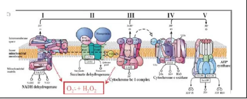
areas of the brain with consequent effects on neurological functions (Beal M F et al, 2004; Imarisio S et al, 2008). Different biochemical studies have also revealed the existence of major defects in the energetic metabolism of HD patients characterized by mitochondrial dysfunction. Mitochondria of HD patients are affected by alterations in electron transport chain (ETC) function, in which complexes II and III are affected, prompting a significant decrease in succinate oxidation and ATP synthesis. Complex IV (cytochrome oxidase) is also affected, albeit to a lesser extent. Other defects seem more selectively distributed, such as in the case of complex I (NADH dehydrogenase) and pyruvate dehydrogenase complex (Brouillet E et al, 2005)(Figure 3).
To investigate HD both genetic and neurotoxic animal models that reproduce some behavioural and neuropathological aspects of the human disease have been developed. Transgenic mice expressing exon 1 of the human HTT gene with an expanded CAG repeat develop a progressive neurologic disorder (Mangiariniet L al, 1996). These R6/2 mice have CAG repeat lengths of 141±157 (normal, 35) under control of the human HD promoter.
The most widely used models for studying neurodegenerative processes in the specific case of HD have been non-genetic models because they are easy to use, control and acquire. Basically, these models induce cell death through alteration of mitochondrial metabolism. In a chemical model for HD, treatment with 3-nitropropionic acid (3-NP), an irreversable inhibitor of succinate dehydrogenase, inhibits mitochondrial complex II activity. This results in a reduction in ATP concentrations, and this defective energy metabolism is associated with increased generation of free radicals, mitochondrial compromise ultimately leading to the formation of striatal lesions, possibly via an excitotoxic mechanism (Brouillet E et al, 1999). Although the mechanisms by which the gene defect responsible for HD leads to neuronal degeneration remains incompletely understood, the involvement of oxidative stress is well established. In postmortem HD patients, the levels of 8-oxo-dG were increased in nuclear DNA in comparison to samples
from age-matched control subjects (Browne SE et al, 1997), although other authors did not confirm this observation (Alam ZI et al, 2000). Oxidation of mtDNA was found in the parietal cortex of HD patients, but not in frontal cortex or cerebellum (Polidori MC et al, 1999) suggesting that region-specific oxidative damage to mtDNA may play a causative role in the mitochondrial dysfunction observed in HD. Increased concentrations of 8-oxo-dG were also found in the urine, plasma and striatal microdialysates of R6/2 mice (Bogdanov MB et al, 2001). More recently a progressive increase in the level of mtDNA damage in the striatum and cerebral cortex of 7– 12-week-old R6/2 mice was confirmed by QPCR analysis (Acevedo-Torr K et al, 2009). All these findings provide convincing evidence that oxidative damage may contribute to HD pathogenesis.
Figure 3: Electron transport chain (ETC). Mitochondria of HD patients are affected
3.3 Oxidative DNA damage and repeat instability in HD
The number of trinucleotide CAG repeats within the HTT gene expands beyond a range that is compatible with effective protein function and is unstable in germline and somatic cells. Transmission of the mutation to the offspring, particularly by fathers, is
characterized by an expansion bias leading to the phenomenon of anticipation with the disease tending to worsen over successive generations. Although CAG repeat length is the main contributor to intergenerational instability, other genetic factors may play a modifying role (Wheeler VC et al, 2007). In addition to the brain, somatic CAG repeat expansion occurs in several tissues, including lymphoblastoid cells and cell cultures derived from skin and muscle biopsies (Cannella M et al, 2009; Squitieri F et al, 2010).
Interestingly, somatic instability is extensive in striatal neurons but very limited in cerebellar neurons, which are largely spared by the disease. It has been proposed that the length of the CAG repeat is the main determinant of the age of onset of HD (Swami M et al, 2009). Some evidence suggests that DNA repair proteins can influence somatic CAG repeat expansion in HD. Thus two repair pathways, MMR and BER, appear to regulate somatic instability in the brain of HD mouse models. While it is well documented the role of some MMR genes in modulating the somatic CAG repeat length (mostly via the Msh2-Msh3 but not Msh2-Msh6 complexes)(Manley K. et al, 1999; Wheeler V.C et al, 2003; Dragileva E et al, 2009), the
involvement of the BER pathway has been more recently proposed. Kovtun et al. (2007) reported that the age-dependent somatic mutation associated with HD occurs in the process of removing oxidized base lesions by OGG1. In addition the expansion of CAG repeats at the long HTT locus in cultured fibroblasts obtained from HD patients was induced by H2O2-mediated oxidative stress.
Somatic expansion does not require cell division however and can occur in neurons after these cells are terminally differentiated and
mitotic replication has ceased (Gonitel R. et al,2008). The current model proposes that somatic expansion initiates from an OGG1-mediated BER mechanism, does not require cell division, and a ‘toxic oxidation cycle’ escalates with age as oxidative lesions in the brain accumulate and contributes to HD onset and progression. More recently the role of other BER enzymes was investigated in vitro and disruption of pol and FEN1 coordination was found to be the main determinant in CAG repeat expansion (Liu Y et al, 2009). However it has also been shown in R6/2 and R6/1 mice that the stoichiometry of BER enzymes, rather than DNA damage levels, correlates with the tissue selectivity of somatic CAG expansion (Goula A et al, 2009).
The role of an oxidized dNTPs pool in modulating CAG repeats expansion is unknown. Whether a general reduction in the steady state levels of oxidative DNA damage in the striatum provided by modulating hMTH1 levels will decrease the probability of gap formation and attenuate or slow down the CAG expansion responsible for the neuropathological process remains to be ascertained.
4. Oxidative DNA damage and aging
It was recently shown that suppression of hMTH1 is sufficient to induce senescence in early-passage human fibroblasts (Rai et al, 2009). These findings support the existence of a causative link between neurodegeneration and oxidative DNA/RNA damage as well as the contribution of oxidized DNA precursors to brain aging, suggesting a possible role of hMTH1 as a general protective factor against senescence.
Aging is another consequence of oxidative stress induced cellular damage. The free radical theory of aging, advanced in 1956 by D. Harman, proposes that changes associated with aging are initiated by the reaction of ubiquitous active free radicals, normally produced by
endogenous metabolic processes of the organism, with cellular constituents (Harman D, 1956). A condition of chronic oxidative stress, probably the consequence of an imbalance between pro-oxidant and antipro-oxidant systems leads to an accumulation of oxidative damage to macromolecules contributing to the progressive decline in cellular functions and resulting in the aging phenotype (Sohal RS et al, 1996). The levels of oxidative damage to lipid, DNA, and protein have indeed been reported to increase with age in a wide variety of tissues and animal models (Bokov A et al, 2004). However, because of the pleiotropic effects of oxidative damage to different types of macromolecules, it remains undefined whether oxidative damage to DNA alone is a major causal factor in the ageing process (Pérez V et al, 2009; Salmon A et al, 2010).
II. AIM
The aim of this project is to increase the understanding of the relationship between oxidative stress and neurodegeneration occurring in HD. In particular we will study the mechanism(s) by which overexpression of the hMTH1 hydrolase, that destroys harmful oxidized DNA precursors, protects against neurodegeneration. The association of oxidative stress with HD is well established. It is unclear, however, at which point during the neurodegenerative process oxidative stress becomes an important factor. It also remains to be determined whether oxidative stress is causally involved in initiating the neurodegeneration in HD or whether it develops following disease onset. Our observations in a transgenic mouse model (hMTH1−Tg) demonstrate that a high level of hMTH1 expression is associated with reduced steady state levels of oxidative DNA damage in several tissues. Strikingly, hMTH1 overexpression in brain protects the animals against neurotoxicity induced by 3−NP, a chemical model for HD. We plan to use these observations as a basis to explore the possible causative role of oxidation in the development of HD. The neuroprotective role of hMTH1 overexpression will be investigated using newly established in vitro cellular systems and transgenic mouse models. We will examine whether hMTH1 protects mitochondrial DNA from oxidative damage and the mode of oxidation−related neuronal cell death. The study should clarify whether oxidative DNA damage has a causal role in the development of the neuropathological and behavioural phenotype in HD.
III. RESULTS
Characterization of a transgenic mouse overexpressing hMTH1
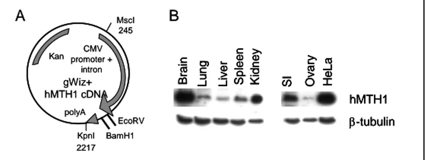
A 509bp hMTH1 cDNA (BamH1-EcoRV fragment) was cloned into the gWIZ vector under the control of the CMV promoter (Fig. 3A) and an MscI-KpnI fragment microinjected into pronuclei of zygotes. One of the founder mice expressing the hMTH1 transgene was selected and either maintained as hemizygous (hMTH1-Tg+/-) or bred to homozygousity (hMTH1-Tg+/+). hMTH1 expression was examined by western blotting in tissue extracts of several organs and the presence of the enzyme was confirmed in brain, lung, liver, spleen, kidney, small intestine and ovary. No signal for the endogenous mMTH1 protein was detected using the anti-human hMTH1 antibody. hMTH1 was found to be particularly highly expressed in brain and kidney (Fig. 3B).
Figure 3: Expression of hMTH1 in a transgenic hMTH1 mouse. (A)
BamH1-EcoRV fragment (509 bp) derived from pcDEB encoding the hMTH1 cDNA was subcloned into the gWIZ vector under the control of the CMV promoter. The MscI-KpnI fragment (2481bp) was used in the construction of the transgenic mouse. (B) Western blot analysis of transgene expression. Total proteins (20-40 µg) from a range of tissues were separated by SDS polyacrylamide electrophoresis, blotted and probed with an antibody against hMTH1. -tubulin was used as a loading control.
gWiz+ hMTH1 cDNA CMV promoter + intron Kan MscI 245 KpnI 2217 EcoRV polyA BamH1 hMTH1 Brain Lun g Splee n Kidne y -tubulin SI Liv e r Ov a ry He L a A B gWiz+ hMTH1 cDNA CMV promoter + intron Kan MscI 245 KpnI 2217 EcoRV polyA BamH1 hMTH1 Brain Lun g Splee n Kidne y -tubulin SI Liv e r Ov a ry He L a hMTH1 Brain Lun g Splee n Kidne y -tubulin SI Liv e r Ov a ry He L a A B
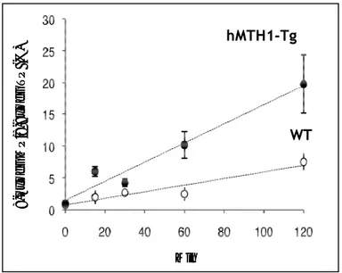
Since the level of the hMTH1 protein was particularly high in the brain, hMTH1 activity was also assayed. Conversion of 8-oxodGTP into 8-oxodGMP was threefold higher in brain extracts from transgenic when compared to wild-type animals (Fig. 4).
Figure 4: hMTH1 activity. hMTH1 enzymatic activity in cell-free extracts from the
brain of hMTH1-Tg (n=4) and wild-type animals (n=4).
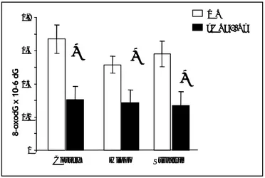
This increased enzymatic activity was associated with a significant decrease in 8-oxo-dG levels in the cortex, hippocampus and striatum of hMTH1-Tg as compared to wild-type mice (2.1- 1,7 and 2.1 fold; p<0.05) (Fig. 5). 8-o x od GM P /8-8xo dGT P (% ) Min hMTH1-Tg WT 8-o x od GM P /8-8xo dGT P (% ) Min hMTH1-Tg WT
Figure 5: Steady-state levels of 8-oxodG in different brain areas. Levels of DNA
8-oxodG were measured in the indicated brain areas from hMTH1-Tg (n=6) and wild-type mice (n=7), by HPLC-EC. Data are indicated as mean+SE. Data groups were compared by t-tests and two-way Anova P-values (* ≤0.05).
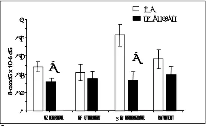
The transgene also provided protection against endogenous oxidation and steady-state levels of DNA 8-oxodG in heart, muscle, small intestine and liver of hMTH1-Tg mice were 1,5, 1,1, 2,4 and 1,1- fold lower than in the same tissues of wild-type animals (p values were 0.02 for heart and 0.006 for small intestine respectively, Student’s t-test) (Fig 6).
These findings indicate that mMTH1 activity is normally limiting in several mouse tissues, including brain. The protection conferred by hMTH1 in untreated animals indicates further that oxidized deoxynucleoside triphosphates are an important source of DNA damage in several organs.
hMTH1-Tg 0 0,2 0,4 0,6 0,8
Cortex Hippo Striatum
WT 8-oxodG x 10-6 dG
*
*
*
hMTH1-Tg 0 0,2 0,4 0,6 0,8Cortex Hippo Striatum
WT 8-oxodG x 10-6 dG
*
*
*
Figure 6: Steady-state levels of oxodG in various organs. Levels of DNA
8-oxodG were measured in several organs from hMTH1-Tg (n=6) and wild-type (n=7), by HPLC-EC. Data are indicated as mean+SE. Data groups were compared by t-tests and two-way Anova P-values (* ≤0.05).
Protection by hMTH1 against HD-like neurodegeneration induced by 3-NP
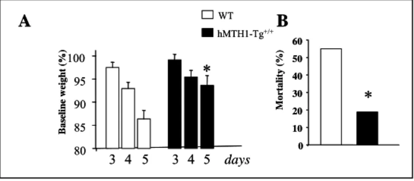
To examine the effect of hMTH1 expression on HD-like neurodegeneration, transgenic and wild-type animals were treated with the mitochondrial toxin 3-NP. This inhibitor of succinate dehydrogenase, selectively causes the death of striatal neurons and induces symptoms similar to HD. These include progressive weight loss, neurological abnormalities such as foot and limb dystonia and, ultimately, death.
Expression of hMTH1 in the transgenic animals protected against 3-NP-induced neurodegeneration. The first evidence of this protective
0 0,2 0,4 0,6 0,8 1
Heart Muscle Small Int Liver
8-oxod G x 10 -6 d G
*
*
hMTH1-Tg WT 0 0,2 0,4 0,6 0,8 1Heart Muscle Small Int Liver
8-oxod G x 10 -6 d G
*
*
0 0,2 0,4 0,6 0,8 1Heart Muscle Small Int Liver
8-oxod G x 10 -6 d G
*
*
hMTH1-Tg WTeffect was a significantly attenuated weight loss at day 5 of treatment in hMTH1-Tg mice (Fig 7A). hMTH1 activity was also associated with a striking decrease of mortality. While at 5 days 55% (11/20) of wild type mice had died, the great majority of hMTH1-Tg animals (13/16) remained alive (Fig 7B).
Figure 7: 3-NP-induced toxicity in wild-type and hMTH1-Tg transgenic mice.
Groups of wild-type (n = 20) and hMTH1-Tg (n = 16) mice were injected i.p. twice daily for 5 days with 60 mg/kg 3-NP. A) Body weight, measured immediately before the first injection on the indicated days, is expressed as a percentage of the pretreated body weight. B) Mortality measured at 5 days post 3-NP treatment in WT and hMTH1-Tg mice. The asterisks indicate a P,0.05 vs wild-type according to One-way Anova and Tukey multiple comparison post-hoc test for panels A and to x2 test for panels B.
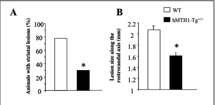
Postmortem examination of 3-NP-treated mice revealed detectable striatal lesions in 77.7% of wild-type animals. These lesions were present in only 30% of hMTH1-Tg animal (Fig 8A). In animals showing a detectable striatal lesion, a reduction in the mean lesion area was found in hMTH1-Tg (3515 ± 305 2, P<0.05 vs wild type) vs wild type mice (5262 ± 528 2)(data not show).
Furthermore, the rostrocaudal extension of the lesions was significantly reduced in hMTH1-Tg versus WT mice (Fig 8B). Thus,
A
B
hMTH1-Tg+/+ WT 80 85 90 95 100 days B a seline w eig ht (% ) 3 4 5 3 4 5*
0 M o rt a lity ( % ) 10 20 30 40 50 60*
A
B
hMTH1-Tg+/+ WT 80 85 90 95 100 days B a seline w eig ht (% ) 3 4 5 3 4 5*
0 M o rt a lity ( % ) 10 20 30 40 50 60*
hMTH1 expression significantly protects the animals from the behavioural and neuropathological effects of 3-NP.
Figure 8: Striatal lesion formation. (A) The percentage of animals with detectable
postmortem striatal lesions is shown. (B) Size of striatal lesions. Postmortem measurements of striatal lesions along the rostrocaudal axis. The asterisks indicate a P,0.05 vs wild-type according to One-way Anova and Tukey multiple comparison post-hoc test for panels A and to x2 test for panels B.
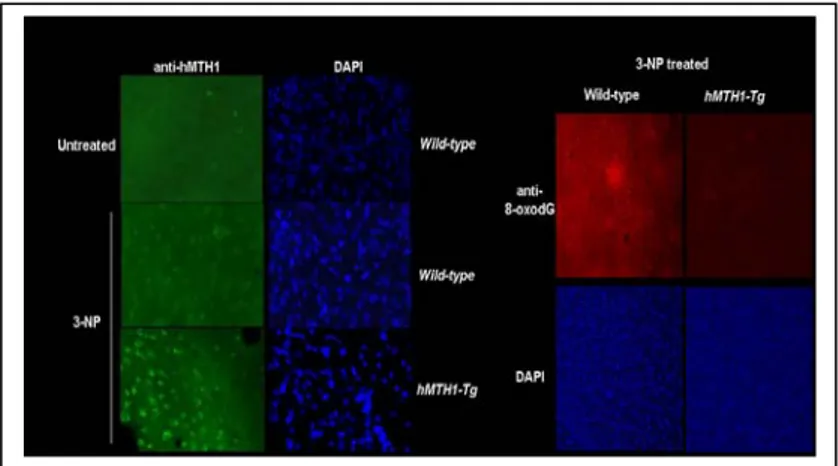
Although endogenous mMTH1 is normally undetectable, it can be visualized by immunofluorescence in the striatum of wild-type mice following 3-NP-treatment (Fig 9, left panel). This suggests that the murine protein is induced in oxidatively stressed striatal cells. As expected, a progressively increasing signal for hMTH1 was observed in hMTH1-Tg animals.
hMTH1 expression significantly reduced DNA 8-oxodG levels in the major target area, the striatum, and increasing protection was observed in hMTH1-Tg animals (Fig 9 right panel).
A
B
L es ion s ize alon g the r o st rocaudal a xi s ( m m )*
1 1.2 1.4 1.6 1.8 2 2.2 An im als with s triatal le si ons (% )*
0 20 40 60 80 100 hMTH1-Tg+/+ WTA
B
L es ion s ize alon g the r o st rocaudal a xi s ( m m )*
1 1.2 1.4 1.6 1.8 2 2.2 An im als with s triatal le si ons (% )*
0 20 40 60 80 100 0 20 40 60 80 100 hMTH1-Tg+/+ WTFigure 9: MTH1 expression and 3-NP-induced oxidative DNA damage in the brain. Immunofluorescence of MTH1 in the striatum of untreated wild-type mice (top
panel, left) or 3 NP-treated wild-typeand hMTH1-Tg animals. (right panels) 8-oxodG immunoreactivity in the striatum of 3-NP-treated wild-type and hMTH1-Tg animals. Nuclei of striatum counterstained by DAPI are shown in the bottom panels.
These data establish an inverse correlation between the levels of DNA 8-oxo-dG and expression of the hMTH1 in the brain and suggest that, during the course of chemically induced neurodegeneration, a large fraction of this oxidative lesion derives from an oxidized dNTP pool.
It is possible therefore that the reduced levels of 8-oxo-dG pools afforded by the hMTH1 transgene, resulted in a diminished incorporation of 8-oxodG into DNA during repair of 3-NP induced oxidative DNA damage (Deluca et al, 2008).
Expression of hMTH1 in neural progenitor cells expressing the mutant huntingtin gene
3-NP is a chemical model for HD-like striatal degeneration. We also investigated whether hMTH1 also conferred protection in a genetic model for HD. We used neuronal progenitor cell lines established from striatal primordia of wild-type or mutant htt knockin mice (HdhQ7and HdhQ111, respectively) in which the htt gene CAG repeat
length is normal or expanded (Trettel F et al, 2000). These nestin-positive cells have been immortalized with the tsA58 mutant of SV40 large T antigen and at the non-permissive temperature (39°C), they cease proliferation and withdraw from the cell cycle. These cells were trasfected with hMTH1 cDNA and individual clones were chosen for further studies (Figure 10A). As expected hMTH1 expression showed both cytosolic and mitochondrial localization (Kang D et al, 1995). A strong signal for hMTH1 was observed by immunofluorescence both in the nuclei and cytoplasm of the transfectants, while a weak hMTH1 signal in untransfected htt knockin cells showed mostly a nuclear localization.
Figure 10. Expression ofhMTH1 in striatal cells. A) Wild-type HdhQ7 and mutant HdhQ111 striatal cells were transfected with hMTH1 cDNA and analysed by Western
blotting for hMTH1 expression. Total cell extracts were separated, blotted and probed with an antibody againsth MTH1. PCNA was used as a loading control. The human SHSY5S neuroblastoma cell line is shown for comparison. Sub-cellular fractions were prepared from HdhQ111-hMTH1 cells and cytochrome c (Cytc) was used to quantify
mitochondrial cell extracts. B) Intracellular localization of hMTH1 (green fluorescence) in HdhQ111 and HdhQ111-hMTH1. Nuclei were counterstained by DAPI.
HdhQ111 HdhQ111 +hMTH1 hMTH1 DAPI hMTH1 Hdh Q11 1 Hdh Q7+h MT H1 Hdh Q11 1+ hM TH1 hMTH1 PCNA Hdh Q7 SH-S Y 5 Y HdhQ111 + hMTH1 cytos ol mit Cyt c A B HdhQ111 HdhQ111 +hMTH1 hMTH1 DAPI hMTH1 DAPI hMTH1 Hdh Q11 1 Hdh Q7+h MT H1 Hdh Q11 1+ hM TH1 hMTH1 PCNA Hdh Q7 SH-S Y 5 Y HdhQ111 + hMTH1 cytos ol mit Cyt c A B
We then investigated hMTH1 activity by measuring conversion of 8-oxo-dGTP into 8-oxo-dGMP in cell-free extracts from these cultures (Bialkowski K. et al, 2009). Expression of the human hydrolase in HdhQ111-hMTH1transfectant increased MTH1 enzymatic activity by
two-fold (Figure 11).
Figure 11: 8-Oxo-dGTPase activity in HdhQ111 and HdhQ111-hMTH1 cells. Following incubation for increasing time periods of 8-oxo-dGTP with 30-kDa ultrafiltrates cell extracts, production of 8-oxo-dGMP was measured by HPLC. HdhQ111-hMTH1 striatal cells (squares) express a 2-fold increased enzymatic activity
in comparison to parental cells (circles).
HdhQ111 HdhQ111+hMTH1 0 2 4 6 8 10 12 14 16 0 20 40 60 80 100 120 140 min 8o x o d G M P /8 ox od GT P ( % ) HdhQ111 HdhQ111+hMTH1 0 2 4 6 8 10 12 14 16 0 20 40 60 80 100 120 140 min 8o x o d G M P /8 ox od GT P ( % )
Protective role of hMTH1 against cell death mediated by the mutant huntingtin gene
As previously reported proliferating HdhQ111 striatal cells expressing
mutant htt are more sensitive than HdhQ7 cells to killing by 3-NP as
measured by clonal assays. It has been shown that this is due to a non-apoptotic form of cell death caused by mitochondrial membrane depolarization (Ruan Q et al, 2004). Expression of hMTH1 protected HdhQ111 cells against 3-NP but had no significant effect in cells
expressing a wild-type htt gene (p=0.02; Anova test)(Figure 12A). The hMTH1-mediated protection against cell killing was not limited however to this mitochondrial toxin but could be extended to a more generalized oxidant such as H2O2. Also in this case exposure to H2O2
was more toxic in HdhQ111 than in HdhQ7 cells and hMTH1
expression provided full protection against killing induced by H2O2
(Figure 12B).
Figure 12: Protection conferred by hMTH1 against cytotoxicity induced by oxidizing agents. Survival was determined by clonal assays 10 days after a 24h or
15min exposure to 3NP (A) and H2O2 (B), respectively (mean+ SE, n = 3).
A B 1 10 100 0 1 2 3 4 5 3-NP (mM) HdhQ111 HdhQ7/Q7 HdhQ111/Q111+hMTH1 surv iva l (% ) 1 10 100 0 200 400 600 800 1000 H2O2(mM) su rviv al (% ) A B 1 10 100 0 1 2 3 4 5 3-NP (mM) HdhQ111 HdhQ7/Q7 HdhQ111/Q111+hMTH1 surv iva l (% ) 1 10 100 0 200 400 600 800 1000 H2O2(mM) su rviv al (% )
To investigate the selective mechanism(s) underlying these protective effects, we measured oxidative DNA damage at the steady state in nuclei as well as in mitochondria. In all cell lines, basal levels of 8-oxo-dG were found to be 1.5-2 fold higher in mtDNA in comparison to nuclear DNA (Figure 13). In addition expression of the mutant htt gene in HdhQ111 cells leads to a 1.5 fold increase in
both nuclear and mtDNA oxidation.
hMTH1 expression in HdhQ111-hMTH1 transfectant decreased DNA
8-oxo-dG to wild-type levels in both cellular compartments.
Figure 13: Steady-state levels of 8-oxo-dG in nuclear and mtDNA. DNA was
extracted from untreated HdhQ7, HdhQ111 and HdhQ111-hMTH1 cells, digested to
nucleosides and 8-oxo-dG was separated and quantified by HPLC-EC. Values are the mean+ SD of 6 independent determinations.
We then measured the levels of 8-oxo-dG after treatment of the striatal cell lines with 3-NP and H2O2. 3-NP induces higher levels of
DNA 8-oxodG in mitochondrial DNA in comparison to nuclear DNA. In addition expression of mutant htt is associated with a particularly pronounced effect in the mitochondrial compartment. Finally hMTH1 decreases DNA 8-oxo-dG in mtDNA, while
0 0,4 0,8 1,2 8 -o xo dG x 10-6 d G mtDNA nuclearDNA HdhQ111 HdhQ7 HdhQ111 +hMTH1 0 0,4 0,8 1,2 8 -o xo dG x 10-6 d G mtDNA mtDNA nuclearDNA HdhQ111 HdhQ7 HdhQ111 +hMTH1 HdhQ111 HdhQ7 HdhQ111 +hMTH1
genomic levels of this oxidized base seems to be unaffected by its expression (Fig 14 A).
Figure 14: Levels of oxidized guanine following oxidant treatment. 8-oxo-dG in
nuclear and mtDNA were measured by HPLC-EC in DNA extracted from striatal cells 24 hr after continuous exposure to 2,5 and 10 mM 3-NP or exposed to the indicated concentration of H2O2 for 15 min. Values are the mean+ SD of 6 independent
determinations 2,5mM control 10mM 0 1 2 3 4 5 6 8-ox odG x 10 -6 dG 2,5mM control 10mM nuclearDNA 3-NP (mM) mtDNA control 0,25mM 1mM 8-ox odG x 10 -6 dG 0 1 2 3 4 nuclearDNA mtDNA control 0,25mM 1mM H2O2(mM) B HdhQ111 HdhQ7 HdhQ111 +hMTH1 2,5mM control 10mM 0 1 2 3 4 5 6 0 1 2 3 4 5 6 8-ox odG x 10 -6 dG 2,5mM control 10mM nuclearDNA 3-NP (mM) mtDNA control 0,25mM 1mM 8-ox odG x 10 -6 dG 0 1 2 3 4 nuclearDNA mtDNA control 0,25mM 1mM H2O2(mM) control 0,25mM 1mM 8-ox odG x 10 -6 dG 0 1 2 3 4 nuclearDNA mtDNA control 0,25mM 1mM H2O2(mM) B HdhQ111 HdhQ7 HdhQ111 +hMTH1 HdhQ111 HdhQ7 HdhQ111 +hMTH1
We conclude from these data that sanitization of the oxidized pool by hMTH1 provides a general protective mechanism affecting both nuclear and mitochondrial compartments and this results in resistance to cell death induced by oxidizing agents.
In vivo measurements of 8-oxodG in nuclear and mitochondrial DNA in several organs of wild-type and hMTH1-Tg mice are currently ongoing.
We show here some preliminary data on mitochondrial DNA oxidation in WT and hMTH1-Tg animals obtained from animals exposed for 5 days to 3-NP and untreated controls. Examination of the striatum, the target tissue for toxicity induced by 3-NP, was unfortunately not possible because of the low amount of recovered material.
Although hMTH1-dependent variations are not statistically significant because of the low number of examined animals, there is a general trend showing that, at steady-state level, hMTH1 provides a general protective mechanism against 8-oxodG accumulation in muscle, heart and hippocampus (Fig 15). Treatment with 3-NP increases DNA oxidation in all the analysed organs and hMTH1 expression decreases levels of DNA 8-oxodG by 1.5, 1.6, 1.5 and 1.2 in muscle, heart, hippocampus and cortex, respectively (Fig 15). We conclude that the degree of protection provided by hMTH1 towards mitochondrial DNA varies depending on the organ and acts both in untreated and treated animals.
Figure 15: Levels of oxidized guanine in mitochondrial DNA isolated from several organs. 8-oxo-dG in mtDNA were measured in the muscle and heart (A) and
in indicated brain areas from hMTH1-Tg (n=6) and wild-type (n=7), by HPLC-EC. Data are indicated as mean+SE.
muscle 0 0,5 1 1,5 2 2,5 3 basal 3-NP basal 3-NP 8-ox o -dG x 10-6 d G hippocampus 0 0,5 1 1,5 2 2,5 3 basal 3-NP basal 3-NP 8-oxo-d G x 10-6 dG heart cortex
A
B
hMTH1-Tg+/+ WT muscle 0 0,5 1 1,5 2 2,5 3 basal 3-NP basal 3-NP 8-ox o -dG x 10-6 d G hippocampus 0 0,5 1 1,5 2 2,5 3 basal 3-NP basal 3-NP 8-oxo-d G x 10-6 dG heart cortexA
B
hMTH1-Tg+/+ WTIV. DISCUSSION
The accumulation of oxidative damage in brain DNA is a common feature of several neurodegenerative diseases, although evidence for a causal contribution of these DNA lesions to the disease process has been lacking.
Our experiments with the transgenic mouse expressing the human 8-oxo-dGTPase hMTH1 indicate that oxidized dNTPs are important contributors to basal levels of DNA oxidation in vivo. Thus, hMTH1 represents a general mechanism of defense against accumulation of oxidized purines in nucleic acids produced either endogenously or by an exogenous oxidative stress.
Experiments with knockout Mth1-/- mice established a connection
between mMTH1 activity and the levels of DNA and RNA 8-oxodG in dopaminergic neurons following exposure to a selective neurotoxin in a PD model (Yamaguchi K et al, 2006) and in hippocampal microglia during kainate-induced excitotoxicity (Kajitani K, et al, 2006). In those animals, abrogation of mMTH1 expression had no measurable impact on the disease, however. In contrast, transgenic hMTH1 expression revealed important connections between nucleotide pool oxidation, HD-like neurological degeneration in a targeted area of the brain, and neurological symptoms. Neurological symptoms that resemble HD were produced in vivo by treating animals with 3-NP, an inhibitor of complex II of the mitochondrial respiratory chain. Overexpression of the human MTH1 protein in brain of hMTH1-transgenic animals conferred a striking protection against neurological and behavioural HD-like symptoms. The dramatic attenuation of HD symptoms, including weight loss, dystonia and gait abnormalities, was reflected in a significantly reduced size of the chemically induced striatal lesions as well as in increased survival. Thus hMTH1 expression protects against HD-like neurodegeneration in vivo and this is associated with decreased levels of DNA 8-oxodG in the striatum.
The accumulation of 8-oxodG in neurodegerative diseases such as PD, AD or ALS is paradoxically accompanied by up-regulation of repair enzymes involved in the control of oxidative DNA damage. Thus, increased levels of hMTH1 (Shimura-Miura H et al, 1999), hMYH (Arai T et al, 2006), or the mitochondrial form of hOGG1(Fukae J et al, 2005) have been reported in the mitochondria of neurons from substantia nigra of PD patients. This up-regulation of several DNA repair enzymes has been interpreted as a general marker of oxidative stress associated with this disease. We observed increased immunostaining for mMTH1 in the affected areas of the brain of wild-type mice induced by 3-NP to show HD- like neurodegeneration. This suggests that in this experimental model of HD, similarly to other neurodegenerative diseases, increased levels of 8-oxodG are accompanied by an upregulation of MTH1 expression.
Striatal neurodegeneration in 3-NP experimental model of HD most likely occurs in terminally differentiated, non-dividing neurons. Thus any impact of hMTH1 on nuclear DNA replication is unlikely to be significant. Mitochondrial DNA stability is a plausible alternative since impaired mitochondrial respiration and ATP production play a central role in HD (Lin MT, et al, 2006). In a mouse model for PD induced by systemic administration of 1-methyl-4-phenyl-1,2,3,6 tetrahydropyridine, Mth1-/- mice accumulated higher levels of
8-oxodG in mitochondrial DNA of the striatum than wild-type mice and this triggered neuronal dysfunction (Yamaguchi K et al, 2006). Here we showed that a large fraction of oxidative damage induced by 3-NP and identified by immunostaining resides in mitochondrial DNA. A comparison of 8-oxodG levels in nuclear and mitochondrial DNA indicates that in vivo 3-NP predominantly oxidize the mitochondrial compartment of several organs and hMTH1 expression significantly reduces this oxidation. We suggest that hMTH1 is a defensive mechanism against mitochondrial degeneration induced by the neurotoxin in this experimental model of HD.