...
Celiac disease and reproductive
disorders: meta-analysis of
epidemiologic associations and
potential pathogenic mechanisms
Chiara Tersigni
1, Roberta Castellani
1, Chiara de Waure
2,
Andrea Fattorossi
1, Marco De Spirito
3, Antonio Gasbarrini
4,
Giovanni Scambia
1, and Nicoletta Di Simone
1,*1Department of Obstetrics and Gynecology, Universita` Cattolica Del Sacro Cuore, Policlinico A. Gemelli, Largo Agostino Gemelli 8, 00168
Rome, Italy2Institute of Public Health, Universita` Cattolica Del Sacro Cuore, 00168 Rome, Italy3Institute of Physics, Universita` Cattolica
Del Sacro Cuore, 00168 Rome, Italy4Department of Internal Medicine, Universita` Cattolica Del Sacro Cuore, Policlinico A. Gemelli, 00168 Rome, Italy
*Correspondence address. Tel:+3906-30154298; Fax: +39-06-3051-160; E-mail: [email protected] Submitted on September 16, 2013; resubmitted on January 9, 2014; accepted on February 13, 2014
table of contents
† Introduction
† CD and reproductive disorders: systematic review and meta-analysis
Methods Results
† Current hypotheses of CD-induced mechanisms of obstetric failures
Malabsorption and nutrient deficiency Autoimmune mechanisms
† Conclusions
background:
An increased risk of reproductive failures in women with celiac disease (CD) has been shown by several studies but a com-prehensive evaluation of this risk is lacking. Furthermore, the pathogenic mechanisms responsible for obstetric complications occurring in CD have not been unraveled.methods:
To better define the risk of CD in patients with reproductive disorders as well as the risk in known CD patients of developing ob-stetric complications, we performed an extensive literature search of Medline and Embase databases. Odds ratio (OR) and relative risk (RR) with 95% confidence intervals (95% CI) were used in order to combine data from case – control and cohort studies, respectively. All data were analyzed using Review Manager software. In addition, we summarized and discussed the current hypotheses of pathogenic mechanisms potentially respon-sible for obstetric complications occurring in CD.results:
Patients with unexplained infertility, recurrent miscarriage or intrauterine growth restriction (IUGR) were found to have a signifi-cantly higher risk of CD than the general population. The OR for CD was 5.06 (95% CI 2.13 – 11.35) in patients with unexplained infertility, 5.82 (95% CI 2.30 – 14.74) in women experiencing recurrent miscarriage and 8.73 (95% CI 3.23 – 23.58) in patients with IUGR. We did not observe an increased risk of CD in women delivering small-for-gestational age or preterm babies. Furthermore, we found that in celiac patients, the risk of miscarriage, IUGR, low birthweight (LBW) and preterm delivery is significantly higher with an RR of 1.39 (95% CI 1.15 – 1.67), 1.54 (95% CI 1.22 – 1.95), 1.75 (95% CI 1.23 – 2.49) and 1.37 (95% CI 1.19 – 1.57), respectively. In addition, we observed that the risk for IUGR, LBW and preterm delivery was significantly higher in untreated patients than in treated patients. No increased risk of recurrent miscarriage, unexplained&The Author 2014. Published by Oxford University Press on behalf of the European Society of Human Reproduction and Embryology. All rights reserved. For Permissions, please email: [email protected]
Advanced Access publication on March 11, 2014 doi:10.1093/humupd/dmu007
stillbirth or pre-eclampsia was found in celiac patients. In vitro studies have provided two main pathogenic models of placental damage at the feto-maternal interface. On the embryonic side of the placenta, a direct binding of anti-transglutaminase (-TG) antibodies to trophoblast cells and, thus, invasiveness reduction via an apoptotic damage, has been proposed. Anti-TG antibodies may also be detrimental to endometrial angiogenesis as shown in vitro in human endometrial endothelial cells (cultures and in vivo in a murine model). The angiogenesis inhibition seems to be the final effect of anti-TG antibody-mediated cytoskeletal damage in endometrial endothelial cells.
conclusions:
Physicians should investigate women with unexplained infertility, recurrent miscarriage or IUGR for undiagnosed CD. Women with CD show an increased risk of miscarriage, IUGR, LBW and preterm delivery. However, the risk is significantly reduced by a gluten-free diet. These patients should therefore be made aware of the potential negative effects of active CD also in terms of reproductive per-formances, and of the importance of a strict diet to ameliorate their health condition and reproductive health. Different mechanisms seem to be involved in determining placental tissue damage in CD patients.Key words: celiac disease / pregnancy / trophoblast / endometrium / angiogenesis
Introduction
Celiac disease (CD) is an autoimmune enteropathy caused by an abnor-mal immune response to dietary gluten, the protein fraction of wheat, barley and rye, in genetically susceptible individuals. The genetic suscep-tibility to develop CD has been shown to be conferred by HLA class II molecules DQ2 or DQ8, responsible for presenting disease-related pep-tides to T lymphocytes. The exposure of the immune system to the im-munogenic and toxic peptides of gliadin, the alcohol-soluble fraction of gluten, can promote an inflammatory reaction. Undigested molecules of gliadinin in conditions of increased intestinal permeability pass through the epithelial barrier of the intestine and interact with
antigen-presenting cells (APCs) in the lamina propria (Sollid, 2002). The adaptive
immune response is mediated by CD4+T lymphocytes in the lamina
propria that recognize gliadin peptides, bound to HLA-DQ2 and -DQ8 molecules on APCs, leading to production of the
pro-inflammatory cytokine interferon-g (Salvati et al., 2005) and to a B
lymphocyte response that results in production of autoantibodies like endomysial, anti-transglutaminase (TG) and anti-gliadin antibodies (Jabri and Sollid, 2006).
CD occurs in adults and children at rates approaching 1% of the general population but only 20 – 50% of affected individuals have
subject-ive symptoms (Fasano et al., 2003;Ma¨ki et al., 2003;West et al., 2003;
Bingley et al., 2004;Tatar et al., 2004). The symptoms of the classical form of childhood CD are malabsorption related and include chronic diarrhea, steatorrhea, abdominal distension, fatigue, nausea, vomiting, anemia and growth retardation. However, CD can present with several non-gastrointestinal symptoms and it may escape timely
recogni-tion (Eliakim and Sherer, 2001;Murray et al., 2003;Ma¨ki et al., 2004).
Thus, given the heterogeneity of clinical presentation, many atypical cases of CD go undiagnosed, leading to a risk of long-term complications. Among atypical symptoms of CD, disorders of fertility, such as delayed menarche, early menopause, amenorrhea or infertilit, and pregnancy complications, such as recurrent abortions, intrauterine growth restric-tion (IUGR), small for gestarestric-tional age (SGA) babies, low birthweight
(LBW) babies or preterm deliveries, must be factored (Eliakim and
Sherer, 2001) (see more below).
Endomysial and anti-TG antibodies are considered the most sensitive serologic test to screen for CD and the sensitivity of the tests for both
autoantibodies is greater than 90% (Rostom et al., 2005). Currently, a
test for either marker is considered to be the best means of screening
for CD and to identify the individuals to be referred for endoscopy (Rostom et al., 2005). The gold-standard treatment for CD relies on a lifelong gluten-free diet (GFD), which interrupts the immune response
triggered by gluten (Tack et al., 2010).
A closer examination of pathogenic mechanisms and clinical manage-ment of CD goes beyond the scope of this discussion and the reader is invited to refer to the increasing number of excellent reviews on the
field (Ciccocioppo et al., 2005; Sollid and Jabri, 2005; Green and
Cellier, 2007).
In this review, we focus on the impact of CD on the reproductive health of women, providing a comprehensive review and meta-analysis of the literature investigating the effect of CD on pregnancy outcomes as well as on the incidence of undiagnosed CD in cohorts of women with a history of obstetric failures. Furthermore, we extensively discuss current hypotheses of the pathogenic mechanisms involved in the occur-rence of placental-related complications in women with CD.
CD and reproductive disorders:
systematic review and
meta-analysis
During the last decades, the association between CD and a wide range of reproductive disorders has been described in a growing number of papers and it is now well recognized that CD may have implications on women’s reproductive health.
A shorter duration of the fertile life span in women with untreated CD, because of an older age of menarche and a younger age of menopause, and an increased prevalence of secondary amenorrhea, have been
shown in several studies (Ferguson et al., 1982;Sher and Mayberry,
1996; Smecuol et al., 1996; Santonicola et al., 2011) and only one
study failed to confirm this observation (Sferlazzas et al., 2008).
Interest-ingly, the contraction of the reproductive period seems to be directly related to the activity of CD, since before-and-after case – control studies have shown that celiac women on a long-term GFD show a
dur-ation of fertile life span analogue to healthy women (Ferguson et al., 1982;
Smecuol et al., 1996;Santonicola et al., 2011).
To date, it is also widely accepted that untreated CD also represents a risk for a short-breastfeeding period. In a case – control before – after study, Ciacci et al. demonstrated that the duration of breastfeeding was 2.5 times shorter in untreated celiac mothers than in healthy
women. Again, a central role for gluten exposition and CD activity has been proposed for this reproductive disorder, since the authors also demonstrated that the introduction of a GFD increased the duration of breastfeeding by 2.4 times, restoring it to the average value of the
general female population (Ciacci et al., 1996).
Several authors have also investigated the fertility rate of women affected by CD. In particular, in a large Swedish population-based cohort study assessing fertility in women with CD compared with con-trols, the overall fertility rate of the two groups was similar but the fertility of celiac women was decreased in the 2 years preceding CD diagnosis (Zugna et al., 2010).
Consistent with this, Sher and Mayberry observed that the mean number of children born to celiac patients was significantly less when compared with controls before diagnosis while, after diagnosis and treat-ment, patients had a number of children similar to controls. The authors concluded that the overall difference in fertility between celiac women and controls was due to relative infertility prior to diagnosis and its
cor-rection by a GFD (Sher and Mayberry, 1996). These data strongly
indi-cate that reduced fertility is more common in patients with active CD when a GFD is unlikely to have been initiated.
In addition to the reproductive disorders considered above, most of the research has focused on the association between CD and adverse pregnancy outcomes. Indeed, celiac women have been found to have a higher risk of pregnancy complications in their reproductive life and several studies have shown that CD is detectable at an increased fre-quency in some high-risk groups of patients with a history of reproductive failures.
Among all of the reproductive disorders referable to CD, unexplained infertility, recurrent pregnancy loss, stillbirth, IUGR and LBW of babies have been most investigated.
Since the evidence of these associations has come mainly from case – control and cohort studies and results arising from these studies are still controversial, we performed a meta-analysis of the studies published in international journals to clarify the relationship between CD and increased risk of reproductive failures. We investigated both the preva-lence of CD in women with reproductive disorders, as well as, from another point of view, the incidence of obstetric complications in women with CD compared with controls.
Methods
We performed an extensive literature search of Medline and Embase Current Contents databases from inception until December 2012, using a broad combination of search terms for CD (e.g. CD, TG anti-bodies, gluten, gliadin) and selected adverse reproductive outcomes (e.g. unexplained infertility, miscarriage, recurrent miscarriage, unexplained stillbirth, IUGR, LBW, SGA) that are more frequently associated with CD and for which enough data were available to make a significant comparison between different studies. We also reviewed the reference lists of all identified studies and review articles to search for additional references.
The eligibility of the studies was independently assessed by two researchers (C.T. and C.W.) in order to select case – control studies reporting the occurrence of CD in women with and without reproduct-ive disorders and cohort studies yielding the incidence of reproductreproduct-ive disorders in women with and without CD. Duplicates of studies were removed. In the first step, the studies were excluded considering only
the information presented in the title and abstract. In the second step, the full texts of the articles not previously excluded were assessed to de-termine their eligibility for the review, using the same set of criteria. In the third step, the full texts were re-evaluated to determine the eligibility for
meta-analyses (Fig.1).
To assess the comparability of the selected studies, we verified that the same definitions of the outcomes analyzed were used.
Unexplained infertility defines the absence of pregnancy after at least 2 years of regular sexual intercourse with: normal semen analysis from the
husband (World Health Organization criteria, 1999); normal ovulation
assessed by progesterone values and/or premenstrual endometrial biopsy; normal post-coital test results (for cervical factor of infertility); normal serum LH, FSH and PRL levels; normal tubal patency assessed by hysterosalpingography/laparoscopic chromotubation and normal results of diagnostic laparoscopy.
Miscarriage is defined as a spontaneous pregnancy loss occurred within the first 24 weeks of gestation. Recurrent miscarriage refers to the occurrence of two or more spontaneous pregnancy losses. Unexplained stillbirth is the birth of a new born after 24 completed weeks of gestation that did not show any signs of life after delivery in the absence of a recognizable cause. IUGR defines, according to the na-tional growth curves of the Countries where the studies were conducted, a fetal weight for age ,2 standard deviations (SD) from the mean. Infants with a birthweight below the 10th percentile weight for gestational age with respect to the reference birthweight values are defined as SGA. LBW is used to define a birthweight ,2500 g at term.
We excluded non-English language papers. We did not include confer-ence abstracts where more detailed papers describing the same study were unavailable. Studies designed without a control group were not included in the meta-analysis. Studies were classed as higher quality and worthy of selection if they satisfied all of the selection criteria. There were 15 case – control and 9 cohort studies included in the meta-analysis. Because of the small number of studies available, a quality assessment of the single studies was not performed. The risk of publication bias was evaluated by funnel plots.
Figure 1 Flow chart showing the search strategy and steps of study selection for the meta-analysis.
Data extraction from selected papers was performed independently by two researchers as follows. For case – control studies papers were first divided into groups depending on the reproductive disorder evalu-ated (seven groups: IUGR, recurrent miscarriage, SGA, unexplained still-birth, unexplained infertility, pre-eclampsia, preterm delivery), then data were extracted as the number of celiac cases in the total number of women with a specific reproductive disorder and as the number of celiac cases in the total number of control women. For cohort studies the papers were first divided according to the considered reproductive disorder (eight groups: SGA, IUGR, recurrent miscarriage, miscarriage, unexplained stillbirth, LBW, preterm delivery, pre-eclampsia) and data were extracted as the number of cases of a specific reproductive disorder among celiac women in the total number of celiac women and as the number of cases of the disorder among control women in the total number of control women. Odds ratio (OR) or relative risk (RR) with 95% confidence intervals (95% CI) was used in order to analyze the data from case – control and cohort studies, respectively. Several forest plots were obtained with respect to the different end-points considered in this review.
In the combination of case – control studies, the OR of having CD was calculated for each of the end-points, whereas, in the analysis of cohort studies, RRs were calculated for each single end-point for celiac patients in comparison with controls.
The analysis of cohort studies was also stratified according to the current treatment of patients; for these analyses only studies reporting data for both treated and untreated patients were used.
The Mantel – Haenszel fixed-effects model was applied to combine data if heterogeneity was not shown. On the contrary, the DerSimonian
and Laird random-effects model was used. The Q test and the I2statistics
were used to assess heterogeneity: a P-value , 0.10 or an I2. 50% was
considered indicative of substantial heterogeneity. Analyses were per-formed using Review Manager software.
Results
The combination of data from case – control studies was performed with respect to all cases considered together in comparison with controls
(Fig.2A) and for patients with unexplained infertility (Fig.2B), recurrent
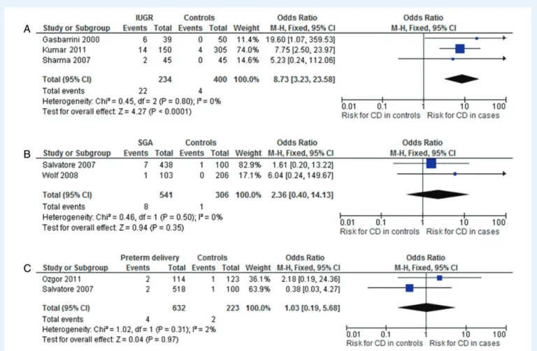
miscarriage (Fig.2C), IUGR (Fig.3A), SGA babies (Fig.3B) or preterm
delivery (Fig.3C). Overall, the OR for CD in all cases was 4.97 (95%
CI 2.88 – 8.57). All the considered reproductive failures, except cases of SGA and preterm delivery, were shown to be at a significant higher risk of CD. The OR for CD was 5.06 (95% CI 2.13 – 11.35) in patients with unexplained infertility, 5.82 (95% CI 2.30 – 14.74) for women ex-periencing recurrent miscarriage and 8.73 (95% CI 3.23 – 23.58) for patients with IUGR. No heterogeneity was found in any analysis.
The prevalence of undiagnosed CD has been investigated also in patients with eclampsia by Wolf et al. Women with a history of pre-eclampsia were tested for anti-TG and anti-endomysium antibodies seropositivity but an increased incidence of CD compared with controls
was not found (Wolf et al., 2008). Because of the few number of studies
available, definitive conclusions cannot be found.
With respect to cohort studies, the risks of miscarriage (Fig.4A),
re-current miscarriage (Fig. 4B), unexplained stillbirth (Fig.4C), IUGR
(Fig.5A), LBW (Fig.5B), preterm delivery (Fig.5C) and pre-eclampsia
(Fig.5D) in celiac patients in comparison with controls was obtained
through data combination. The risks of miscarriage, IUGR, LBW and
preterm delivery were shown to be significantly higher in celiac patients with RRs of 1.39 (95% CI 1.15 – 1.67), 1.54 (95% CI 1.22 – 1.95), 1.75 (95% CI 1.23 – 2.49) and 1.37 (95% CI 1.19 – 1.57), respectively. On the contrary, no increased risk of recurrent miscarriage, unexplained still-birth or pre-eclampsia was found in celiac patients. The heterogeneity was slight with regard to IUGR, LBW and preterm delivery but high in the analysis on recurrent miscarriage. In all those cases, a random-effect model was used.
After stratifying the analyses for current treatment of the disease, it emerged that the risks of IUGR, LBW and preterm delivery were
signifi-cant higher in untreated patients but not in patients on a GFD (TableI).
Finally, Khashan et al. investigated the risk in celiac women of delivering SGA babies in a well-designed population-based cohort study, and observed that women with untreated CD delivered smaller babies, showing a higher risk of SGA infants compared with women without CD. Furthermore, they showed that women with CD on a GFD had no increased risk of SGA compared with women without CD, high-lighting the cause – effect relationship between exposure to gluten, and then of the activity of CD, and the considered adverse pregnancy
outcome (Khashan et al., 2010). Unfortunately, no comparable studies
have been found and that is currently the only study supporting this observation.
The main limits of these meta-analyses were the potential for selec-tion bias within each of the studies and the lack of quality assessment of these single studies. Furthermore, the analysis of publication bias by funnel plots was not exhaustive because of the small number of studies for each end-point. Publication bias could only be excluded for the overall occurrence of adverse pregnancy outcomes in the
case – control studies (Fig.2A).
Current hypotheses
of CD-induced mechanisms
of obstetric failures
The pathogenesis of reproductive disorders in CD is unclear, but some hypotheses have been suggested. Those hypotheses may be clas-sified under two main headings: nutrient deficiency and autoimmune mechanisms.
Malabsorption and nutrient deficiency
Nutrient deficiency, often occurring in active CD, has historically been considered the main cause of gynecologic disorders and adverse preg-nancy outcomes associated with the disease. Indeed, the abnormal villous structure of the small intestine, characteristic of CD, generally results in malabsorption and can lead to minor hematologic abnormal-ities, anemia and other selective nutrient deficiencies, such as zinc,
selen-ium and folic acid (Jameson, 1976;Yu¨ce et al., 2004;Haapalahti et al.,
2005;Singhal et al., 2008;Ho¨gberg et al., 2009), which play significant roles in pregnancy and fetal development.
Zinc deficiency has been shown to cause impaired synthesis and secre-tion of luteinizing hormone (LH) and follicle-stimulating hormone (FSH), which may subsequently cause an abnormal ovarian axis, secondary
amenorrhea, spontaneous abortions and pre-eclampsia (Bedwal and
Bahuguna, 1994). Selenium deficiency also affects the synthesis and
se-cretion of FSH and LH (Bedwal and Bahuguna, 1994). Finally, it is well
recognized that folic acid is an essential vitamin in nucleic acid
metabolism, and that deficiency of it has an impact on rapidly proliferating tissues, such as the embryo, especially in its neuronal development. However, Dickey et al., investigating the possibility that maternal CD might be a risk factor for the occurrence of fetal neural tube (NTD) related to folic acid deficiency, found that the majority of NTD were
not associated with maternal CD (Dickey et al., 1996), which means
that an increased incidence of CD in this population of a nutrient deficiency-related fetal abnormality could not be confirmed.
Unfortunately, nutritional studies in CD during pregnancy are very limited. Most of studies investigating malabsorption and nutrient deficit in CD have been completed in children, so that, available data do not seem to offer a definitive explanation for reproductive disorders in women with CD.
The nutritional status has been emphasized by Kotze as an important and relevant factor in determining pregnancy outcome. The severity of malnutrition directly correlated with the frequency of gynecologic and Figure 2 Overall risk of CD in cases of adverse pregnancy outcomes (cases) in comparison with healthy women (controls) (n ¼ 15 studies) (A). Risk of CD in patients with unexplained infertility (n ¼ 7 studies) (B) or with history of recurrent miscarriage (n ¼ 6 studies) (C) in comparison with controls. OR, odds ratio; 95% CI, 95% confidence intervals.
obstetric disorders, and, as expected, adherence to a GFD was shown to significantly ameliorate reproductive performances of celiac women (Kotze, 2004). On the other hand, women with infertility associated with total and subtotal villous atrophy were often shown to have neither
severe malnutrition nor signs of trace element deficiency (Wilson et al.,
1976;Collin et al., 1996;Meloni et al., 1999;Shamaly et al., 2004). Clearly, the current knowledge does not point to nutrient deficiency as the main pathologic condition responsible for reproductive failures oc-curring in CD. Further studies are needed to precisely define the role of altered absorption and resultant nutritional changes on female fertility in untreated CD, as well as the effects of a GFD, especially with restor-ation of normal nutritional status.
Autoimmune mechanisms
Since the increased incidence of obstetrics failures in CD cannot be explained by malabsorption alone, new pathogenic mechanisms have been investigated during recent years to demonstrate a direct immune-mediated impairment of the physiologic processes occurring during embryo implantation and placental development in women with CD.
Anti-TG antibodies induce trophoblast apoptosis
Patients with CD on a gluten-containing diet generally show increased
levels of serum autoantibodies, in particular, of anti-TG antibodies (
Diet-erich et al., 1997;Ma¨ki, 1997). It is has been firstly hypothesized that
circulating anti-TG antibodies, produced in active CD, could be not only a diagnostic marker of CD, but also directly involved in placental-related pregnancy complications.
Indeed, it is noteworthy that anti-TG antibodies are not only an epi-phenomena in CD, but are also reported to cause the intestinal and neurologic damage, interfering with the cell cycle of human enterocytes
and inducing the apoptosis of neuronal cells, respectively (Caputo et al.,
2010;Cervio et al., 2007).
The rationale of a possible direct binding of circulating TG anti-bodies to placental cells in vivo is supported by the evidence that the enzyme TG is expressed in many different tissues and organs, and it is found intracelluarly as well as extracellularly. In particular, it has been demonstrated that TG is expressed in endometrial cells as well as in stromal and trophoblast placental cells, with higher levels in late
preg-nancy (Robinson et al., 2006).
Since TG, in the extracellular environment, is involved in extracellular matrix assembly and cell adhesion, spreading and migration in diverse
tissues (Zemskov et al., 2006;Park et al., 2010), it is probable that one
or more of the mentioned TG-mediated cellular activities are likely to play a critical role in the implantation process. Thus, TG on syncytiotro-phoblasts may be a target of maternal autoantibodies in CD, and in par-ticular, it is conceivable that the binding of circulating anti-TG antibodies to placental cells could be an immunologic mechanism by which CD may interfere with pregnancy outcome.
Figure 3 Risk of CD in patients with intrauterine growth restriction (IUGR) (A) (n ¼ 3 studies), small for gestation age (SGA) babies (B) (n ¼ 2 studies) or preterm delivery (C) (n ¼ 2 studies) in comparison with controls. OR, odds ratio; 95% CI, 95% confidence intervals.
Normal development and function of the placenta requires invasion of the maternal decidua by extravillous trophoblast (EVT), followed by
abundant and organized vascular growth (Helige et al., 2008). EVTs
produce large amounts of basic proteins and hormones involved in main-tenance of the pregnancy. Therefore, it is likely that increased apoptosis of EVT may contribute to the pathophysiology of human miscarriage and
IUGR (Hadziselimovic et al., 2007;Minas et al., 2007), which are
charac-teristic placental-related complications, found at higher frequency in women with active CD than in the general population. Supporting this
hy-pothesis,Hadziselimovic et al. (2007) showed increased apoptosis of
EVT in placentas of celiac women noncompliant to a GFD, which is con-sistently linked to the low birthweight of newborns.
Interestingly, Anjum et al. have shown that anti-TG antibodies of IgA class are able to directly bind to the syncytial surface of the placenta,
sig-nificantly inhibiting TG activity (Anjum et al., 2009). Anti-TG
antibody-mediated inhibition of syncytial TG is an intriguing hypothesis to
explain a functional impairment of placental development.
However, Anjum et al. only evaluated the effect of IgA class auto-antibodies on placental cells and, even if IgA are the class of immuno-globulins secreted at highest concentration in active CD, it is well known that the IgG class is the only one able to cross the placental barrier and to potentially determine a direct effect at the fetal site of the placenta.
Based on these preliminary observations, we hypothesized that anti-TG antibodies could bind to TG expressed on trophoblast cells in vivo, determining a functional impairment by affecting the invasive potential.
To assess our hypothesis, we isolated both IgG and IgA polyclonal fraction from sera of patients with active CD, not on a GFD, and with
a high titer of anti-TG antibodies (Di Simone et al., 2010). Human
primary trophoblasts cells provide a reliable model for studying the mo-lecular mechanisms of the pathologic conditions affecting the placenta (Di Simone et al., 2005). Thus, trophoblast cell cultures were exposed to increasing concentration of IgA and IgG anti-TG antibodies, both com-mercially available and isolated from celiac women. We specifically focused on the effect of anti-TG antibodies on placental invasiveness, ac-tivity of cellular matrix metalloproteases (MMPs) and cellular apoptosis, as indicators of trophoblast damage.
We observed that both the polyclonal fractions of anti-TG antibodies and the commercial monoclonal anti-TG antibody (CUB7402) were able to directly bind to trophoblast cells, and significantly reduce trophoblast
invasiveness through apoptotic damage (Di Simone et al., 2010). In
add-ition to the anti-TG antibody binding-mediated increase in trophoblast apoptosis, a significant decrease in MMPs activity was observed in this study, and this could be an indirect effect of the increase in trophoblast
apoptosis (Di Simone et al., 2010) (Fig.6A).
Figure 4 Risk of miscarriage (A) (n ¼ 4 studies), recurrent miscarriage (B) (n ¼ 2 studies) or unexplained stillbirth (C) (n ¼ 2 studies) in celiac patients in comparison with controls. RR, relative risk; 95% CI: 95% confidence intervals.
In general, these studies provide a pathogenic model of immune-mediated placental damage potentially occurring in vivo in women with active CD and a high titer of circulating anti-TG antibodies, giving a ration-ale for the increased incidence of placental-related obstetric failures in celiac women.
Anti-trasglutaminase antibodies affect human endometrial
angiogenesis
Endometrial angiogenesis and decidualization, as well as trophoblast in-vasion, are fundamental prerequisites for a successful implantation and a good outcome of pregnancy. In the pregnant uterus, critical angiogenic Figure 5 Risk of intrauterine growth restriction (IUGR) (A) (n ¼ 5 studies), low birthweight (LBW) (B) (n ¼ 2 studies), preterm delivery (C) (n ¼ 3 studies) or pre-eclampsia (D) (n ¼ 2 studies) in celiac patients in comparison with controls. RR, relative risk; 95% CI: 95% confidence intervals.
...
Table I Risk for IUGR, LBW, preterm delivery in treated and untreated celiac patients in comparison with controls (RR with 95% CI).
Untreated patients Treated patients
IUGR (Nørga˚rd et al., 1999;Greco et al., 2004;Ludvigsson et al., 2005) 1.98 (95% CI 1.12 – 3.52); I2: 52% 1.28 (95% CI 0.93 – 1.76); I2: 0% LBW (Nørga˚rd et al., 1999;Ludvigsson et al., 2005) 2.47 (95% CI 1.86 – 3.29); I2: 0% 1.22 (95% CI 0.91 – 1.63); I2: 0% Preterm delivery (Ludvigsson et al., 2005;Khashan et al., 2010) 1.62 (95% CI 1.05 – 2.51); I2: 81% 1.20 (95% CI 0.97 – 1.48); I2: 0%
signals are likely to be produced by the decidualizing endometrial cells acting on the endothelial cells to promote their proliferation and differ-entiation. After stimulation by angiogenic factors, the basement mem-brane is degraded by MMPs and the proteolytic enzymes secreted by endothelial cells. Then the cells invade, migrate and proliferate into the
underlying interstitial matrix and form new capillary structures (Taylor
et al., 1992;Murray and Lessey, 1999). Thus, angiogenesis induces funda-mental changes in the endometrium, enabling it to accept the blastocyst and initiate the process of implantation.
We hypothesized that, together with a direct apoptotic damage to trophoblast cells, circulating anti-TG antibodies could be responsible for an additional mechanism of impairment of placental development, this time at the maternal site of the placenta: endometrial angiogenesis. To assess this hypothesis, we firstly isolated human endometrial endo-thelial cells (HEECs) from placental explants through immune selection and put them in culture. After incubation of HEEC cultures with both IgA and IgG polyclonal immunoglobulins isolated from the sera of celiac patients, and with commercial monoclonal anti-TG IgG (CUB 7402), we demonstrated a direct binding of the anti-TG antibodies to cell membrane of HEECs and a consequent decrease in cellular TG
activ-ity (Di Simone et al., 2013). This binding was followed by a striking
de-crease of in vitro angiogenesis, in terms of the number and total length
of capillary-like tubes formed by HEECs (Di Simone et al., 2013). To
confirm this observation in vivo, we also evaluated the effect of anti-TG antibodies in a murine model of angiogenesis, confirming the
autoantibody-mediated inhibiting effect obtained in vitro (Di Simone
et al., 2013).
The specific role of cellular TG as target of anti-TG antibodies was defined by investigating the effect of its down-regulation by siRNA on this mechanism of inhibition. We found that treatment of TG-silenced HEEC with polyclonal or monoclonal anti-TG antibodies did not cause a reduction in cell differentiation in contrast with HEECs with normal ex-pression of TG, confirming the supposed role of this enzyme expressed
on HEEC membranes as a target for the anti-TG antibodies (Di Simone
et al., 2013).
To identify the molecular mechanisms involved in the inhibition of angiogenesis, we evaluated the activity of MMP-2 in HEEC culture in the presence of polyclonal and monoclonal anti-TG2 antibodies and observed a significant reduction of both pro- and active MMP-2 protein levels. Thus, it is likely that, among the possible molecular mechanisms responsible for the anti-TG antibody-induced angiogenesis inhibition is a reduction of MMP secretion and of extracellular matrix deg-radation. Surprisingly, the functional impairment of HEEC angiogenesis was not associated with an increase in cell apoptosis. In addition, having not found a reduction of vascular endothelial growth factor secre-tion in the medium of HEEC cultures after anti-TG antibody binding, we also supposed that a mechanism other than cell apoptosis or decreased pro-angiogenic factors secretion could be involved in this antibody-mediated angiogenesis inhibition.
Thus, since a negative effect of anti-TG antibodies on human umbilical vein endothelial cell (HUVEC) cytoskeleton organization has been
shown (Myrsky et al., 2008), we hypothesized that anti-TG antibodies,
by binding to TG on cells surface, may exert their effects interacting with actin fibers of cytoskeleton, closely connected with cell membranes. Figure 6 Representative image of the two main mechanisms of anti-TG antibody-mediated placental damage proposed. Anti-TG antibodies from ma-ternal blood circulation bind to trophoblast cells inducing an apoptotic damage (A). At the mama-ternal site, anti-TG antibodies binding to endometrial endo-thelial cells (HEEC) may cause a dramatic disarrangement of the F-actin cytoskeleton impairing the angiogenic process (B). MMP, matrix metalloproteinase.
Indeed, it is well known that the cytoskeleton mediates a variety of cel-lular functions, such as cell – substrate and cell – cell adhesion, which is fundamental for cellular replication and migration, and that a proper actin dynamics and cytoskeleton rearrangement of endothelial cells are recognized to play a pivotal role in the angiogenic process. Hence, we investigated whether a specific autoantibody-induced impairment of HEEC F-actin fiberes organization might explain the inhibited angiogen-esis observed after anti-TG antibody exposure in the in vitro and in vivo models.
We documented a dramatic disarrangement of the F-actin cytoskel-eton in anti-TG antibodies-treated HEEC both directly, by visualization through confocal microscopy, and indirectly, by detecting an increase in the cytoskeleton stiffness and a reduction of fluidity and cell adhesivi-ness of HEEC membranes, which represents the functional counterpart of cytoskeleton modification. Since membrane fluidity reflects the struc-ture of lipids in the membrane, while adhesiviness strongly depends on the cytoskeleton architecture, our results shed light on a functional
inter-play between membrane lipids and the cytoskeleton (Khurana, 2000;
Chichili and Rodgers, 2009) (Fig.6B).
To better understand the intracellular mechanisms regulating the changes in TG-mediated HEEC motility and cytoskeletal organization during the process of angiogenesis, we also examined the effect of anti-TG antibodies on FAK and ERK activation, the key kinases of the intracellular pathway regulating cytoskeleton arrangement and the
tran-scription of pro-angiogenic factors (Okajima and Thorgeirsson, 2000;
Huang et al., 2004). The activation of FAK results in phosphorylation of downstream target molecules, including ERK, which are localized at focal adhesion sites, thus ensuring cell contact with the extracellular matrix and providing the structural links between the extracellular matrix and polymerized actin filaments involved in focal adhesion, cell
shape and motility (Hanks and Polte, 1997; Weisberg et al., 1997;
Small et al., 2002). Furthermore, angiogenic factors directly stimulate angiogenesis by activating the ERK- and FAK-dependent signaling path-ways and the inhibition of these cascade suppresses their angiogenic
ac-tivities (Lee et al., 2006;Chung et al., 2009;Namkoong et al., 2009). We
demonstrated that anti-TG antibodies inhibited the intracellular pro-angiogenic signal mediators ERK and FAK, providing an additional molecular mechanism by which TG antibodies may cause their anti-angiogenic activity on endometrial cells.
In conclusion, antigenic structures suitable for anti-TG antibodies are present on HEEC and the binding of autoantibodies to endometrial endothelial cells and their consequent functional inhibition might repre-sent a key mechanism by which anti-TG antibodies could affect embryo implantation and placentation at the maternal site of the fetal – maternal unit. Because endometrial angiogenesis is essential for placental develop-ment and fetal growth, this could be considered a novel pathogenic mechanism contributing to the adverse pregnancy outcomes occurring in CD.
Conclusions
CD is the most common autoimmune disease with a prevalence of 1% in the general population worldwide. Our meta-analysis demonstrates that patients with unexplained infertility, recurrent miscarriage or IUGR have a nearly 5-, 6- or 8-fold, respectively, increased risk of being affected from CD compared with the general population. Often, patients with the above reproductive disorders have no overt symptoms of CD, or at
most, fatigue associated with iron deficiency anemia. As a result, irregu-lar menstruation, reduced fertility and/or adverse pregnancy outcomes may be the initial clinical feature that ultimately results in a diagnosis of CD.
Thus, a serologic screening for CD, performed by testing the patients’ sera for endomysial and anti-TG antibodies is strongly suggested in cases of unexplained infertility, recurrent miscarriage and IUGR. However, not enough evidence is available to recommend a screening for CD to women with a history of SGA, preterm birth or pre-eclampsia.
We also observed that celiac women have a significantly higher risk of experiencing miscarriage (RR 1.39 – 95% CI 1.15 – 1.67), IUGR (RR 1.54 – 95% CI 1.22 – 1.95), LBW (RR 1.75 – 95% CI 1.23 – 2.49) or preterm delivery (RR 1.37 – 95% CI 1.19 – 1.57) compared with healthy women. However, no significantly increased risk of recurrent miscarriage, unexplained stillbirth or pre-eclampsia was found in celiac patients.
Before – after studies have shown that the risk for IUGR, LBW and preterm delivery in celiac women was significantly reduced by adherence to a GFD. As a consequence, it is mandatory for physicians to make celiac women aware of the potential negative effects of active CD disease, also in terms of reproductive performances, and of the importance of a strict GFD to ameliorate their general and reproductive health.
Concerning the pathogenic mechanisms indicated to be involved in adverse pregnancy outcomes occurring in CD, we believe that the close link between CD activity and a higher risk of reproductive failures may strongly suggest a central role of the immune system in causing ob-stetric failures. In vitro studies have provided two main pathogenic models of placental damage at the feto-maternal interface. On the embryonic side of the placenta, a direct binding of anti-TG antibodies to trophoblast cells, with a reduction in trophoblast invasiveness due to apoptotic damage, has been proposed. Furthermore, on the maternal side of the placenta, anti-TG antibodies may also be detrimental to endometrial angiogenesis by impairing the cytoskeleton structure in endometrial endothelial cells.
In aggregate, these studies support a role of gluten exposure in eliciting immune responses likely responsible for the occurrence of some preg-nancy complications.
Further studies are required to provide more details about the complex interference of this autoimmune disease in human reproduction.
Acknowledgements
We thank Prof. Carolina Ciacci, Department of Medicine and Surgery, University of Salerno, Italy, for her support to data retrieval for meta-analysis; Dr Marco Silano, Department of Veterinary Public Health and Food Safety, Istituto Superiore di Sanita`, Rome, Italy, for pro-viding gliadin peptides for our experiments; Dr Daniela Pedicino for her helpful support in drawing figures.
Authors’ roles
C.T., A.F., N.D.S., M.D.S., A.G. and G.S. were responsible for the study concept, design and supervision. C.T. and C.d.W. performed the litera-ture searches and extraction of data. N.D.S., R.C., A.F. and C.T. con-ducted the basic research experiments. N.D.S., C.T., A.F. and C.d.W. were responsible for the analysis and interpretation of the data. C.T., A.F., C.d.W. and N.D.S. drafted the manuscript.
Funding
No funding was provided for the study.
Conflict of interest
There are no competing interests.
References
Anjum N, Baker PN, Robinson NJ, Aplin JD. Maternal celiac disease autoantibodies bind directly to syncytiotrophoblast and inhibit placental tissue transglutaminase activity. Reprod Biol Endocrinol 2009;19:7 – 16.
Bedwal RS, Bahuguna A. Zinc, copper and selenium in reproduction. Experientia 1994; 50:626 – 640.
Bingley PJ, Williams AJ, Norcross AJ, Unsworth DJ, Lock RJ, Ness AR, Jones RW., Avon Longitudinal Study of Parents and Children Study Team. Undiagnosed coeliac disease at age seven: population based prospective birth cohort study. BMJ 2004; 328:322 – 323.
Bustos D, Moret A, Tambutti M, Gogorza S, Testa R, Ascione A, Prigoshin N. Autoantibodies in Argentine women with recurrent pregnancy loss. Am J Reprod Immunol 2006;55:201 – 207.
Caputo I, Barone MV, Lepretti M, Martucciello S, Nista I, Troncone R, Auricchio S, Sblattero D, Esposito C. Celiac anti-tissue transglutaminase antibodies interfere with the uptake of alpha gliadin peptide 31 – 43 but not of peptide 57-68 by epithelial cells. Biochim Biophys Acta 2010;1802:717 – 727. First published 27 May 2010. doi:10.1016/j.bbadis.2010.05.010
Cervio E, Volta U, Verri M, Boschi F, Pastoris O, Granito A, Barbara G, Parisi C, Felicani C, Tonini M et al. Sera of patients with celiac disease and neurologic disorders evoke a mitochondrial-dependent apoptosis in vitro. Gastroenterology 2007;133:195 – 206.
Chichili GR, Rodgers W. Cytoskeleton – membrane interactions in membrane raftstructure. Cell Mol Life Sci 2009;66:2319 – 2328.
Choi JM, Lebwohl B, Wang J, Lee SK, Murray JA, Sauer MV, Green PH. Increased prevalence of celiac disease in patients with unexplained infertility in the United States. J Reprod Med 2011;56:199 – 203.
Chung EJ, Yoo S, Lim HJ, Byeon SH, Lee JH, Koh HJ. Inhibition of choroidal neovascularisation in mice by systemic administration of the multikinase inhibitor, sorafenib. Br J Ophthalmol 2009;93:958 – 963.
Ciacci C, Cirillo M, Auriemma G, Di Dato G, Sabbatini F, Mazzacca G. Celiac disease and pregnancy outcome. Am J Gastroenterol 1996;91:718 – 722.
Ciccocioppo R, Di Sabatino A, Corazza GR. The immune recognition of gluten in coeliac disease. Clin Exp Immunol 2005;140:408 – 416.
Collin P, Vilska S, Heinonen PK, Ha¨llstro¨m O, Pikkarainen P. Infertility and coeliac disease. Gut 1996;39:382 – 384.
Dickey W, Stewart F, Nelson J, McBreen G, McMillan SA, Porter KG. Screening for coeliac disease as a possible maternal risk factor for neural tube defect. Clin Genet 1996;49:107 – 108.
Dieterich W, Ehnis T, Bauer M, Donner P, Volta U, Riecken EO, Schuppan D. Identification of tissue transglutaminase as the autoantigen of celiac disease. Nature Med 1997;3:797 – 801.
Di Simone N, Raschi E, Testoni C, Castellani R, D’Asta M, Shi T, Krilis SA, Caruso A, Meroni PL. Pathogenic role of anti-beta 2-glycoprotein I antibodies in antiphospholipid associated fetal loss: characterisation of beta 2-glycoprotein I binding to trophoblast cells and functional effects of anti-beta 2-glycoprotein I antibodies in vitro. Ann Rheum Dis 2005;64:462 – 467.
Di Simone N, Silano M, Castellani R, Di Nicuolo F, D’Alessio MC, Franceschi F, Tritarelli A, Leone AM, Tersigni C, Gasbarrini G et al. Anti-tissue transglutaminase antibodies from celiac patients are responsible for trophoblast damage via apoptosis in vitro. Am J Gastroenterol 2010;105:2254 – 2261.
Di Simone N, De Spirito M, Di Nicuolo F, Tersigni C, Castellani R, Silano M, Maulucci G, Papi M, Scambia G, Gasbarrini A. Potential new mechanisms of placental damage in celiac disease: anti-transglutaminase antibodies impair human endometrial angiogenesis. Biol Reprod 2013;89:88.
Eliakim R, Sherer DM. Celiac disease: fertility and pregnancy. Gynecol Obstet Invest 2001; 51:3 – 7.
Fasano A, Berti I, Gerarduzzi T, Not T, Colletti RB, Drago S, Elitsur Y, Green PH, Guandalini S, Hill ID et al. Prevalence of celiac disease in at-risk and not-at-risk groups in the United States: a large multicenter study. Arch Intern Med 2003; 163:286 – 292.
Ferguson R, Holmes GK, Cooke WT. Coeliac disease, fertility, and pregnancy. Scand J Gastroenterol 1982;17:65 – 68.
Gasbarrini A, Torre ES, Trivellini C, De Carolis S, Caruso A, Gasbarrini G. Recurrent spontaneous abortion and intrauterine fetal growth retardation as symptoms of coeliac disease. Lancet 2000;356:399 – 400.
Greco L, Veneziano A, Di Donato L, Zampella C, Pecoraro M, Paladini D, Paparo F, Vollaro A, Martinelli P. Undiagnosed coeliac disease does not appear to be associated with unfavourable outcome of pregnancy. Gut 2004;53:149 – 151. Green PH, Cellier C. Celiac disease. N Engl J Med 2007;357:1731 – 1743.
Haapalahti M, Kulmala P, Karttunen TJ, Paajanen L, Laurila K, Ma¨ki M, Mykka¨nen H, Kokkonen J. Nutritional status in adolescents and young adults with screen-detected celiac disease. J Pediatr Gastroenterol Nutr 2005;40:566 – 570. Hadziselimovic F, Geneto R, Buser M. Celiac disease, pregnancy, small for gestational
age: role of extravillous trophoblast. Fetal Pediatr Pathol 2007;26:125 – 134. Hanks SK, Polte TR. Signaling through focal adhesion kinase. Bioessays 1997;
19:137 – 145.
Helige C, Ahammer H, Hammer A, Huppertz B, Frank HG, Dohr G. Trophoblastic invasion in vitro and in vivo: similarities and differences. Hum Reprod 2008; 23:2282 – 2291. First published 11 July 2008. doi:10.1093/humrep/den198. Ho¨gberg L, Danielsson L, Jarleman S, Sundqvist T, Stenhammar L. Serum zinc in small
children with coeliac disease. Acta Paediatr 2009;98:343 – 345.
Huang C, Jacobson K, Schaller MD. MAP kinases and cell migration. J Cell Sci 2004; 117:4619 – 4628.
Jabri B, Sollid LM. Mechanisms of disease: immunopathogenesis of celiac disease. Nat Clin Pract Gastroenterol Hepatol 2006;3:516 – 525.
Jackson JE, Rosen M, McLean T, Moro J, Croughan M, Cedars MI. Prevalence of celiac disease in a cohort of women with unexplained infertility. Fertil Steril 2008; 89:1002 – 2004.
Jameson S. Zinc deficiency in malabsorption states: a cause of infertility? Acta Med Scand Suppl 1976;593:38 – 49.
Khashan AS, Henriksen TB, Mortensen PB, McNamee R, McCarthy FP, Pedersen MG, Kenny LC. The impact of maternal celiac disease on birthweight and preterm birth: a Danish population-based cohort study. Hum Reprod 2010;25:528 – 534. Khurana S. Role of actin cytoskeleton in regulation of ion transport: examples from
epithelial cells. J Membr Biol 2000;178:73 – 87.
Kolho KL, Tiitinen A, Tulppala M, Unkila-Kallio L, Savilahti E. Screening for coeliac disease in women with a history of recurrent miscarriage or infertility. Br J Obstet Gynaecol 1999;106:171 – 173.
Kotze LM. Gynecologic and obstetric findings related to nutritional status and adherence to a gluten-free diet in Brazilian patients with celiac disease. J Clin Gastroenterol 2004;38:567 – 574.
Kumar A, Meena M, Begum N, Kumar N, Gupta RK, Aggarwal S, Prasad S, Batra S. Latent celiac disease in reproductive performance of women. Fertil Steril 2011; 95:922 – 927. First published 24 Nov 2010. doi:10.1016/j.fertnstert.2010.11.005. Lee C, Dixelius J, Thulin A, Kawamura H, Claesson-Welsh L, Olsson AK. Signal
transduction in endothelial cells by the angiogenesis inhibitor histidine-rich glycoprotein targets focal adhesions. Exp Cell Res 2006;312:2547 – 2556. Ludvigsson JF, Montgomery SM, Ekbom A. Celiac disease and risk of adverse fetal
outcome: a population-based cohort study. Gastroenterology 2005;129:454 – 463. Ma¨ki M. Tissue transglutaminase as the autoantigen of coeliac disease. Gut 1997;
41:565 – 566.
Ma¨ki M, Mustalahti K, Kokkonen J, Kulmala P, Haapalahti M, Karttunen T, Ilonen J, Laurila K, Dahlbom I, Hansson T et al. Prevalence of celiac disease among children in Finland. N Engl J Med 2003;348:2517 – 2524.
Ma¨ki M, Lohi O. Enteropathy. In: Walker WA, Goulet O, Kleinman R, Sherman P, Sheinder B, Sanderson I (eds). Pediatric Gastrointestinal Disease. Hamilton, ONT, Canada: BC Decker Inc., 2004, pp. 932 – 943.
Martinelli P, Troncone R, Paparo F, Torre P, Trapanese E, Fasano C, Lamberti A, Budillon G, Nardone G, Greco L. Coeliac disease and unfavourable outcome of pregnancy. Gut 2000;46:332 – 335.
Martinelli D, Fortunato F, Tafuri S, Germinario CA, Prato R. Reproductive life disorders in Italian celiac women. A case – control study. BMC Gastroenterol 2010;10:89. Meloni GF, Dessole S, Vargiu N, Tomasi PA, Musumeci S. The prevalence of coeliac
disease in infertility. Hum Reprod 1999;14:2759 – 2761.
Minas V, Jeschke U, Kalantaridou SN, Richter DU, Reimer T, Mylonas I, Friese K, Makrigiannakis A. Abortion is associated with increased expression of FasL in decidual leukocytes and apoptosis of extravillous trophoblasts: a role for CRH and urocortin. Mol Hum Reprod 2007;13:663 – 673.
Molteni N, Bardella MT, Bianchi PA. Obstetric and gynecological problems in women with untreated celiac sprue. J Clin Gastroenterol 1990;12:37 – 39.
Murray MJ, Lessey BA. Embryo implantation and tumor metastasis: common pathways of invasion and angiogenesis. Semin Reprod Endocrinol 1999;17:275 – 290. Murray JA, Van Dyke C, Plevak MF, Dierkhising RA, Zinsmeister AR, Melton LJ III.
Trends in the identification and clinical features of celiac disease in a North American community, 1950 – 2001. Clin Gastroenterol Hepatol 2003;1:19 – 27. Myrsky E, Kaukinen K, Syrjanen M, Korponay-Szabo´ IR, Ma¨ki M, Lindfors K. Coeliac
disease-specific autoantibodies targeted against transglutaminase 2 disturb angiogenesis. Clin Exp Immunol 2008;152:111 – 119.
Namkoong S, Kim CK, Cho YL, Kim JH, Lee H, Ha KS, Choe J, Kim PH, Won MH, Kwon YG. Forskolin increases angiogenesis through the coordinated cross-talk of PKA-dependent VEGF expression and Epac-mediated PI3 K/Akt/eNOS signaling. Cell Signal 2009;21:906 – 915.
Nørga˚rd B, Fonager K, Sørensen HT, Olsen J. Birth outcomes of women with celiac disease: a nationwide historical cohort study. Am J Gastroenterol 1999; 94:2435 – 2440.
Okajima E, Thorgeirsson UP. Different regulation of vascular endothelial growth factor expression by the ERK and p38 kinase pathways in v-ras, v-raf, and v-myc transformed cells. Biochem Biophys Res Commun 2000;270:108 – 111.
Ozgo¨r B, Selimog˘lu MA, Temel I, Sec¸kin Y, Kafkaslı A. Prevalence of celiac disease in parents of preterm or low birthweight newborns. J Obstet Gynaecol Res 2011; 37:1615 – 1619.
Park D, Choi SS, Ha KS. Transglutaminase 2: a multifunctional protein in multiple subcellular compartments. Amino Acids 2010;39:619 – 631.
Robinson NJ, Glazier JD, Greenwood SL, Baker PN, Aplin JD. Tissue transglutaminase expression and activity in placenta. Placenta 2006;27:148 – 157.
Rostom A, Dube´ C, Cranney A, Saloojee N, Sy R, Garritty C, Sampson M, Zhang L, Yazdi F, Mamaladze V et al. The diagnostic accuracy of serologic tests for celiac disease: a systematic review. Gastroenterology 2005;128:S38 – S46.
Salvati VM, Mazzarella G, Gianfrani C, Levings MK, Stefanile R, De Giulio B, Iaquinto G, Giardullo N, Auricchio S, Roncarolo MG et al. Recombinant human IL-10 suppresses gliadin-dependent T-cell activation in ex vivo cultured coeliac intestinal mucosa. Gut 2005;54:46 – 53.
Salvatore S, Finazzi S, Radaelli G, Lotzniker M, Zuccotti GV. Premacel Study Group. Prevalence of undiagnosed celiac disease in the parents of preterm and/or small for gestational age infants. Am J Gastroenterol 2007;102:168 – 173.
Santonicola A, Iovino P, Cappello C, Capone P, Andreozzi P, Ciacci C. From menarche to menopause: the fertile life span of celiac women. Menopause 2011;18:1125–1130. Sferlazzas C, Arrigo T, Salzano G, Pellegrino S, La Fauci G, Rulli I, Magazzu` G, De Luca F.
Menarcheal age in celiac disease may not be delayed and may be irrespective of age at diagnosis and dietary management. J Endocrinol Invest 2008;31:432 – 435. Shamaly H, Mahameed A, Sharony A, Shamir R. Infertility and celiac disease: do we need
more than one serological marker? Acta Obstet Gynecol Scand 2004;83:1184 – 1188. Sharma KA, Kumar A, Kumar N, Aggarwal S, Prasad S. Celiac disease in intrauterine
growth restriction. Int J Gynaecol Obstet 2007;98:57 – 59.
Sheiner E, Peleg R, Levy A. Pregnancy outcome of patients with known celiac disease. Eur J Obstet Gynecol Reprod Biol 2006;129:41 – 45.
Sher KS, Mayberry JF. Female fertility, obstetric and gynaecological history in coeliac disease: a case control study. Acta Paediatr Suppl 1996;412:76 – 77.
Singhal N, Alam S, Sherwani R, Musarrat J. Serum zinc levels in celiac disease. Indian Pediatr 2008;45:319 – 321.
Small JV, Geiger B, Kaverina I, Bershadsky A. How do microtubules guide migrating cells? Nat Rev Mol Cell Biol 2002;3:957 – 964.
Smecuol E, Maurino E, Vazquez H, Pedreira S, Niveloni S, Mazure R, Boerr L, Bai JC. Gynaecological and obstetric disorders in coeliac disease: frequent clinical onset during pregnancy or the puerperium. Eur J Gastroenterol Hepatol 1996;8:63 – 89. Sollid LM. Coeliac disease: dissecting a complex inflammatory disorder. Nat Rev
Immunol 2002;2:647 – 655.
Sollid LM, Jabri B. Is celiac disease an autoimmune disorder? Curr Opin Immunol 2005; 17:595 – 600.
Tack GJ, Verbeek WHM, Schreurs MWJ, Mulder CJJ. The spectrum of celiac disease: epidemiology, clinical aspects and treatment. Nat Rev Gastroenterol Hepatol 2010; 7:204 – 213. First published Epub 9 March 2010. doi:10.1038/nrgastro.2010.23. Tata LJ, Card TR, Logan RF, Hubbard RB, Smith CJ, West J. Fertility and
pregnancy-related events in women with celiac disease: a population-based cohort study. Gastroenterology 2005;128:849 – 855.
Tatar G, Elsurer R, Simsek H, Balaban YH, Hascelik G, Ozcebe OI, Buyukasik Y, Sokmensuer C. Screening of tissue transglutaminase antibody in healthy blood donors for celiac disease screening in the Turkish population. Dig Dis Sci 2004; 49:1479 – 1484.
Taylor CM, McLaughlin B, Weiss JB, Maroudas NG. Concentrations of endothelial-cell-stimulating angiogenesis factor, a major component of human uterine angiogenesis factor, in human and bovine embryonic tissues and decidua. J Reprod Fertil 1992;94:44544 – 9.
Weisberg E, Sattler M, Ewaniuk DS, Salgia R. Role of focal adhesion proteins in signal transduction and oncogenesis. Crit Rev Oncog 1997;8:343 – 358.
West J, Logan RF, Hill PG, Lloyd A, Lewis S, Hubbard R, Reader R, Holmes GK, Khaw KT. Seroprevalence, correlates, and characteristics of undetected coeliac disease in England. Gut 2003;52:960 – 965.
Wilson C, Eade OE, Elstein M, Wright R. Subclinical celiac disease and infertility. Br Med J 1976;2:215 – 216.
Wolf H, Ilsen A, van Pampus MG, Sahebdien S, Pena S, Von Blomberg ME. Celiac serology in women with severe pre-eclampsia or delivery of a small for gestational age neonate. Int J Gynaecol Obstet 2008;103:175 – 177 First published 15 July 2008. doi:10.1016/j.ijgo.2008.05.024.
World Health Organization. Laboratory Manual for the Examination of Human Semen and Semen-Cervical Mucus Interaction, 4th edn. New York, USA: Cambridge University Press, 1999, pp. 1 – 126.
Yu¨ce A, Demir H, Temizel IN, Koc¸ak N. Serum carnitine and selenium levels in children with celiac disease. Indian J Gastroenterol 2004;23:87 – 88.
Zemskov EA, Janiak A, Hang J, Waghray A, Belkin AM. The role of tissue transglutaminase in cell – matrix interactions. Front Biosci 2006;1:1057 – 1076. Zugna D, Richiardi L, Akre O, Stephansson O, Ludvigsson JF. A nationwide
population-based study to determine whether coeliac disease is associated with infertility. Gut 2010;59:1471 – 1475.