DOCTORAL SCHOOL IN BIOLOGY
Biology Applied to Human Health
XXVII DOCTORAL PROGRAM
Functional characterization of SslE, a novel
protective antigen involved in Escherichia coli
translocation of mucosal surfaces
Relatore Dottrando
Prof. Paolo Visca Maria Valeri
Tutor esterno
DOCTORAL SCHOOL IN BIOLOGY
Biology Applied to Human Health
XXVII DOCTORAL PROGRAM
Functional characterization of SslE, a novel
protective antigen involved in Escherichia coli
translocation of mucosal surfaces
MARIA VALERI
__________________
Dottoranda
PAOLO VISCA
__________________
Docente Guida/Tutor: Prof.
PAOLO VISCA
__________________
Coordinatore: Prof.
MARCO SORIANI
__________________
Cotutor Esterno: Dr.
I TABLE OF CONTENTS ... I LIST OF FIGURES ... II ABBREVIATIONS ... III Abstract ... VI Riassunto ... VII
Chapter 1. Introduction and aims ... 1
1.1. E. coli: A Versatile Species ... 2
1.2. Evolution of pathogenic E. coli ... 3
1.3. Extraintestinal pathogenic E. coli ... 5
1.4. Pathogenesis of ExPEC ... 6
1.5. Intestinal pathogenic E. coli ... 7
1.6. Pathogenesis of InPec ... 8
1.7. E. coli and host interactions in the gut epithelial barrier ... 13
1.8. Vaccine against pathogenic E.coli ... 14
1.9. The reverse vaccinology approach ... 16
1.10. Identification of protective vaccine candidates against pathogenic E. coli ... 19
Chapter 2. SslE elicits functional antibodies that impair in vitro mucinase activity and in vivo colonization by both intestinal and extraintestinal Escherichia coli strains ... 30
Chapter 3. Pathogenic E. coli exploits ssle mucinase activity to translocate through the mucosal barrier and get access to host cells ... 44
Chapter 4. Concluding remarks ... 65
Appendix ... 72
II
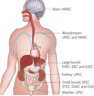
Figure 1-1: Sites of pathogenic E. coli colonization ... 3
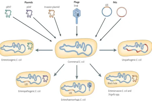
Figure 1-2: Horizontal gene transfer contribution to the evolution of E. coli pathotypes... 4
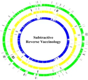
Figure 1-3: The subtractive reverse vaccinology approach ... 20
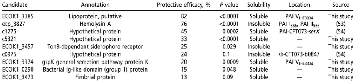
Figure 1-4: The evolutionary relationship and distribution of protective antigens among sequenced E. coli strains ... 21
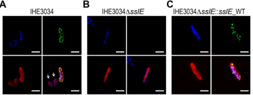
Figure 2-1: SslE surface localization on the ExPEC strain IHE3034 ... 32
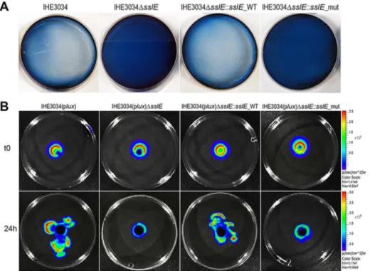
Figure 2-2: SslE mucinolytic activity. ... 33
Figure 2-3: Anti-SslE antibodies impair translocation of E. coli through a mucin-gel matrix. ... 34
Figure 2-4: Phylogenetic tree of SslE from a panel of E. coli isolates ... 35
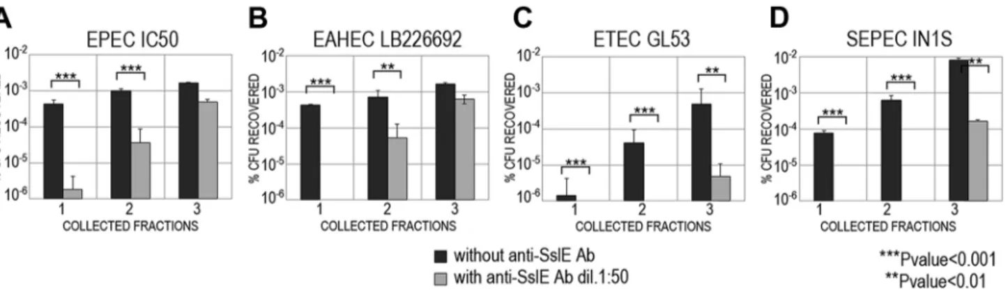
Figure 2-5: Cross-inhibition of E. coli translocation through a mucin-gel matrix by anti-SslEIHE3034 (belonging to variant I) antibodies ... 36
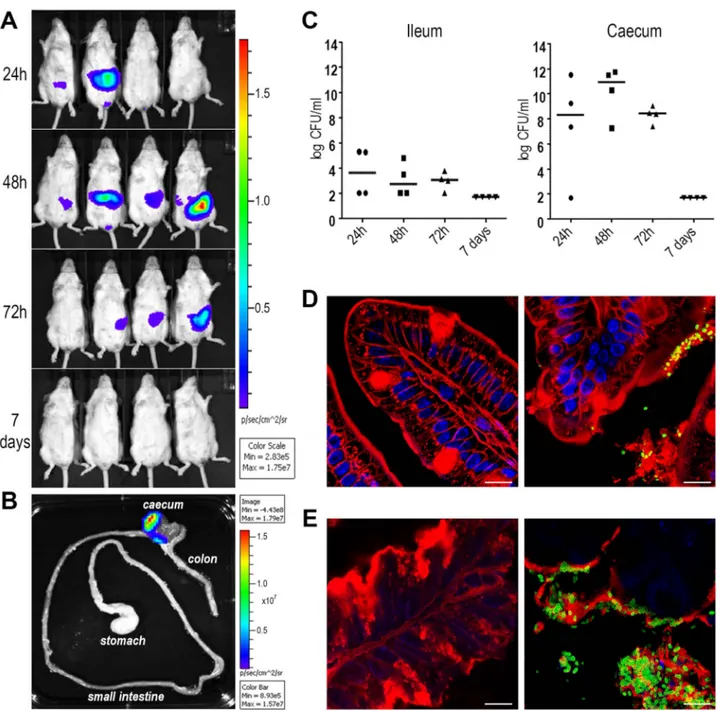
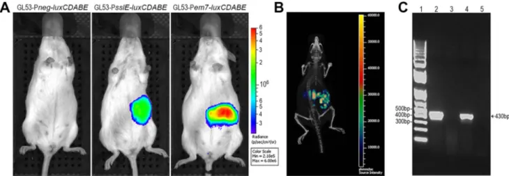
Figure 2-6: ETEC strain GL53 is able to colonize the mouse intestine. ... 37
Figure 2-7: The sslE promoter is functional in an intestinal model of colonization ... 38
Figure 2-8: SslEIHE3034 induces cross-protection in intestinal colonization, UTI and sepsis models ... 38
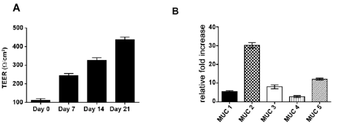
Figure 3-1: Kinetics of transepithelial electrical resistance in HT29-MTX cells over a 21 day period of differentiation ... 51
Figure 3-2: Modulation of SslE gene expression upon interaction with intestinal epithelial cells ... 52
Figure 3-3: Growth curves of strain IHE3034 in the presence of mucin ... 53
Figure 3-4: E. coli growth rate in association with HT29-MTX cells ... 53
Figure 3-5: SslE contributes to the capacity of IHE3034 strain to reach the surface of mucus-producing epithelial cells ... 54
Figure 3-6: SslE induces IL-8 secretion and stimulates neutrophil chemotaxis iratory burst ... 55
Figure 3-7: Schematic representation of the contribution of SslE to E.coli pathogenesis ... 58
Figure S3-1: Characterization of the extracellular mucus layer of HT29-MTX cells ... 59
III
AIDS: acquired immune deficiency syndrome BBB: blood-brain barrier
BFP: bundle forming pili BSA: bovine serum albumin
CAECAM: carcinoembryonic antigenrelated cell adhesion molecule cDNA: complementary deoxyribonucleic acid
CDT: cytolethal distending toxin CF: Colonization factors CFU: colony forming unit
CNF-1: cytotoxic necrotizing factor 1 CpG: cytosine phosphate guanine
DAEC: diffusely adherent Escherichia coli DAF: decay-accelerating factor
DNA: deoxyribonucleic acid
EAEC: enteroaggregative Escherichia coli EAF : EPEC adherence factor
EDTA: ethylenediaminetetraacetic acid Efb: extracellular fibrinogen-binding protein EGTA: ethylene glycol tetraacetic acid EHEC: enterohemorrhagic Escherichia coli Ehly: enterohemolysin
EIEC: enteroinvasive Escherichia coli EL: erythrocyte lysis
ELISA: enzyme-linked immunosorbent assay EPEC: enteropathogenic Escherichia coli ETEC: enterotoxigenic Escherichia coli
ExPEC: extraintestinal pathogenic Escherichia coli FACS: fluorescence-activated cell sorting
FdeC: Factor adherence E. coli FH: factor H
FITC: fluorescein isothiocyanate
FMLP: formyl-methionyl-leucyl-phenylalanine GBS: group B streptococcus
GEMS: Global Enteric Multi-Center Study GTP: guanosine triphosphate
HBSS: Hank’s balanced salt solution HC: hemorrhagic colitis
IV
HSA: human serum albumin HUS: hemolytic uremic syndrome IBC: intracellular bacterial community Ig: immunoglobulin
IgA-BP: immunoglobulin A-binding protein IL: interleukin
InPEC: intestinal pathogenic Escherichia coli IVIS: In Vivo Imaging System
LB: Luria-Bertani
LBSS: luminol-balanced salt solution LEE: locus of enterocyte effacement LP: lectin pathway
LPS: lipopolysaccharide LT: heat-labile enterotoxin MGL : mucus gel layer
mRNA: messenger ribonucleic acid MUC: mucin
NAC: N-acetyl cysteine NHS: normal human serum
NMEC: neonatal meningitis Escherichia coli OD: optical density
OMP: outer membrane protein ORF: open reading frame PAI: pathogenicity island PBS: phosphate buffered saline PCR: polymerase chain reaction PE: protective efficacy
pEAF: enteropathogenic Escherichia coli adhesion-factor plasmid pENT: enterotoxin-encoding plasmid
PLC: phospholipase C
PMN: polymorphonuclear leukocyte RNA: ribonucleic acid
RPMI: Roswell Park Memorial Institute
RT-PCR: reverse transcription-polymerase chain reaction Sat: secreted autotransporter toxin
SC: secretory component SD: standard deviation
SDS-PAGE: sodium dodecyl sulphate-polyacrylamide gel electrophoresis SEM: standard error of the mean
V
ST : heat-stable enterotoxin
STEC: Shiga-toxin producing E. coli Stx: Shiga toxin
TEER: Trans Epithelial Electrical Resistance THB: Todd-Hewitt broth
TIR: translocated initmin receptor TMD: transmembrane domain TNF-α: tumor necrosis factor α tRNA: transfer ribonucleic acid T2SS: type II secretion system TTP: thrombocytopenic purpura UPEC: uropathogenic Escherichia coli UTI: urinary tract infection
VI
Pathogenic Escherichia coli are responsible for a heterogeneous group of disorders, including diarrhoea, urinary tract infections, sepsis and neonatal meningitis, that collectively cause significant morbidity, lost productivity and high healthcare costs. Considering the incidence and also the increasing antibiotic resistance of E. coli strains, the prevention of infections is of pressing concern from both the public health and economic perspectives. Since conventional attempts to develop a highly immunogenic, safe and polyvalent vaccine had failed, we decided to apply the reverse vaccinology approach for the identification of protective and broadly conserved vaccine antigens. Although some of the protective candidates have been previously described, most of them have just putative or hypothetical functions assigned and, therefore, their characterization could contribute to the understanding of E. coli pathogenesis. In this study, vaccine antigen EKO_K1 3385, formally known as SslE (Secreted and surface-associated lipoprotein from Escherichia coli) has been characterized.
By applying a number of in vitro bioassays and comparing wild type, knockout mutant and complemented strains, we have demonstrated that SslE specifically contributes to degradation of mucin substrates, typically present in the intestine and bladder. Mutation of the zinc metallopeptidase motif of SslE dramatically impaired E. coli mucinase activity, confirming the specificity of the phenotype observed.
SslE can be divided into two main variants, we proved that antibodies raised against SslE variant I are able to inhibit translocation of E. coli strains expressing a different variant through a mucin-based matrix, suggesting that SslE induces cross-reactive functional antibodies that affect the metallopeptidase activity. To test this hypothesis, we used well established animal models and demonstrated that immunization with SslE variant I significantly reduced gut, kidney and spleen colonization by strains producing variant II SslE and belonging to different pathotypes.
Furthermore by exploiting a human in vitro model of mucus-secreting cells, we demonstrated that bacteria expressing SslE have a metabolic benefit which results in an increased growth rate postulating the importance of this antigen in enhancing E. coli fitness.
The results presented in this work conclusively designate SslE as an important colonization factor favouring E. coli access to both metabolic substrates and target cells and reinforce the use of this antigen as a component of a protective vaccine against pathogenic E. coli species.
VII
I ceppi patogeni di Escherichia coli sono causa di un ampio spettro di malattie, quali diarrea, infezioni del tratto urinario, sepsi e meningite neonatale, che, complessivamente, sono responsabili di elevati tassi di morbilità, perdita di produttività e ingenti costi sanitari. A causa della crescente incidenza delle infezioni provocate da questi ceppi e dell’aumento della resistenza antibiotica, la prevenzione delle infezioni riveste un’importanza sempre maggiore sia in ambito sanitario sia in ambito economico. Poiché gli approcci convenzionali si sono dimostrati inadeguati allo sviluppo di un vaccino polivalente, sicuro e altamente immunogenico, è stato scelto di applicare la “reverse vaccinology” per l’identificazione di antigeni protettivi e ampiamente conservati. Sebbene alcuni dei candidati selezionati siano stati in precedenza descritti in letteratura, alla maggior parte di essi è stata assegnata soltanto una funzione putativa o ipotetica, pertanto la loro caratterizzazione può costituire uno strumento utile all’elucidazione dei meccanismi di patogenesi. In questo studio, è stato caratterizzato l’antigene vaccinico EKO_K1 3385, noto formalmente come SslE (Secreted and surface-associated lipoprotein from Escherichia coli).
Utilizzando differenti test biologici e confrontando ceppi wild type, knockout e complementanti, abbiamo dimostrato che SslE contribuisce alla degradazione di substrati mucosi, tipicamente presenti nell’ intestino e nella vescica. Mutazioni del motivo metallopeptidasico compromettono l’attività enzimatica di questa proteina, confermando il fenotipo osservato.
Esistono due diverse varianti di SslE, nel presente studio abbiamo dimostrato che anticorpi diretti contro la variante I possono inibire la traslocazione attraverso una matrice mucosa di ceppi di E. coli che esprimono la variante II, suggerendo che SslE induce anticorpi funzionali cross-reattivi che interferiscono con l’ attività metalloproteasica. Per testare questa ipotesi, abbiamo utilizzato modelli animali ben validati e abbiamo dimostrato che l’immunizzazione con SslE variante I riduce in modo statisticamente significativo la colonizzazione del cieco, dei reni e della milza da parte di ceppi che producono la variante II e appartenenti a differenti patotipi.
Inoltre, utilizzando un modello in vitro di cellule umane che producono muco, abbiamo provato che SslE conferisce un beneficio metabolico ai batteri che la esprimono che porta a un aumentato tasso di crescita, postulando l’importanza di questo antigene nell’ aumentare la fitness di E. coli.
I risultati presentati in questo lavoro designano in modo conclusivo SslE come un importante fattore di colonizzazione che favorisce l’accesso di E. coli sia ai substrati metabolici che alle cellule target, rinforzando l’ uso di questo antigene come componente di un vaccino protettivo contro i ceppi patogenici di
1
2
Chapter 1
Introduction and aims
Escherichia coli is a gram-negative bacterium commonly found in the intestine of humans and
other mammals. Most E. coli strains are harmless commensals, however, pathogenic E. coli strains also exist and these isolates are typically categorized based on their mechanisms of disease and clinical outcomes [1].
For many years, E. coli pathotypes have been studied in isolation instead of addressing E. coli as a single microorganism responsible for human and animal infections. Considering the increasing antibiotic resistance present among pathogenic strains, which is derived from an uncontrolled use of antibiotics in humans and in the veterinary field, vaccination is the most promising approach to control disease. Comparative genome analysis and reverse vaccinology led to the identification of nine antigens capable of inducing protective immune responses against ExPEC strains, several of which are also prevalent in different intestinal E. coli pathotypes [2].
The aim of my PhD stems from this previous research and by functionally characterize SslE (EKO_K1 3385), the most protective vaccine candidate identified during the screening, try to define the molecular mechanisms of in vivo protection.
1.1. E. coli: A Versatile Species
Theodor Escherich first reported the isolation and characterization of slender short rods from infant stool, which he named Bacterium coli commune, in his 1885 publication [3]. Over 125 years later,
Escherichia coli is known as a harmless commensal of the gastrointestinal tract in warm-blooded animals
and is used as the colloquial laboratory workhorse. The bacterial species E. coli is a member of the family Enterobacteriaceae, located taxonomically within the gamma subdivision of the phylum Proteobacteria [1,4].
Normally, E. coli colonizes the infant gut within hours of birth and establishes itself as the most abundant facultative anaerobe of the human intestinal microflora for the remainder of life, equipped with the abilities to grow in the ever-changing environment in the gut and cope with the mammalian host interaction. Nevertheless, E. coli can survive in many different ecological habitats, including abiotic environments, and is considered a highly versatile species. Known habitats of E. coli include soil, water, sediment, and food. However, there are several highly adapted E. coli clones that have acquired specific virulence attributes, which confers an increased ability to adapt to new niches and allows them to cause a broad spectrum of disease [1,5-9].
Pathogenic E. coli strains can be divided into intestinal pathogenic E. coli (InPEC) and extraintestinal pathogenic E. coli (ExPEC), depending on the site of infection. Both are further subcategorized into distinct pathotypes, defined as a group of strains of a single species with certain pathogenic traits. Pathotype classification is based on the clinical manifestation of disease, the virulence factors (VFs) involved, and the phylogenetic background. Among the intestinal pathogens there are six well-described categories: enteropathogenic E. coli (EPEC), enterohaemorrhagic E. coli (EHEC),
3
enterotoxigenic E. coli (ETEC), enteroaggregative E. coli (EAEC), enteroinvasive E. coli (EIEC) and diffusely adherent E. coli (DAEC) [10]. UTIs are the most common extraintestinal E. coli infections and are caused by uropathogenic E. coli (UPEC). An increasingly common cause of extraintestinal infections is the pathotype responsible for meningitis and sepsis — meningitis-associated E. coli (MNEC) [11].
1.2. Evolution of pathogenic E. coli
As a population, E. coli strains can be assigned phylogenetically to 5 main clusters, i.e., A, B1, B2, D, and E, with Shigella forming a different group [12]. Commensal isolates mostly group in phylogroup A; however, not all E. coli pathotypes group together. Intestinal pathogenic E. coli strains derive from phylogenetic groups A, B1, D or from ungrouped lineages and are seldom found in the fecal flora of healthy individuals. These obligate pathogens are generally unable to cause extraintestinal disease and have evolved a special ability to induce colitis or gastroenteritis if ingested with contaminated food or water. Usually, mere acquisition of these bacteria by the naïve host is sufficient for disease to ensue. Each intestinal pathotype possesses a characteristic combination of virulence traits which allows the colonization of specific niches and results in a unique diarrheal syndrome (Figure 1-1) [13,14].
Unlike commensal and intestinal pathogenic E. coli, ExPEC strains belong predominantly to phylogenetic group B2 and, to a lesser extent, group D and have acquired various virulence genes that allow them to induce extraintestinal infections in both normal and compromised hosts. ExPEC are incapable of causing gastrointestinal disease, but they can asymptomatically colonize the human intestinal tract and may represent the predominant strain in approximately 20% of normal individuals. Therefore, although they are best known for their virulence behavior, ExPEC actually associate with the host primarily in a commensal fashion, causing disease only when they exit the gut and enter a normally sterile body site (Figure 1-1) [14,15].
Figure 1-1: Sites of pathogenic E. coli colonization (Croxen and Finlay, 2010).
Pathogenic E. coli colonize various sites in the human body. Enteropathogenic E. coli (EPEC), enterotoxigenic E. coli (ETEC) and diffusely adherent E. coli (DAEC) colonize the small bowel and cause diarrhea, whereas enterohemorrhagic E. coli (EHEC) and enteroinvasive E. coli (EIEC) cause disease in the large bowel; enteroaggregative E. coli (EAEC) can colonize both the small and large bowels. Uropathogenic E. coli (UPEC) enter the urinary tract and travel to the bladder to cause cystitis and, if left untreated, can ascend further into the kidneys to cause pyelonephritis. Septicemia can occur with both UPEC and neonatal meningitis E. coli (NMEC), and NMEC can cross the blood– brain barrier into the central nervous system, causing meningitis.
4
Genome sizes of E. coli can differ by a million base pairs between commensals and pathogenic variants, and this extra genetic content can contain virulence and fitness genes. Comparative genomics have shown that E. coli genomes are split between a shared, conserved set of genes, called the core genome, and a flexible gene pool. The pathogenic ability of E. coli is therefore largely afforded by the flexible gene pool through the gain and loss of genetic material at a number of hot spots throughout the genome [12]. DNA can be moved between prokaryotic hosts through mechanisms such as conjugation, transformation, and transduction, encoded by mobile genetic elements, resulting in horizontal gene transfer (HGT). Mobile genetic elements, such as transposons, insertion sequences, bacteriophages, and plasmids, can exist either integrated into the chromosome or through self-replication within the new host to provide new traits and fitness advantages[16]. A recent comparison of 186 E. coli genomes found approximately 1,700 homolog gene clusters shared in all genomes and a pangenome of about 16,400 gene clusters [17]. The pathogenic ability of E. coli is therefore largely afforded by the flexible gene pool through the gain and loss of genetic material at a number of hot spots throughout the genome (Figure 1-12) [12].
ExPEC differ from IPEC, because these facultative pathogens were traditionally already regarded as derived from different phylogenetic groups, illustrated for instance by their diversity of serotypes. Additionally, they do not host an unambiguous distinctive repertoire of VFs characteristic for a specific type of disease. Many ExPEC virulence-associated features are also present in commensal E. coli. Genome sequencing projects revealed extensive genome diversity among ExPEC, but also identified some pathotype-specific genes including toxins, iron acquisition systems, adhesins, lipopolysaccharides (LPS), polysaccharide capsules, proteases, and invasins. Again, these factors are frequently encoded on mobile elements [18,19].
Figure 1-2: Horizontal gene transfer contribution to the evolution of E. coli pathotypes (Ahmed et al., 2008).
The uptake of mobile genetic elements (phages, virulence plasmids and pathogenicity islands), as well as the loss of chromosomal DNA regions in different E. coli lineages, has enabled the evolution of separate clones, which belong to different E. coli pathotypes and are associated with specific disease symptoms. LEE, locus of enterocyte effacement; PAI, pathogenicity island; pEAF, enteropathogenic E. coli adhesion-factor plasmid; pENT, enterotoxin-encoding plasmids; Stx, Shiga-toxin-encoding bacteriophage.
5
1.3. Extraintestinal pathogenic Escherichia coli
Extraintestinal pathogenic E. coli represent a major but little-appreciated health threat. Although ExPEC strains have not captured the public’s attention as have intestinal pathogenic E. coli, probably because extraintestinal infections do not occur in a sensational food-borne epidemic fashion, their medical importance cannot be ignored. By virtue of their numerous virulence traits, ExPEC clearly possess a unique ability to cause disease outside the host intestinal tract and are responsible for a heterogeneous group of disorders that collectively cause considerable morbidity, lost productivity and increased healthcare costs [15]. Indeed, ExPEC is the most common cause of urinary tract infections (UTIs) in ambulatory and hospital settings. It is responsible for 85 to 95% of the cases of uncomplicated cystitis and for over 90% of the episodes of uncomplicated pyelonephritis in premenopausal women. An estimated 40 to 50% of women will experience at least one case of UTI due to E. coli during the lifetime and one fourth will experience a recurrent infection within 6 months of initial infection. ExPEC is also responsible for 25 to 35% of the episodes of catheter-associated UTIs. Furthermore, along with group B Streptococcus (GBS), E. coli is one of the leading causes of neonatal meningitis, accounting for an estimated 20 to 40% of the cases, with a fatality rate ranging from 25 to 40% and with neurological sequelae affecting 33 to 50% of survivors. E. coli also accounts for 17% of the cases of severe sepsis, with a mortality rate of approximately 30%. In addition, ExPEC can be associated with intra-abdominal infections and nosocomial pneumonia and occasionally participates in other extraintestinal infections, such as osteomyelitis, cellulitis and wound infections [13,20,21].
ExPEC strains possess a broad range of virulence factors that are distinct from those found in the intestinal pathotypes and that allow them to colonize host mucosal surfaces, avoid or subvert local and systemic host defense mechanisms, scavenge essential nutrients such as iron, injure or invade the host, and stimulate a noxious inflammatory response [15]. Most of these virulence factors have been acquired by mobile genetic elements.
PAIs carry many of the virulence factors characteristic of ExPEC strains. PAIs are large clusters (10-200 kb) of virulence genes that are present in the genomes of pathogenic strains but absent from the genomes of non-pathogenic members of the same or related species. They are typically associated with t-RNA genes, have a different G+C content and a different codon usage compared to the core genome and often carry cryptic or functional genes that encode mobile elements [1].
Among the ExPEC virulence factors frequently harboured by PAIs, a fundamental role is played by adhesins, which allow the strict interaction of the pathogen with the host, facilitating the colonization and invasion processes and avoiding clearance by the host immune defences. Despite the similarity in their tertiary structure, each adhesin recognizes a specific host receptor [22]. For example, type 1 fimbriae of UPEC strains recognize manno-oligosaccharides naturally present on glycoprotein molecules in the human urinary tract and participate in bacterial adhesion, invasion and formation of biofilms. P fimbriae recognize a digalactoside component of the P blood group antigen abundantly present on the surface of uroepithelial cells and are strictly related to bacteria ascending to the kidneys and causing acute pyelonephritis [23,24].
The presence of group 2 and 3 capsules confers additional selective advantages to ExPEC strains. Indeed, their molecular mimicry to host tissue components helps the bacteria to evade the immune response, providing protection against phagocytic engulfment and complement-mediated bactericidal activity [25,26].
6
Some virulence factors do not remain attached to the bacterial outer membrane, but are released into the extracellular milieu. The most important secreted factor of ExPEC strains is α-hemolysin (HlyA), a toxin with a promiscuous spectrum of target cells, including not only erythrocytes, but also leucocytes, endothelial cells and renal epithelial cells. It is intracellularly activated by fatty acylation and extracellularly activated by calcium, allowing the insertion into the cell membrane followed by pore formation and disruption of the phospholipid bilayer [27-30]. Other secreted proteins also play important roles in ExPEC pathogenesis, for example: cytotoxic necrotizing factor 1 (CNF-1), which interferes with polymorphonuclear phagocytosis and evokes apoptotic death of bladder epithelial cells [31]; secreted autotransporter toxin (Sat), a serine protease autotransporter with vacuolating activity on bladder and kidney cells [32,33]; Pic and PicU, other type V secreted proteins with serine protease activity [34,35]; cytolethal distending toxin (CDT), responsible for the arrest of cell cycle by inducing DNA double-strand breaks and preventing the transition between G2 and M phases [36-39].
Growth of ExPEC strains in iron-limited conditions, such as urine, requires successful mechanisms for the scavenging of iron, which rely on siderophores and iron-complex receptors [40,41]. Several iron and siderophore receptors, which are highly expressed during infection of the urinary tract, have already been described in E. coli, for example the salmochelin siderophore receptor IroN [42], the enterochelin siderophore receptor FepA [43], the hemoglobin and hemin receptor ChuA [44], the ferric yersiniabactin receptor FyuA [45], FitA [46] and IreA [47].
1.4. Pathogenesis of ExPEC
Among ExPEC strains, uropathogenic E. coli and neonatal meningitis E. coli are characterized by different molecular mechanisms of pathogenicity.
Urinary tract infection usually begins with the colonization of the bowel with an uropathogenic strain in addition to the commensal flora. This strain, by virtue of its virulence factors, is able to colonize the periurethral area and to ascend the urethra to the bladder. Between 4 and 24 hours after infection, the new environmental conditions in the bladder select for the expression of type 1 fimbriae that allow the adhesion to the uroepithelium [1]. This attachment is mediated by fimbrial adhesin H (FimH), which is located at the tip of type 1 pili. FimH binds to mannose moieties of the receptors uroplakin Ia and IIIa that coat terminally differentiated superficial facet cells in the bladder, stimulating also unknown signalling pathways that induce invasion and apoptosis . Bacteria internalization is also mediated by FimH binding to α3 and β1 integrins that are clustered with actin at the sites of invasion, as well as by microtubule destabilization. These interactions trigger local actin rearrangement by stimulating kinases and Rho-family GTPases, which results in the envelopment and internalization of the attached bacteria. Once internalized, UPEC can rapidly replicate and form biofilm-like complexes called intracellular bacterial communities (IBCs), which act as transient, protective environments. UPEC can also leave the IBCs through a fluxing mechanism and enter again the lumen of the bladder. Filamentous UPEC has also been observed fluxing out of an infected cell, looping and invading surrounding superficial cells in response to innate immune responses. During infection, the influx of polymorphonuclear leukocytes (PMNs) causes tissue damage, while apoptosis and exfoliation of bladder cells can be induced by UPEC attachment and invasion, as well as by sublytic concentrations of the pore-forming toxin HlyA. This breach of the superficial facet cells
7
temporarily exposes the underlying transitional cells to UPEC invasion and dissemination. Invading bacteria are trafficked in endocytic vesicles enmeshed with actin fibers, where replication is restricted. Disruption of host actin allows rapid replication, which can lead to IBC formation in the cytosol or fluxing out to the cell. This quiescent state may act as a reservoir that is protected from host immunity and may, therefore, permit long-term persistence in the bladder, as well as recurrent infections [9].
In strains causing cystitis, type 1 fimbriae are continuously expressed and the infection is confined to the bladder. In strains that are able to cause pyelonephritis, the invertible element that controls type 1 fimbriae expression turns to the “off” position and type 1 pili are less well expressed. This releases the UPEC strain from bladder epithelial cell receptors and allows the microorganism to ascend through the ureters to the kidneys, where it can attach by P fimbriae to digalactoside receptors that are expressed on the kidney epithelium. At this stage, hemolysin could damage the renal epithelium inducing an acute inflammatory response with the recruitment of PMNs to the infection site. Hemolysin has also been shown to cause calcium oscillations in renal epithelial cells, resulting in increased production of interleukin-6 (IL-6) and -8 (IL-8). Secretion of the vacuolating cytotoxin Sat damages glomeruli and is cytopathic for the surrounding epithelium. In some cases, bacteria can cross the tubular epithelial cell barrier and penetrate the endothelium to enter the bloodstream, leading to bacteremia [1].
The pathogenesis of NMEC strains is a complex mechanism, as the bacteria must enter the bloodstream through the intestine and ultimately cross the blood-brain barrier (BBB) into the central nervous system, which leads to meningeal inflammation and pleocytosis, that means presence of a higher number of cells than normal, in the cerebrospinal fluid. Bacteria can be acquired perinatally from the mother and, after the initial colonization of the gut, they can translocate to the bloodstream by transcytosis through enterocytes. The progression of disease is dependent on high bacteremia (>103 colony forming units per ml of blood), therefore survival in the blood is crucial. NMEC is protected from the host immune responses by its K1 antiphagocytic capsule, made up of a homopolymer of polysialic acid, and by outer membrane protein A (OmpA), which confers serum resistance through manipulation of the classical complement pathway. NMEC has also been shown to interact with immune cells: invasion of macrophages and monocytes prevents apoptosis and chemokine release, providing a niche for replication before dissemination back into the blood. Bacterial attachment to the BBB is mediated by FimH binding to CD48 and by OmpA binding to its receptor, ECGP96. Invasion of brain microvascular endothelial cells involves CNF-1 binding to the 67 kDa laminin receptor (67LR), which leads to myosin rearrangement, as well as OmpA and FimH binding to their receptors, which results in actin rearrangement. The K1 capsule, which is found in approximately 80% of NMEC isolates, also has a role in invasion by preventing lysosomal fusion and thus allowing delivery of live bacteria across the BBB. Collectively, these mechanisms allow NMEC to penetrate the BBB and gain access to the central nervous system, where they cause edema, inflammation and neuronal damage [9].
1.5. Intestinal pathogenic E. coli
Diarrheal illness causes much mortality worldwide, particularly in children under the age of 5 [48] and particularly in countries in sub-Saharan Africa and South Asia, whose children suffer many diarrhea-related deaths. Recent data from the Global Enteric Multi-Center Study (GEMS) illustrate that
8
enterotoxigenic E. coli and Shigella are among two of the four main causative agents of moderate to severe diarrhea among children in these areas [49]. The pathogenic E. coli isolates share many virulence strategies. Adhesion to host cells is a requirement for all pathovars and is frequently achieved through long appendages called fimbriae or pili. Following attachment, E. coli must subvert host cell processes, often using secreted proteins. Hijacking and manipulating host cell signalling pathways can result in the coordinated invasion of host cells, evasion of host immune responses and efficient colonization, and ultimately leads to disease. Each pathovar has its own characteristic mechanisms of attaching to and exploiting host cells although they often target the same host machinery [10].
One main feature of the different intestinal E. coli pathotypes is the presence of pathotype-specific plasmids that often encode toxins [50]. Concentrations of important intracellular messengers, such as cyclic AMP, cyclic GMP and Ca2+, can be increased, which leads to ion secretion by the actions of the heat-labile enterotoxin (LT), heat-stable enterotoxin (STa) and heat-stable enterotoxin b (STb), respectively — all of which are produced by different strains of ETEC [51]. The Shiga toxin (Stx) of EHEC cleaves ribosomal RNA, thereby disrupting protein synthesis and killing the intoxicated epithelial or endothelial cells [52]. The cytolethal distending toxin (CDT) has DNaseI activity that ultimately blocks cell division in the G2/M phase of the cell cycle [36]. The Map protein of EPEC and EHEC has at least two independent activities — stimulating Cdc42-dependent filopodia formation and targeting mitochondria to disrupt membrane potential in these organelles [53].
Additionally, large so-called colicin plasmids seem to contain several gene clusters, including the salmochelin determinant, that can also be found within chromosomal PAIs in E. coli and closely related species. Nearly one-quarter of the EAEC strain 042 genomic content is made up of genomic islands [54], similar to the percentage of unique genomic islands found in STEC O157:H7 strain EDL933 [55].
1.6. Pathogenesis of InPec
Enteropathogenic E. coli (EPEC)
Enteropathogenic E. coli is a leading cause of potentially fatal infant diarrhoea in developing countries and also an important cause of intestinal infection in industrialized countries. EPEC infection is primarily a disease in infants younger than 2 years. Sporadic disease also occurred in some adults with compromising conditions. Once defined only on O and H serotyping, they are currently defined by pathogenic features as those diarrheagenic E. coli that induce attaching and effacing (A/E) lesions on intestinal cells and do not produce Shiga toxins. They could be divided into typical EPEC and atypical EPEC based on the presence of EPEC adherence factors (EAF) plasmid. Molecular detection and differentiation of EPEC could be based on the eae gene (A/E lesions) and bfp gene (which resides on EAF plasmids and encodes bundle- forming pilus). Typical EPEC contain both eae and bfp genes, while atypical EPEC contain only eae gene. Typical EPEC infections are common in developing countries while atypical
E. coli seems to predominate in the industrialized countries. Not like typical EPEC which are found only in
human so far, atypical EPEC have been isolated from a variety of animal species such as cattle, goats, sheep, chickens, pigeons and gulls [56,57].
9
Pathogenesis of EPEC is currently considered to include four stages: expression of adhesion factors, initial localized adherence, signal transduction and intimate contact, cytoskeletal rearrangement and pedestal formation. Initially the bacteria attach to intestinal epithelial cells by adhesive fimbriae called bundle forming pili (BFP) or EPEC adherence factor (EAF) [58]. EPEC also adhere to epithelial cells by expressing intimin (encoded by eae gene) and surface–associated filament EspA. After initial binding, EPEC utilize type III secretion system to inject into host cells translocated initmin receptor (TIR) and several effector molecules, which activate cell signaling pathways and actin polymerization and depolymerization to alter cytoskeletal structure. TIR is then phosphorylated and inserted into the host cell membrane for later intimate contact. Activated host cell signal transduction pathway causes extensive rearrangement of actin, which results in the formation of the characteristic attaching and effacing lesions [59]. The membrane under the bacteria forms a pedestal due to host cell cytoskeletal rearrangement. The microvilli are lost due to depolymerization of actin filament in microvilli. The effector proteins also influence membrance permeability and cause diarrhea-associated symptoms. Virulence genes in EPEC are mostly located in a pathogenic island called locus of enterocyte effacement (LEE).
Enterohaemorrhagic E. coli (EHEC)
The EHEC group is also referred to Shiga-toxin producing E. coli (STEC) because its pathogenicity is largely attributed to the production of Shiga toxins. It should be noted that some researchers prefer to use EHEC only for those STEC containing LEE pathogenic island, while others use them exchangeably. Among six categories of diarrheagenic strains, EHEC strains distinguish themselves by their ability to cause severe life-threatening complications, such as hemolytic uremic syndrome (HUS) and thrombotic thrombocytopenic purpura (TTP). Other symptoms of EHEC infection include bloody diarrhea and hemorrhagic colitis (HC). Children and the elderly are more susceptible to severe STEC infections than healthy adults. Outbreaks and sporadic cases of EHEC infections are frequently reported worldwide, indicating the great threat that EHEC could pose for human health [60-62]
EHEC can be disseminated through a variety of means. Most human infections are caused by consumption of contaminated foods. Domestic and wild ruminant animals, in particular cattle, are considered as the main reservoir of EHEC and the main source for contamination of the food supply [63-65]. Food products derived from these animals can be contaminated with EHEC during slaughter and further processing. In addition, vegetables contaminated with cattle manure have been also implicated in many cases and outbreaks of EHEC infections [66]. EHEC have been also isolated from other food animals such as pigs and poultry, but whether these animals represent real hosts or are just temporarily colonized with EHEC is not clear [67,68].
The mechanisms of EHEC infection in humans are not fully understood. The major virulence factors implicated are potent Shiga toxins, which are classified into two groups: Stx1 and Stx2. In each group, variants that differ in toxicity, toxin receptor, and amino acid sequences have been described. Few variants (Stx1c, Stx1d) were found for Stx1, whereas Stx2 contains several variants including Stx2c, Stx2d, Stx2e, Stx2f, Stx2g [69-74]. Shiga toxin types were suggested to correlate with the clinical symptoms of EHEC infection. Some Stx types, such as Stx2, Stx2c and Stx2d-activatable, have been associated with high virulence and ability to cause HUS, while Stx1, Stx1c, Stx2e occurred mainly in milder diarrhea patients or
10
asymptomatic carriers [75]. In addition to Stx production, other (putative) virulence factors that could contribute to the pathogenicity have been discovered. The eae gene, which is located in a pathogenic island in the chromosome called the locus of enterocyte effacement (LEE), is the best characterized virulence loci other than stx. The eae gene encodes the adherence factor intimin, an outer membrane protein involved in the attachment of E. coli to the enterocyte. In addition, many pathogenic EHEC possess a large plasmid that harbors several putative virulence factors such as hlyA, which encode for a cytolytic EHEC-hemolysin. EHEC hemolysin is strongly associated with EHEC isolates causing severe infections in human, but its exact role in pathogenesis is still yet to be known.
More than 400 serotypes of EHEC strains have been implicated in human infections. Although E.
coli O157: H7 is considered the principal EHEC in the U. S., infections due to non-O157 EHEC occur and
are thought to be underreported [76,77]. In some other countries, such as Germany, Australia, and the UK, non-O157 EHEC infections predominate [78,79]. Globally, only a limited numbers of serotypes were frequently observed and are responsible for the majority of EHEC infections.
Enterotoxigenic E. coli (ETEC)
ETEC are defined as those E. coli strains that contain at least one of two defined groups of enterotoxins: heat stable toxin (ST) and heat labile toxin (LT). ETEC is an important cause of childhood diarrhea in developing countries due to poor sanitary conditions. It is also a common cause of diarrhoea in travellers to developing countries. It is estimated that around 650 million cases of ETEC infection occur each year, which include 800,000 deaths mostly in young children [80]. It causes watery diarrhoea ranging from mild form to severe purging disease. The diarrhoea persists for 3-4 days and is usually self-limiting, however, diarrhoea may be fatal in young children and infants. Epidemiologic studies found that contaminated food and water serves as the most common vehicles for ETEC infections [81].
Colonization factors (CF) and one or more enterotoxins that induce a secretory diarrhoea are the major determinants of ETEC virulence. CFs are proteinaceous fimbrial and afimbrial structures that enable bacteria to attach to intestinal mucosa. More than 20 CFs have been identified and characterized in ETEC [82]. Other adhesion factors such as TibA (an afimbrial adhesion) and Tia (an outer membrane protein) are also involved and implicated in the attachment of ETEC[83].
Having established contact with epithelial cells, ETEC can produce one or more ST or LT. ST have been divided into two distinct groups: methanol soluble STI (or STa) and methanol insoluble STII (or STb) [84]. STa toxins have two genetic variants STh and STp, described originally in association with strains isolated from human and pigs, respectively. However, new studies found that both variants could be found in ETEC strains of human origin. STa binds to guanylate cyclase C receptor and activates its guanylate cyclase domain, which results in an increase in intracellular cGMP level. Increase cGMP influences ion pumps, resulting in enhanced salt and water secretion and inhibition of Na+ absorption. STb is most associated with porcine strains of ETEC.
LT is an oligomeric protein composed by a ring of five identical B subunits with one A subunit. The B subunits bind to a GTP binding protein (ganglioside receptor), while the A subunit is responsible for the enzymatic activity of the toxin [85]. Based on type of cell surface receptor to which B subunits bind, LT could be divided into LT-I (bind to ganglioside receptor GM1) and LT-II (bind to ganglioside receptor GD1). A subunit ADP-ribosylates the alpha subunit of the GTP-binding protein Gs, leading to activation of
11
adenylate cyclase in the enterocyte and accumulation of cyclic AMP. The increase in intracellular cAMP increases Cl- secretion in crypt cells and decreases absorption of Na+ and Cl- by villus tip cells. Other toxins that may contribute to ETEC infection include a novel heat stable enterotoxin EAST1 and a serine protease autotransporter, EatA, and a pore–forming toxin, ClyA.
Enteroinvasive E. coli (EIEC)
EIEC strains are genetically and pathogenically related closely to Shigella spp.[86]. EIEC and
Shigella are highly invasive pathogens that use the intracellular milieu of intestinal epithelial cells (IECs) in
the large intestine as their replicative niche. These pathogens are readily adaptable to the various environmental challenges they face during the course of infection, including low gastric pH, temperature changes, oxygen availability, and oxidative stress, as well as osmolarity [87]. Successful infection is dependent on essential virulence determinants that are encoded by both chromosomal and plasmid loci. Key plasmid encoded virulence factors include components of the T3SS needle complex (Mxi-Spa proteins), chaperones (IpgA, IpgC, IpgE, and Spa15), transcriptional regulators (VirF, VirB, and MxiE), translocators (IpaB, IpaC, and IpaD), and approximately 25 effector proteins [88]. Bacterial infection is a multistep process involving penetration of the epithelial barrier, induction of macrophage cell death, IEC invasion, suppression of the immune response, intra and intercellular movement, and modulation of epithelial integrity.
In general, EIEC and Shigella employ the same strategies to invade host cells. Nevertheless, EIEC exhibits reduced virulence compared to that of Shigella, including reduced expression of virulence genes, less efficient macrophage killing, reduced cell-to-cell spread, and decreased induction of a proinflammatory host response which correlates with the less severe disease induced by EIEC [89,90].
Enteroaggregative E. coli (EAEC)
EAEC are defined as E. coli that do not produce LT or ST and that adhere to HEp-2 cells in a pattern described as autoaggregative. EAEC have been increasingly recognized as an important causative agent of persistent diarrhoea in children and adults in both developing and developed countries. EAEC mostly cause sporadic cases, but several outbreaks have been reported [91]. Prior to 2011, there were only been a few reports of Shiga toxin-producing EAEC causing bloody diarrhoea and haemolytic uremic syndrome (HUS) [92,93], but these are now more appreciated due to the E. coli O104:H4 outbreak in Germany [94]. Characterization of isolates from this outbreak identified key virulence features belonging to different pathotypes, such as an aggregative adhesive phenotype in vitro, lack of the LEE PAI, and expression of a Shiga toxin [95]. Therefore, these isolates can be considered a hybrid of both EAEC and EHEC (a subset of STEC), and it has been suggested that the STEC O104:H4 strain associated with the 2011 German outbreak be called enteroaggregative haemorrhagic E. coli (EAHEC) [96]. Additionally, genome analysis of LEE-negative STEC has uncovered homologs and subunits of ETEC toxins in some isolates [97], further demonstrating the potential for the emergence of novel pathogenic E. coli hybrids. For non-Stx variants, EAEC strain 042 has been used as a prototypical strain to study virulence factors and pathogenicity of diarrheagenic EAEC, as it causes diarrhoea in the majority of volunteers. However, the encoding genes for numerous adhesins, toxins, and proteins associated with virulence are highly variable among strains [1,10,98-101]. Even the site of infection in the gastrointestinal tract is not uniform. For
12
example, the EAEC 042 strain has been isolated from the jejunum in infected volunteers, and in tissue culture it adheres strongly to samples of jejunal, ileal, and colonic mucosa [102,103]. In a controlled study looking at five different non-Stx EAEC isolates from children, each strain had a different affinity for the jejunal, ileal, and colonic mucosae [104]. Despite the heterogeneity among the different non-Stx EAEC strains, a general three-part model has emerged for non-Stx EAEC pathogenesis: (i) adherence to the intestinal mucosa, (ii) production of enterotoxins and cytotoxins, and (iii) mucosal inflammation[100]. EAEC express aggregative adherence fimbriae (AAF) I, II and III and outer membrane adhesion proteins. Adherence is described as a stacked-brick shape. EAEC elaborate enteroaggregative heat stable toxin (EAST) and a cytotoxin that is responsible for pathological effects. Infections usually lead to mucoid stool and persistent diarrhoea (often more than 14 days).
Diffusely adherent E. coli (DAEC)
The diffusely adherent E. coli (DAEC) pathotype describes diarrheagenic E. coli strains that attach to cells but do not fall into classical patterns of adherence, such as localized or A/E [10]. They have now emerged as a unique group and are considered distinct from other pathotypes, but because of difficulties in classification and identification, the designation of DAEC as a distinct enteric E. coli pathotype [105] requires further epidemiological studies. DAEC has been classically defined by its diffuse adherence (DA) to cultured epithelial HEp-2 cells, where bacterial adherence occurs over the entire surface of the cell in a scattered pattern [106]. The prototypical strain C1845 is a DAEC strain that encodes Afa/Dr adhesins. The Afa/Dr adhesins are a class of adhesins that includes the AfaE-I, AfaE-II, AfaE-III, AfaE-V, Dr, Dr-II, F1845, and NFA-I adhesins[107]. In Afa/Dr DAEC, the Dr and the F1845 adhesins bind to brush border-associated decay-accelerating factor (DAF), a molecule that is highly expressed on the apical surface of polarized epithelial cells. After binding, cytoskeleton rearrangement is induced, destroying or partially rearranging microvilli [107-112]. Some Afa/Dr DAEC strains also bind the human carcinoembryonic antigenrelated cell adhesion molecule (CAECAM) family of receptors in a process that leads to internalization into undifferentiated epithelial cells [113]. In addition to binding DAF and the CAECAM family of receptors, Afa/Dr DAEC has also been shown to bind type IV collagen through the Dr adhesin, an interaction that is important for urinary tract infections caused by Afa/Dr DAEC. After binding DAF, disassembly of F actin and villin results in brush border lesions. This eventually leads to a loss of microvilli due to defective expression of brush border-associated functional intestinal proteins [109]. Rearrangement of the tight-junction- associated proteins ZO-1 and occludin after infection by Afa/Dr DAEC strains leads to increased paracellular permeability but does not affect transepithelial electrical resistance[111]. The proinflammatory cytokine IL-8 is produced through flagellar stimulation of Toll-like receptor 5 (TLR5), resulting in activation of mitogen activated protein kinase, extracellular signal-regulated kinases 1 and 2 (ERK1/2), P38, and Jun-C kinase [114-118].
While the pathogenesis of typical Afa/Dr DAEC is beginning to be characterized, much remains to be discovered for atypical diarrheagenic DAEC strains. There are two different subclasses of atypical DAEC. One subclass contains all the adhesins typical of the Afa/Dr family of adhesins in another E. coli background, such as diffusely adherent EPEC. In the other subclass of atypical DAEC, the bacterium does not bind DAF and expresses a different array of adhesins on its surface, including AfaE-VII, AfaE-VIII,
13
AAF-I, AAF-II, and AAF-III [108]. In this subclass of atypical DAEC, IL-8 is still stimulated by DAEC strains that have been internalized by an uncharacterized mechanism, suggesting that it may elicit pathogenesis mechanisms similar to those of typical Afa/Dr DAEC [119].
1.7. E. coli and host interactions in the gut epithelial barrier
The gastrointestinal epithelium is covered by a mucus gel layer (MGL) synthesized and secreted by host goblet cells. The MGL is an integral structural component of the mammal intestine, acting as a medium for protection and transport between the luminal content and the epithelium lining. The major function of the MGL is to lubricate and to protect the intestinal epithelium from damage caused by food and digestive secretions. Moreover, the MGL acts as a trap for microorganisms, including pathogens, preventing their access to the epithelia [120].
Studies by Holm and colleagues showed that there are two mucus layers in the stomach and colon: an outer “loose” layer that was easy to aspirate and an inner mucus layer that was “firmly” attached to the epithelium [121] . These mucus layers are organized around the highly glycosylated MUC2 mucin, forming a large, net-like polymer that is secreted by the goblet cells. The inner mucus layer is dense and does not allow bacteria to penetrate, thus keeping the epithelial cell surface free from bacteria. The inner mucus layer is converted into the outer layer, which is the habitat of the commensal flora. The outer mucus layer has an expanded volume due to proteolytic activities provided by the host but probably also caused by commensal bacterial proteases and glycosidases. This is in contrast to the small intestine, where the mucus is discontinuous and less well defined. The mucus is secreted at the top of the crypts and then moves upward between the villi. Thus, the tips of the villi are not always covered with mucus [122] .
The commensal bacteria in colon live and thrive in the outer loose mucus layer. This is possible after the Muc2 mucin network has expanded in volume, such that it allows the bacteria to penetrate into the mucin network. Once inside the mucus gel, the commensal bacteria can use its large number of glycan-degrading enzymes that release one monosaccharide at a time from the mucin glycans [123].
Pathogenic strains of E. coli, without exception, must first colonize the host gastrointestinal tract before causing disease [1]. Freter postulated that successful competition for nutrients allows intestinal bacteria to colonize, which we define as the ability to achieve and maintain a stable population without reintroduction. Freter’s nutrient niche hypothesis theoretically explains the succession of community members of the intestinal microbiota, as well as the ability of enteric pathogens to overcome colonization resistance and thereby invade the microbiota [124]. How invading pathogens compete for nutrients with the established microbial residents is an open question.
A large and growing body of evidence indicates that E. coli grows in the intestine on nutrients acquired from mucus. Fluorescence in situ hybridization of intestinal thin slices showed that E. coli BJ4 [125] and MG1655 [126] are dispersed in the mucus layer. E. coli BJ4 grows rapidly in the mouse intestine, with a generation time of 40–80 min [127]. In vitro, rapid growth (30-min generation time) occurs in intestinal mucus, but not in luminal contents [128,129] . Among mutants unable to colonize the mouse intestine are those that fail to penetrate mucus, have difficulty surviving in mucus, or have difficulty growing on mucus [130-132].
14
Virulence factors such as proteases, glycosidases, and mucus secretagogues are produced by these organisms and are believed to be responsible for disruption and depletion of the mucus gel [133]. Enzymes, such as Pic, a serine protease that degrades mucin [134]; StcE, a zinc metalloprotease that cleaves mucin-type O-glycosylated proteins [135]; Hap, a zinc metalloprotease [136] ; TagA, a Hap homolog with metalloprotease activity that is distinct from Hap [137] or mucin-degrading enzymes[138] , degrade mucin oligosaccharides, reduce mucus viscosity and hamper the release of antimicrobial peptides.
The personal repertoire of expression of mucin core proteins and their glycans, mucin allele length, and transient changes in mucin expression and glycosylation in response to infection or stress, as well as variations in environmental conditions may all affect microbial interaction with host mucins and the pathogenic consequences of microbial colonization. Rather than a static barrier, mucins should be considered as a dynamic responsive component of the mucosal barrier that interacts with and responds to other elements of innate and adaptive immunity.
1.8. Vaccines against pathogenic E. coli
The prevention of E. coli infections is of pressing concern from both the public health and economic perspectives [14]. Indeed, the absence of a broadly protective vaccine against pathogenic E. coli strains is a major problem for modern society since some of the diseases caused by these bacteria are associated with high costs to healthcare systems. The overall problem is exacerbated by the increasing antibiotic resistance and the number of recurrent infections [139]. Attempts to develop a broadly protective and safe vaccine against E. coli have not been successful so far. The large antigenic and genetic variability of pathogenic E. coli species has been a major obstacle to the development of an effective vaccine. Indeed, the difficulty in predicting vaccine coverage and the lack of a correlate of protection, has led to numerous promising pre-clinical data not being confirmed by human studies [140,141].
For many years, E. coli pathotypes have been studied in isolation instead of addressing E. coli as a single microorganism responsible for human and animal diseases.
Many efforts have already been done to assess protein moieties as putative vaccine candidates against extraintestinal E. coli infections. These efforts were logically concentrated on proteins that are surface-exposed and have a potential role in pathogenesis, such as adhesins, iron-regulated outer membrane proteins (OMPs) and toxins [139]. Antibodies directed against adhesins have the promise to enhance the bactericidal activity mediated by complement and professional phagocytes and also to inhibit bacterial binding to host structures, a critical step in the pathogenesis of infection [139]. Systemic immunization with purified P fimbriae [142,143] and synthetic peptides corresponding to the protective epitope of the P fimbrial major subunit PapG [144] conferred protection in a murine pyelonephritis model; immunization with purified P fimbriae or with purified PapDG-complex also conferred protection in a nonhuman primate model [145,146]. Several iron-regulated OMPs have also been assessed to date as potential vaccine candidates, given that many are surface-exposed and iron acquisition is a requisite for pathogenesis [139]. The pore-forming toxin α-hemolysin has been demonstrated to be highly conserved [147]. In a mouse model of pyelonephritis, systemic immunization with purified HlyA was associated with decreased renal damage, but did not affect clearance of E. coli. However, its combination with digalactoside-binding pilus was able to prevent both bacterial colonization and renal injury [139,142].
15
Several whole-cell vaccine approaches have also been tried to prevent extraintestinal E. coli infections. Four standardized whole-cell vaccine formulations (Urovac®, OM-89 or Uro-Vaxom®, Urvakol®, Urostim®) have been tested for their efficacy in preventing UTIs [139,148,149]. Although for most of the formulations data are far from convincing and many studies lack of scientific rigor, the potentiality of whole-cell vaccines is high, since they could present multiple antigens, elicit antibodies against conformational and linear epitopes and possess natural adjuvants.
Vaccines are being developed to prevent some of the serious sequel and complications associated with E. coli-induced diarrheal illness. Only one vaccine (Dukoral® produced by SBL Sweden) is currently available for the prevention of ETEC diarrhea. This vaccine has been recommended to prevent 'travellers' diarrhea' in people visiting endemic regions from developed countries [150]. Dukoral is primarily designed and licensed to prevent diarrhea due to Vibrio cholera, but it contains a recombinant B subunit of the cholera toxin that is antigenically very similar to the LT of ETEC [151]. In an early clinical trial, using a prototype of this vaccine which contained purified cholera B subunit rather than the recombinant form, significant cross protection against ETEC diarrhea was demonstrated [152]. Many alternative vaccine candidates designed specifically to protect people against ETEC are now at various stages of clinical development. These vaccine candidates can be broadly categorized in to two groups: inactivated vaccines containing killed whole cells, purified CF antigens, or inactivated LT; and live attenuated vaccines containing genetically modified, non-pathogenic strains of ETEC, or alternative carrier bacteria expressing the important ETEC antigens [153]. Given the number of antigenically different strains of ETEC, it is likely that a vaccine formulation capable of providing broad protection would need to contain a combination of the most commonly expressed antigens [153].
The spread of infections due to EPEC has been well documented with numerous case studies in hospitals and nurseries [10]; however, no vaccines are currently available to control its spread. Antibodies against EPEC O antigens and outer membrane proteins, such as Bfp and intimin, as well as the secreted proteins EspA, have been found in breast milk [154,155], and protection from mother to infant can be transmitted through colostrum IgA. Purified recombinant versions of EspB and BfpA were capable of eliciting an antibody response in rabbits and showed antigenic potential in humans when reacted with secretory IgA (sIgA) present in the stools of diarrheic pediatric patients [156],indicating that an immune response to these potential vaccine subunits can be produced at an early age [157]. Recently, bacterial ghosts devoid of cytoplasmic contents but expressing all EPEC surface components were constructed and used in vaccination challenge experiments with mice [158]. Vaccinated mice showed 84 to 90% protection when challenged with wild-type EPEC, compared to no protection in control mice. Homologous re-challenge with wild-type EPEC resulted in a reduced severity of disease but had no effect on incidence of diarrhea [159].
Several vaccine strategies have been used against EHEC with variable success in a number of animal models.The strategies have involved the use of recombinant virulence proteins such as Stx, intimin and E. coli secreted protein A (EspA) [160]or peptides [161] or fusion proteins of A and B subunits of Stx2 and Stx1 such as Stx2Am-Stx1B [162] or avirulent ghost cells of EHEC O157:H7 [163]. The application of live attenuated bacteria such as Salmonella as a carrier for vaccine proteins against mucosal pathogens including EHEC have obvious advantages [164]. As well as protein-based vaccines, DNA vaccines are a
16
recent development in EHEC prevention, providing encouraging results in a mouse model. Immunization trials carried out with a DNA vaccine expressessing a nontoxic Stx2 mutated form, alone or in combination with another DNA vaccine encoding granulocyte-macrophage colony-stimulating factor, resulted in systemic Stx-specific antibody responses. These antibodies showed toxin neutralization activity in vitro and, more importantly, conferred partial protection to Stx2 challenge in vivo.
The development of an effective EIEC or Shigella vaccine would constitute another preventive and sustainable approach to eliminate the disease burden of bacillary dysentery. Briefly summarized, recent research pursues the design of a multivalent vaccine protecting against the most prevalent serotypes and subserotypes, including S. dysenteriae 1, S. sonnei, and all 14 types of S. flexneri. Multiple strategies were implemented to engineer both parenteral and mucosal candidate vaccines that have shown various levels of efficacy in clinical trials. The most promising candidates include live attenuated strains of S. flexneri 2a, S.
sonnei, and S. dysenteriae 1, as well as inactivated whole-cell vaccines derived from inactivated S. sonnei
and S. flexneri 2a strains. Subunit-based approaches involve covalent and noncovalent O-polysaccharide protein conjugates targeting S. flexneri, S. sonnei, and S. dysenteriae, LPS-Ipa-protein complexes protecting against S. flexneri 2a and S. sonnei, and S. flexneri 2a-directed outer membrane vesicles. These candidates, together with major efforts to increase the immunogenicity of mucosal vaccines as well as the selection and design of potent adjuvants and antigen carriers, promise fast progress toward a long-awaited safe and powerful vaccine against Shigella
As EAEC proteins are antigenic, it remains possible that a vaccine could be developed, but as of yet, there is none. In vitro treatment with lactoferrin inhibits EAEC enteroadhesion and biofilm formation, making it a potential but untested nonantibiotic treatment for the prevention of EAEC [165,166].
In summary, the development of a polyvalent subunit vaccine or a genetically engineered killed whole-cell vaccine will be challenging. However, achieving this goal is important because of the medical and economic burden attributable to ExPEC infections [139].
1.9. The reverse vaccinology approach
Vaccine development followed the same basic principles for more than two centuries. When Edward Jenner inoculated James Phipps with a bovine poxvirus to induce protection against the closely related human pathogen smallpox virus in 1976 and then, almost a century later, Pasteur developed a live attenuated vaccine against rabies, the basic principles for vaccine development were established [167]. These approaches served as guidelines for the development of vaccines throughout the twentieth century, conferring protection against many once lethal infectious diseases. Indeed, all existing vaccines were developed using at least one of the following approaches: killed (inactivated), live attenuated and subunit vaccines, including protein-conjugated capsular polysaccharides, toxoids, cell-free extracts, recombinant proteins and stand-alone capsular polysaccharides. Thanks to those basic principles, several infectious diseases can be prevented by vaccines now. Conventional approaches led to great achievements, such as the eradication of smallpox and the virtual disappearance of diseases like diphtheria, tetanus, poliomyelitis, pertussis, measles, mumps, rubella and invasive Haemophilus influenzae B, increasing the life quality and expectancy [168]. Conventional approaches were important to provide the basis of vaccinology, but showed to be time-consuming, leading to years or even decades of research. Inactivation and attenuation were the