Page | 1 SAPIENZA Università di Roma
Facoltà di Scienze Matematiche Fisiche e Naturali DOTTORATO DI RICERCA
IN BIOLOGIA CELLULARE E DELLO SVILUPPO PH. D. IN CELL AND DEVELOPMENTAL BIOLOGY
Cycle: XXXI (A.Y.: 2017/2018)
Analysis of the R451C Neuroligin3 Knock-In mouse, a model of a monogenic form of autism
Ph. D. Student Laura Trobiani Supervisor Dr. Antonella De Jaco Coordinator
Page | 2 INDEX
Abstract ______________________________________ 3
Sommario ____________________________________ 5
General Introduction ___________________________ 7
Aims of the work ______________________________ 13
CHAPTER I: ________________________________ 15
UPR activation specifically modulates glutamate
neurotransmission in the cerebellum of a mouse
model of autism_______________________________15
CHAPTER II: ________________________________ 27
Screening of chemical compounds to restore the
defective trafficking of R451C Neuroligin3 ________ 27
Introduction ___________________________________ 27 Results _______________________________________ 31 Discussion _____________________________________ 47 Methods ______________________________________ 53 References ____________________________________ 59 Supplementary _________________________________ 69General conclusions and perspectives _____________ 71
LIST OF PUBBLICATIONS ____________________ 77
Page | 3 Abstract
Autism Spectrum Disorders (ASDs) are neurodevelopmental syndromes, in which several environmental risk factors act on a vulnerable genetic background. Among genes whose mutations have been associated with ASDs, the R451C substitution in the synaptic protein Neuroligin3 (NLGN3) has been highly characterized.
It is known from in vitro studies, that the mutation affects folding of the extracellular domain of the protein, causing its retention in the Endoplasmic Reticulum (ER) and the activation of the Unfolded Protein Response (UPR). It has been shown both in vitro and in vivo, that only ~10% of the mutant protein reach the synapse, causing loss of NLGN3 on the cell surface and leading to alterations in synaptic neurotransmission.
In this work, we have evaluated whether UPR was activated in vivo, in the brain of the knock-in mouse model carrying the R451C mutation in the endogenous NLGN3. We showed a selective increase of UPR markers levels in the cerebellum of the R451C mice, along with an increase in the frequency of the miniature excitatory currents in the Purkinje cells, that resulted to be UPR-dependent.
At the same time, in order to find a strategy to rescue NLGN3 folding and expression on the cell surface, we have generated and characterized a new cell-based model system that allowed studying NLGN3 protein trafficking. By using this system, we have screened an FDA-approved library of compounds for improving impaired protein folding. Among the compounds that have been tested, several members of the glucocorticoid
Page | 4
family showed efficacy in increasing mutant protein trafficking and restoring membrane localization.
Collectively, our data indicated that the ER-retention of R451C NLGN3 in vivo, caused UPR activation and alterations of synaptic function in the cerebellum of a mouse model of a monogenic form of autism. Furthermore, we identified compounds improving NLGN3 folding and rescuing impaired trafficking.
Page | 5 Sommario
Negli ultimi anni, un sempre maggior numero di mutazioni associate ai Disturbi dello Spettro Autistico (DSA) è stato rinvenuto in proteine coinvolte nella formazione e nella funzione delle sinapsi, come la famiglia delle proteine post-sinaptiche Neuroligins (NLGNs). In particolare, la mutazione R451C in NLGN3 causa un parziale malripiegamento del dominio extracellulare della proteina, portando al suo trattenimento nel Reticolo Endoplasmatico (RE). L’accumulo nel RE della proteina R451C NLGN3, in vitro, causa attivazione dell’Unfolded Protein Response (UPR) ed è noto che l’espressione di questa proteina mutata porta ad alterazioni delle funzioni sinaptiche in diverse aree cerebrali.
In questo lavoro abbiamo investigato l’attivazione dell’UPR in un modello murino di autismo che esprime in maniera endogena la proteina R451C NLGN3. I nostri dati mostrano attivazione di tale risposta cellulare in maniera specifica nel cervelletto dei topi R451C KI ed un aumento della frequenza della corrente eccitatoria spontanea nelle cellule del Purkinje, che è dipendente dall’attivazione della UPR.
In parallelo, al fine di trovare una strategia per recuperare il corretto ripiegamento della proteina mutata e la sua espressione sulla superficie cellulare, abbiamo generato e caratterizzato un nuovo sistema modello cellulare che ci ha permesso di studiare il trafficking di R451C NLGN3 e di effettuare lo screening di una libreria di composti approvati dalla Food and Drug Administration americana. Tra i composti testati, diversi membri della famiglia dei glucocorticoidi sembravano in grado di aumentare il
Page | 6
trafficking e la localizzazione in membrana della proteina R451C NLGN3.
Complessivamente, i nostri dati indicano che la mutazione R451C in NLGN3 porta all’attivazione dell’UPR in vivo, nel cervelletto di un topo modello di autismo, dove provoca alterazioni delle funzioni sinaptiche. Inoltre, abbiamo identificato un gruppo di composti in grado di ripristinare il corretto ripiegamento della proteina mutata e il suo traffico sulla superficie cellulare in un sistema modello in vitro.
Page | 7 General Introduction
Autism Spectrum Disorders (ASDs) are a group of congenital developmental impairments of the central nervous system (Kawada et al., 2018), which are characterized by abnormal behaviour and communication skills emerging during childhood. Approximatively, 1% of children are affected by ADSs, with a male:female ratio of 4:1 (de la torre-Ubieta et al., 2016).
The strong heterogeneity in both genetic and environmental risk factors associated with these syndromes makes difficult to understand their etiology.
In the last years, more than five hundred genetic variants have been correlated with ASDs (Campisi et al., 2018). It is noteworthy that there are several affected cellular pathways among which the synaptic one. In fact, several susceptibility genes codify for synaptic proteins, highlighting that alterations in formation and function of the synapse play a major role in the development of psychiatric and neurological disorders (Taoufik et al., 2018; Ribeiro et al., 2018). Moreover, several environmental factors can act on this genetically vulnerable background to contribute to the ASDs pathogenesis (Li et al., 2012; Campisi et al., 2018).
Among the synaptic genes, a relevant role belongs to the Neuroligins (NLGNs) family of proteins. The post-synaptic NLGNs play a role in the stabilization and function of the synapse through the formation of a trans-synaptic bridge with pre-synaptic partner proteins of the Neurexins (NRXNs) family (Sudhof, 2008; Bang and Owczarek, 2013). NLGNs form a not-covalent homo- or heterodimer, whose interaction with NRXNs is regulated by several factors, such as
Page | 8
alternative splicing, glycosylation state and presence of Ca2+ ions (Fabrichny et al., 2007).
In humans, the NLGNs family comprises five members, whose genes are located on different chromosomes: NLGN1 (3q26), NLGN2 (17p13), NLGN3 (Xq13), NLGN4 (Xp22.3) and NLGN5 (also known as NLGN4Y).
The NLGNs show a non-overlapping distribution in the central nervous system, with NLGN1 at excitatory synapses and NLGN2 and NLGN4 at inhibitory synapses. Only NLGN3 makes an exception, since it is expressed on both types of synapses (Bemben et al., 2015). Little is known about NLGN5, which is not expressed in rodents.
Among the NLGNs genes, the highest number of ASDs-associated mutations have been found in NLGN3 and NLGN4 (Jamain et al., 2003; Talebizadeh et al., 2006; Yan et al., 2005), though several mutations in ASDs patients have been recently mapped on NLGN1 gene (Nakanishi et al., 2017). The most characterized ASDs-associated mutation in the NLGNs family is a point mutation, resulting in a Cys substitution of an Arg residue at position 451 (R451C) in the extracellular protein domain of NLGN3. This mutation has been found in two Swedish brothers, one with autism and one with Asperger syndrome (Jamain et al., 2003).
Studies conducted in vitro have shown that the R451C mutation affects protein folding and causes partial retention of NLGN3 in the Endoplasmic Reticulum (ER), leading to its degradation via proteasome (De Jaco et al., 2010). Moreover, it has been shown that the amount of R451C NLGN3 reaching the cell surface shows reduced binding to its pre-synaptic partner NRNX1β (Comoletti et al., 2004).
Page | 9
generate a cellular stress condition, which can trigger a complex signalling, the Unfolded Protein Response (UPR). This response activates transcription of target genes in the nucleus, with the aim to restore ER homeostasis (Chambers and Marciniak, 2014; Almanza et al., 2018).
The UPR comprises three branches: PERK, IRE1 and ATF6. The first line of response is mediated by PERK, which inhibits general protein synthesis by direct phosphorylation of the translation factor eIF2α (McQuiston and Diehl, 2018). Moreover, IRE1, through its endonuclease activity, mediates the alternative splicing of the transcription factor XBP1, generating its active form (XBP1s) (Ni et al., 2018). Furthermore, in response to ER stress, ATF6 undergoes proteolytic cleavage, generating a transcription factor, ATF6n, which activates transcription of XBP1 and other UPR target genes (Hillary and FitzGerald, 2018).
Besides the role played by UPR in pathological conditions, UPR targets have been recently implicated in the regulation of neuronal physiology (Martinez et al., 2018). For instance, it has been demonstrated that the axis IRE1-XBP1 plays a role in memory formation, through the transcriptional activation of BDNF (Martinez et al., 2016). Moreover, the regulation of protein synthesis mediated by PERK-eIF2α is implicated in synaptic plasticity (Di Prisco et al., 2014).
Several studies have associated UPR activation with neurodegenerative diseases, such as Alzheimer’s, Parkinson’s and prion diseases (Mercado et al., 2016; Roussel et al., 2013), but little is known about the correlation between UPR and neurodevelopmental disorders. Only recently, Kawada and Mimori showed an up-regulation of UPR markers in the brain of mice born from VPA-injected mothers (Kawada and
Page | 10
Mimori, 2018). Valproic acid (VPA) administration during pregnancy is a known risk factor of ASDs pathogenesis in the offspring (Christensen et al., 2013). In the same year, Crider and colleagues found increased levels of UPR markers in the middle frontal cortex of ASDs patients (Crider et al., 2017). These data are indicating a possible role of ER stress and UPR activation in the pathogenesis of ASDs. In this context, our group has focused on studying UPR activation triggered by the R451C NLGN3 mutant protein. We have shown activation of all UPR branches in neuronal-like cells stably expressing R451C NLGN3 (Ulbrich et al., 2016). In particular, we have analysed both the activation state of the three UPR effectors and the expression levels of ER chaperones, such as BiP and GRP94, which are commonly up-regulated following UPR activation. Our work has correlated, for the first time, an ASDs-associated mutation to ER stress, putting the basis for further studies on the effects of UPR activation in vivo, in the context of the monogenic form of ASDs due to the mutation R451C in NLGN3.
In 2007, the research group headed by prof. Südhof has generated a knock-in (KI) mouse carrying the R451C mutation in the endogenous NLGN3 gene (Tabuchi et al., 2007). This is considered a model of a monogenic form of ASDs, since it shows ASDs-like behaviours and alterations of synaptic neurotransmission in several brain areas, such as hippocampus, cerebral cortex and dorsal striatum (Tabuchi et al., 2007; Etherton et al., 2011; Martella et al., 2018).
This KI mouse model is a useful tool for studying the effects of the human R451C NLGN3 mutation in vivo and to test possible treatment for the monogenic form of ASDs caused by it. Indeed, a crucial issue of ASDs is the lack of appropriate
Page | 11
cures, mostly due to its heterogeneity. Treatments developed so far act only at alleviating symptoms, such as aggressive behaviour and lack of attention. These pharmacological treatments are usually supported by psychological therapy, aiming at improving language and social communication. A better understanding of ASDs etiology would be the first step in developing targeted treatments solving the cause rather than the symptoms.
In the last years, several studies aimed at developing treatments both for protein misfolding and attenuation of UPR activation using both in vitro and in vivo disease model systems. Some of them reached the stage of clinical trials in some cases of cancer and amyloidogenic diseases (Cortez and Sim, 2014; Valenzuela et al., 2016, 2018; Almanza et al., 2018).
In the context of misfolded proteins, the proteostatic function of chaperones, makes them a tempting therapeutic tool. Whereas molecular and chemical chaperones play a role at helping any other protein to fold, pharmacological chaperones are molecules specifically design to bind a protein of interest and induce its refolding (Cortez and Sim, 2014).
On the other hand, in the last years many pharmacological and gene therapy approaches have been developed to modulate each UPR effector. At the moment, this research field is pointing at better understand the biochemical interaction between UPR factors and drug modulators, with the aim to generate more and more specific treatments (Almanza et al., 2018).
The above-mentioned strategies would represent valid approaches to contrast the effects of the R451C mutation in NLGN3, with the result of both restoring proper protein
Page | 12
localization and favouring protein escape from the ER. Moreover, reducing the ER overload, would alleviate UPR activation and mitigate its downstream effects on neuronal physiology and function.
To conclude, finding molecules specifically helping protein folding would represent an avenue for treating diseases characterized by loss of function due to protein misfolding, or to diseases characterized by gain of function effects due to the retention of the misfolded protein in the ER.
Page | 13 Aims of the work
Among the susceptibility genes whose mutations have been correlated to Autism Spectrum Disorders (ASDs), our interest is focused on the Neuroligins (NLGNs) family. NLGNs are postsynaptic cell-adhesion molecules interacting with a presynaptic partner of the Neurexins (NRXNs) family and supporting synapse formation and function.
The most studied ASDs-associated mutation in NLGNs is the Arg451 to Cys (R451C) substitution in NLGN3. From in vitro studies, it is known that the mutation causes a misfolding of the extracellular protein domain, leading to a partial retention of NLGN3 in the Endoplasmic Reticulum (ER), to the activation of the Unfolded Protein Response (UPR) and its degradation through the proteasome.
The generation of the knock-in (KI) mouse carrying the R451C substitution in the endogenous NLGN3 has allowed studying the effects of the mutation in vivo. The R451C KI mouse shows autistic-like behaviors and alterations of the synaptic neurotransmission in several brain areas, causing an excitatory/inhibitory imbalance, which is considered a hallmark of autism.
In this thesis, we pursued two principal aims. The first aim was to better characterize the R451C KI mouse model. In particular, we aimed to further analyze both WT and R451C NLGN3 protein expression and degradation in vivo. Moreover, we sought to verify whether the mutant protein was retained in the ER and was able to induce UPR activation in a specific brain region of the R451C KI mouse.
And whether UPR was activated, in one or more brain regions that we analyzed, we would evaluate electrophysiological
Page | 14
properties of those regions, since it is known from literature data that UPR can modulate synaptic transmission.
The second aim of this thesis was to investigate for a strategy to restore correct protein folding of R451C NLGN3, in order to favor its escape from the ER and to rescue its proper localization on the cell surface and reduce the ER overload. To this purpose, we generated and characterized a new cell-based model system for screening a library of compounds approved by the American Food and Drug Administration (FDA) for improving protein trafficking and restoring proper protein localization and possibly lower UPR activation.
Page | 15 CHAPTER I:
Page | 27 CHAPTER II:
Screening of chemical compounds to restore the defective trafficking of R451C Neuroligin3
Introduction
The Endoplasmic Reticulum (ER) is the cellular compartment responsible for the synthesis of one-third of the proteome, through the action of ER-resident chaperones (Halperin et al., 2014). Furthermore, several post-translational modifications take place in the ER, mostly represented by glycosylation and disulfide bond formation (Takayanagi et al., 2013). The folding and glycosylation state of proteins is monitored by the quality control system of the organelle, which allows only proteins correctly folded to undertake the secretory pathway; than otherwise would be shuttled for the ER-mediated degradation (ERAD) (Corazzari et al., 2017; Hebert and Molinari, 2007).
To ensure these functions, the ER requires a fine homeostatic state to avoid perturbations that can lead to ER stress conditions. In case of stress, a highly conserved signaling pathway, the Unfolded Protein Response (UPR) is activated with the aim of restoring ER homeostasis by attenuating protein synthesis, enhancing protein folding capacity of the ER and protein degradation through the ERAD (Chambers and Marciniak, 2014).
The accumulation of misfolded/unfolded proteins and UPR activation are hallmarks of several neurological and non-neurological diseases, collectively called protein misfolding
Page | 28
disorders, such as Alzheimer’s, Parkinson’s, diabetes and cancer (Mercado et al., 2016; Corazzari et al., 2017).
Recently, a growing interest is focused on developing small-molecule drugs with chaperone-like activity, able to restore protein folding, by reducing the accumulation of misfolded proteins and re-establish normal trafficking to the appropriate subcellular localization.
Numerous pharmacological and chemical chaperones have been tested both in vitro and in vivo in disease model systems and some of them reached the stage of clinical trials (Cortez and Sim, 2014). The well-known chemical chaperones, sodium 4-phenylbutyrate (4-PBA) and Trimethylamine N-oxide dihydrate (TUDCA), have been shown to reduce ER stress in a mouse model of type II diabetes by increasing folding capacity (Ozcan et al., 2006). 4-PBA has also been tested in vivo, in mouse models of cerebral ischemic injury and spinal cord ischemia, showing positive effects on reducing ER stress and alleviating neurological damages (Qi et al., 2004; Mizukami et al., 2010). Among the neurological disorders, in the last years, several cellular pathways have been characterized for underlying Autism Spectrum Disorders (ASDs). Mutations in genes encoding for synaptic proteins have been studied at the molecular level for possible folding and trafficking impairments. Among these, the synaptic family of Neuroligins (NLGNs) proteins represent a group of proteins well-characterized (de la Torre-Ubieta et al., 2016; Baig et al., 2017). NLGNs are post-synaptic cell-adhesion molecules, playing a role in synaptic organization and function, through the interaction with the pre-synaptic family of proteins Neurexins (NRXNs) and with scaffolding proteins (Südhof 2008, 2017).
Page | 29
NLGN3 and NLGN4 were initially considered the most interested by ASDs-associated mutations. However, several mutations have been recently found in the NLGN1 gene (Nakanishi et al., 2017)
It is noteworthy that most of these mutations impact on the folding of the protein extracellular domain, occurring in the ER, resulting in reduced trafficking of the mutant protein to the cell surface. The lack of these proteins at their proper localization, affect synaptic functions and cause behavioral defects (Ribeiro et al., 2018).
Among the NLGNs mutations, the most studied is the substitution Arg451Cys (R451C) in NLGN3, which causes a local misfolding of the extracellular protein domain (De Jaco et al., 2010). The extent of the misfolding is sufficient to cause the retention of the mutant NLGN3 in the ER, leading to the activation of the UPR in PC12 Tet-on cell lines with inducible expression of NLGN3 (De Jaco et al., 2006; Ulbrich et al., 2016).
The Knock-In (KI) mouse carrying the R451C mutation in the endogenous NLGN3 gene, represents a model of a monogenic form of ASDs for its autistic-like behaviors and imbalance in the excitatory/inhibitory neurotransmission ratio (Tabuchi et al., 2007; Etherton et al., 2011). We have shown a drastic reduction in mutant NLGN3 protein levels in the whole brain during development and in the adulthood, supporting the effect of the mutation on protein stability in vivo (Trobiani et al., 2018). The mutant protein is also retained in the ER in the whole brain of the R451C KI mouse and UPR is selectively activated in the cerebellum, where it modulates neurotransmission (Trobiani et al., 2018).
Page | 30
In this work, we aimed at generating an easy-to-use model system for the screening of chemical compounds with chaperone-like activity acting at improving protein folding in the ER in order to favor the escape of the mutant NLGN3 from the organelle, restoring its physiological expression to the cell surface and alleviating UPR.
This would represent a possible strategy to treat the monogenic form of ASDs caused by the R451C mutation in NLGN3, but it would be potentially useful for other protein misfolding diseases due to the retention of mutant proteins in the ER.
Page | 31 Results
Generation of HEK293 cell lines expressing truncated and fluorescent NLGN3
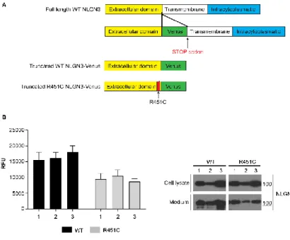
Previous studies showed alterations of the full-length R451C NLGN3 protein trafficking through the secretory pathway. Indeed, the mutant protein was shown to be retained in the ER failing to reach the cell surface (De Jaco et al., 2006, 2010). We have generated a new cell-based model system to study NLGN3 trafficking by using HEK293 cell lines stably expressing a truncated form of NLGN3 made of the extracellular protein domain, lacking the transmembrane and intracellular protein domains, and C-terminally fused to the Venus Fluorescent Protein (Giepmans et al., 2006) (Fig. 1A). These cell lines produce a fluorescent NLGN3, either WT or R451C, that is secreted by the cell, allowing to evaluate protein trafficking by measuring fluorescence levels in the cell culture medium. We have generated several clones expressing the truncated protein forms that were characterized for the levels of fluorescence in the medium and the levels of protein expression (Fig. 1B).
Page | 32
Figure 1. Generation of HEK293 cell lines expressing truncated and fluorescent NLGN3
A. Schematic representation of the vectors encoding for the NLGN3 truncated and fluorescent protein forms.
B. Fluorescence levels (RFU) and representative western blot of cell medium and lysates from three stably transfected clones (mean ± SEM; n= 4).
R451C NLGN3-Venus shows reduced secretion and increased degradation rate
To further characterize the newly generated cell-system, we compared basal levels of fluorescence in the medium of WT and R451C NLGN3-Venus selected clones and found a consistent and significative reduction of the R451C
NLGN3-Page | 33
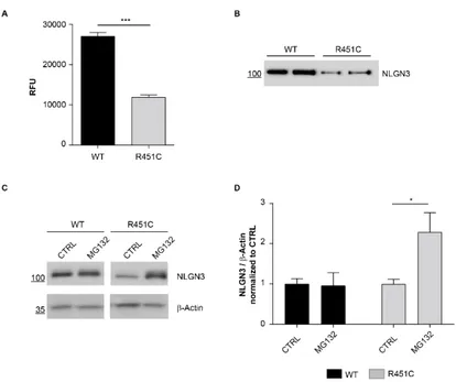
Venus protein secretion (~50%) compared to the WT protein (Fig. 2A), that was also confirmed by western blot analysis (Fig. 2B). To assess if the reduced levels of the R451C NLGN3-Venus are due to degradation through the ERAD, as it was previously shown for the full-length protein (De Jaco et al., 2010), we blocked the proteasome with the inhibitor MG132 and observed the accumulation of the mutant protein in the cell, not detectable for the WT protein (Fig. 2C-D).
Figure 2. Characterization of WT and R451C NLGN3-Venus selected clones
A. Fluorescence levels (RFU) in medium from WT and R451C NLGN3-Venus expressing cells (mean ± SEM; n= 12; P<0.001; Mann Whitney test).
Page | 34
B. Representative western blot of NLGN3-Venus levels in medium from WT and R451C expressing cells.
C. NLGN3-Venus levels in the medium from WT and R451C expressing cells after treatment with MG132 or in untreated condition.
D. Densitometric analysis of NLGN3 protein levels in the lysates after treatment with MG132, normalized and compared (for each genotype) to untreated control condition (mean ± SEM; n= 4; one-way ANOVA *P< 0.05).
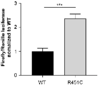
ER-retained R451C NLGN3-Venus activates UPR Previous work from our group showed the activation of all three branches of the UPR in the PC12 Tet-On cell system with inducible expression of R451C NLGN3 (Ulbrich et al., 2016). Thus, we studied UPR activation in the newly-generated cell system by investigating ATF6 activity by luciferase assay. HEK293 cells expressing either WT or R451C NLGN3-Venus were transfected with a reporter using the Firefly luciferase gene under the control of the ERSE (ER stress sequence elements), found in the promoter of ATF6-target genes. Co-transfection with a Renilla luciferase reporter has been used to assess transfection efficiency. Cells expressing R451C NLGN3-Venus showed significantly higher activation levels of ATF6 compared to WT (Fig. 3).
Page | 35
Figure 3. UPR activation in HEK293 NLGN3-Venus cells ATF6 activation in HEK293 cells expressing either WT or R451C NLGN3-Venus, shown as ratio Firefly/Renilla luciferases normalized and compared to WT condition (mean ± SEM; n= 6; Student’s t-test ***P< 0.001).
4-PBA improves NLGN3-Venus trafficking
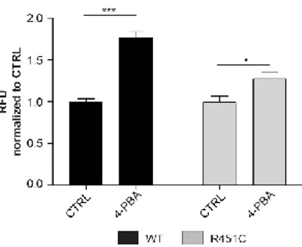
It is notable that known chemical chaperones, such as 4-PBA, can stabilize protein folding and increase protein trafficking in vitro (Uggenti et al., 2016). We treated WT and R451C NLGN3-Venus expressing cells with 4-PBA (500 µM) and measured fluorescence levels in the medium. We detected a significant increase of fluorescence for the mutant protein, indicating a positive effect of 4-PBA on improving defective protein trafficking and secretion (Fig. 4). However, this increase was observed also in the medium of the cells expressing the WT protein, denoting a generalized effect of this chemical chaperone on protein folding.
Page | 36
Figure 4. Effects of 4-PBA on WT and R451C NLGN3-Venus expressing cells
Fluorescence levels (RFU) after treatment with 4-PBA (500 µM), normalized and compared (for each genotype) to untreated control condition (mean ± SEM; n= 12; one-way ANOVA *P<0.05 ***P<0.001).
Screening of an FDA-approved library of compounds
A library of 2662 compounds was screened for enhancing secretion of mutant R451C NLGN3-Venus protein. All the compounds were previously approved by the American Food and Drugs Administration (FDA) for treatment of a variety of neurological and non-neurological diseases.
The screening was performed at a concentration of 2 µM on HEK293 cells expressing R451C NLGN3-Venus. The fluorescence in the medium was measured after 60 hours from treatment, to allow the mutant protein to accumulate to levels detectable by a fluorimeter. From this initial screening, we selected 12 compounds, which showed a 2-fold increase in the fluorescence levels in comparison to the untreated condition
Page | 37
(Fig. 5A). It is noteworthy that most of these compounds belonged to the glucocorticoid family, which is known to increase the levels of ER-resident chaperones (Das et al., 2013).
See supplementary Fig. 1 for a more detailed list of positive compounds.
To confirm data obtained in this first screening and to exclude that some contaminants were present in the library and were interfering with the results, we performed a second screening using a new batch of each compound. However, we chose only 5 of them, 3 glucocorticoids (Prednisolone sodium phosphate, Desonide, Alclometazone dipropionate) and 2 belonging to other chemical families (Quinestrol, Phenytoin sodium) (Table 1) for this further characterization. We tested them on both WT and R451C NLGN3-Venus cells and DMSO was used as vehicle control condition. Only treatments with glucocorticoids showed to significantly increase trafficking of the mutant protein in comparison to control untreated condition, while Quinestrol and Phenytoin sodium had no effect, such as DMSO (Fig. 5C). Glucocorticoids had no effect on the WT NLGN3-Venus (Fig. 5B), indicating a specific effect of the compounds only on the mutant form of the protein.
The significant increase in protein secretion was confirmed by western blot analysis on the cell media (Fig. 6A). It is noteworthy that the amount of NLGN3-Venus protein was unchanged in cell lysates after treatment (Fig. 6B). This was indicating that treatment didn’t affect protein synthesis but rather induced a higher proportion of R451C NLGN3-Venus protein to be released in the cell medium.
Page | 38 Table 1
Selected compounds that resulted positive from the first screening. The amount of increased fluorescence in the cell medium of R451C NLGN3-Venus expressing cells and the chemical class to which the single compounds belong are indicated.
Page | 39
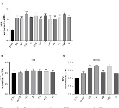
Figure 5. Screening of an FDA-approved library of compounds
AD: alclometazone dipropionate; BA: betamethasone acetate; CBX: carbenoxolone sodium; D: desonide; DES: desoxymetasone; EH: ethylnorepinephrine hydrochloride; IA: isoflupredone acetate; MH: milnacipran hydrochloride; PA: phenylethyl alcohol; PS: phenytoin sodium; PSP: prednisolone sodium phosphate; Q: quinestrol
A. Fluorescence levels (RFU) in the medium of HEK293 R451C NLGN3-Venus treated with indicated compounds, normalized and compared to untreated control condition (CTRL) (mean ± SEM; n= 4; one-way ANOVA ***P<0.001). B. Fluorescence levels (RFU) in the medium of HEK293 expressing WT NLGN3-Venus treated with selected compounds or with vehicle control (DMSO), normalized and
Page | 40
compared to control condition (CTRL) (mean ± SEM; n= 7; one-way ANOVA).
C. Fluorescence levels (RFU) in the medium of HEK293 expressing R451C NLGN3-Venus treated with selected compounds or with vehicle control (DMSO), normalized and compared to control condition (CTRL) (mean ± SEM; n= 7; one-way ANOVA **P<0.01).
Page | 41
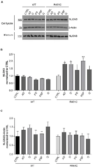
Figure 6. Quantification of NLGN3-Venus protein after treatment
AD: alclometazone dipropionate; D: desonide; PS: phenytoin sodium; PSP: prednisolone sodium phosphate; Q: quinestrol
Page | 42
A. Representative western blot of NLGN3-Venus levels in cell lysates and in medium from WT and R451C expressing cells after treatment with indicated compounds
B. Densitometric analysis of NLGN3-Venus levels in cell medium from WT and R451C expressing cells after treatment with indicated compounds, normalized and compared to CTRL condition (mean ± SEM; n= 4; one-way ANOVA *P<0.05 **P<0.01).
C. Densitometric analysis of NLGN3-Venus levels in cell lysates from WT and R451C expressing cells after treatment with indicated compounds, normalized and compared to CTRL condition (mean ± SEM; n= 4; one-way ANOVA).
Selected compounds improve trafficking of full - length R451C NLGN3
Compounds selected for enhancing trafficking of the truncated and fluorescent form of NLGN3 were then tested on cells expressing the full-length form of the protein. HEK293 cells were transiently transfected with the pcDNA3.1 vector encoding for R451C NLGN3 full-length and co-localization staining with the ER marker calreticulin was analyzed by confocal microscopy (Fig. 7A) in order to evaluate the exit of the mutant R451C protein from the ER. The effect of the compounds to decrease the amount of co-localization was used as a parameter of their activity. HEK293 cells transiently transfected with WT NLGN3 full-length normally expressed on the cell surface were used as control. As expected, this condition showed a significant lower co-localization with calreticulin in comparison to cells expressing mutant NLGN3 (Fig. 7B). R451C NLGN3 expressing cells were treated with 5
Page | 43
of the selected compounds and co-localization with calreticulin was assessed 48 h after treatment. We considered as positive those compounds able to decrease all three coefficients calculated. It was confirmed that Alclometazone dipropionate and Prednisolone sodium phosphate, resulted the most effective in improving trafficking of both the truncated and full-length R451C NLGN3 mutant forms. The remaining compounds seem not to have any effect on decreasing ER-localization of the full-length mutant protein (Fig. 7C-D-E).
Page | 44
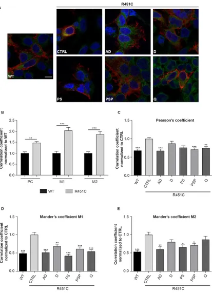
Figure 7. Compounds effect on localization of full length NLGN3
AD: alclometazone dipropionate; D: desonide; PS: phenytoin sodium; PSP: prednisolone sodium phosphate; Q: quinestrol
Page | 45
A. Representative confocal images of NLGN3 (red) and Calreticulin (green) staining in HEK293 cells transfected with either WT or R451C NLGN3 treated with selected compounds (scale bar 1µm).
B. Quantification of NLGN3-calreticulin co-localization in cells expressing either WT or R451C NLGN3, normalized and compared to WT condition. Pearson’s coefficient (PC) and both Mander’s overlap coefficients M1 and M2 were measured (mean ± SEM; n= 54 cells/3 independent experiments; one-way ANOVA **P<0.01 ***P<0.001). C. Pearson’s coefficient measured in cells expressing either WT or R451C NLGN3 and treated with indicated compounds, normalized and compared to CTRL condition (mean ± SEM; n= 50 cells/3 independent experiments; one-way ANOVA **P<0.01 ***P<0.001).
D. Mander’s overlap coefficient M1 analysis in cells expressing either WT or R451C NLGN3 and treated with indicated compounds, normalized and compared to CTRL condition (mean ± SEM; n= 50 cells/3 independent experiments; one-way ANOVA **P<0.01 ***P<0.001). E. Analysis of Mander’s overlap coefficient M2 in cells expressing either WT or R451C NLGN3 and treated with indicated compounds, normalized and compared to CTRL condition (mean ± SEM; n= 55 cells/3 independent experiments; one-way ANOVA *P<0.05 **P< 0.01).
Compounds fail to lower UPR activation induced by full length R451C NLGN3
We evaluated the effect of the compounds on alleviating UPR activation due to the retention of the full length R451C
Page | 46
NLGN3 in the ER. To this purpose, we used the UPR reporter that measures the activation of the ATF6 branch. HEK293 cells were transiently transfected with pcDNA3.1 expressing full length NLGN3, either WT or R451C, and with the luciferase reporter vectors previously used (Fig. 3). Cells expressing R451C NLGN3 were treated with 5 selected compounds and luciferase activity was measured, in each condition, 48 h after treatment. HEK293 cells expressing WT NLGN3 represented the control condition. The treatment with the compounds failed to decrease the activation of ATF6 in cells expressing R451C NLGN3 comparing to untreated condition (Fig. 8).
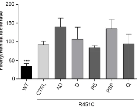
Figure 8. Effects of compounds on UPR activation
AD: alclometazone dipropionate; D: desonide; PS: phenytoin sodium; PSP: prednisolone sodium phosphate; Q: quinestrol Firefly/Renilla luciferases activities in HEK293 cells expressing either WT or R451C NLGN3 and treated with indicated compounds, compared to CTRL condition (mean ± SEM; n= 5; one-way ANOVA).
Page | 47 Discussion
In the last years, the research field on diseases correlated to the accumulation of misfolded proteins has been greatly expanding. Collectively called protein-misfolding diseases, they include common neurological and non-neurological disorders, such as Alzheimer’s, Parkinson’s, Cystic Fibrosis, Cancer and Diabetes (Koss and Platt, 2017; Sarvani et al., 2017; Scheper and Hoozemans, 2016). In these disorders, the common underlying mechanism is represented by the accumulation of misfolded proteins. The ER is specialized in recognizing and retaining not properly folded proteins that undertake the secretory pathway. This might result in a double effect: a loss-of-function due to the mis-localization of the protein, and the accumulation of the protein in the organelle leading to a toxic gain-of-function and in most cases to ER stress conditions. In this contest, the research for small molecules facilitating protein folding and their escape from the ER is becoming of growing interest. Chemical chaperones are useful tools in helping protein folding, but their lack of specificity limits their potential as therapeutics. On the other hand, pharmacological chaperones have a higher specificity, being designed to bind a selected protein (Cortez and Sim, 2014).
Our work focused on the biological effects caused by the Arg451 to Cys substitution (R451C) in the extracellular domain of the post-synaptic protein Neuroligin3 (NLGN3). This mutation, found in patients with ASDs, causes the retention of the protein in the ER and its loss at the synapse, where it plays a crucial role as a cell-adhesion molecule (Baig et al., 2017). Our recent work established the first evidence
Page | 48
between the retention of the R451C NLGN3 in the ER and the activation of UPR in vitro (Ulbrich et al., 2016). Moreover, mice carrying the R451C mutation in the endogenous NLGN3 gene show altered neurotransmission in several brain areas correlated to ASDs (Tabuchi et al., 2009; Etherton et al., 2001; Trobiani et al., 2018). However, it is not clear if those alterations are due to the loss of NLGN3 proper localization on the cell surface, or if it is a consequence of the ER stress condition due to the retention of the protein in the organelle (Trobiani et al., 2018). In fact, it is possible that, in almost completely absence of NLGN3 on the cell surface, the pre-synaptic partner NRXN1β associates with other ligands, thus inducing a gain-of-function. Besides, this hypothesis does not exclude the possibility that the UPR, induced by the ER-retention of R451C NLGN3, represents an additional factor participating to the gain-of-function phenotype.
The effect of restoring proper protein localization to rescue phenotype, has been shown by Mei and colleagues in the Shank3:Cre conditional Knock-In (KI) mouse, a model of ADSs (Mei et al., 2016). Untreated mice showed a phenotype characterized by molecular, electrophysiological and behavioural alterations. Treatment with Tamoxifen activates the Cre-recombinase and re-establishes expression of Shank3 at the post-synaptic site, allowing an almost complete recovery of the phenotype. Therefore, restoration of proper protein expression and cellular disposition might represent a strategy to treat diseases characterized by protein mis-localization. In this prospective, helping the mutant R451C NLGN3 to exit the ER would be beneficial in two ways: restoring proper protein localization for its function, and alleviating the overload of the ER, reducing ER stress and UPR activation.
Page | 49
We generated stable cell lines expressing a truncated form of either WT or R451C NLGN3, tagged with the fluorescent protein Venus and demonstrated that their trafficking was impaired similarly to what was previously shown for the full- length form of the protein (De Jaco et al., 2006, 2010). We report a ~50% reduction of R451C protein release compared to the WT, increased protein levels following the inhibition of proteasome activity and activation of the UPR. Our cell model system responded to the treatment with 4-PBA, as showed by the increase of protein trafficking for both WT and R451C NLGN3-Venus proteins, suggesting a generalized effect of 4-PBA on all proteins passing through the ER. However, our interest was focused on identifying compounds selective for the mutant R451C NLGN3 form.
We screened a library of compounds, approved by the FDA for treatment of a variety of other diseases. The screening used a concentration 250-fold lower than the one used for 4-PBA and closer to normally used pharmacological concentrations. We found 12 compounds able to enhance R451C NLGN3-Venus trafficking, doubling its amount in the cell medium. It is noteworthy that those compounds had no effect on trafficking of the WT NLGN3-Venus. Two out of the three compounds which resulted the most effective on the truncated NLGN3-Venus, Alclometazone dipropionate and Prendisolone sodium phosphate, showed a specific activity in reducing the retention of the full-length protein in the ER.
Unfortunately, none of the compounds showed an effect at reducing the activation of the ATF6 branch of the UPR caused by the retention of the full-length R451C NLGN3 in the ER. However, it is possible that they modulate other branches of this cellular response or that the amount of mutant NLGN3
Page | 50
escaping from the ER in response to treatment, is not enough to reduce ER stress and UPR activation. Although, the selective effect on the mutant NLGN3, with no effect on the WT form, is indicating that the compounds were not affecting the generalized protein trafficking through the secretory pathway. Instead, they were improving protein folding, probably by increasing levels of ER chaperones (Das et al., 2013; Fujii et al., 2006). It has been shown both in vitro and in vivo, that the administration of exogenous glucocorticoids, was able to increase levels of ER chaperones, such as Calreticulin, BiP and Hsp70, and induce the escape of an ER-retained protein from this organelle (Das et al., 2013; Duzgun et al., 2013; Fujii et al., 2006; Nair et al., 2018). Moreover, it has been shown that dexamethasone, a synthetic glucocorticoid used in clinic, has a role in modulating the half-life of CFTR by attenuating its interaction with a ubiquitin E3 ligase in vitro (Caohuy et al., 2009).
Glucocorticoids mediate their effects by binding the intracellular receptor GR, which translocates to the nucleus and activates transcription of target genes, by binding specific glucocorticoids response elements (GREs) (Meijsing 2015). By this way, glucocorticoids regulate several cellular processes.
In this contest, our findings strongly suggest a role for glucocorticoids in the regulation of protein folding and trafficking and establish a new prospective for treatment of misfolding protein diseases and for the monogenic form of autism due to the R451C substitution in the synaptic protein NLGN3.
Based on these data, in the next future, we aim at elucidating the mechanism of action of glucocorticoids for improving
Page | 51
protein trafficking in our model systems. Moreover, we want to investigate the ability of glucocorticoids at increasing NLGN3 protein expression in vivo. Besides, we want to assess the activity of these compounds on other mutant proteins, in order to verify whether this effect is specific for the R451C NLGN3 or is common to ER-retained proteins that are not structurally correlated.
Page | 53 Methods
Plasmids generation
Truncated rat NLGN3 was generated by introducing a stop codon right after the residue Tyr640 in the cDNA encoding for full-length NLGN3 either WT or carrying the mutation Arg451Cys (R451C). The cDNAs were subcloned into a vector encoding an N-terminal FLAG octopeptide and were additionally tagged at the C-terminus with Venus, an analog of the Green Fluorescent protein.
Selection of stable clones of HEK293 cells expressing truncated NLGN3-Venus
pcDNA3.1 plasmid, encoding truncated NLGN3-Venus either WT or R451C, has been used for transfecting HEK293 cells, using polyethylenimine (PEI) procedure (Sigma-Aldrich). Transfected cells were cultured in selective medium composed by DMEM (Dulbecco’s Modified Eagle’s Medium, Sigma), 5% Fetal Bovine Serum (FBS, Sigma-Aldrich) and 500 µg/ml Geneticin (G418, Corning). Resistant cells have been plated at clonal density in 96-well plate and expanded to obtain clones, which were screened for NLGN3 protein expression by quantifying fluorescence levels in the culture medium and by Western blot on both cell lysates and medium. Stable cell lines are maintained in medium containing 500 µg/ml G418 at 37°C and 5% CO2.
The proteasome-inhibitor MG132 (Z-Leu-Leu-Leu-al, #C2211 Sigma-Aldrich) at concentration of 2.5 µM was added in the medium for the last 48 h of culture before harvesting.
Page | 54 Luciferase assay
To assess ATF6 activation, 800.000 HEK293 cells were seeded in a 6-well plate. At 24 h after plating, cells were transfected with 2 µg of total DNA according to the Lipofectamine2000 protocol (Thermo Fisher Scientific). For HEK293 cells stably expressing NLGN3-Venus, total DNA was represented by two vectors: pATF6(5 x)-Luc (Firefly) and pTK (Renilla) in a 50:1 ratio (Davies et al., 2009). Otherwise, besides these two reporter vectors, pcDNA3.1 expressing full length NLGN3, either WT or R451C, was added to the transfection mix. Moreover, in the latter case, 3 h after transfection, HEK293 cells expressing R451C NLGN3 were treated with selected compounds at 2 µM concentration. Transfection was carried out for 48 h, then cells were harvested and activity of both luciferases was assessed by the Dual-luciferase Reporter Assay kit (Promega) and detected by the Glomax multi+ detection system (Promega).
Treatment with chemical chaperones
HEK293 cells expressing either WT or R451C NLGN3-Venus were plated at a density of 600.000 cells in 24-well plate. 24 h after seeding, medium was changed to Optimem (Gibco) and sodium 4-phenylbutyrate (4-PBA, Sigma-Aldrich) was added at the concentration of 500 µM in the cell culture medium. For the screening of the FDA-approved compounds, 75.000 cells from both lines have been plated in 96-well plate, in 150 µl Optimem medium added with 2% FBS.
The 2662 compounds of the library were dissolved in 10% DMSO (Sigma-Aldrich) and used at final concentration of 2 µM. Compounds were added in the cell culture medium 24
Page | 55
hours after seeding and fluorescence levels were measured after 60 h of treatment. Cells treated with 0,3% DMSO were used as vehicle control.
Quantification of protein secretion by fluorimetric measurements
To evaluate NLGN3-Venus protein trafficking, 100 µl of medium from cells either untreated or treated with selected compounds was transferred in a black 96-well plate and fluorescence was measured by a fluorimeter (excitation wavelength 485 nm, emission wavelength 535 nm).
Preparation of protein samples
Lysates of HEK293 cells were obtained using lysis buffer (150 mM NaCl, 10 mM Tris pH 8.0, 0.5% Nonidet P40) supplemented with proteases inhibitor cocktail (Sigma-Aldrich). Lysis was performed for 15 min on ice, followed by centrifugation at 17000 g for 15 min at 4°C. Protein concentration in the samples was determined by the Bradford assay.
SDS-PAGE and Western blot
Equal amount of cell medium or 30 µg of cell lysate from NLGN3-Venus (WT or R451C) cells were subjected to SDS-PAGE (10% v/v polyacrylamide gel) in running buffer (25 mM Tris pH 8.3, 0.19 M Glycine, 0.1% Sodium dodecyl sulfate) and transferred to PVDF membrane (Merck-Millipore) in transfer buffer (25 mM Tris pH 8.3, 0.19 M Glycine, 10%
Page | 56
Methanol). The membrane was blocked with 5% w/v non-fat milk in PBS (2.68 mM KCl, 1.47 mM KH2PO4, 0.137 M
NaCl, 10.16 mM Na2HPO4) and stained with anti-FLAG M2
(1:1000, mouse, Sigma-Aldrich #F3165) or anti-β-Actin (1:1000, mouse, Millipore #MAB1501) primary antibodies diluted in PBS containing 1% BSA (Sigma-Aldrich). HRP (horseradish peroxidase)-conjugated anti-mouse (goat, Sant Cruz Biotechnologies #SC-2005) secondary antibody was diluted 1:10000 in 5% milk in PBS. All the incubations were performed for 1 h at room temperature. The HRP signal has been detected by using ECL (HyGLO, Denville Scientific Inc.) and visualised by a ChemiDoc system (BioRad).
Immunocytochemistry
HEK293 cells (100.000 cells/well) were plated on glass coverslips coated with 0.5 mg/ml poly-D-lysin (Sigma-Aldrich) in 24-well plates. Cells were transfected with cDNA NLGN3 full-length either WT or R451C using the calcium phosphate method and treated with selected compounds for 48 h. Untreated cells cultured in the same conditions were used as control. Cells were washed with PBS and fixed with 4% paraformaldehyde (PFA, Sigma-Aldrich) for 20 minutes at room temperature, followed by fixation with pre-chilled methanol (Sigma-Aldrich) for 15 minutes at -20°C.
The blocking step was performed for 1 h at room temperature using a solution containing 2% normal sheep serum (NSS), 0.1% Triton-X100, 0.02% sodium azide in PBS. The same solution has been used for diluting primary and secondary antibodies. Anti-FLAG (1:5000, Rabbit, Sigma-Aldrich #F7425) and anti-calreticulin (1:500, Mouse, Enzo
ADI-Page | 57
SPA601) were incubated in combination, overnight at 4°C, while fluorescent secondary antibodies Cy3-anti-rabbit (donkey, Jackson Immuno Research #711-165-152) and Alexa Fluor 488-anti-mouse (donkey, Jackson Immuno Research #715-545-151) were diluted 1:500 and incubated 1 h at room temperature. Nuclei were stained with Hoechst (Thermo Fisher Scientific) 1:1000 in PBS for 10 min. Fluorescent signal was detected by confocal microscope (Zeiss) at 63x magnification and co-localization analysis was obtained on Z-stack images using the JACoP plugin of the NIH software ImageJ as previously described (Bolte and Cordelières, 2006). Pearson’s correlation coefficient and Mander’s overlap coefficients M1 and M2 have been calculated for at least 15 cells/experiment.
Statistical analysis
All experiments were performed at least three times on independent biological samples, as indicated in the figure legends. Student’s t-test, Mann Whitney test and one-way ANOVA with Bonferroni multiple comparison test were used for statistical analysis by using Prism5 (Graph-Pad 5 Software Inc.): *p<0.05, **p<0.01, ***p<0.001.
Page | 59 References
Almanza A, Carlesso A, Chintha C, Creedican S, Doultsinos D, Leuzzi B, Luís A, McCarthy N, Montibeller L, More S, Papaioannou A, Püschel F, Sassano ML, Skoko J, Agostinis P, de Belleroche J, Eriksson LA, Fulda S, Gorman AM, Healy S, Kozlov A, Muñoz-Pinedo C, Rehm M, Chevet E, Samali A (2018). Endoplasmic reticulum stress signalling - from basic mechanisms to clinical applications. FEBS J. Epub ahead of print
Baig DN, Yanagawa T, Tabuchi K (2017). Distortion of the normal function of synaptic cell adhesion molecules by genetic variants as a risk for autism spectrum disorders. Brain Res Bull. 129, 82-90
Bang ML, Owczarek S (2013). A matter of balance: role of neurexin and neuroligin at the synapse. Neurochem Res. 38(6), 1174-89
Bemben MA, Shipman SL, Nicoll RA, Roche KW (2015). The cellular and molecular landscape of neuroligins. Trends Neurosci. 38(8), 496-505
Bolte S, Cordelières FP (2006). A guided tour into subcellular colocalization analysis in light microscopy. J Microsc. 224(Pt 3), 213-32
Campisi L, Imran N, Nazeer A, Skokauskas N, Azeem MW (2018). Autism spectrum disorder. Br Med Bull. 127(1), 91-100
Caohuy H, Jozwik C, Pollard HB (2009). Rescue of DeltaF508-CFTR by the SGK1/Nedd4-2 signaling pathway. J Biol Chem. 284(37), 25241-53
Page | 60
Chambers JE, Marciniak SJ (2014). Cellular mechanisms of endoplasmic reticulum stress signaling in health and disease. 2. Protein misfolding and ER stress. Am J Physiol Cell Physiol. 307(8), C657-70
Christensen J, Grønborg TK, Sørensen MJ, Schendel D, Parner ET, Pedersen LH, Vestergaard M (2013). Prenatal valproate exposure and risk of autism spectrum disorders and childhood autism. JAMA. 309(16), 1696-703
Comoletti D, De Jaco A, Jennings LL, Flynn RE, Gaietta G, Tsigelny I, Ellisman MH, Taylor P (2004). The Arg451Cys-neuroligin-3 mutation associated with autism reveals a defect in protein processing. J Neurosci. 24(20), 4889-93
Corazzari M, Gagliardi M, Fimia GM, Piacentini M (2017). Endoplasmic Reticulum Stress, Unfolded Protein Response, and Cancer Cell Fate. Front Oncol. 7, 78
Cortez L, Sim V (2014). The therapeutic potential of chemical chaperones in protein folding diseases. Prion. 8(2)
Crider A, Ahmed AO, Pillai A (2017). Altered Expression of Endoplasmic Reticulum Stress-Related Genes in the Middle Frontal Cortex of Subjects with Autism Spectrum Disorder. Mol Neuropsychiatry. 3(2), 85-91
Das I, Png CW, Oancea I, Hasnain SZ, Lourie R, Proctor M, Eri RD, Sheng Y, Crane DI, Florin TH, McGuckin MA (2013). Glucocorticoids alleviate intestinal ER stress by enhancing protein folding and degradation of misfolded proteins. J Exp Med 210(6), 1201-16
Davies MJ, Miranda E, Roussel BD, Kaufman RJ, Marciniak SJ, Lomas DA (2009). Neuroserpin polymers activate
NF-Page | 61
kappaB by a calcium signaling pathway that is independent of the unfolded protein response. J Biol Chem. 284(27), 18202-9 De Jaco A, Comoletti D, Kovarik Z, Gaietta G, Radic Z, Lockridge O, Ellisman MH, Taylor P (2006). A mutation linked with autism reveals a common mechanism of endoplasmic reticulum retention for the alpha,beta-hydrolase fold protein family. J Biol Chem. 281(14), 9667-76
De Jaco A, Dubi N, Comoletti D, Taylor P (2010). Folding anomalies of neuroligin3 caused by a mutation in the alpha/beta-hydrolase fold domain. Chem Biol Interact. 187(1-3), 56-8
De Jaco A, Lin MZ, Dubi N, Comoletti D, Miller MT, Camp S, Ellisman M, Butko MT, Tsien RY, Taylor P (2010). Neuroligin trafficking deficiencies arising from mutations in the alpha/beta-hydrolase fold protein family. J Biol Chem. 285(37), 28674-82
De Jaco A, Dubi N, Camp S, Taylor P (2012). Congenital hypothyroidism mutations affect common folding and trafficking in the α/β-hydrolase fold proteins. FEBS J. 279(23), 4293-305
de la Torre-Ubieta L, Won H, Stein JL, Geschwind DH (2016). Advancing the understanding of autism disease mechanisms through genetics. Nat Med. 22(4), 345-61
Di Prisco GV, Huang W, Buffington SA, Hsu CC, Bonnen PE, Placzek AN, Sidrauski C, Krnjević K, Kaufman RJ, Walter P, Costa-Mattioli M (2014). Translational control of mGluR-dependent long-term depression and object-place learning by eIF2α. Nat Neurosci. 17(8), 1073-82
Page | 62
Duzgun A, Bedir A, Ozdemir T, Nar R, Kilinc V, Salis O, Alacam H, Gulten S (2013). Effect of dexamethasone on unfolded protein response genes (MTJ1, Grp78, Grp94, CHOP, HMOX-1) in HEp2 cell line. Indian J Biochem Biophys. 50(6), 505-10
Etherton M, Földy C, Sharma M, Tabuchi K, Liu X, Shamloo M, Malenka RC, Südhof TC (2011). Autism-linked neuroligin-3 R451C mutation differentially alters hippocampal and cortical synaptic function. Proc Natl Acad Sci U S A. 108(33), 13764-9
Fabrichny IP, Leone P, Sulzenbacher G, Comoletti D, Miller MT, Taylor P, Bourne Y, Marchot P (2007). Structural analysis of the synaptic protein neuroligin and its beta-neurexin complex: determinants for folding and cell adhesion. Neuron. 56(6), 979-91
Falivelli G, De Jaco A, Favaloro FL, Kim H, Wilson J, Dubi N, Ellisman MH, Abrahams BS, Taylor P, Comoletti D (2012). Inherited genetic variants in autism-related CNTNAP2 show perturbed trafficking and ATF6 activation. Hum Mol Genet. 21(21), 4761-73
Fujii Y, Khoshnoodi J, Takenaka H, Hosoyamada M, Nakajo A, Bessho F, Kudo A, Takahashi S, Arimura Y, Yamada A, Nagasawa T, Ruotsalainen V, Tryggvason K, Lee AS, Yan K (2006). The effect of dexamethasone on defective nephrin transport caused by ER stress: a potential mechanism for the therapeutic action of glucocorticoids in the acquired glomerular diseases. Kidney Int. 69(8), 1350-9
Page | 63
Giepmans BN, Adams SR, Ellisman MH, Tsien RY (2006). The fluorescent toolbox for assessing protein location and function. Science. 312(5771), 217-24
Halperin L, Jung J, Michalak M (2014). The many functions of the endoplasmic reticulum chaperones and folding enzymes. IUBMB Life. 66(5), 318-26
Hillary RF, FitzGerald U (2018). A lifetime of stress: ATF6 in development and homeostasis. J Biomed Sci. 25(1), 48
Jamain S, Quach H, Betancur C, Råstam M, Colineaux C, Gillberg IC, Soderstrom H, Giros B, Leboyer M, Gillberg C, Bourgeron T; Paris Autism Research International Sibpair Study (2003). Mutations of the X-linked genes encoding neuroligins NLGN3 and NLGN4 are associated with autism. Nat Genet. 34(1), 27-9
Kawada K, Mimori S (2018). Implication of Endoplasmic Reticulum Stress in Autism Spectrum Disorder. Neurochem Res. 43(1), 138-143
Kawada K, Mimori S, Okuma Y, Nomura Y (2018). Involvement of endoplasmic reticulum stress and neurite outgrowth in the model mice of autism spectrum disorder. Neurochem Int. 119, 115-119
Koss DJ, Platt B (2017). Alzheimer's disease pathology and the unfolded protein response: prospective pathways and therapeutic targets. Behav Pharmacol. 28(2 and 3-Spec Issue), 161-178
Kosuge Y, Sakikubo T, Ishige K, Ito Y (2006). Comparative study of endoplasmic reticulum stress-induced neuronal death
Page | 64
in rat cultured hippocampal and cerebellar granule neurons. Neurochem Int. 49(3), 285-93
Li X, Zou H, Brown WT (2012). Genes associated with autism spectrum disorder. Brain Res Bull. 88(6), 543-52
Martella G, Meringolo M, Trobiani L, De Jaco A, Pisani A, Bonsi P (2018). The neurobiological bases of autism spectrum disorders: the R451C-neuroligin 3 mutation hampers the expression of long-term synaptic depression in the dorsal striatum. Eur J Neurosci. 47(6), 701-708
Martínez G, Vidal RL, Mardones P, Serrano FG, Ardiles AO, Wirth C, Valdés P, Thielen P, Schneider BL, Kerr B, Valdés JL, Palacios AG, Inestrosa NC, Glimcher LH, Hetz C (2016). Regulation of Memory Formation by the Transcription Factor XBP1. Cell Rep. 14(6), 1382-1394
Martínez G, Khatiwada S, Costa-Mattioli M, Hetz C (2018). ER Proteostasis Control of Neuronal Physiology and Synaptic Function. Trends Neurosci. 41(9), 610-624
McQuiston A, Diehl JA (2018). Recent insights into PERK-dependent signaling from the stressed endoplasmic reticulum. F1000Res. 6, 1897
Mei Y, Monteiro P, Zhou Y, Kim JA, Gao X, Fu Z, Feng G (2016). Adult restoration of Shank3 expression rescues selective autistic-like phenotypes. Nature. 530(7591), 481-4 Meijsing SH (2015). Mechanisms of Glucocorticoid-Regulated Gene Transcription. Adv Exp Med Biol. 872, 59-81 Mercado G, Castillo V, Soto P, Sidhu A (2016). ER stress and Parkinson's disease: Pathological inputs that converge into the secretory pathway. Brain Res. 1648(Pt B), 626-632
Page | 65
Mihailidou C, Panagiotou C, Kiaris H, Kassi E, Moutsatsou P (2016). Crosstalk between C/EBP homologous protein (CHOP) and glucocorticoid receptor in lung cancer. Mol Cell Endocrinol. 436, 211-23
Mizukami T, Orihashi K, Herlambang B, Takahashi S, Hamaishi M, Okada K, Sueda T (2010). Sodium 4-phenylbutyrate protects against spinal cord ischemia by inhibition of endoplasmic reticulum stress. J Vasc Surg. 52(6), 1580-6
Hebert DN, Molinari M (2007). In and out of the ER: protein folding, quality control, degradation, and related human diseases. Physiol Rev. 87(4), 1377-408
Nair M, Romero J, Mahtabfar A, Meleis AM, Foty RA, Corbett SA (2018). Dexamethasone-Mediated Upregulation of Calreticulin Inhibits Primary Human Glioblastoma Dispersal Ex Vivo. Int J Mol Sci. 19(2)
Nakanishi M, Nomura J, Ji X, Tamada K, Arai T, Takahashi E, Bućan M, Takumi T (2017). Functional significance of rare neuroligin 1 variants found in autism. PLoS Genet. 13(8), e1006940
Ni H, Rui Q, Xu Y, Zhu J, Gao F, Dang B, Li D, Gao R, Chen G (2018). RACK1 upregulation induces neuroprotection by activating the IRE1-XBP1 signaling pathway following traumatic brain injury in rats. Exp Neurol. 304, 102-113 Nosyreva E, Kavalali ET (2010). Activity-dependent augmentation of spontaneous neurotransmission during endoplasmic reticulum stress. J Neurosci. 30(21), 7358-68
Page | 66
Ozcan U, Yilmaz E, Ozcan L, Furuhashi M, Vaillancourt E, Smith RO, Görgün CZ, Hotamisligil GS (2006). Chemical chaperones reduce ER stress and restore glucose homeostasis in a mouse model of type 2 diabetes. Science. 313(5790), 1137-40
Qi X, Hosoi T, Okuma Y, Kaneko M, Nomura Y (2004). Sodium 4-phenylbutyrate protects against cerebral ischemic injury. Mol Pharmacol. 66(4), 899-908
Ribeiro LF, Verpoort B, de Wit J (2018). Trafficking mechanisms of synaptogenic cell adhesion molecules. Mol Cell Neurosci. 91, 34-47
Roussel BD, Kruppa AJ, Miranda E, Crowther DC, Lomas DA, Marciniak SJ (2013). Endoplasmic reticulum dysfunction in neurological disease. Lancet Neurol. 12(1), 105-18
Sarvani C, Sireesh D, Ramkumar KM (2017). Unraveling the role of ER stress inhibitors in the context of metabolic diseases. Pharmacol Res. 119, 412-421
Scheper W, Hoozemans JJ (2015). The unfolded protein response in neurodegenerative diseases: a neuropathological perspective. Acta Neuropathol. 130(3), 315-31
Südhof TC (2017). Synaptic Neurexin Complexes: A Molecular Code for the Logic of Neural Circuits. Cell. 171(4), 745-769
Südhof TC (2008). Neuroligins and neurexins link synaptic function to cognitive disease. Nature. 455(7215), 903-11 Sun L, Zhao Y, Zhou K, Freeze HH, Zhang YW, Xu H (2013). Insufficient ER-stress response causes selective mouse
Page | 67
cerebellar granule cell degeneration resembling that seen in congenital disorders of glycosylation. Mol Brain. 6, 52
Tabuchi K, Blundell J, Etherton MR, Hammer RE, Liu X, Powell CM, Südhof TC (2007). A neuroligin-3 mutation implicated in autism increases inhibitory synaptic transmission in mice. Science. 318(5847), 71-6
Takayanagi S, Fukuda R, Takeuchi Y, Tsukada S, Yoshida K (2013). Gene regulatory network of unfolded protein response genes in endoplasmic reticulum stress. Cell Stress Chaperones. 18(1), 11-23
Talebizadeh Z, Lam DY, Theodoro MF, Bittel DC, Lushington GH, Butler MG (2006). Novel splice isoforms for NLGN3 and NLGN4 with possible implications in autism. J Med Genet. 43(5), e21
Taoufik E, Kouroupi G, Zygogianni O, Matsas R (2018). Synaptic dysfunction in neurodegenerative and neurodevelopmental diseases: an overview of induced pluripotent stem-cell-based disease models. Open Biol. 8(9) Trobiani L, Favaloro FL, Di Castro MA, Di Mattia M, Cariello M, Miranda E, Canterini S, De Stefano ME, Comoletti D, Limatola C, De Jaco A (2018). UPR activation specifically modulates glutamate neurotransmission in the cerebellum of a mouse model of autism. Neurobiol Dis. 120, 139-150
Tsai PT (2016). Autism and cerebellar dysfunction: Evidence from animal models. Semin Fetal Neonatal Med. 21(5), 349-55
Uggenti C, Briant K, Streit AK, Thomson S, Koay YH, Baines RA, Swanton E, Manson FD (2016). Restoration of mutant