SAPIENZA
Università di Roma
Facoltà di Scienze Matematiche Fisiche e Naturali DOTTORATO DI RICERCA
IN GENETICA E BIOLOGIA MOLECOLARE XXXI Ciclo
(A.A. 2017/2018)
Characterisation of lnc-G4, a long noncoding
RNA that regulates skeletal muscle
differentiation through translational repression
of G-quadruplex containing mRNAs
Dottorando Davide Mariani Docente guida Prof. Irene Bozzoni Tutore
Dr Gianluca Canettieri Coordinatore
Cover image: differentiated C2C12 mouse myoblasts after immunostaining for Myosin Heavy Chain (red); cell nuclei are stained with DAPI (blue).
INDEX
1. INTRODUCTION 7
1.1. A brief history of non-coding RNA 7
1.2. Long non coding RNAs: definition and features 9
1.3. Nuclear long non-coding RNAs 13
1.4. Cytoplasmic long non-coding RNAs 14
1.5. Not all long non-coding RNAs are strictly non-coding 17 19 1.6. Molecular regulation of skeletal muscle differentiation 20 1.7. Non-coding RNAs in skeletal muscle differentiation 22
1.8. G-quadruplexes in RNA biology 23
2. AIM OF THE THESIS 27
3. RESULTS 29
3.1. Criteria for lnc-G4 selection 29
3.2. Characterization of lnc-G4 30
3.3. lnc-G4 downregulation affects proper myogenesis 33
3.4. lnc-G4 has a complex molecular interactome 37
3.4.1. lnc-G4 co-precipitates with several mRNAs 38 3.4.2. lnc-G4 directly interacts with MLX mRNA 41 3.4.3. lnc-G4 interacts with DHX36 RNA helicase, and forms a molecular complex together with MLX g mRNA 44
3.5. lnc-G4 and DHX36 do not affect MLX mRNA stability and the
total protein levels 47
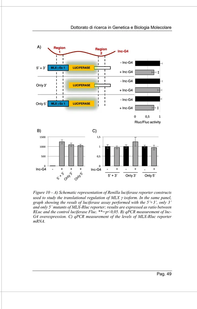
3.6. lnc-G4 specifically regulates MLX g translation 48 3.7. MLX g interacting region can fold into a G-quadruplex structure
52 3.8. lnc-G4 dependent MLX g modulation affects the subcellular
localization of total MLX protein 54
3.9. lnc-G4 could regulate the translation of many other mRNAs 57 3.10. Possible extensibility of lnc-G4 molecular mechanism 60
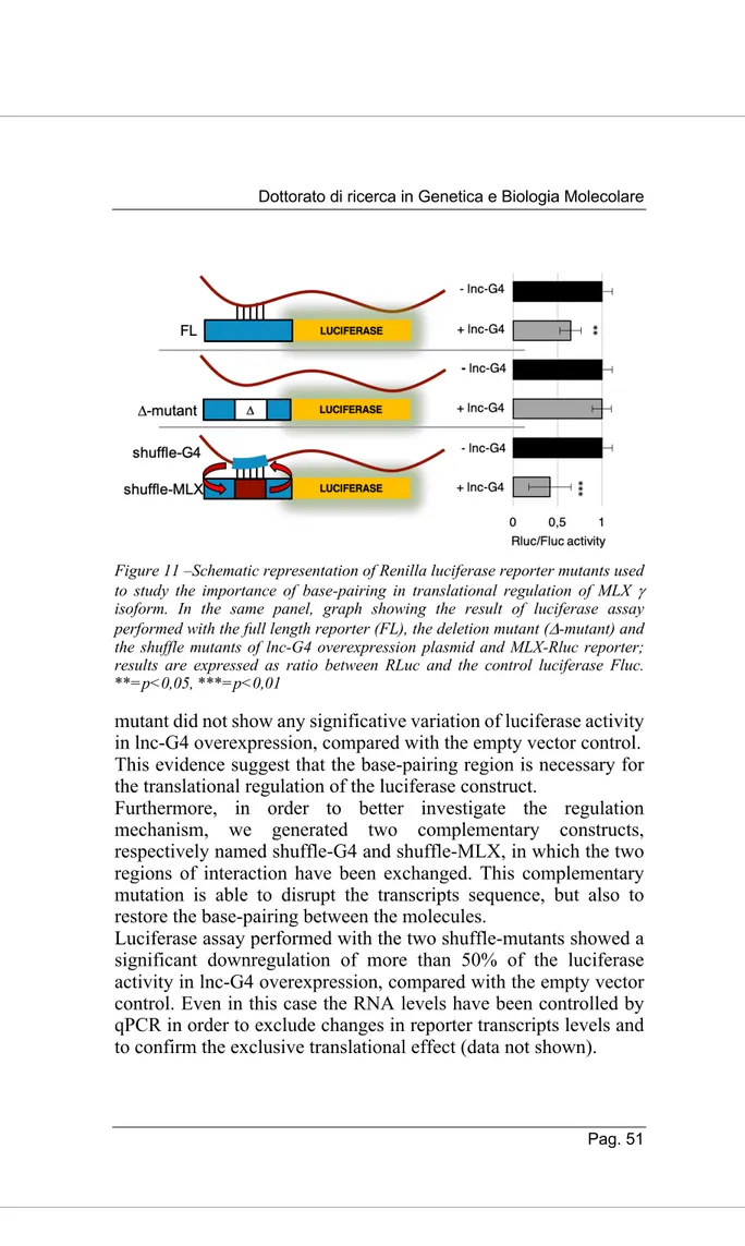
4. DISCUSSION 63
5.1 Cell culture and treatment methods 69
5.1.1 siRNA treatment 69
5.1.2 Plasmid transfection for overexpression experiments 70
5.2 RNA isolation and analysis 70
5.2.1 RNA purification 70
5.2.2. RNA retrotranscription 71
5.2.3 Semiquantitative RT-PCR 71
5.2.4. Quantitative PCR 71
5.2.5 RNA sequencing after lnc-G4 knockdown 71
5.3 Protein isolation and analysis 72
5.3.1 Protein extraction 72
5.3.2 Western Blot 72
5.4 Native RNA pulldown 73
5.4.1 RNA preparation for sequencing 74 5.4.2 Protein preparation for mass spectrometry 75
5.5 Psoralen-crosslinked RNA pulldown 75
5.6 DHX36 immunoprecipitation 76
5.7 C2C12 Immunostaining 77
5.7.1 Myosin heavy Chain immunostaining 77
5.7.2 MLX immunostaining 77
5.8 Luciferase assay 78
5.8.1 Constructs generation 78
5.8.2 Luciferase assay 79
5.9 In-gel G-quadruplex staining 80
5.10 Ribosome Profiling 81 5.11 Appendix tables 82 6. GLOSSARY 85 7. BIBLIOGRAPHY 89 8. LIST OF PUBLICATIONS 99 9. ACKNOWLEDGEMENTS 101
SUMMARY
Long non-coding RNAs are crucial regulators of the fine tuning of gene expression. Their role has been widely studied especially in developmental processes such as skeletal muscle differentiation. In particular, a novel cytoplasmic long non-coding RNA, called lnc-G4, has a relevant role in promoting murine C2C12 myoblast differentiation. The analysis of the interactors of this long non-coding RNA showed its ability to base-pair with many mRNAs thanks to a repeated element embedded in its sequence; among the interactors, we focused on MLX mRNA, which encodes for a myogenic transcription factor. We demonstrated that lnc-G4 directly interacts with the three splicing isoforms of MLX mRNA, while it is able to specifically inhibit the translation of only MLX g isoform; this translational regulation could depend on the recruitment of the RNA helicase DHX36. Interestingly, the effect of lnc-G4 on MLX g regulates the subcellular localization of the other isoforms, and this has an impact on the transcriptional activation of MLX targets. Taken together, these evidences suggest that lnc-G4 could be a key factor in post-transcriptional gene regulation during the early phases of myogenesis through the translational regulation of MLX g. Moreover, lnc-G4 interacts with other mRNAs, and the regulation mechanism could be extended to many other targets.
1. INTRODUCTION
1.1. A brief history of non-coding RNA
In every representation of the central dogma of molecular biology (first stated by Francis Crick in 1958), RNA lies at the heart of the genetic information flow; its central role came to light in 1961, when Jacob and Monod established for the first time the concept of messenger RNA (mRNA) as the connection between genetic information and protein synthesis (Jacob & Monod 1961). Later in the ‘60s, a population of heterogeneous nuclear RNA (hnRNA) was identified in higher eukaryotes, leading to the formulation of extensive RNA-dependent regulatory networks that could also be at the bases of complex organisms’ evolution (Britten & Davidson 1969).
Despite these early discoveries and hypotheses, a protein-centric view of molecular biology persisted for the following decades, in which the regulation of gene expression was ascribed to the exclusive action of transcription factors. However, as it was written by Thomas Cech and Joan Steitz:
“The seeds of a revolution are invariably sown decades before it erupts. And so it is with the revolution in noncoding RNAs.” In fact, between 1970 and 2000, the way to think about RNA biology was overthrown, and the established rules were step by step replaced by new rules; some examples are here reported:
1. The description of the first protein-independent RNA catalysis (Kruger et al. 1982) opened the ribozyme field, counteracting the idea that only proteins can have an enzymatic activity; ribosomal RNA itself was shown to be responsible for the catalysis of peptide bond formation.
2. The discovery of introns and of the splicing machinery (in which small nuclear RNAs are major components) showed that RNA transcription is not a straight-forward process, and later the evidences of alternative splicing demonstrated that exon rearrangement is responsible for the complexity of higher eukaryotes transcriptome.
3. The well-established model of proteins as unique regulators of transcription and translation was unhinged by the characterization of the first microRNAs (Lee et al. 1993) and the description of the RNA interference pathway.
Together, these milestones of RNA biology shed light on the pleiotropic and multivalent role of this molecule, that has even been indicated as the primordial constituent of life according to the “RNA world hypothesis” (Gilbert 1986).
Moreover, during the ‘90s it became clear that there was a correlation between the number and size of introns and intergenic sequences and the developmental complexity of organisms, and that these sequences could have evolved in order to express a wide spectrum of trans-acting regulatory RNAs. In fact, soon after the Human Genome Project conclusion, it was observed that the transcriptional activity of human chromosomes 21 and 22 was an order of magnitude higher than accounted for by the predicted and characterized exons (Kapranov et al. 2002).
The non-coding RNA revolution was completed by the advent of genome tiling arrays and the evolution of deep sequencing technologies that characterized the last decade. In particular, the Encyclopaedia of DNA Elements (ENCODE) project produced the first complete view of regions of transcription, binding of transcription factors and chromatin dynamics of the human genome, showing that 80.4% of it participates in at least one biochemical RNA- and/or chromatin-associated event in at least one cell type (Dunham et al. 2012). However, it was also observed that 74.7% of the human genome is transcribed in at least one of the 15 cell lines analysed (Djebali et al. 2012), while the estimated 20.687
protein-coding genes cover only the 2.94% of the sequence (ENCODE). With the increase of the number of publicly available sequencing data, pervasive transcription has been widely accepted as a common feature of eukaryotic genomes spanning from yeast to mammals. These recent studies have enlighted the complexity of transcriptomes, where the so called “junk DNA” regions are not transcriptionally silent and give rise to a plethora of non-coding RNAs which could originate a repertoire of new functions.
Nevertheless, even if the existence of a huge amount of non-coding transcripts is incontrovertible nowadays, the question is whether such transcriptional activities serve any biological function.
Figure A – Graphical representation of the most important milestones of molecular biology of RNA (from Rinn and Chang 2012)
1.2. Long non coding RNAs: definition and features
Long non-coding RNAs (lncRNAs) are a class of transcripts that mirrors the length and structure of mRNAs, including 5’ cap, 3’ polyA tail and splicing dynamics, though they do not have a clear coding potential. The operative definition of lncRNAs describes them as RNA molecules larger than 200 nt, in order to clearly distinguish these transcripts from small regulatory RNAs (Rinn & Chang 2012).Before the advent of deep sequencing, a few dozen lncRNAs that play a role in development, imprinting (e.g. H19) and X chromosome inactivation (e.g. Xist) had been identified by biochemical approaches and genetic screenings.
Nowadays, RNA sequencing allows a comprehensive visualization of the transcriptome thanks to the reconstruction of each transcript isoform at single-nucleotide resolution (Garber et al. 2011). On the other side, the analysis of epigenetic marks of Polymerase-II initiation (H3K4me3) and elongation (H3K36me3) by ChIP-seq approach consents the mapping of novel transcriptional units outside form known protein-coding loci (Guttman et al. 2009).
The combination of these high-throughput techniques led to the identification and annotation of more than 15000 lncRNAs encoded in the human genome, and at least 12000 in the mouse genome (Uszczynska-Ratajczak et al. 2018).
Despite the structural similarities between them and mRNAs, lncRNAs possess unique features regarding their genomic organization and their pattern of expression. First of all, lncRNAs can be classified based on their localization and orientation within the genome in five different categories:
1. Sense lncRNAs, which originate from the same strand of protein coding genes, and may overlap them partially or completely;
2. Antisense lncRNAs, which are transcribed from the opposite strand of a protein-coding gene and overlap at least one coding exon. Antisense transcription regards as many as the 87% of coding transcripts in the mouse genome (Ma et al. 2013)
3. Divergent lncRNAs, that share their promoter with protein-coding genes and are transcribed in opposite direction;
4. Intronic lncRNAs, which are entirely embedded in introns of protein-coding genes without any preference in directionality;
5. Intergenic lncRNAs, also called lincRNAs, that are encoded by totally independent transcriptional units,
and are usually 5 kb away from other genes (Guttman et al. 2009)
As a main trend, the expression level of lncRNAs is lower than the one of mRNAs. Several studies have estimated the abundance of the two species starting from RNA-seq data, quantifying differences of the median expression level spanning from 3-fold (Guttman et al. 2010) to 10-fold (Guttman et al. 2009).
Moreover, the vast majority of lncRNAs exhibit expression patterns that are limited to one or a few tissues, independently from their expression ranges. In particular, 78% of human lincRNAs are tissue-specific, relative to only ~19% of coding genes (Cabili et al. 2011) Tissue specificity of lncRNAs goes hand in hand with their role as molecular signals, since their transcription occurs at a very specific time and place to integrate developmental cues and interpret cellular context (Wang & Chang 2011).
The reconstruction of full-length gene structures of lncRNAs allows to study their evolutionary sequence conservation. Excluding the Telomeric repeat-containing RNA Terra, which is strongly conserved from yeast to mammals, mammalian lncRNAs lack any known orthologue in species outside the vertebrates (Ulitsky & Bartel 2013). Generally, lncRNA sequences appear to be more conserved than mRNA introns, while they are less conserved than coding sequences (Rinn & Chang 2012). The low sequence conservation is due to a high evolutionary rate: for example, within rodents only 60% of the lncRNAs (compared to >90% of mRNAs) expressed in Mus musculus liver have alignable counterparts expressed in the livers of Mus castaneus and rat (Kutter et al. 2012). Despite undetectable sequence conservation, genomic location and intron-exon structure are more prone to be maintained in different organisms, as well as the secondary structure of the lncRNA (Ulitsky & Bartel 2013). Finally, the fourth dimension of lncRNA conservation is at the level of the function, so that different molecules in different organisms can be responsible for the same phenotypic trait or molecular ac
A single lncRNA is able to interact with other nucleic acids through base-pairing, and to be recognised by protein interactors thanks to secondary structures and specific binding sequences; this is mainly due to the modular structure of these molecules, that allows them to be the central core of interaction networks between DNA, RNA and protein effectors (Guttman & Rinn 2012).
Summarising current evidences, the functionality of lncRNAs depends on three main mechanistic themes:
1. Decoy activity: lncRNAs can sequester other factors, either proteins or RNAs, and thus compete for their binding to other interactors. For example the lncRNA Gas5, which is induced upon starvation, is able to bind the glucocorticoid receptor through a hairpin sequence motif and thus competing for binding and activation of metabolic genes (Kino et al. 2010).
2. Scaffolding activity: lncRNAs can act as adaptors to bring together two or more proteins into discrete complexes (Spitale and Tsai 2011). A well described mechanism of scaffolding RNA is TERC, the RNA component of the telomerase complex, which is responsible for template synthesis, binding and catalytic activity of the protein component TERC (Collins 2008). In fact, mutations that affect the RNA scaffold structure result in impaired telomerase activity and in the onset of dyskeratosis congenita (Chen & Greider 2004).
3. Guide activity: many lncRNAs are required for the localization of protein complexes, and to target them on specific DNA or RNA sequences. For example, lincRNA-p21 is induced in a p53-dependent manner after DNA damage, and is able to binding the nuclear factor hnRNP-K and mediate its recruitment on the promoters of genes that have to be transcriptionally repressed (Huarte et al. 2010). These general mechanisms of action of long noncoding RNAs allow them to act both as transcriptional or post-transcriptional regulation, and this distinction is mainly depending on their preferential localization within the nucleus or in the cytoplasm.
1.3. Nuclear long non-coding RNAs
A significant fraction of lncRNAs is preferentially localised in the nucleus, and in several cases they have been shown to be crucial regulators of transcription and RNA processing, but also of chromatin architecture and organization of nuclear domain. The recruitment of lncRNAs on chromatin depends on protein-mediated interactions, RNA-RNA base pairing or RNA-DNA direct interface by formation of triple helixes (Rinn & Chang 2012).
The identification of their molecular mechanisms has been strongly promoted by newly developed techniques, like CHIRP (Chu et al. 2012) or RAP (Engreitz et al. 2015), that allow the capture of specific RNAs and the detection of their chromatin association sites in a genome-wide manner. lncRNAs can act on chromatin organization by recruiting epigenetic remodelers: Oct4P4 lncRNA interacts with the SUV39H1 to direct the silencing of the Oct4 gene through deposition of H3K9me3 and HP1a on its promoter, leading to reduced mESC self-renewal (Scarola et al. 2015).
The orchestration of chromatin remodelling is not the only way by which lncRNAs affect transcription. For example, the lncRNA PACER positively regulates the expression of COX2 by sequestering the p50 repressive subunit of NF-kB away from its promoter (Krawczyk & Emerson 2014).
Moreover, long noncoding RNAs are major organisers of nuclear structure. The best characterised example is Xist, that is not only responsible for X-chromosome inactivation by recruiting the PRC2 repressive complex, but is also interacts directly with the Lamin B receptor in order to relocalize the entire inactivated X chromosome next to the nuclear lamina (Chen et al. 2016).
Finally, nuclear lncRNAs are also necessary to nucleate and maintain specific nuclear domains. NEAT1 RNA transcription is essential for the structure of paraspeckles (nuclear bodies in which specific Alu- containing mRNAs are retained) (Mao et al. 2011), while MALAT1 has been shown to localize serine/arginine (SR) splicing factors to a compartment called nuclear speckles, where they can be stored and modified by phosphorylation (Bernard et al. 2010). These few examples of functional nuclear-retained lncRNAs
show how their ability to interact with both protein and nucleic acid interaction is at the base of several different mechanisms that allow to regulate transcription at different levels.
1.4. Cytoplasmic long non-coding RNAs
Perhaps the most common misperception of lncRNAs is that they are mainly localized in the nucleus. Studied based on subcellular RNA-seq are often biased since the distribution is quantified relative to mRNA abundance. Keeping in mind that some lncRNAs might act in the nucleus before making their way to the cytoplasm, the current picture is that many lncRNAs spend most of their time in the cytoplasm, which is frequently their site of action (Ulitsky & Bartel 2013). In the cytoplasm, lncRNAs take part to the multiple layers of post-transcriptional regulation by affecting the half-life and the translation of messenger RNAs; in this way, lncRNAs can quickly regulate gene expression by directly intervening on already transcribed mRNAs, a feature that assumes great importance in development and differentiation.
Starting from mRNA stability modulation, there are several examples of cytoplasmic lncRNAs that are able to induce specific mRNA decay. One of the pathways on which they act is STAU1-mediated decay (SMD), that involves the degradation of translationally active mRNAs whose 3' UTRs contain stem structures bound by STAU1. However, not all SMD targets that possess comparable structures in their 3’UTR: in some cases, a STAU1 binding site can be formed by Alu sequence-mediated base-pairing between so called half-STAU1-binding site lncRNAs (1/2 sbsRNAs) and their target mRNAs, committing them to degradation (Gong & Maquat 2011). Another example of cytoplasmic lncRNA-mediated decay is represented by the DNA-damage induced RNA Gadd7; this transcript associates to TDP43 protein, interfering with its interaction with the cyclin-dependent kinase 6 (Cdk6) mRNA, thus leading to Cdk6 mRNA instability and G1 phase arrest of the cell cycle (Liu et al. 2012).
On the other side, cytoplasmic lncRNAs can have a role as mRNA stabilizer, in both protein dependent and independent manner.
In the former case, a good example is represented by TINCR, a lncRNA involved in epidermal differentiation which is able to interact with several mRNAs of genes associated with epidermal barrier formation, thanks to a unique, 25-nt sequence motif called TINCR-box (Kretz et al. 2013). Notably, TINCR-mediated stabilization relies on the direct binding of the already cited STAU1 protein, that is this case has a positive effect on mRNA stability. In other cases, the base-pairing of the RNA alone is sufficient to trigger the regulation mechanism. For example, the conserved noncoding BACE1-AS antisense transcript stabilises BACE1 mRNA by forming a sense-antisense duplex, as demonstrated by RNase protection assay (Faghihi et al. 2008); the binding of the lncRNA has been shown to mask the miR-485-5p binding site on BACE1 mRNA, preventing its miRNA-induced repression (Faghihi et al. 2010). In this case, the presence of the RNA alone is sufficient to trigger the regulation mechanism.
Another layer of regulation in which lncRNAs seem to have a major role in translational control. A paradigmatic example is lincRNA-p21, already cited for its nuclear role; in the cytoplasm, this transcript is associated with CTNNB1 and JUNB mRNAs by imperfect base-pairing in their 3’UTR regions. The formation of lincRNA-p21–mRNA complex recruits the translational repressors RCK as well as Fragile X mental retardation protein (FMRP), thus reducing ribosome occupancy on the mRNAs (Yoon et al. 2012). Translational repression mediated by lncRNAs has also a validated role in cancer progression: treRNA stimulates tumour invasion in
vitro and metastasis formation in vivo by downregulating the
translation of the epithelial marker E-cadherin (Gumireddy et al. 2013). Even in this case, the scaffolding activity of the lncRNA recruits protein regulators, such as hnRNP K, FXR1 and 2, to form a novel ribonucleoprotein complex required to exert its function. An example of positive regulation of translation is represented by Uchl1-AS, that is an antisense transcript to mouse ubiquitin carboxy-terminal hydrolase L1 (Uchl1), sharing a 5′ overlapping sequence that contains an embedded inverted SINEB2 element with its sense transcript (Carrieri et al. 2012). Under stress signalling
control, Uchl1-AS migrates to the cytoplasm, interacts with the mRNA via the SINEB2 sequence, and recruits the protein coding transcript to the heavy polysomes increasing its translation.
In the crowded environment of the cytoplasm, not only RNA-protein, but also RNA-RNA crosstalk between different RNA species becomes an important layer of regulation. In particular, a huge variety of transcript contain miRNA binding sites; competition for the interaction with shared microRNAs is then a possibility for a new layer of regulation (Tay et al. 2014). In fact, the expression of lncRNAs containing miRNA binding sites in specific tissues and windows of time can impact the stoichiometry of microRNA-mRNA interaction, as demonstrated by several studies.
PTENP1, a noncoding transcript expressed from a pseudogene, regulates the tumour suppressor PTEN expression both at transcriptional level, by recruiting DNMT3 and EZH2 on its promoter, and post-transcriptional level, acting as a decoy for PTEN-targeting miRNAs (Johnsson et al. 2013). Moreover, sequestration of miRNAs has been shown to be important for embryonic stem cell maintenance: the lncRNA linc-RoR is an effective sponge for miR-145, whose targets OCT4, SOX2 and NANOG are core regulators of pluripotency (Wang et al. 2013). Another peculiar example of competing endogenous RNA is represented by ciRS-7, a circular RNA that contains 73 selectively conserved target sites for miR-7 in which a mismatch in the central part of the duplex prevents miRNA-mediated endocleavage (Hansen et al. 2013).
Finally, in the recent years several lncRNAs have been identified as regulators of post-translational modification of cytoplasmic proteins. Just to cite an example, lincRNA-p21 has an additional role in hypoxic condition response, binding both the transcription factor 1a and the ubiquitin ligase VHL, preventing HIF-1a ubiquitination and degradation and promoting glycolysis under hypoxia (Yang et al. 2014).
Taken together, all the cited examples show how cytoplasmic lncRNAs are core components of a complex network of RNA-protein and RNA-RNA interactions that consent them to exert
important functions in the post-transcriptional regulation of gene expression, especially during stimuli response and developmental programs.
1.5. Not all long non-coding RNAs are strictly
non-coding
As it has been reported above, pervasive translation of complex eukaryotes genomes gives rise to thousands of non-coding transcripts, beside the already known protein-coding gene products. However, canonical open reading frames (ORFs) have historically been identified as sequencing coding for peptides longer than 100 codons, based on the assumption that short peptides are unable to fold into secondary structure and thus to perform biological functions. Recently, the introduction of Ribosome Profiling, a powerful high-throughput technique based on deep sequencing of ribosome protected fragments after RNase footprinting, have permitted the quantitative study of translation by estimating the ribosomal engagement of transcripts (Ingolia et al. 2009). Several Ribosome Profiling works have showed that ribosomes occupy many bona fide non-coding regions of transcriptomes, including lncRNAs, thus enlarging the complexity of the mammalian translatome. These data have been confirmed by the analysis of the distribution of thousands of lncRNAs in polysome profiling after sucrose fractionation (Van Heesch et al. 2014). Taken together, these studies suggest that many lncRNAs could be translated in order to produce small, previously unidentified peptides, despite of their predicted low coding potential. There are in fact examples of transcripts, formerly identified as long non-coding RNAs, which are in fact coding for short functional peptides (Rion & Rüegg 2017). The first to be identified was the Drosophila tarsal-less/polished rice transcript, which encodes peptides of 11 to 32 amino acids whit a role in post-translational regulation of the Shavenbaby transcription factor (Kondo et al. 2010). More recently, other short regulatory peptide have been identified in mammals; for instance, Myoregulin and DWORF are ultraconserved micropeptides that fold into transmembrane a-helixes and interact with the SERCA calcium
channel, regulating calcium uptake in skeletal muscle (Anderson et al. 2015; Nelson et al. 2016). Nevertheless, these examples could be interesting exceptions, while it is still unclear at what extent lncRNAs could be translation templates. In fact, ORFs in lncRNAs might act as upstream ORFs (uORFs) to prevent ribosome scanning or translation in downstream regions of the transcripts, thereby enabling the lncRNAs to perform noncoding functions in the cytoplasm without interference from the ribosome. lncRNA ORFs might also tether factors to ribosomes or modulate the stability of the lncRNA by influencing RNA decay pathways, some of which depend on translation, such as non-sense mediated decay (Ulitsky & Bartel 2013).
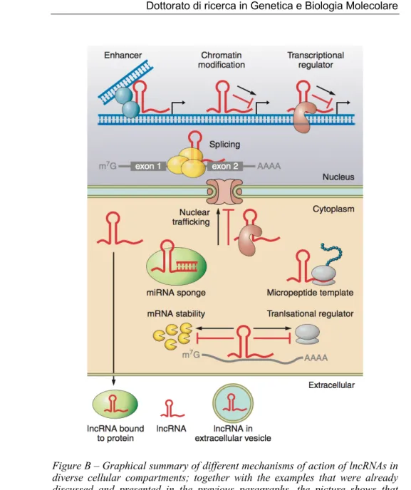
Figure B – Graphical summary of different mechanisms of action of lncRNAs in diverse cellular compartments; together with the examples that were already discussed and presented in the previous paragraphs, the picture shows that lncRNAs can also be embedded in extracellular vesicles or circulate freely in biological fluids, with a speculative role on cell-to-cell communication (adapted from Beerman et al, 2016)
1.6. Molecular regulation of skeletal muscle
differentiation
In mammalian embryos, the formation of skeletal muscle of trunk and limbs starts from the somites, which are derivatives of the antero-posterior segmentation of paraxial mesoderm. In particular, somitic cells subsequently distribute into a ventral mesenchymal sclerotome, the precursor of vertebral column and ribs, and a dorsal dermomyotome, that gives rise to both dorsal dermis and all the skeletal muscles of limbs and trunk. Head muscles formation follows a different path, taking origin from the mesodermal core of branchial arches (Buckingham and Rigby 2014). However, myogenesis is not limited to embryonic development and occurs also in adult vertebrate as a response to muscle injury. Muscle regeneration is the formation of new muscle fibers on the template of the extracellular matrix following injury; this function is exerted by satellite cells, somite-derived myogenic progenitors located under the basal lamina that surrounds the multinucleated fibre. Once they are activated by injury signalling, satellite cells leave their niche and undergo asymmetric divisions to form new fibers and maintain a stem cell pool in the same time.
Transcriptional control is fundamental in both embryonic and adult skeletal muscle differentiation, and it mainly depends on a core unit of four Myogenic Regulatory Factors (MRFs) that belong to MyoD family (Buckingham and Rigby 2014). The role and molecular circuitries in which these basic helix-loop-helix (bHLH) factors are involved have been widely elucidated in the last 30 years: for instance, overexpression of MyoD in non-muscle cells leads to activation of myogenic program and suppression of other cell fates (Weintraub et al. 1991). Mutations of the MRFs in mouse models have shown that MyoD, Myf5 and Mrf4 act as essential myogenic determination factors, while Myogenin is a differentiation factor necessary to control the differentiation program (Moncaut et al. 2013).
The upstream controllers of MRFs activations are Pax3 and Pax7, members of the family of paired domain transcription factors. Pax3
expression begins in the presomitic paraxial mesoderm, and it identifies migrating myogenic progenitors that have not yet activated the determination genes (Buckingham & Relaix 2007).In somite multipotent cells, the balance between Pax3 and Foxc2 is necessary for self-renewal, but a relative increase in Pax3 level in the trigger for Myf5 activation and therefore for the beginning of myogenic program. On the other side, Pax7 is expresses in the central domain of the dermomyotome; while it is not necessary for pre-natal myogenesis, it becomes the dominant factor in adult progenitors. Pax7 is necessary for satellite cell quiescence, since it is an activator of the HLH inhibitor Id3 (Kumar et al. 2009), but also to trigger the initiation of differentiation by direct MyoD activation (Hu et al. 2008) and chromatin remodelling of Myf5 locus through the recruitment of Wdr5– Ash2L–MLL2 histone methyltransferase (HMT) complex (McKinnell et al. 2008).
The induction of skeletal muscle differentiation requires the downregulation of Pax3/Pax7 and the subsequent activation of MRF network. Briefly, Myf5 expressing cells, called myoblasts, undergo a proliferative phase under FGF and Notch signalling, until MyoD activation leads to cell cycle exit. MRF factors act as obligate heterodimers with E proteins, thus binding E-boxes containing promoters and enhancers of a wide plethora of genes controlling muscle structure, contraction and metabolism. MRF-dependent gene activation requires also a complete chromatin remodelling. For instance, MyoD interacts with the BAF60c subunit of the SWI/SNF chromatin-remodelling complex and recruits it on the promoters of inactive genes creating poised chromatin domains ready for rapid transcription in response to differentiation stimuli (Forcales et al. 2012). MRFs do not act in isolation but they work in concert with other transcription factors, like members of the Mef2 family (Molkentin et al. 1995). After the proliferation arrest and lineage-specific transcriptional activation, myoblast transition towards the myocyte state is characterised by a reduction in cell motility and the precise orientation and positioning of muscle cells in response to chemotactic gradients (Griffin et al. 2010). The formation of myotubes, multinucleated syncytia with contraction ability, requires extensive actin cytoskeleton rearrangement; moreover, myoblast
fusion requires the presence of the small, muscle-specific peptide Myomixer, that mediated plasma membrane fusion through the association with the fusogenic protein Myomaker (Bi et al. 2017).
1.7. Noncoding RNAs in skeletal muscle differentiation
Transcriptional regulation of the onset of skeletal muscle formation is modified by post-transcriptional mechanisms that affect the presence and function of the transcription factors concerned (Buckingham and Rigby 2014). This fine regulation of differentiation timing mainly depends on a complex network of long and small non-coding RNAs. First of all, myogenic microRNAs are fundamental to repress self-renewal factors and to prevent early expression of late myogenic markers. For example, Pax3/Pax7 are targets of miR-206, and their downregulation contributes to the induction of differentiation; miR-1 maintains the repression during the late phases of the developmental program (Horak et al. 2016). Moreover, miR-31 is required for translational repression of late myogenic markers such as dystrophin (Cacchiarelli et al. 2011). Together with the cited microRNA circuitries, also lncRNAs work as crucial regulators of skeletal muscle differentiation thanks to their tissue-specific expression and their fine temporal regulation, acting at different layers of gene expression both in the nucleus and the cytoplasm. In some cases, the way of action of myogenesis-involved lncRNAs is strictly connected to microRNAs; H19, the first long noncoding RNA to be identified, harbours both canonical and non-canonical binding sites for microRNAs of the let-7 family, and its depletion causes precocious muscle differentiation (Kallen et al. 2013). Furthermore, linc-MD1 is a muscle-specific, MyoD-induced competing endogenous RNA that governs the timing of myogenesis. linc-MD1originates from the miR-206/133b locus and acts as a decoy for miR-133 and miR-135, derepressing their mRNA targets, including MAML1 and MEF2C, which encode crucial myogenic factors required for the activation of muscle- specific genes (Cesana et al. 2011). Moreover, the interplay between linc-MD1 and the protein HuR in the early phases of differentiation prevents the Drosha cleavage of the lncRNA, allowing its sponging activity;when this feedforward regulatory loop is interrupted from external stimuli, linc-MD1 is processed for miR-133b production triggering the progression to late differentiation stages (Legnini et al. 2014). Recently, a study based on RNA-seq characterized 30 novel lncRNA species, including previously unannotated transcripts, which undergo modulated expression during in vitro differentiation of C2C12 murine myoblasts (Ballarino et al. 2015). These newly identified transcripts have been classified based on their expression dynamics in growth condition (GM) versus differentiating condition (DM), their subcellular localization between nucleus and cytoplasm and their expression in tissues withdrawn from 2-month-old wild type and mdx mice (mdx mouse is the principal animal model for Duchenne Muscular Dystrophy, carrying a point mutation in exon 23 of DMD gene; it is characterised by increased muscle damage and regeneration). Some of this new lncRNAs have been further characterised in their molecular mechanism of action in the last years. For instance, Charme is a nuclear lncRNA that takes contact with the nctc locus, which contains Igf2 and the troponins Tnnt3 and Tnni2 genes, and is essential for its organization; the depletion of the lncRNA causes the disassembly of the chromatin domain and the downregulation of the genes contained therein.
Moreover, Charme-/- mouse shows severe cardiac remodelling and has shorter lifespan (Ballarino et al. 2018).
Another case is represented by lnc-31, a cytoplasmic lncRNA that is mainly expressed in growing condition and is required to sustain myoblast proliferation. lnc-31 interacts with both Rock1 mRNA and YB-1 protein, and increases Rock1 translation thanks to a localized effect of YB-1 stabilization and to the counteraction of YB-1 proteasome degradation (Dimartino et al. 2018).
1.8. G-quadruplexes in RNA biology
The identification of nucleic acid structures with a high guanine abundance became apparent in the early ’60s, and later in the ‘80s it was discovered that these formations were at the base of telomere formation (Henderson et al. 1987). Nowadays, structural analyses
have given the G-quadruplex name to these peculiar DNA and RNA assemblies.
G-quadruplexes formed by guanine-rich nucleic acid sequences are non-canonical structures organized by stacking of tetrads or G-quartets, in which four guanines are assembled in a planar arrangement by Hoogsteen hydrogen bonding. In order to fold into a G-quadruplex structure, the linear sequence of DNA or RNA must contain four guanine stretches of 2-5 G, intercut with variable loops from 1 to 7 nt. The stability of this structure depends on the presence of a monovalent cation, typically potassium, that “coordinates” the structure by positioning within or in between the plane of the tetrads (Millevoi et al. 2012).
The human genome contains as many as 376,000 G-quadruplex-forming sequences which are not randomly distributed (Todd et al. 2005). At the DNA level, quadruplexes are enriched at the telomere, where they inhibit the activity of telomerase, and they are associated with ~40% of human promoter, supporting their role in transcriptional regulation. Nevertheless, an increasing number of evidences in the last few years support the view that RNA G-quadruplexes are also crucial, intrinsic biological regulators. Additionally, RNA G-quadruplexes have been demonstrated to be more stable than the DNA counterpart, with faster association dynamics and slower dissociation (Saccà et al. 2005). Increasing evidences show that RNA quadruplexes at the 5’ and 3’ ends and/or other locations in the RNAs have functional role in pre-mRNA processing (including splicing and polyadenylation), RNA stability and turnover, mRNA translation and subcellular targeting.
Starting from the regulation of mRNA splicing, G-quadruplexes placed in the vicinity of splicing sites of a growing list of genes, can act as cis- regulatory elements of alternative exon inclusion or exclusion. For instance, a G-quadruplex located in intron 3 of the TP53 gene favours splicing of the adjacent intron 2; mutation of the G-quadruplex leads to intron 2 retention (Marcel et al. 2011). More interestingly, computational analysis revealed that G-quadruplexes forming motifs are overrepresented in the 5′-UTRs, which are key elements in the initiation of mRNA translation. The presence of these secondary structures in the 5′-UTR, can interfere
with translation either by cis-acting effects (steric blockade) or through the binding of trans-acting mRNA binding proteins that may impact on the recruitment of eIFs to the mRNAs (e.g., eIF4F to the cap) or also affect the scanning process or the recognition of the initiation codon. Nonetheless, G-quadruplexes located in 5′-UTR are able to activate translation: G-quadruplex-forming motifs are present in IRES elements, and a mutational analysis has shown that the G-quadruplex within the VEGF IRES is essential for IRES function (Morris et al. 2010).
Even if there is a positional bias toward the presence of G-quadruplexes in the 5′-UTR of mRNAs, these structural elements can also be found in 3′-UTR and open reading frames (ORF). The involvement of G-quadruplex-binding proteins is particularly evident for mRNA translational regulation, where G-quadruplex structures needs to be unfolded to relieve translational repression. In particular, the RNA helicase DHX36, also named RHAU, is a crucial actor for its global G-quadruplex unwinding activity and its extreme specificity in the recognition of these structures (Lattmann et al. 2011). This helicase has an important role in mRNA G-quadruplex resolution, but also in AU-elements mediated decay for the regulation of mRNA turnover. A good example of this mechanism is represented by the regulation of Nkx2.5 cardiac-specific mRNA: in this case, DHX36 is able to promote translation of Nkx2.5 through the resolution of a G-quadruplex in the 5’ UTR on the mRNA, while on the other side it induces the decay of the transcript by binding a AU-rich element in the 3’ UTR (Nie et al. 2015).
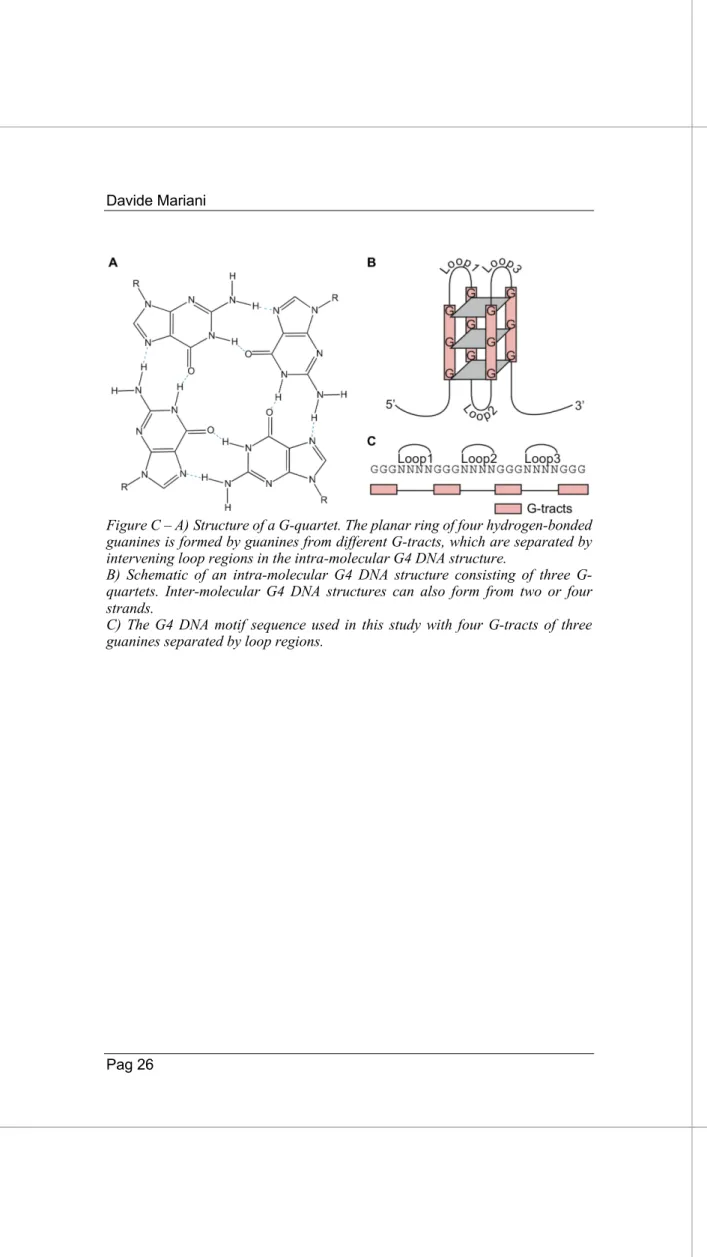
Figure C – A)Structure of a G-quartet. The planar ring of four hydrogen-bonded guanines is formed by guanines from different G-tracts, which are separated by intervening loop regions in the intra-molecular G4 DNA structure.
B) Schematic of an intra-molecular G4 DNA structure consisting of three G-quartets. Inter-molecular G4 DNA structures can also form from two or four strands.
C) The G4 DNA motif sequence used in this study with four G-tracts of three guanines separated by loop regions.
2. AIM OF THE THESIS
In 1988, Francis Crick wrote:“Almost all aspects of life are engineered at the molecular level, and without understanding molecules we can only have a very sketchy understanding of life itself.”
Crick has always been a pioneer, or a sort of “prophet” of molecular biology future directions. In particular, he always had a complete and wide vision of organisms as complex systems that are finely regulated at the molecular level.
The technological advances made in the last years revolutionized the protein-centric paradigm of gene expression regulation. Nowadays, the eukaryotic genome is considered as a “RNA machine” in which different species of non-coding RNAs cooperate to modulate gene expression in a very fine way.
In this landscape, long non-coding RNAs have been identified and described as essential regulators of developmental and differentiation processes. Thanks to their versatility, modularity and cell- and tissue-specific expression, these molecules have been selected during evolution to exert a wide variety of functions and increase the complexity of gene expression modulation.
Long non-coding RNAs are core regulators of every step of mRNA processing, from transcription to translation and turnover, by acting both in nuclear and cytoplasmic compartments. Even if a strong effort has been made to reveal and enlighten the ways of action of nuclear lncRNAs, the molecular mechanisms by which these molecules influence mRNA life in the cytoplasm are still poorly understood, with few complete examples.
The aim of my thesis is to add knowledge on the role of cytoplasmic long non-coding RNAs in post-translational regulation during skeletal muscle differentiation.
Since many years, the research group of Prof. Irene Bozzoni, in which I prepared my thesis, is interested in understanding the mechanisms of regulation of skeletal muscle differentiation in both
physiological and pathological conditions, particularly focusing on the involvement of non-coding RNA species.
In particular, during my thesis I worked on the characterization of long non-coding RNAs differentially expressed during skeletal muscle differentiation of C2C12 murine myoblasts, previously identified by the group of Dr. Monica Ballarino (Ballarino et al. 2015). Among the cytoplasmic candidates, we selected lnc-G4 for further investigation due to its muscle-specific expression, its abundance and the interesting phenotype on muscle differentiation after its knockdown. Using a RNA pulldown approach coupled with high-throughput techniques, I identified and validated the protein and RNA interactors of this long non-coding RNA.
My work allowed me to describe a molecular mechanism in which lnc-G4 physically interacts with the g isoform of MLX mRNA in a G-quadruplex containing region. This interaction is sufficient to repress MLX g translation, and to thus to modulate the subcellular localization of the total level of MLX protein.
Moreover, I showed that the C-rich region embedded in lnc-G4 could be able to interact with many other G-quadruplex containing mRNAs, thus increasing the number of putative target of lnc-G4 activity during skeletal muscle differentiation.
3. RESULTS
3.1. Criteria for lnc-G4 selection
As previously described, we started our work with a candidate selection from the lncRNAs identified from Ballarino et al. 2015, according to the following criteria:
• Prevalent cytoplasmic localization, because we were interested on the characterization of novel cytoplasmic lncRNA-mediated mechanisms of gene expression regulation;
• Expression level: we focused on quite abundant molecules, selecting the lncRNAs with a relevant expression (quantified as a FPKM value between 7 and 50) in at least one time point of C2C12 differentiation;
• Tissue specificity: we selected lncRNAs that are upregulated or exclusively expressed in skeletal muscle tissues of wt and
mdx mice.
According to these criteria, 6 out of 30 lncRNAs were chosen for further characterization.
The identified candidates were then subjected to a siRNA screening in order to select molecules with an interesting phenotype on myoblast proliferation and/or differentiation in vitro.
The phenotypic screening part was performed by dr. Sama Shamloo, a former PhD student of the laboratory; the details of this preliminary work will not be presented and discussed in this manuscript.
My thesis project started form the best candidate identified in this screening, afterwards named lnc-G4.
3.2. Characterization of lnc-G4
Lnc-G4 is a multiexonic long non-coding RNA of 1409 nt which is transcribed from the negative strand of an intergenic locus of Mus
musculus chromosome X (Fig. 1A). It can be classified as an
intergenic long non-coding RNA (lincRNA) since it is expressed from a totally independent transcriptional unit, far more than 100 kb from the flanking genes (Bcor and Atp6ap2 respectively).
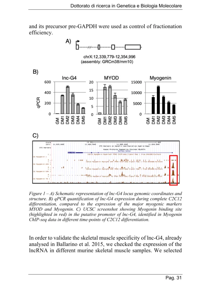
The timing of expression of lnc-G4 was analysed during time-course experiments of C2C12 differentiation, from proliferating myoblasts (condition named GM, Growth Medium) to terminally differentiated myotubes (condition named DM5, after 5 days from switch to Differentiation Medium). While the transcript is almost not detectable in GM condition, lnc-G4 expression dramatically increases during the early phases of C2C12 differentiation, with a peak in DM2, and drops down during the late phases of the differentiation process (Fig. 1B). This expression pattern is coherent with the RNA sequencing data published by Ballarino et al. 2015, where lnc-G4 expression reaches the highest FPKM value (17,25) in the DM3 sample.
Interestingly, the expression pattern of lnc-G4 recapitulates the one of Myogenin, a master regulator of skeletal muscle differentiation. To evaluate a putative role of Myogenin as a transcriptional regulator of lnc-G4 expression, publicly available ChIP-seq datasets from the mouse ENCODE project have been analysed (Fig. 1C). In Myogenin ChIP-seq experiments performed in C2C12 myoblasts and differentiated myotubes, there is a strong recruitment of the transcription factor on the putative lnc-G4 promoter; the maximum peak is reached in the early phases of myogenesis (60 hours), while Myogenin is less enriched in terminally differentiated myotubes (7 days).
The subcellular localization of lnc-G4 was then determined by nuclear-cytoplasmic fractionation of C2C12 cells at day 2 of differentiation, when lnc-G4 is expressed at its maximum. Around 70% of the transcript is localized in the cytoplasm, suggesting a prevalent cytoplasmic function for the lncRNA (Fig. 2A); GAPDH
and its precursor pre-GAPDH were used as control of fractionation efficiency.
Figure 1 – A) Schematic representation of lnc-G4 locus genomic coordinates and structure. B) qPCR quantification of lnc-G4 expression during complete C2C12 differentiation, compared to the expression of the major myogenic markers MYOD and Myogenin. C) UCSC screenshot showing Myogenin binding site (highlighted in red) in the putative promoter of lnc-G4, identified in Myogenin ChIP-seq data in different time-points of C2C12 differentiation.
In order to validate the skeletal muscle specificity of lnc-G4, already analysed in Ballarino et al. 2015, we checked the expression of the lncRNA in different murine skeletal muscle samples. We selected
gastrocnemius, masseter, extensor digitorum longus (EDL) and tongue of both two-month-old wild type (wt) and mdx in order to analyse a sample set of muscles with different fibre composition coming from different body districts. As shown in Fig. 2B, lnc-G4 is expressed in all the analysed samples and it is upregulated in mdx dystrophic condition, in which muscle regeneration is strongly activated. Taken together, these data characterize lnc-G4 as a mostly cytoplasmic lncRNA with a strong skeletal muscle specificity, mainly expressed in the early phases of C2C12 differentiation. Figure 2 – A) RT-PCR performed on RNA resulting from subcellular fractionation of C2C12 at day 2 of differentiation, showing that most of lnc-G4 transcript is
localized in the cytoplasm. Pre-GAPDH and GAPDH were used as controls of fractionation efficiency. B) RT-PCR analysis of lnc-G4 expression in skeletal muscle samples from two-month-old wt and mdx mice. Gastro=gastrocnemius, EDL=extensor digitorum longus. GAPDH was used as loading control.
3.3. lnc-G4 downregulation affects proper myogenesis
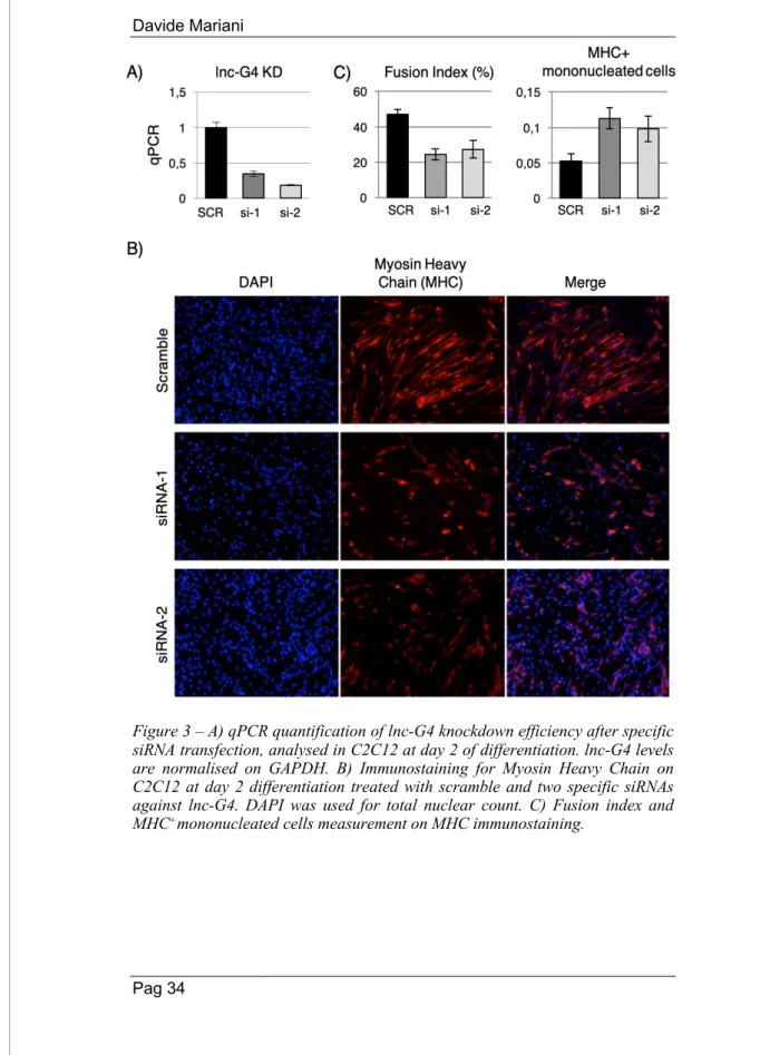
In order to identify the potential role of lnc-G4 in the establishment of proper muscle differentiation, a RNA interference loss-of-function approach has been chosen. Knockdown of lnc-G4 has been performed in C2C12 cells at day 2 of differentiation, using two different specific siRNAs, obtaining a consistent downregulation of ~80% of the lncRNA level as measured by qPCR (Fig. 3A). The impact of lnc-G4 downregulation on myogenesis was evaluated through Myosin Heavy Chain (MHC) immunostaining on C2C12 cells treated with scramble and the two different specific siRNAs. As shown in Fig. 3B, lnc-G4 depletion had a clear phenotypic effect on C2C12 differentiation, with a lower expression of MHC protein marker and a reduced formation of mature, correctly oriented myotubes with respect to the scramble-treated condition.To quantitatively analyse the effect of lnc-G4 downregulation on skeletal muscle differentiation, two different parameters were taken in account:
• Fusion index, defined as the fraction of the total number of fibre-embedded nuclei to the total number of nuclei; lnc-G4 depletion causes a reduction of the fusion index of 50%. • MHC+ mononucleated index, defined as the fraction of the
mononucleated MHC+ cells to the total of MHC+ cells; lnc-G4 depletion causes a marked increase of cells that already express Myosin Heavy Chain without beginning the fusion process.
Figure 3 – A) qPCR quantification of lnc-G4 knockdown efficiency after specific siRNA transfection, analysed in C2C12 at day 2 of differentiation. lnc-G4 levels are normalised on GAPDH. B) Immunostaining for Myosin Heavy Chain on C2C12 at day 2 differentiation treated with scramble and two specific siRNAs against lnc-G4. DAPI was used for total nuclear count. C) Fusion index and MHC+mononucleated cells measurement on MHC immunostaining.
To better understand and characterize the molecular pathways modulated after lnc-G4 knockdown, RNA isolated from C2C12 cells transfected with scramble or specific siRNAs was subjected to Illumina RNA-sequencing. The results were analysed by Dr. Alessio Colantoni (Sapienza University of Rome) with a pipeline focused on the identification of differential expression (details are reported in Material and Methods).
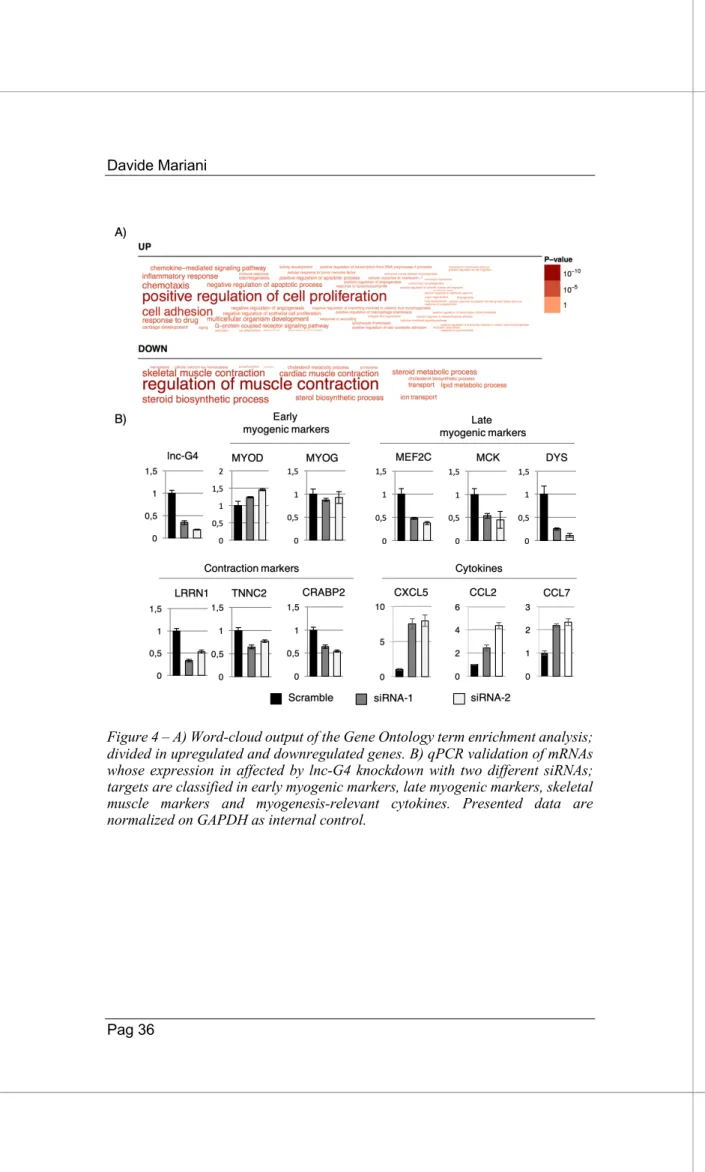
The bioinformatics analysis for showed that the expression of more than 50 genes was affected by lnc-G4 depletion. Gene Ontology analysis revealed the upregulation of genes involved in myoblast proliferation, chemotaxis and cytokine production and response, together with the downregulation of gene products involved in regulation of muscle contraction, muscle metabolism and calcium homeostasis, coherently with the observed phenotype (Fig. 4A). Moreover, a subpopulation of the most downregulated and upregulated genes was subjected to qPCR validation of the RNA-sequencing results. In particular, Figure 4B shows that lnc-G4 knockdown with both siRNAs do not affect early myogenic markers expression (Myogenin, MYOD), while it negatively affects the expression of late myogenic transcription factors (MEF2C), muscle-specific proteins (MCK, Dystrophin), and contraction markers (LRRN1, TNNC2, CRABP2). Furthermore, lnc-G4 downregulation positively impacts on the expression of a subset of cytokines (CXCL5, CCL2, CCL7) that have a validated role in myoblast proliferation and survival.
Taken together, these findings suggest that lnc-G4 strongly promotes skeletal muscle differentiation, and its depletion impacts on myocyte fusion ability and mature myotube formation. The phenotypic effect of lnc-G4 downregulation is recapitulated by the transcriptomic changes analysed by RNA-sequencing.
Figure 4 – A) Word-cloud output of the Gene Ontology term enrichment analysis; divided in upregulated and downregulated genes. B) qPCR validation of mRNAs whose expression in affected by lnc-G4 knockdown with two different siRNAs; targets are classified in early myogenic markers, late myogenic markers, skeletal muscle markers and myogenesis-relevant cytokines. Presented data are normalized on GAPDH as internal control.
3.4. lnc-G4 has a complex molecular interactome
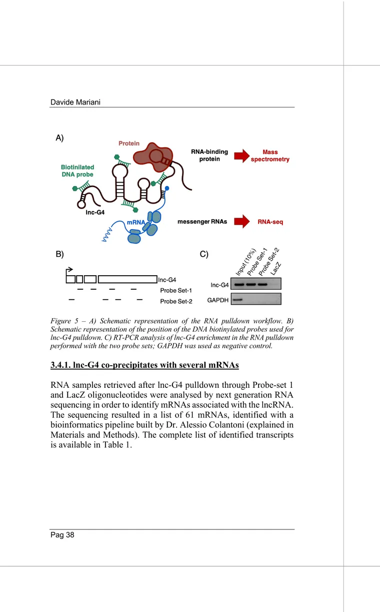
As treated in the Introduction, cytoplasmic long non coding RNAs can influence gene expression thanks to many different mechanisms of post-transcriptional regulation. However, all these diverse ways of action rely on the ability of lncRNAs to interacts with other RNA species, especially mRNAs and microRNAs, and with a wide variety of RNA binding proteins.The molecular interactome of lnc-G4 has been inspected with a novel endogenous RNA pulldown approach, coupled with high-throughput techniques in order to identify mRNA and protein interactors. Briefly, DNA antisense biotinylated probes designed against lnc-G4 sequence have been used to isolate the lncRNA from a cytoplasmic extract of C2C12 at day 2 of differentiation (Fig. 5A). Eight 20-nt probes have been designed on the complete sequence of lnc-G4, excluding repeated parts and regions which were predicted to be strongly structured in order to increase the accessibility; the selected probes were subsequently divided in two different sets of four probes each, and RNA pulldown was performed independently with both the sets (Fig. 5B). A third set of four probes, designed against LacZ mRNA sequence, was used as a negative control since it is not expressed in C2C12 cellular system. As shown in Figure 5C, the obtained pulldown efficiency for lnc-G4 was around 10% of the input with both the probe sets. The developed RNA pulldown approach allowed the isolation of both mRNA and protein putative interaction, and their subsequent identification through RNA sequencing and mass spectrometry, respectively.
Figure 5 – A) Schematic representation of the RNA pulldown workflow. B) Schematic representation of the position of the DNA biotinylated probes used for lnc-G4 pulldown. C) RT-PCR analysis of lnc-G4 enrichment in the RNA pulldown performed with the two probe sets; GAPDH was used as negative control.
3.4.1. lnc-G4 co-precipitates with several mRNAs
RNA samples retrieved after lnc-G4 pulldown through Probe-set 1 and LacZ oligonucleotides were analysed by next generation RNA sequencing in order to identify mRNAs associated with the lncRNA. The sequencing resulted in a list of 61 mRNAs, identified with a bioinformatics pipeline built by Dr. Alessio Colantoni (explained in Materials and Methods). The complete list of identified transcripts is available in Table 1.
Gene name P-Value Gene name P-Value Gene name P-Value lnc-G4 9,50E-09 21 Acad8 7,90E-04 42 Asb1 3,79E-03
1 Rps9 7,36E-05 22 Snord72 8,37E-04 43 Eef1g 3,79E-03
2 Mybph 1,80E-03 23 Rpl37 8,37E-04 44 Dclk1 3,79E-03
3 Mir503 4,94E-03 24 Glis3 8,54E-04 45 Cdkn1a 3,89E-03
4 Mir322 4,94E-03 25 Tysnd1 8,66E-04 46 Eef2 4,71E-03
5 Six1 1,14E-05 26 Rps8 9,86E-04 47 Rpl18 4,82E-03
6 Uba52 5,20E-06 27 Rps7 1,07E-03 48 Rn7sk 5,76E-03
7 Kxd1 5,20E-05 28 Myh3 1,41E-03 49 Tnnc1 5,91E-03
8 Arf5 1,14E-04 29 Rpl8 1,45E-03 50 Rpl19 5,93E-03
9 Atf2 1,46E-04 30 Acsl6 1,54E-03 51 Myl1 6,02E-03
10 Ndrg4 2,60E-04 31 Cdc26 1,55E-03 52 AC092404.1 7,54E-03
11 Slc15a4 5,50E-04 32 Mlx 1,65E-03 53 Rpl13 8,11E-03
12 Rpl18a 1,25E-04 33 Myo1c 1,85E-03 54 Mir7079 8,11E-03
13 Snora68 1,25E-04 34 Tnnt3 1,88E-03 55 Vti1b 8,14E-03
14 Rps20 1,62E-04 35 Cnpy2 1,89E-03 56 Mylpf 8,51E-03
15 Spire1 2,04E-04 36 Gm22009 2,08E-03 57 Cd63 8,62E-03
16 Coq2 2,70E-04 37 Ahnak 2,12E-03 58 Rps6 9,07E-03
17 mt-Co1 3,08E-04 38 Eef1a1 2,12E-03 59 Rps27a 9,20E-03
18 Zfp532 3,42E-04 39 Rbm45 2,18E-03 60 Sqstm1 9,21E-03
19 Hist1h2bg 4,38E-04 40 Lars 2,27E-03 61 Rps19 9,21E-03
20 Myeov2 6,59E-04 41 Usp10 2,58E-03
Table 1. List of mRNAs enriched in lnc-G4 pulldown identified from the RNA sequencing. RNAs are listed based on their p-value (calculated based on the sequencing replicates and the enrichment towards the LacZ negative control) from lower to higher value. Lnc-G4 is the first result since it is the most enriched transcript in its own pulldown.
Moreover, a bioinformatic prediction of the base-pairing interaction between the identified mRNAs and lnc-G4 was performed with the IntaRNA bioinformatics tool, which is specific for the prediction of RNA-RNA interactions among long RNA molecules (Mann et al. 2017). In particular, only interactions with a free energy lower or equal to -20 Kcal/mol have been considered, in order to select only the most stable and consistent base-pairings.
Interestingly, 22 out of 62 mRNAs were predicted to interact in a specific region of 250 nt at the beginning of lnc-G4 exon 4, within a C-rich sequence which is part of a LINE-L2 repeated element (Fig. 6A). Among them, we selected the 7 most promising interactors (i.e. the candidates with the best interaction energy) for further analysis. RT-PCR validation of the selected mRNA candidates identified from the RNA sequencing was performed on different replicates of lnc-G4 pulldown performed with both the probe sets, confirming the efficient precipitation of the identified RNAs; GAPDH was used as negative control (Fig.6B).
Figure 6. A) Graph showing the number of interactors for lnc-G4 20-nt sequence slots. The majority of interactions fall in a 250-nt region at the beginning of lnc-G4 exon 4. B) RT-PCR validation of mRNA candidate enrichment in lnc-lnc-G4 pulldown performed with the two probe sets; GAPDH was used as negative control.
The different pull-down efficiency among different mRNAs can be due to both differences in the length of the base-pairing regions between the lncRNA and the mRNAs and in the energy of interaction: ta good and strong interaction promotes a higher efficiency of pulldown. Due to the technical limitations of working with many candidates, we chose MLX mRNA for further investigation, because of its good enrichment and its well-studied role in regulation of gene expression during myogenesis.
3.4.2. lnc-G4 directly interacts with MLX mRNA
MLX, which stands for MAX-like protein X, is a basic helix-loop-helix component of the MYC/MAX network of transcription factors. The factors involved in this network integrate diverse cellular stimuli, like nutrient sensing and metabolic stress, in order to regulate proliferation and/or differentiation by dimerization and binding to E-box elements in target promoters (Diolaiti et al. 2015). Interestingly, the output resulting from the activation of this network of transcription factors depends of the dynamics of dimerization: for example, MLX can heterodimerize with MXD proteins to triggers proliferation arrest and differentiation, or it can bind to MONDO-A/B factors for glucose sensing activity. In the particular panorama of skeletal muscle differentiation, MLX has been shown to promote myogenesis in a glucose-depending manner: the heterodimerization with the MONDO-A glucose-sensing factors causes MLX translocation to the nucleus, with subsequent activation of several cytokine genes by binding to the carbohydrate responsive elements in their promoters (Hunt et al. 2015). Moreover, MLX is expressed in three different splicing isoforms:
• MLX a, which lacks exon 3; • MLX b, which contains exon 3;
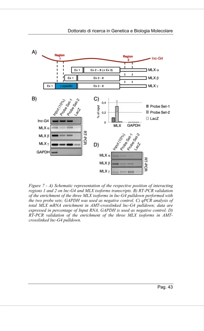
Using the already cited IntaRNA bioinformatic suite, we predicted the possible base-pairing interaction between lnc-G4 and the three different annotated isoform of MLX mRNA. We were able to find two different regions of interaction, named Region 1 and Region 2 respectively. In particular, Region 1 is restricted to the MLX g isoform, since it is included in the specific part of exon 1 within its coding sequence, while Region 2 is common to the three isoforms since it falls into the common 3’UTR sequence (Fig. 7A). The presence of all the three isoforms in lnc-G4 pulldown was validated through RT-PCR analysis using specific primers able to distinguish between the different transcripts (Fig. 7B).
In order to check whether the binding between lnc-G4 and MLX mRNA isoforms was direct or not, we optimized the RNA pulldown protocol including a AMT-crosslinking step. AMT (4’-aminomethyl-4,5’,8-trimethylpsoralen) is a chemical compound which is able to form covalent bonds amid base-pairing RNA molecules after UV-irradiation at 365 nm; the covalent crosslinking is revertible under UV-irradiation at 254 nm. As shown in Figure 7C, MLX total mRNA was enriched with both probe sets in lnc-G4 pulldown performed with AMT crosslinking, while GAPDH was used as negative control. Moreover, RT-PCR analysis showed that all the three MLX mRNA isoforms were enriched in AMT-crosslinked pulldown (Fig 7D).
Taken together, these data indicate that MLX mRNA is a valid interactor of lnc-G4; in particular, lnc-G4 directly bind to the three isoforms of MLX mRNA, presumably thanks to the two predicted binding regions.
Figure 7 - A) Schematic representation of the respective position of interacting regions 1 and 2 on lnc-G4 and MLX isoforms transcripts. B) RT-PCR validation of the enrichment of the three MLX isoforms in lnc-G4 pulldown performed with the two probe sets; GAPDH was used as negative control. C) qPCR analysis of total MLX mRNA enrichment in AMT-crosslinked lnc-G4 pulldown; data are expressed in percentage of Input RNA, GAPDH is used as negative control. D) RT-PCR validation of the enrichment of the three MLX isoforms in AMT-crosslinked lnc-G4 pulldown.
3.4.3. lnc-G4 interacts with DHX36 RNA helicase, and forms a molecular complex together with MLX g mRNA
Protein samples isolated from lnc-G4 native pulldown were analysed by mass spectrometry in order to identify putative lnc-G4 protein partners. Specifically, enriched proteins were cleaved into peptides with trypsin and analysed by high-resolution LTQ Velos Pro instrument. We were able to identify 375 proteins in all of the samples (172 in Probe-set 1 sample, 199 in Probe-set 2 sample, and 221 for the LacZ). Among the proteins that were common in lnc-G4 pulldown performed with the two sets of probes, only 14 proteins were significantly enriched compared to LacZ control (Table 2).
Probe set 1 Probe set 2 LacZ
Sc or e Co ve ra ge # P ep ti d es Sc or e Co ve ra ge # P ep ti d es Sc or e Co ve ra ge # P ep ti d es PUR-B 108,3 49 9 87,3 52 8 24,6 29 3 DHX36 56 17 10 26,9 9 5 0 0 0 CSRP1 37,3 38 4 26,4 28 3 18 20 2 RFA1 42,6 14 6 21,8 11 4 3,2 2 1 CPSF7 14,3 5 1 27,1 8 2 6 5 1 PFKAM 20,7 8 4 16,3 8 4 4,3 2 1 ALBU 8,1 5 2 18,7 5 2 3,7 2 1 CAZA2 7,2 12 2 13,7 19 3 3,5 6 1 RS11 9,4 11 1 17,7 11 1 5,2 11 1 Hnrnp0 16,4 7 1 3,9 5 1 0 0 0 RS8 8,8 7 1 8,6 7 1 0 0 0 MYL4 3,7 10 1 7,7 23 2 0 0 0 CCD30 3,1 2 1 3,4 2 1 0 0 0 SE1L1 3,5 2 1 3,2 2 1 0 0 0
Table 2 – List of proteins enriched in lnc-G4 pulldown with the two probe sets with respect to LacZ control. Score=sum of individual peptide counts. Coverage=percentage of protein sequence covered by identified peptides. #Peptides=number of distinct and uniquely assignable peptides.
Due to its good enrichment in both Probe-set 1 and 2 pulldowns and its complete absence in LacZ control, we chose DHX36 for further validation. DHX36, also known as RHAU (RNA helicase associated with AU-rich element) and G4R1, is a member of the DExH/D family of ATP-dependent RNA helicases. Thanks to its flexible, partially unstructured N-terminal domain, DHX36 has a strong specificity for unwinding both DNA and RNA G-quadruplex structures. It has been shown that DHX36 is able to regulate both stability and translation efficiency of G-quadruplex containing mRNAs (Nie et al. 2015); its role in post-transcriptional gene regulation made it a good candidate to study.
First of all, we validated the presence of DHX36 in different replicates of lnc-G4 pulldown through Western Blot, confirming the results of the mass spectrometry (Fig. 8A). To further confirm the interaction between DHX36 and lnc-G4, we performed RNA immunoprecipitation of the protein and checked for the enrichment of lnc-G4 through RT-PCR. Interestingly, we could demonstrate that also MLX is immunoprecipitated together with DHX36, while GAPDH was used as a negative control (Fig. 8B). This evidence allowed us to speculate that lnc-G4, MLX mRNA and DHX36 are part of the same cytoplasmic molecular complex.
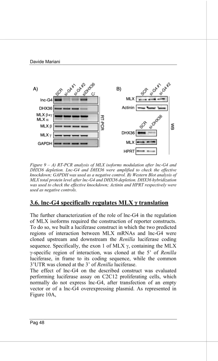
To understand whether lnc-G4 is required to recruit DHX36 on MLX mRNA, we performed DHX36 immunoprecipitation on extracts of C2C12 cell transfected with scramble and lnc-G4 siRNA. The results showed that, in scramble condition, both lnc-G4 and MLX total mRNA were enriched in DHX36 pulldown, compared to the positive control WBP4 (Lattmann et al. 2011) and the negative control GAPDH; however, no significative changes in MLX enrichment were detected after lnc-G4 knockdown with the specific siRNA (Fig. 8C).
Since lnc-G4 had been shown to interact with the three MLX mRNA isoforms, we verified their enrichment in DHX36 immunoprecipitation in both scramble and lnc-G4 depletion conditions. The RT-PCR analysis revealed that DHX36 specifically interacts with the MLX g isoform, while a and b isoforms resulted to be not enriched; also in this case, lnc-G4 knockdown did not affect the enrichment (Fig. 8D).
Taken together, these data describe DHX36 as a good protein interactor for lnc-G4; moreover, DHX36 is also able to specifically interact with MLX g isoform in a lnc-G4 independent manner.