ALMA MATER STUDIORUM UNIVERSITA' DEGLI STUDI DI BOLOGNA
SCUOLA DI SCIENZE
Corso di laurea magistrale in Biologia Marina
Daily modulation of the Heat shock proteins (Hsps) in
three different species of scleractinian corals
Tesi di laurea in adattamenti degli animali all’ambiente marino
Relatore
Presentata da
Prof. Elena Fabbri
Andrea Stefano De Simone
Correlatore
Prof. Paolo Galli
III sessione
INDEX
1. Introduzione
1.1 Coral reefs: habitat and functions 1.2 Stressors and coral bleaching 1.3 Principali stress factors
1.3.1 Temperature 1.3.2 Solar radiation 1.3.3 Salinity
1.4 Molecular and cellular response to stress in corals 1.5 Le Heat shock proteins (Hsps)
1.5.1 The mitochondrial 60-kDa Heat shock protein (Hsp60) 1.5.2 The cytoplasmic 70-kDa Heat shock protein (Hsp70)
2. Aim Of The Study
3. Framework of the study area
3.1 Indo-Pacific status 3.2 Republic of Maldives
3.3 Faafu atoll and Maghoodoo island
4. Materials and methods
4.1 Sampling design: study area and selection of the coral colonies 4.2 Coral species identification
4. 3 Coral collection 4.4 Protein analyses
4.4.1 Protein extraction and dosage 4.4.2 SDS-PAGE
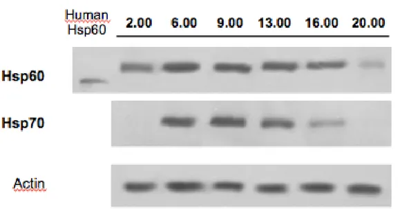
4.4.3 Western Blot and Immunodecoration
4.5 Densitometric and statistical analysis
5. Results
5.1 Daily variation Temperature/Illuminance
5.2 Assessing the absence of Symbiodinium contamination 5.3 Statistical relevance
5.4 Hsps modulation in different corals species 5.4.1 Acropora tenuis
5.4.2 Echinopora lamellosa 5.4.3 Porites lobata
5.5 Modulation of Hsp60 and Hsp70
6. Discussion and Conclusion
1. INTRODUCTION
1.1 Coral reefs: habitat and functions
Coral reefs are tropical coastal ecosystems mainly composed and built by organisms which belong to one of the lowest metazoan phyla, the Cnidaria, class of Anthozoa and order Scleractinia. For these reasons, these organisms are commonly called scleractinian corals or hermatypic corals. They represent the major building blocks of the reefs creating some of largest and diverse ecological communities on the Earth. Coral reefs cover only 0.2% of the ocean surface but they contain almost the 25% of the total marine species (Hoegh-Guldberg, 1999; Roberts, 2003; Hoegh-Guldbergh et al., 2007), supporting an extraordinary biomass and diversity. Until today, about 4000 fish species and 800 coral species which inhabit the reefs worldwide are classified (Groombridge and Jenkins, 2000). Scleractinian corals live at an average depth of 40-50 and in a range of latitude between 30°N and 30°S, which constitute the tropical latitudes. In this region, the oligotrophic and clear water with a low sedimentation rate, a sea temperature between 20 to 30°C during the whole year and a salinity range between 32 and 40 ‰ have an essential role to permit the development of this organism (Veron, 1986).
Scleractinian corals are colonial animals composed by many genetically identical units called polyps which are covered by a hard skeleton produced by themselves and formed by calcium carbonate. In the coral colony the polyps live in symbiosis with a diverse microbiota encompassing virus (Wilson et al. 2005; Davy et al. 2006; Marhaver et al. 2008; Claverie et al. 2009), fungi (Rypien and Baker 2009; Kirkwood et al. 2010; Rivest et al. 2010), protozoa (Croquer et al. 2006; Dong et al. 2009; Sebastian et al. 2009), bacteria (Rosenberg et al. 2007; Smith et al. 2007; Kimes et al. 2010) and archaea (Rohwer and Kelley 2004; Wegley et al. 2004). All the microbial components are recognized as fundamental for the functionality of corals (Knowlton and Rohwer, 2003). However, the most important symbiotic organisms for scleractinian coral are represented by the dinoflagellate photosinthetic algae called zooxanthellae (Symbiodinium spp.). Symbiodinium cells inhabit the coral gastrodermal tissues in a concentration of millions of cells per square centimetre and they use the sunlight to get energy via photosynthesis. The zooxanthellae provide a metabolic benefit to their coral hosts by translocating carbohydrates, amino acids, and lipids to the host (Trench, 1971) representing the majority of daily carbon requirements for coral growth and skeletal deposition
(Gates et al., 1995). Through the photosynthetic activity, more than 90% of the energy required by the corals is satisfied. In exchange, the host provides the algae with inorganic nitrogen, phosphorus and carbon, the products of the metabolism and respiration of corals, as well as an environment characterized by high light exposure for photosynthesis and protection from predation (Venn et al., 2008; Yellowlees et al., 2008). This nutrients exchange also gives to the coral the energy required for an extensive calcification of the skeleton. The
Symbiodinium algae are classified into 7 different phylogenetic clades (Rodriguez-Lanetty,
2003). Each coral colony could hosts one or several individual clades of Symbiodinium and the host-symbiont specificity can be stable to the environmental perturbations (Baker, 2003; Thornhill et al., 2006). Overall, the coral and the closely associated microbial assemblages and zooxanthellae is collectively described with the term coral holobiont (Lesser et al., 2004; Ritchie, 2006; van Oppen et al., 2009).
Coral reefs represent one of the most important ecosystem in the world, both from a biological and ecological point of view and from a social and economic role. In fact, first of all coral reefs protect the natural coastal environment from the wave action. In fact, the coral reefs structure mitigates the violence of the waves and tropical hurricanes, protecting the coast by reducing and slowing the coastal erosion. In this way, the coral reefs give the possibility to other linked ecosystems, as the seagrasses beds and mangrove forests, to survive and develop (Cesar, 2000). Coral reefs support a very high biodiversity comparable only to that of tropical forests also representing a nursery for many juvenile fish species which will constitute the new fish stocks.
Furthermore, coral reefs provide profits and benefits, being one of the first source of income for hundred million of people which live close to this ecosystem. In all the world, about ten thousand people partly or totally depends for their sustenance from the coral reefs and about 15% of world population lives within 100 km from the reef (Pomerance, 1999). Coral reefs are a vital resource particularly for local people, who exploit them, sometimes excessively, to provide food (fishing and mariculture), medicines, feed for livestock, pharmaceuticals and material to build (Obura, 2003). In particular, 20-25% of the fish caught by the countries in the developing world lives on coral reefs and the people of the Pacific get about 90% of their protein requirements from their fishing (Cesar et al., 2003). This ecosystem represents a strong attractive for tourists thus also supporting foreign exchanging (Moberg and Folke, 1999). Only the tourism generates billions of dollars for each country that has these natural ecosystems, absolutely unique in the world for its beauty and spectacular and renowned for high levels of biodiversity and productivity (Cesar, 2000). Finally, coral reefs are also an importance resource for the detection of new anti-tumoral biomolecules used in the pharmaceutical industry (Carte, 1996).
1.2 Stressors and coral bleaching
Despite their importance and persistence through the geological ages (Hoegh-Guldberg, 1999), coral reefs are among the most vulnerable and delicate ecosystems in the world. Since corals are sessile organisms they are steadily subjected to variable environmental conditions typical of the marine habitat (Feder and Hofmann, 1999). Coral reefs are confined between 30° N and 30° S of latitude and they must fit to relatively stable and positive condition and to seasonal cycles, typical of the tropical regions (Grottoli et al., 2006). This stability explains why these organisms and these ecosystems are vulnerable to extreme or sudden environmental changes (McClanahan et al., 2002). Therefore, coral reefs are among the most threatened ecosystems by the global climate change (Hughes et al., 2007).
To date, the coral reefs health worldwide is seriously threatened by different stress factors, both biotic and abiotic, as the abnormally elevated and low ocean temperatures, high solar and UV radiations, severe changes in the salinity values, destructive storms and hurricanes, pollution, predation events (e.g. crown of thorn starfish Acanthaster plancii, Drupella spp., corallivorous fishes) and the imminent sea rise due to the thermal expansion of the seawater and glacial melting (Dustan, 1999; Marshal and Schuttemberg, 2006). Closely related to global warming is also the progressive raising of the CO2 atmospheric concentration which cause the oceans acidification causing the dissolution of all the carbonate structures, such as the corals skeletons (Marubini et al., 2008; Pandolfi et al., 2011). Other stresses and disturbs are represented by the anthropogenic activities such as the overfishing and the destructive fishing (with bombs or poison), the wastes, the increasing of the sedimentation rate induced by an extreme exploitation and development of the coastal zone, damage caused by anchors, invasive species and unsustainable tourism, operated synergistically in the last years causing a severe degradation of the health, resistance and resilience of the coral reefs worldwide (Grimsditch and Salm, 2006).
According to a recent summary, the 20% of the world’s reefs has been irreparably damaged and a further 24% is threatened by anthropogenic pressures such as destructive fishing practices, coastal development, agricultural land-use, increasing nutrient and sedimentation load run-off and eutrophication (Wilkinson, 2004; Wilkinson, 2008). Abiotic and biotic stresses often work together in driving the physiological ecology of the intertidal communities on coral reefs (Lang and Chornesky, 1990; Karlson, 1999).
The ability of a coral reef to support a high productivity depends primarily on the symbiotic relationship between the polyps and the zooxanthellae, which provide to the polyps more than 90% of their energy requirements through the photosynthetic activity. Furthermore, these
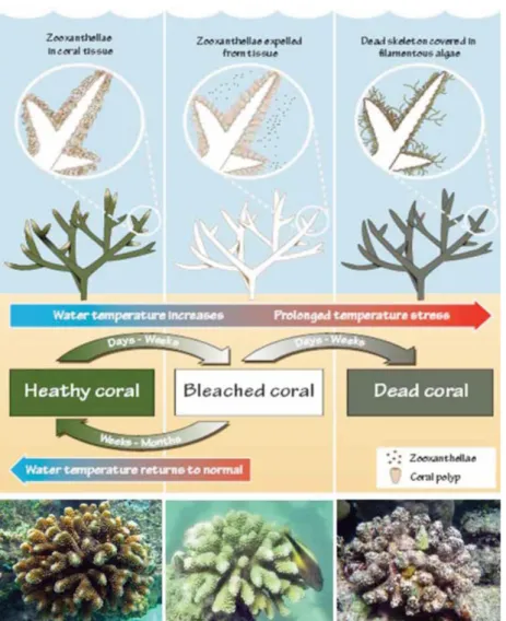
unicellular algae are also the responsible of the bright colouring of corals. Under adverse circumstances and environmental conditions, the equilibrium between the partners of the coral holobiont could be compromised often leading to the phenomenon known as coral bleaching, which is considered one of the main causes of the reefs degradation worldwide (Hughes et al., 2003). Coral bleaching is defined as the process by which the symbiosis between corals and algae (Symbiodinum spp.) is broken with a decrease in the density of
Symbiodinium and/or photosynthetic pigments (chlorophyll a) in the polyps tissues
(Hoegh-Guldberg, 1989; Glynn, 1993; Douglas, 2003). Losing the algae, polyps remain transparent and the coral shows his white calcareous skeleton (Marshall and Schuttenberg, 2004). In according with Glynn (1996) during a coral bleaching event, about 60-90% of the coral’s zooxanthellae are lost and the remaining algae may lost about 50-80% of their photosynthetic pigments. The bleached corals slowly and gradually die since they lack the main source of energy, unless the symbiosis is not restored following the return to normal and favorable environmental conditions. In this case, zooxanthellae can repopulate coral’s tissues and they can survive the bleaching event and recover their normal colour and metabolic activity (Brown, 1997). Thus, in some cases bleaching is a transient altered physiological state; the coral quickly recovers and the health of the individual is relatively un-impacted. Otherwise, if stressful conditions subside soon enough, zooxanthellae can repopulate coral’s tissues and they can survive the bleaching event and recover their normal colour and metabolic activity (Marshall and Schuttenberg 2004).
Coral bleaching may occur on local scale, affecting some reefs or reef portions in the same geographical area, on regional scale, affecting different but close geographical areas, to become events widespread worldwide, the mass coral bleaching events (Glynn, 1993; Hoegh-Guldberg and Salvat, 1995; Brown, 1997). Phenomenon of coral bleaching was observed and described in detail by Vaughan in 1914 as a loss of pigmentation due to low salinity and light exclusion. Yonge and Nicolas in 1931 observed a low cellular concentration of zooxanthellae in corals exposed to high temperature in Lowe Island, Great Coral Reef, Australia. Since 1979 mass coral bleaching events of tens of thousand of km2 were recorded, with an increasing of the frequency and range of the phenomenon (Hoegh-Guldberg, 1999).
Bleaching can be induced by various factors, including heat stress, cold stress, elevated irradiance, increased UV radiation, prolonged absence of light, low salinity, heavy sedimentation, general exposure to pollutants (herbicides, pesticides, heavy metals), starvation, and bacterial infection (Goreau, 1964; Hoegh-Guldberg and Smith, 1989; Lesser et al., 1990; Glynn, 1991; Coles and Fadlallah, 1991; Muscatine et al., 1991; Fang et al., 1995; Brown et al., 2000; Owen et al., 2002; Jones 2005). Buddemeir and Fautin (1993) and Ware et al., (1996), hypothesized that coral bleaching could represent an adaptive strategy, knows as the adaptive bleaching hypothesis. In fact, expelling the zooxanthellae, the corals permit to
other algae more tolerant to the environmental stresses to rebuild the symbiosis, making the coral more resistant to the stressor. According to this hypothesis, the phenomenon of coral bleaching could represent only an evolutionary strategy in order to adapt the organism to the increasing temperatures.
The maintenance of photosynthetic algae within coral cells involves constant cellular communication between the two partners (Weis, 2008), but the cellular mechanisms and molecular pathways involved in the intake and retention of unicellular algae are largely unresolved. Different hypotheses have been suggested regarding the breakdown of the symbiosis. They include, in situ degradation of the symbiont and its photosynthetic pigments (Brown et al., 1995, Fitt and Warner, 1995, Ainsworth and Hoegh-Guldberg, 2008), exocytosis of the symbiont cells (Brown et al., 1995), detachment of the symbiont-containing animal cell (Gates et al., 1992; Brown et al.,1995; Fitt et al., 2001), necrosis and apoptosis of both host and symbiont cells (Dunn et al., 2004, 2007; Lesser and Farrell, 2004), and digestion of the symbiont by the coral host (Brown et al., 1995).
Fig. 1.1 Rising of sea surface temperature may cause brokage of symbiosis with zooxanthellae conferring pale colouring to the colony until coral bleaching. Corals bleached may survive and restore to original condition recovering symbiont algae, if stress conditions are extend in time coral led to die.
1.3 Principali stress factors
Among the abiotic stressor, the temperature, the light irradiation and the salinity are the principal driving forces which are able to influence the structure, the composition and the health status of a coral reef ecosystem since they represent the major causes of the coral bleaching events
1.3.1 Temperature
The majority of the mass coral bleaching events have been correlated with elevated sea surface temperatures (SST), (Hoegh-Guldberg and Jones, 1999). Corals are stenotherm and exist within a relatively narrow temperature range, often within 2-3°C from their upper thermal limit (Podesta and Glynn, 1997; Berkelmans and Willis, 1999), and just an increase of 1°C for some days may trigger an event of mass coral bleaching (Hoegh-Guldberg et al., 1997). Short term SST increase of 3-4°C as well as long term SST increase of 1-2° beyond the normal tolerance threshold could lead to coral bleaching (Jokiel and Coles, 1990). If temperature remains above a threshold for several weeks, the coral is unable to meet nutritional requirements through feeding alone, cannot retrieve or maintain sufficient densities of zooxanthellae, and mortality can occur (Brown, 1997; Hoegh-Guldberg, 1999, 2004). Coral bleaching has become common since 1983, however, the 1997-98 mass coral bleaching event remain the most geographically extensive and severe resulted in 90% mortality to 16% of the worldwide (Hoegh-Guldberg, 2004). Large-scale bleaching events, caused primarily by heat stress have been associated with the El-Niño Southern Oscillation (ENSO) phenomenom (Wilkinson et al., 1999; Mumby et al., 2001; Hoegh-Guldberg, 2004). ENSO is a phenomenon that occur when in mid-low latitude a complex of high pressure depress the wind systems reducing the cloud coverage and the evaporation rate which is responsible of the sea surface temperature cooling both locally and regionally (Coles and Brown, 2003). El-Niño Southern Oscillation has been known since centuries, as periodic inversion of the flow in corresponding of the Perù and Ecuador coasts. El-Niño is generated by the inversion of the sea flow which carries the superficial hot Pacific water from east to west, linked to the disintegration of the Alisei winds in the southern hemisphere that are interrupted or reversed becoming westerly winds. The immediate consequences of this event are the worming of the East Pacific for thousand of km and the pressure reversal between South America and Indonesia (Southern Oscillation), with an increase of the rain in Perù and Ecuador and drought events in the Indonesian Archipelagos (Collins, 2000). Since 1980 all the major mass coral bleaching
events happened in correspondence with the ENSO, such those generated in 1982-83 and in 1997-98 (Obura, 2005).
However, the events of coral bleaching are likely to increase in frequency and scale over time and becoming yearly phenomenon by 2040. In fact with global climate change sea surface are warming and was increased about 0.3°C per decade in the past 30 years and will increase about 1-3°C by 2050 (IPCC, 2007; Hoegh-Guldberg, 1999; Pittock, 1999). Mass coral bleaching were registred also in Maldive with a mortality rate about 80-90% (Berkelmans and Oliver, 1999; McClanahan and Mangi, 2000). Other events occurred in the Pacific Ocean in 2000 and in 2002 (Berkelmans et al., 2004) and in Indian Ocean in 2005 and in 2007 (McClanahan et al., 2007; Montano et al., 2010) and a sever event in the Caribbean in 2005 (Donner et al., 2007).
Since the IPPC’s (Intergovernmental Panel on Climate Change) predictions also show that the terrestrial temperature will raise about 1.4-5.8 °C before 2100, the coral bleaching events may represent in the future the main phenomenon which will define the health condition of the future coral reefs (Grottoli at al., 2006).
1.3.2 Solar radiation
Several studies have also implicated high rates of solar radiations, which include the photosynthetically active radiation (PAR) and the ultraviolet radiation (UVR 290-400 nm), as a potential cause of coral stress (Shick et al., 1996; Lesser, 2000). In fact, together with high SSTs, which is the main cause of coral bleaching, other environmental factors may act synergistically to lower the coral resistance to stressor (Lesser, 2006). Laboratory experiments (Hoegh-Guldber and Smith, 1989; Glynn and D’Croz, 1990) and field studies (Thailandia: Brown, 1997; Papua Nuova Guinea: Davies et al., 1997; French Polinesia: Gleason, 1993; East Pacific: Glynn, 1984; Glynn, 1988) show that high temperatures and high level of solar radiation may enhance the coral bleaching. Bleaching of corals has been attributed to also the high levels of solar radiation as these events usually coincide with periods of calm winds, resulting in increased penetration of solar radiation (Glynn, 1996, Wilkinson, 1998). In fact, the most of coral bleaching events happened in condition of calm wind, calm sea and limpet seawater, promoting the UVR penetration (Glynn, 1993). Furthermore, corals start to bleach from the tips, which are more exposed to solar radiation and corals placed in the shade are less affected by bleaching than those exposed to full solar radiation (Hoegh-Guldberg, 1999).
The combination elevated SSTs and long summer days of intense UV exposure is commonly agreed to be the predominant trigger for mass bleaching episodes (Lesser et al. 1990, Gleason and Wellington 1993, Glynn 1993).
Especially in the photosynthetic organisms such as corals, the elevated solar UV may triggers the production of toxic reactive oxygen species (ROS) which cause cellular damage to the organism, in particular to proteins, DNA and membranes (Lesser et al., 1990; Lesser, 1997) also inducing the photoinhibition of the photosynthesis process (Lesser, 1996).
Recently has been observed that the synergic effect of high temperature and high level of UVR may set off in polyps and in symbiont algae oxidative stress, producing and storing of
reactive oxygen speacies (ROS), defeating protection mechanism mainly dial by pigment
such as carotenoids (Glynn, 1996; Brown, 1997; Jones et al., 1998) and reducing the
photosynthetic active radiation (PAR). ROS may cause serious damage to cellular membrane,
to the proteins, to DNA, loss of function of cellular organelles, mutations, enzymatic inactivation, reduction of metabolic efficiency and photosynthetic capacity of zooxanthellae. In despite of this the absence of coral bleaching during period of high UVR and high temperature show that the UVR is not a fundamental condition but play only a secondary role in coral bleaching, depressing only the density of pigmentation but not the dissolution of symbiosis (Lesser, 1996; Hoegh-Guldberg, 1989).
In particular several solar radiation were suspected to be involved in that phenomenon for several reason: first of all, several bleaching are preceded by relative calm and clear water, with high penetration of UVR (e.g. Great Barrier Reef 1982-83, Harriott, 1985; French Polynesia 1993, Drollett et al., 1994), secondly because corals star to bleach first from their surface portion and exposed by solar radiation, finally several studies have shown by manipulation of UVR and PAR may led corals to bleach (Gleason and Welllington, 1993). This phenomenon is generally linked to photoinhibition (Walker, 1992). High light intensity may promote the forming of potentially damage product, such as free radicals, which are detossificated by specific enzymes. Principal damage that free radicals may cause is at level of photosystem II (PSII). Zooxanthellae photoinhibition may be a sever risk, hermatipic corals and their symbionts appear having several defensive mechanisms called as quenching, such as changing in xanthophyll pigments, to develop high radiance impact (Brown et al., 1999) or producing mycrosporin-like amino acids (MAAs). Therefore, high light intensity may cause a lowering of growth rate in zooxanthellae and reduction in concentration of pigments such as chlorophyll a, however only photoinhibition can’t explain mass coral bleaching.
1.3.3 Salinity
Although coral reefs commonly inhabit areas where salinity is generally permanent and stable over long timescales (Coles and Jokiel, 1992), they can experience extreme changes in salinity levels of varying duration. Heavy rainy seasons which are common in tropical regions, hurricanes, storms and coastal freshwater runoff can significantly reduce surface and depth
salinity in the short term (min to h) or over longer periods (days to weeks), (Van Woesik et al., 1995; Moberg et al., 1997; Devlin et al., 1998; Berkelmans and Oliver, 1999; Porter et al., 1999). At the other extreme, a high rate of evaporation in tide pools during low tides and prolonged drought may cause salinities to rise to stressful levels (Wells, 1957; Lirman et al., 2008), and also lead in this case to the physiological damage of reef organisms.
Osmotic stress, which occurs when the cell experiences volume and osmolyte fluctuations which compromise macromolecular structures and metabolic function (Mayfield and Gates, 2007), has long been recognized as a limiting physical parameter for marine organisms. Corals are generally considered stenohaline, with a limited ability to adapt to or survive salinity changes (Wells, 1957; Coles and Jokiel, 1992), even if some species are euryhaline and can withstand significant changes in external osmolarity (Muthiga and Szmant, 1987; Coles, 1992; Manzello and Lirman, 2003; Mayfield and Gates, 2007). In particular, reef-building corals, like the majority of marine invertebrates, are osmoconformers. Since they lack the capability of osmoregulation, they do not possess a constant cellular osmolarity but respond to dynamic changes in their environment rapidly by absorbing water to become iso-osmotic with their surroundings (Rankin and Davenport, 1981; Mayfield and Gates, 2007). Sudden deviations in ambient salinity affect many physiological processes in marine organisms, such as intracellular transport, feeding rates, excretion, osmoregulatory capacity (Normant and Lamprecht, 2006), and negatively influence the basal metabolic functions of the corals inducing changes in respiratory pathways and symbiont photosynthetic efficiency (Alutoin et al., 2001; Kerswell and Jones, 2003). In this context, the majority of studies have analyzed the effects of reduced salinity on coral metabolism (Nystrom et al., 1997; Tytlianov et al., 2000; Downs et al., 2009). For example, following hyposaline treatments, a significant drop in net photosynthesis of Porites furcata has been reported (Manzello and Lirman, 2003). Decreased rates of gross photosynthesis due to cellular damage to algae have also been observed in Siderastrea siderea, Porites lutea and Pocillopora damicornis (Muthiga and Szmant, 1987; Moberg et al., 1997). In the long term, the salinity stress condition can imply decreased growth potential (Coles, 1992) and also give rise to higher-order physiological diseases, such as gamete abnormalities and reduced viabilities, that affect fecundity, settlement success and larval survivorship (Richmond, 1993; True, 2012) or even lead to death (Coles and Jokiel, 1992; Hoegh-Guldberg and Smith, 1989).
1.4 Molecular and cellular response to stress in corals
Marine environment vary in biotic and abiotic conditions and in the amplitude and frequency of fluctuations in these conditions (Brown, 1997). Due to the physical characteristics of
seawater, the aquatic environment can be extremely stressful to its inhabitants (Feder & Hofmann, 1999). In particular, as sessile organisms, scleractinian corals inhabit several niches in the intertidal zone and undergo constant direct exposure to the surrounding environmental conditions. Extreme or unexpected short or long-term environmental fluctuations could be very stressful for marine organisms, causing cell damage. Organisms in nature have developed several mechanisms to withstand environmental stresses, such as behavioral adaptations, morphological changes, physiological regulations and biochemical and cellular specializations (Feder and Hofmann, 1999). Nevertheless, corals lack any developed physiological regulatory system and for this reason they are expected to possess well-developed cellular adaptation abilities (Brown, 1997).
Furtermore, the coral susceptibility to stress and bleaching appears to be highly variable since different corals display different levels of physiological resistance to environmental stress (Obura, 2001; McClanahan et al., 2007; Montano et al., 2010). Bleaching response is highly species/genus specific but also different colonies of the same species or same coral species living in different geographic areas could show a different tolerance to the environmental disturbances (Marshall and Baird, 2000; McClanahan et al., 2007; Montano et al., 2010). The diversity among species in their susceptibility to disturbances represents a critical aspect of community dynamics, since it can give rise to changes in the community structure and species composition which determine the long-term persistence of coral reefs (Hughes and Connell, 1999). Attempts to understand the differences in the response of reef-building corals to thermal stresses have principally focused on coral morphology, growth and metabolic rates, tissue thickness and host CO2 supply strategies (Gates and Edmunds, 1999; Marshall and Baird, 2000; Loya et al., 2001; Darling et al., 2012; Wooldridge, 2014). Generally, in literature, variation among coral genera has been associated with coral growth and metabolic rates (Gates and Edmunds, 1999). Corals with branching morphology, high growth rates, low tissue thickness, good connection between polyps and low metabolic rates (branching species) would have a lower capacity to acclimatize than corals with massive or sub-massive growing, with a low growing rate, high tissue thickness and high metabolic rate (massive species), (Marshall and Baird, 2000; Loya et al., 2001; McClanahan et al., 2004; Darling et al., 2012). Moreover, the taxa most susceptible to bleaching tend to be those corals that are quick to colonize free space and often short-lived and those corals that have low tissue retractibility and thickness and so high degree of tissue exposure to water column stressor (Hueerkamp et al., 2001, Loya et al., 2001). In particular among the scleractinian corals, acroporids and pocilloporids appear to be taxa most sensitive to bleaching stresses, since these faster growing species typically suffer high mortality during bleaching events (Glynn, 1990; Gleason, 1993; Marshall & Baird, 2000; Edwards et al., 2001; Loya et al., 2001; McClanahan, 2004).
However, the molecular mechanisms which determine the different tolerance and susceptibility in corals to different environmental stressors are not still completely elucidated. The coral stress response involves a wide array of cellular and physiological processes, including heterotrophic plasticity, the production of protective pigments, mycosporine-like amino acids and the expression of fluorescent proteins and antioxidant enzymes such as superoxide dismutases, Catalase, Glutatione peroxidases and Glutatione transferases (Lesser 1997; Downs et al 2000; Richier et al. 2005, 2008; Grottoli et al., 2006; Weis, 2008; Baird et al., 2009; Linan-Cabello 2010). One mechanisms by which coral jointly with the host might reduce bleaching damage is the producing of flourecent pigmants (FPs). FPs produced by host appear to belonging from a single family of proteins, closely related from green fluorescent proteins (Martz et al., 1999). FPs reduce photoinhibition and severity of bleaching by assorbing, scattering and concluding dissipating high energy solar radiation, such as UV, via fluorescence (Salih et al., 2000, 2006). FPs have a stringent role in reducing and preventing bleaching damage, furthermore, its not know whether FPs can provides relief from heat stress alone (Baird et al., 2008). In addition to FPs corals have mycrosporine-like amino acids (MAAs) strategy to protect themselves to high energy of UV. In fact MAAs absorb and dissipate UV energy as heat, without forming toxic intermediates (Shick and Dunlap, 2002). MAAs are synthesized via the shikimic acid pathway, is presumed that symbionts being the source of MAAs in corals, because animals lack this pathway. Reactive oxygen species (ROS), play and essential role in temperature- and in radiation-induced bleaching damage (Weis, 2008). High temperature and irradiace may cause photoinhibition and damage to chloroplast and photosyntetic apparatus in three inter-related ways that act in concert to start bleaching cascade. The D1 protein is part of the water-splitting complex in photosystem II in thylakoid membranes, in consequence of elevated temperature, D1 becomes damage and outpaces the normal repair mechanisms (Warner et al., 1999). Heat and like could compromise also dark reaction of photosynthesis (Jones et al., 1998), damaging Rubisco (Lesser, 1996). This results in reduced consumption of ATP and NADH coming from the light reaction that in turn leads to the dysfunction of photosystem II via backup of excitation energy (Jones et al., 1998; Venn et al., 2008). Tchernov et al. (2004) have shown that heat and high light damage directly the thylakoid membraes, having the results that photosynthetic apparatus continue to generate electrons but not ATP and NADPH caused by and uncoulped transport of electrons. The build up of electrons under those mechanisms causes the generation of ROS. Excess electrons react with O2 forming superoxide (O2-) (Tchernov et al., 2004) this reactive specie of oxygen can be reduced by SOD into H2O2 (Jones et al., 1998, Lesser, 2006). H2O2 can react with Fe2+ forming the most reactive ROS .OH. In addition excess of electrons can react photochemically with pigments forming 1O2 (Lesser, 2006). Furthermore, ROS begin to diffuse into the host tissue where the damage continues ad
ultimately leads to bleaching. Into bleaching cascade, caused by ROS, nitric oxide (NO) plays a pivotal role (Weis, 2008). In according with Fang (2004), NO acts as both a cytotoxic and a signaling molecule in animals including host-pathogen interactions. It can react with O2 -forming the potent and high diffusible oxidant peroxynitrite, ONOO- (Pacher et al., 2007). In according with Bouchard and Yamasaki (2008) and Trapido-Rosenthal et al. (2001), there is the evidence that NO is a direct signaling molecule between the partners of symbiosis that could initiate bleaching cascade. Superoxide dismutases catalyze the reaction of superoxide ions and two protons to form hydrogen peroxide and O2 (Fridovich, 1995). Manganese and Glutathione play an essential role in antioxidant detoxification pattern (Downs et al., 2001; Klaassen et al., 1999). During oxidative stress reduce glutathione (GSH), act in different antioxidant pattern. GSH may form GSSG via glutathione peroxidase reacting with hydrogen peroxide, or reacting, in a major antioxidant cyclic patheay, with ascorbate as an essential component of the Asada-Halliwell pathway, or moreover GSH may act as an hydroxyl and superoxide quencher (Asada and Chen, 1988; Halliwell, 1999). Fridovich (1995) shows that ManganeseSOD is located in mitochondria in eukaryotic cells and it’s up-regulated under oxidative stress to reduce the stress. Another strategy that corals adopt to resist and recover to bleaching events is the heterotrophic plasticity. Grottoli et al. (2006) show that corals able to switch acquiring fixed carbon heterotrophically in bleaching events are ecologically advantage for long-term survival.
There is also evidence that corals have an enhanced tolerance to environmental stress linked to cellular protective mechanisms such as the induction of the Heat shock proteins (Hsps), (Brown et al., 2002; Rosic et al., 2011; Chow et al., 2012; Seveso et al 2013, 2014).
1.5 Le Heat shock proteins (Hsps)
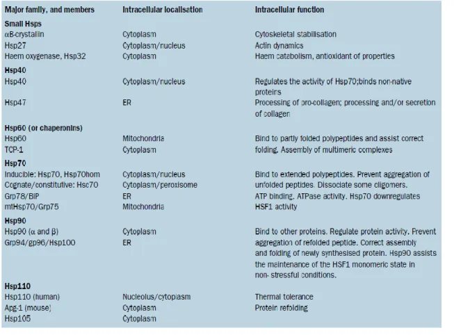
One mechanism of reaction to deleterious environmental conditions is the rapid increase of the induction of a set of stress proteins called Heat shock proteins (Hsps), (Lindquist 1986). This is one of the most important defence mechanism, conserved throughout evolution as the Hsps are ubiquitous, occurring in all organisms from bacteria to humans (Fink 1999, Kregel 2002). Heat shock proteins are categorized into several families that are named on the basis of their approximate molecular mass and specific functions: small Hsps, 40-kDa Hsp, 60-kDa Hsp, 70-kDa Hsp, 90-kDa Hsp and 110-kDa Hsp (Tab. 1.1).
Tab. 1.1 - Heat shock protein families and their intracellular location and function. ER: endoplasmic reticulum. TCP-1: tailless complex polypeptide. Grp: glucose regulated protein. Hsp70 hom: testis-specific Hsp70. BiP: immunoglobulin heavy chain binding protein. Mt: mitochondrial. Apg-1: protein kinase essential for autophagy. From Pockley (2003)
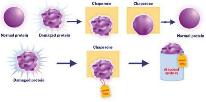
There is substantial evidence that Hsps play important physiological roles both in normal conditions and also in situations involving systemic and cellular stress (Kregel, 2002). Under normal cellular physiological conditions the Hsps mainly function as molecular chaperones. Chaperones are involved in a multitude of proteome-maintenance functions that regulate protein homeostasis in directing the folding and assembly of other proteins (Parsell and Lindquist, 1993; Sanders, 1993; Fink, 1999). In particular, they participate broadly in de novo folding of proteins, refolding of stress-denatured proteins, prevention of oligomeric assembly, protein trafficking and assistance in proteolytic degradation (Fig. 1.2). They also are involved in the intracellular protein transport and in the degradation of damaged proteins (Hightower, 1991; Gething and Sambrook, 1992; Vabulas et al., 2010; Hartl et al., 2011). The chaperones are multicomponent molecular machines that promote folding through ATP- and cofactor-regulated binding and release cycles (Hartl et al., 2011). In fact, Hsps typically function as oligomers, as well as complexes of several different chaperones, co-chaperones, and/or nucleotide exchange factors (Feder and Hofmann 1999).
Fig. 1.2 Role of a molecular chaperone in renaturing of a protein or in its degradation
An induction and an up-regulation of the expression of Hsps constitutes an emergency response and confers tolerance to harsh conditions (Parsell & Lindquist, 1993). Cells respond to stress by increasing either the amount or the activity of a transcription factor that is specific for the heat shock genes. The result is increased transcription of the heat shock genes, which leads to an increase in the concentration of Hsps in the cell (Craig and Gross, 1991). High levels of specific Hsps are maintained throughout the exposure to stress. However, the magnitude of the response and its duration depend on the severity of the stress and the sensitivity of the organism (Lindquist, 1986, Feder, 1999).
Hsps act to protect the organisms from cell protein damage after exposition to stressor such as elevated temperature (Linquist, 1986; Feder and Hofmann, 1999; Krebs and Bettencourt, 1999), cold shock (Nunamaker et al., 1996; Ali et al., 2003), increased UVR (Sanders, 1993; Trautinger, 2001), extreme pH (Kozoil et al., 1996; Tetth and Beuchat, 2003), osmotic stress (Burg et al., 1996; Beck et al., 2000; Spees et al., 2002), heavy metals (Köhler et al, 1992; Sanders, 1993; Vedel and Depledge, 1995; Hall, 2002), crude oil (Sanders, 1993; Wolfe et al., 1999), and other pollution (Linquist, 1986; Blom et al., 1992; Pomerai, 1996; Wiens et al., 1998).
In literature many works also focused on the coral Hsps expression (Fang et al., 1997; Sharp et al., 1997; Branton et al., 1999; Robbart et al., 2004; Rossi and Snyder 2001, Synder and Rossi 2004).
1.5.1 The mitochondrial 60-kDa Heat shock protein (Hsp60)
The 60-kDa heat shock proteins (Hsp60) belongs to the group of the chaperonins and in particular it is a mitochondrial chaperonin. Chaperonins are ring-shaped chaperones that encapsulate nonnative proteins in an ATP-dependent manner (Ritcher et al., 2010). Chaperonins are large double-ring complexes of approximately 800 kDa enclosing a central cavity. They occur in two subgroups that are similar in architecture but distantly related in sequence (Vabulas et al., 2010). Group I chaperonins (also called Hsp60s) occur in bacteria (GroEL), mitochondria, and chloroplasts. The function of type I chaperonins is performed by the cooperation of two proteins, Hsp60 and Hsp10, that function with the help of hydrolyzed ATP, as folding chamber and co-chaperone, respectively (Cheng et al. 1989, Bukau and Horwich 1998). Group II chaperonins exist in archaea (thermosome) and in the eukaryotic cytosol (TRiC/CCT).
Through the extensive study of GroEL, Hsp60’s bacterial homolog, Hsp60 has been deemed essential in mitochondrial biogenesis, in the synthesis and transportation of essential mitochondrial proteins from the cell’s cytoplasm into the mitochondrial matrix, playing a central role in the folding of newly imported and stress-denatured proteins in the mitochondria (Martinus et al., 1995; Hood et al., 2003). Hsp60 possesses two main responsibilities with respect to mitochondrial protein transport. It functions to catalyze the folding of proteins destined for the matrix and maintains protein in an unfolded state for transport across the inner membrane of the mitochondria (Koll et al., 1992). The hydrophobic portion HSP60 is responsible for maintaining the unfolded conformation of the protein for trans-membrane transport (Koll et al., 1992).
Cnidarian heat shock protein 60 is the major mitochondrial chaperonin and functions to mature nuclear-encoded, mitochondrial-imported proteins into their active state (Ellis, 1996). Elevation of this protein signifies that there has been a general shift in the protein chaperoning and degradation within the mitochondria and implies a change in the equilibria of mitochondrial-associated metabolic pathways (Papp et al. 2003).
Hsp60 has also anti-apoptotic ability and can protect against cell death by maintaining mitochondrial oxidative phosphorylation. In addition to its critical role in protein folding, Hsp60 is involved in the replication and transmission of mitochondrial DNA (Arya et al. 2007).
Hsp60 are commonly used as biomarkers in coral health assessment and the expression of Hsps after abiotic stresses such as extreme temperatures (Brown et al., 2002; Kingsley et al., 2003; Chow et al., 2009, 2012; Seveso et al., 2014), elevated light intensity (Downs et al., 2000; Chow et al., 2009, 2012), xenobiotics (Downs et al., 2005), change in salinity (Downs et al., 2009; Seveso et al., 2013) and also after biotic stresses (Seveso et al., 2012) has been reported in literature.
1.5.2 The cytoplasmic 70-kDa Heat shock protein (Hsp70)
Members of the 70-kDa heat shock protein family (HSP70) play an essential role in protein folding, transport of proteins into different cellular compartments, regulation of apoptosis and regulation of the heat shock response (Lindquist, 1986; Sanders, 1993). In eukaryotes, at least three HSP70s are uniquely located in the mitochondrial matrix (Azem et al., 1997). All mitochondrial HSP70 (mt-HSP70) are nuclear encoded, synthesized in cytosol and then imported into the mitochondria. The so-called mt-HSP70 (product of Ssc1 gene in yeast) is the only mt-HSP70 essential for the growth of yeast. It plays a central role in the transport and folding of mitochondrial proteins.
Heat shock protein 70 has in important role in assembling of newly synthesized proteins, refolding of missfolded and aggregated proteins and control of the activities of regulatory proteins (Bukau et al., 2000; Moro et al., 2002). Hsp70 has also a important role in proteins quality control and turnover during both normal and stress conditions (Evans et al., 2010). Hsp70 in expressed in different forms, a conservative form (Hsc70), which maintain normal cell function and stress-inducible form (Hsp70/72), which mainly prevents protein damage or protein aggregation under stress conditions (Mayer and Bukau, 2005). Constitutive form of hsp70 (hsc70) is also well documented. Vaughan (1907), with on field corals trasplantation, demostrate that elevated levels of hsc and/or hsp70 following increased thermal shock (Sharp et al., 1997). Differentiation between these two forms is of interest, as the increased synthesis of the constitutive hsc70 may act to stabilize proteins prior to the induction of an inducible isoform such as hsp70 during the initial stages of a stress event (Thompson and Scordilis, 2001; Thompson et al., 2001; Robbart et al., 2004). Heat shock protein 70 has an ATPase dominion, which is responsible for chaperon and folding activity. Hydrolysis of ATP to ADP is accelerated by Heat shock protein 40 (Hsp40), which also interact directly with unfolded proteins and recruit Hsp70 to protein substrates (Kampinga and Craig, 2010). The regulation of heat shock factor 1 (hsf1) activity, is complex and controlled (in part) by the fundamental properties of the molecule, specifically by thermally induced changes to the molecule's conformation. Craig and Gross (1991) demonstrate that have suggested that heat shock factor1 (hsf1) may detect cellular stress, priory thermal stress, being a sort of cellular thermometer. Several studies have demonstrated that the temperature at which hsf1 is activated is variable, and implies
that extrinsic factors may influence the regulation of hsf1 activity. These factors include hsp40, 70 and 90, which directly interact with hsf1 in a multi-protein complex (Hochachka and Somero, 2002; Tomanek, 2002; Tomanek and Somero 2002).
The relationship between environmental tolerance of organisms and the expression of cytosolic HSP70 has been studied in diverse aquatic and terrestrial organisms (Sanders, 1993; Krebs & Bettencourt, 1999).
In several studies Hsp70 was used as biomarker to investigate heat shock response (HSR) among intertidal organisms in particular Gastropos living in rocky shore (Tomanek and Sanford, 2003; Halpin et al., 2002). Hsp70 was used to investigate the Patterns of Variation from Microscales to Mesoscales, the toxicity of contaminants of concern (Mukhopadhyay et al., 2003 ), how Water flow may influences coral replace (Carpenter et al., 2010), seasonal acclimatization (Dietz and Somero, 1992; Hofmann and Somero, 1995; Roberts et al., 1997; Chapple et al., 1998; Buckley et al., 2001), laboratory acclimation (Hofmann and Somero, 1996a; Roberts et al., 1997; Tomanek and Somero, 1999, 2000, 2002), competition for space (Rossi and Snyder, 2001), food availability and wave exposure (Dahlhoff et al., 2001), and microhabitat (Helmuth and Hofmann, 2001).
2. AIM OF THE STUDY
Coral reefs worldwide are among the ecosystem most vulnerable to the global changes since to date their health is seriously threatened by different stress factors, both biotic and abiotic, including the human disturbances (Dustan, 1999; Hughes et al., 2003; Marshall and Schuttemberg, 2006). Corals inhabit intertidal zone, which is subjected both to cyclical, such as tidals, and sudden and unexpected environmental chances, such as storm and extreme rains. Scleractinian corals are sessile organisms and for this reason they are directly and steadily subjected to typical environmental changes of their habitat (Feder and Hofmann,1999). Since corals are not able to dispel threats, they carry out specific defensive mechanisms (Brown, 1997). Extreme changing in environmental factors or pollution may led coral bleaching. Stress factors which may induce coral bleaching, such as variation in SST, UV radiation, led corals to protect themselves with singular defence mechanisms. In the last years, several studies have investigate the processes involved in the induction of the coral bleaching phenomenons, in order to determinate the difference in sensibility, resilience and resistance of corals (Nyström and Folke, 2001; Obura, 2001; Grottoli et al., 2006; McClanahan et al., 2007; Montano et al., 2010). Mainly mechanisms defence of corals to stressors are expression of heat shock proteins, heterotrophic plasticity, production of protective pigments, mycrosporine-like amino acids and expression of fluorescent proteins and antioxidant enzymes (Lesser, 1997; Downs et al., 2000, 2001; Snyder and Rossi, 2004; Richier et al., 2005, 2008; Grottoli et al., 2006; Weis, 2008; Baird et al., 2009; Linan-Cabello 2010). Difference in bleaching susceptibility may also influence structure community in long-term (Hughes and Connel, 1999).
However, until now these studies have shown that coral resistance and resilience to stress factor is generally species/genera specific (Marshall & Baird, 2000, McClanahan et al., 2007, Montano et al., 2007) and that the different sensibility and susceptibility to coral bleaching were attributed mainly to morphological difference among the different coral genera, rather than those physiological and molecular (Edmundus, 1994, Marshall and Baird, 2000, Loya et al., 2001, van Woesik et al., 2011). In the last years is underlined the importance of corals physiological replace to the stressors and it’s shown the evidence that also cellular mechanisms are species/genera specific (Seveso et al., 2014; Chow et al., 2009; Ferrier-Pagès, 2007). On the other hand, corals possess a molecular stress response which involved a wide array of cellular and physiological process that act in order to protect the organism from cellular degenerations and damages and from the breakage of the symbiosis with the zooxanthellae (Richier et al., 2005, Grottoli et al., 2006, Weis, 2008, Baird et al., 2009).
During the last years many works showed as, also in corals, an important mechanism of defence and protection against harmful environmental conditions was represented by the expression of a set of stress proteins called Heat Shock Proteins (Hsps) (Brown et al., 2002, Rosic et al., 2011, Chow et al., 2012). In fact, in response to environmental stresses, the coral Hsps were up-regulated in order to increase cellular repair and tolerance to preserve the cellular metabolic functions. (Robbart et al., 2004; Mayer, 2010; Eghtesadi-Araghi and Bastami, 2010; Chow et al., 2012). For this reason, Hsps are commonly used as biomarkers to analyse the different susceptibility of reef-building corals to many factor stress (Chow et al., 2009; Fitt et al., 2009). Consequently, the role of Hsps as stress markers is widely recognized (Wahid et al., 2007), although considerable variability in the Hsps endogenous level, kinetic of expression and threshold of stress response depend on species, Hsp family and stressor type (Iwama et al., 2004; Clark and Peck, 2009). However, variation of Hsps under varying of natural conditions have been investigated in very few studies (Feder and Hofmann, 1999) and even in fewer studies have been investigated Hsps variation in short-term under varying natural conditions (Hofmann and Somero, 1995, Nakano and Iwama, 2002; Tomanek and Sanford, 2003). To correct understanding of variation of Hsps expression levels under natural condition is needed to interpret correctly on field variation of Hsps levels. To date, ecological role of Hsps replace in stress events tested in laboratory is not yet tested on field. In particular, three different coral species were chosen and investigated: the branching coral
Acropora tenuis, the massive coral Porites lobata and the foliose coral Echinopora lamellosa.
These species were investigated in their original environmental represented by the coral patches located in the lagoon of Maghodhoo Island, in the South-east part of the Faafu Atoll, Republic of Maldives.
In this study the expression and the modulation of both the cytoplasmic 70-kDa Hsp (Hsp70) and the mithocondrial 60-kDa Hsp (Hsp60) was analyzed in different corals taxa directly in their natural environmental conditions. In particular, three different coral species were chosen and investigated: the branching coral Acropora tenuis, the massive coral Porites lobata and the foliose coral Echinopora lamellosa. These species were investigated in their original environmental represented by the coral patches located in the lagoon of Maghodhoo Island, in the South-east part of the Faafu Atoll, Republic of Maldives. These coral species were selected as they are among the most abundant and widespread scleractinian species in the study area and in particular within the selected sampling site. Furthermore, they were especially chosen as characterized by three different growth morphologies, tissue thickness and growth rates which are involved in determining a different sensitivity, tolerance and resistance to the environmental stresses (Marshall and Baird, 2000; Loya et al., 2001; van Woesik et al., 2011; Chow et al., 2009; Maydlarz et al., 2009; Seveso et al., 2014; Levy et al., 2006).
The present study aimed to examine the in situ modulation of Hsps across diel cycles in A.
tenuis, P. lobata and E. lamellosa. In particular, in order to understand how the expression of
both the Hsp60 and Hsp70 during the normal nictimeral cycle is modulated in these coral, which apparently showed healthy tissues conditions, the levels of such proteins were analyzed during six different sampling time within the 24 h. Thus, the expression of the Hsps in relation to the daily variations of the two main environmental factors which affect the diel cycle, the sea surface temperature and the light intensity, was deepen explored. Several study shows that there is a different susceptibility and resistance to stress factor in correlation with different growth morphology, goal of this study is to understand if to different growth morphology matches also difference in physiological replace shown by different Hsps modulation. Furthermore two types of Hsps is subjected to this study, Hsp60 and Hsp70. Monitoring Hsp60 and Hsp70, which are located in different cellular compartments, give the possibility to understand if there is some difference in replace in depending with cellular location. It’s known that Hsps are expressed at low level also under normal condition to regulate protein folding (Downs et al., 2000). Moreover this study investigates if Hsps may have key role in coral replace to diel variation under normal condition on field.
3. FRAMEWORK OF THE STUDY AREA
3.1 Indo-Pacific status
Indo-Pacific coral reefs are the worldwide areas with higher biodiversity, with about 581 species of scleractinian corals represent 91% of the world’s reef (Veron, 2000). Instead of this, Indo-Pacific area is subjected to a fewer study than in Caribbean area. Caribbean sea presents only 62 coral species (Veron, 2000), but it’s considered an hot-spot for coral disease,
regarding the high number of syndromes described, for the frequency that they are discovered, for increasing of associated epizootic events and for rapid spread of disease to new sites and new hosts. High biodiversity of Indo-Pacific area may have in important role in slowing the propagation rate disease in consequence of that diseases are linked to a specific host. The few studies led in this area, have shown the presence of diseases in Philippines (Raymundo et al., 2005), Guam (Myers and Raymundo, 2009), Great Barrier Reef (Willis et al., 2004), Hawaii (Aeby, 2005), East Africa (McClanahan, 2004), Indonesia (Haapkylä et al. 2007), Eilat Gulf (Barash et al., 2005), Japan (Wiel, 2012) and Maldive (Montano et al., 2012) with emphasis on how corals diseases have become a real threat thus contributing to increase Indo-Pacific reefs to decline.
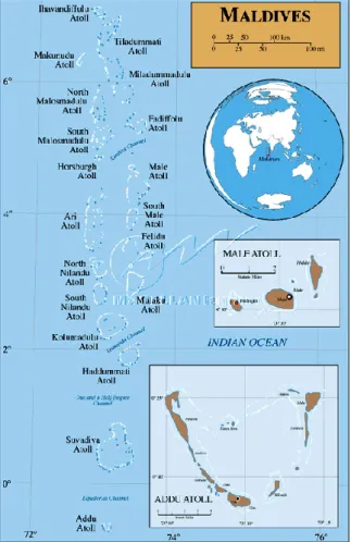
3.2 Republic of Maldives
Republic of Maldives (latitude from 7°06’ N to 00°45’ S e longitude from 72°13’ E to 73°45’ E) is composed by atolls, located under Indian sub-continent and Laccadive Islands far from Sri Lanka about 700 km. Maldives are composed by 26 atolls and about other 1190 corallins structure (Zahir, 2000), with deep lagoon from 40 to 60 m and islands with square from 0.5 to 6 km2, with total area of 90000 km2. Maldivian reefs represent 5% of the worldwide reefs, with a total square of 8920 km2 (Spalding et al., 2001). Maldivian lagoons are called as “Faros”, in which is possible to find little reefs, o patch reefs. In lagoon zone, in presence of shallow water are frequently found sea-grasses which are a refuge area fore juveniles of many organisms (Shakeel and Hudha, 1997). External reefs which delimitate lagoons, have an high slope, dominated in upper zone by corals in symbiosis with zooxanthellae and in deeper by branched etherotrophic corals without zooxanthellae. Maldivian coral reef are explored and studied for first time in 1958 during “Xarifa Expedition” (Wallace and Zahir, 2007). Sheppard
(1987) identified 166 corals species but newest studied have discovered in totally 248 species owning at 57 different genera (Pichon and Benzoni, 2007).
Maldivian climate is typical tropical climate, with temperature around 28°C, with maximum of 32°C and minimum of 25°C. the climate is characterized by two monsonic periods, from April to November and from December to March. Coral reefs are ecosystems which suffer consequently to environmental variation and fluctuation (McClanahan et al., 2002). In 1998 was verified, after El Niño phenomenon, a sever coral bleaching which threat Maldivian reefs with a mortality rate about 100% with different replace in correlation with different species and different location of archipelagh (Bianchi et al., 2003). Living coral after this mass coral bleaching event was above from 2 to 8%, however, Lasagna (2008) described that in the following 8 years corals replace increase living rate corals above from 12 to 37%. Critical event of 1998 changed benthic community causing an ecological shifting from reefs dominated by Acropora to reefs dominated by Porites (Goreau et al., 2000).
In consequence of their position in Indo-Pacific ocean, Maldives, has serious problems to commercial exchanging, basing all their economy on fishing and turism (Naseer, 1997). Increasing of population caused trought the years a gradual deplacment of natural resource and habitat degradation (Zhair, 2010). In consequence to improving of turism is needed to defence costal zone with seawalls and building of harbour in replace to an increasing needed of services (Shareef, 2010).
3.3 Faafu atoll and Maghoodoo island
Faafu atoll, originally called as Northern Nilandhé Atoll, measure 30 km in length and 27km in large, is composed by 23 islands, 5 of those are inhabit with total population above 2000 people.
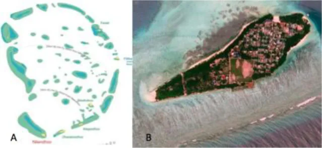
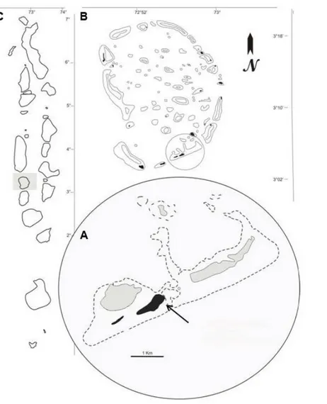
Maghoodoo (3°04′42.92″ N 72°57′52.31″ E) is an island of Faafu atoll (Fig. 3.2), measure 970 m in length and 350 in large. Maghoodoo is located in south of Faafu atoll, is far from Male above 134 km and is free by touristic resort. The principal sources of incoming are fishing, quite agriculture and from resort of the atoll. In Maghoodoo, in 2011 was founded Marine Research and High Education Center (MaRHE Center) of University of Milano-Bicocca. MaRHE Center represent an important base for research in Maldives, which promote research didactive activities in environmental science, marine biology with the goals to understand, teach and divulgate how to protect this such as delicate ecosystem.
4. MATERIALS AND METHODS
4.1 Sampling design: study area and selection of the coral colonies
On March 2014, after previous extensive surveys, a defined sampling area of approximately 150 m2, located about 70 m from the shore and composed of several coral patches was chosen in the lagoon of Magoodhoo Island (3°04’42”N; 72°57’50”E), in the south east part of Faafu Atoll, Republic of Maldives (Fig. 4.1).
The sampling site was selected for both logistic and organizational reasons that ecological and biological. First, it represented an easy and short time access area suitable for rapid sampling activities such as those involving the coral proteins analysis. In fact, in order to preserve the coral proteins we had the necessity that the sampling area is was placed as close as possible to the MaRHE Center laboratories for fast freezing of the coral fragments. For the same reason, we selected an area extended in the same reef flat zone characterized by a stable and constant shallow depth (around 2-3 meters maximum depending on tide ranges) where sampling by snorkeling instead of scuba diving, allowed a quick exit from the sea, also minimizing the risks for the operators. Furthermore, we chose an area situated in the same reef flat zone in order to be subjected to the same environmental parameters along its extension. In fact, just because the study was conducted in the natural condition, we must minimize as much as possible all the environmental variables that can affect the expression of the proteins in order to be sure that the modulation of such proteins was affected exclusively by the diel variations in the temperature and light only and not by other parameters. Thus, the whole sampling area was characterized also by the same typology of substrate and it was subjected to the same tidal range. Finally, the sampling zone selected for the study would be also characterized by coral patches which showed a high level of biodiversity and especially a healthy condition. The health status of these coral patches was judged good in according with the absence of coral bleaching events, physical damages of the colonies, algal overgrowth and coral diseases.
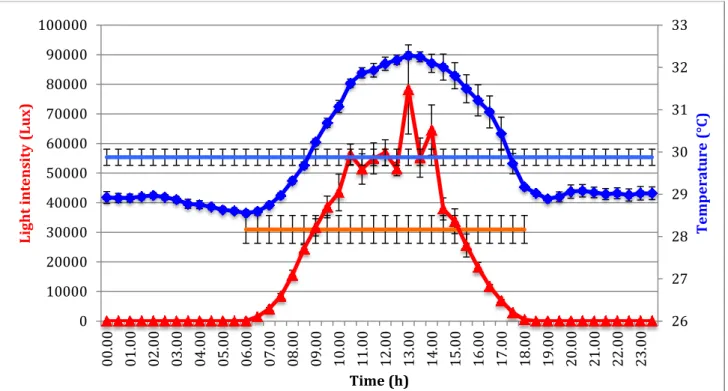
During the surveys conducted prior the sampling activites, an HOBO pendant data logger (Onset, UA-002-64) used for measuring the sea temperature (°C) and the light intensity level (Lux) was placed and maintained until the end of the sampling on the coral patches within the sampling area. Both the data were recorded continuously every 30 minutes for one week in order to analyze the daily means trend of temperature and light affecting the lagoon reef flat
zone. The data logger was placed in a horizontal position on hard substrate using plastic bands in order to permit a proper recording of the light intensity by the specific light sensor. Afterwards, the coral colonies were selected within the area in order to have samples representative of the Magoodhoo reefs and displaying 3 different growth morphology. The 3 coral species selected were A. tenuis (branching growth form), P. lobata (massive growth form) and E. lamellosa (foliose/encrusting growth form). Thus, 5 colonies (which represented the biological replicates) of each coral species and showing similar sizes were randomly selected, tagged and photographed (Canon G11 with Canon housing) by snorkeling within the sampling area. Only healthy colonies exhibiting characteristic color, dark pigmentation, absence of any sign of physical damage and stress were used for the subsequent collection and proteins analyses.
Fig. 4.1 Map of Magoodhoo Island (A), located in Faafu Atoll (B), Republic of Maldives (C). The islands are indicated in black, the deeper part of the lagoons in grey and the dotted line indicates the reef edges. An area of approximately 150 m2 and about 70 m distant from the shore in the Magoodhoo lagoon (3° 04' 45.60" N; 72° 58' 02.20" E), was chosen as sampling site to create an easy and short time access area (indicated by the arrow).
4.2 Coral species identification
To ensure that the sampled colonies owning to the three selected species was needed before sampling each colony belonging to the three taxa were analyzed by a morphological and, when necessary, also by a genetic analysis. For identify species of Porites and Echinopora was enough morphology identification by Veron (2000) supporting by on field survey, photos of colony and detection of morphology of corallites under stereomicroscope. To identify Acropora specie was used PCR.
Morphology identification started from external aspects of colony following by photos analyses comparing with Veron (2000) and ending with laboratory investigation with stereomicroscope, Leica EZ4 HD with Integrated LED Illumination and HD Camera, and manual for coral identification. On field survey have a prior goals to collect photos for each colony subjected to the study, recognizing coral genera and giving a chance to formulate an hypothesis around of coral species. To confirm this hypothesis there is a need of sampling coral of interest and bleach it to remove the live tissue to investigate the corallite under stereomicroscopic. In consequence of hemispherical morphology and smoothy surface observed during previous survey its suggests that Porites may own of the specie of P. lobata. This hypothesis was confirmed with microscope investigation, which confirm it by recognising of corallite morphology. P. lobata may be confused with P. solida, but it’s distinct by having taller pali, especially on the taller pairs of septa. E. lamellosa was individuated on field recognising its growth morphology, by its tiers arrangement of colony structure, and finally microscope investigation validates the hypothesis identifying the small and compact columellae and paliform lobes. To identify taxonomy of Acropora colony subjected to the study was not enough both survey and also stereomicroscope investigation. The knowledge in morphology of Acropora was not enough to recognize unequivocally A. tenuis. To confirm that the specie of Acropora under investigation was A. tenuis, coral DNA was extracted using DNeasy_Tissue kit (QIAGEN, Qiagen Inc., Valencia, CA, USA) and a rDNA region of about 500 bp (spanning the entire ITS1, 5.8S, ITS2 and a portion of 28S and 18S) was amplified and sequenced. Amplification was performed using the coral-specific primer A18S (50 GATCGAACGGTTTAGTGAGG 30) (Takabayashi et al., 1998), and the universal primer ITS4 (50 TCCTCCGCTTATTGATATGC 30) (White et al., 1990). Sequences were compared with known scleractinian corals sequences in GenBank using the BLAST nucleotide search (http://www.ncbi.nlm.nih.gov/BLAST/). BLAST searches showed 98% identity with rDNA sequences of A. tenuis.
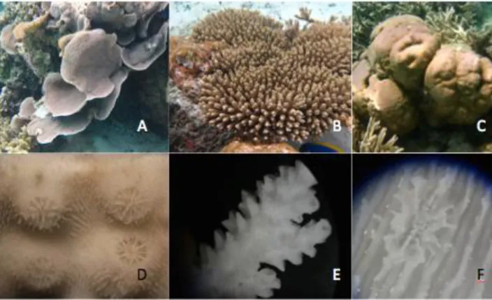
Fig 4.2 Photos of colonies subjected to the study on field, (A) Echinopora lamellosa, (B) Acropora tenuis, (C)
Porites lobata and particular of corallite (D) skeleton of E. lamellosa, (E) skeleton of A. tenuis (F) skeleton of P. lobata
4. 3 Coral collection
Coral collection was conducted by snorkelling at shallow depth using a destructive method in a period between the 19 and 23 of March 2014. Overall, 15 colonies (5 colonies for each species) were selected and collected at 6 different times within the 24h in order to analyze the proteins modulation over a complete daily cycle. Therefore, we totally collected and analyzed 90 coral samples.
The 6 times chosen for the sampling were 06:00, 09:00, 13:00, 16:00, 20:00, 02:00. Three sampling times (09:00, 13:00 and 16:00) were selected as characterized by a full in full light intensity. On the other hand, during the other three sampling times the light level was low (06:00 and 20:00) or completely absent (02:00).
During the sampling activities, small coral fragments were excised from colonies with a hollow-point stainless steel spike (1 cm diameter) by applying constant rotational pressure in order to reduce the size of coral sampled and, at the same time, reduce the amount of sampling stress and limit excessive damage to the colonies allowing rapid coral recovery following the sampling (Bromage et al. 2009). For the sampling of the massive corals (P.
lobata) the use of the hammer and chisel was required. Each coral fragments from each
colony was maintained in a specific plastic tube and was immediately frozen at -80°C using an immersion cooler (FT902, JULABO, Labortechnick GmbH) placed in the MaRHE Centre laboratory of Maghodhoo Island which is located a few meters from the sampling area.
4.4 Protein analyses
4.4.1 Protein extraction and dosage Reagents:
1. Sample buffer (SDS-SB): Tris-HCl 0.0625 (pH 6.8) Glycerol 10%
sodium dodecyl sulfate (SDS) 2.3%
2. Phenylmethylsulfonyl fluoride (PMSF, Sigma): inhibitor of serine and thiol protease, soluble in ethanol and isopropanol. Stock solution 100mM (17.4 mg/ml), used at 2mM
3. Complete EDTA-free cocktail of protease inhibitors (25x, Roche Diagnostic)
Protocol:
In the laboratory, about 1 g of each frozen coral fragment was powdered using mortar and pestle and collected in 2 ml plastic tubes (Eppendorf)
Proteins of the coral holobiont were extracted by homogenizing the tissue powder in 400 ml of the denaturing SDS buffer containing 1 mM PMSF and Complete EDTA-free cocktail of protease inhibitors.
Two different procedures were performed in according to Seveso et al 2013. - One half of the mixture was boiled for 10 min and then centrifuged (15 min at
12,000 rpm, 4 °C). Supernatants were clarified (5 min at 12,000 rpm) and then frozen at -20 °C until used.
- Before boiling, to the other half of the mixture, an equal volume of acid-washed glass beads (425-600 mm, Sigma) was added followed by vigorously shaking on a vortex (10 cycles of 1 min) interspersed with cooling on ice. This latter procedure has been used to prepare extracts containing algal proteins (Weis et al., 2002).